Embed Size (px)
Citation preview

I.J. Image, Graphics and Signal Processing, 2020, 2, 30-41 Published Online April 2020 in MECS (http://www.mecs-press.org/)
DOI: 10.5815/ijigsp.2020.02.04
Copyright © 2020 MECS I.J. Image, Graphics and Signal Processing, 2020, 2, 30-41
Improving Retinal Image Quality Using the
Contrast Stretching, Histogram Equalization, and
CLAHE Methods with Median Filters
Erwin Computer Engineering, Sriwijaya University, Indralaya, Indonesia
Email: [email protected]
Dwi Ratna Ningsih Computer Engineering, Sriwijaya University, Indralaya, Indonesia
Email: [email protected]
Received: 01 December 2019; Accepted: 15 March 2020; Published: 08 April 2020
Abstract—This paper performs three different contrast
testing methods, namely contrast stretching, histogram
equalization, and CLAHE using a median filter. Poor
quality images will be corrected and performed with a
median filter removal filter. STARE dataset images that
use images with different contrast values for each image.
For this reason, evaluating the results of the three
parameters tested are; MSE, PSNR, and SSIM. With the
gray level scale image and contrast stretching which
stretches the pixel value by stretching the stretchlim
technique with the MSE result are 9.15, PSNR is 42.14
dB, and SSIM is 0.88. And the HE method and median
filter with the results of the average value of MSE is
18.67, PSNR is 41.33 dB, and SSIM is 0.77. Whereas for
CLAHE and median filters the average yield of MSE is
28.42, PSNR is 35.30 dB, and SSIM is 0.86. From the
test results, it can be seen that the proposed method has
MSE and PSNR values as well as SSIM values.
Index Terms—Median filter, STARE, contrast stretching,
CLAHE, HE, PSNR, SSIM.
I. INTRODUCTION
Enhancement of the quality of retinal images that have
both noise and noise is the first step in image processing
to help improve the accuracy of the results for image
segmentation and extraction. Images store a lot of
information, but often there is a decrease in quality or
image defects. So that images that have experienced
interference or noise are easily interpreted, then the image
can be manipulated into other images of better quality
using image processing techniques or methods[1]. Retinal
eye images can provide information about pathological
changes caused due to disease[2]. Processing medical
image of the retina in the eye with disease information by
increasing contrast quality and eliminating noise that can
damage the image so that sharpness can be improved and
can facilitate segmentation or extraction of retinal images
[3]. From an image, sharp lines can be sharpened to
increase. Poor and poor image quality cannot be
interpreted directly by the human eye, so we use methods
to improve the quality of contrast stretching images and
median filters[4]. One type of image modeling used for
digital processing, one of which has good results is RGB.
To find out the color frequency in a digital image a
histogram graph is used[5]. In the image, there are bright
and dark noise in the random distribution in the image
which can affect the visual effect of the image as well as
the information in the image[6]. Then the method to
improve the image quality of the median filter and
contrast stretching is applied. It is a method that will
increase contrast by getting new images as well as new
contrast.
In this pre-processing stage, two contrast methods are
used namely contrast stretching and histogram
equalization and CLAHE. Increase contrast by stretching
the existing value in the image at a certain intensity.
Contrast stretching will stretch, increase and decrease the
contrast of the image. And the median filter as a filter for
image enhancement that will filter out noise or noise.
With this increase in image quality, it can be seen which
method is better in improving it. With the parameters of
PSNR, SSIM and MSE to see the image quality and
whether or not accurate.
The test of this research is that it can improve the low
image quality by processing the image with contrast
stretching with the median filter method for noise
removal. Obtaining better image results than the original
image that has not been improved image quality.
Limitation of the problem is limited by researchers,
namely only discussing on improving image quality with
the Contrast Stretching method using Stretchlim, HE and
CLAHE. And noise removal with a median filter. By
using the STARE dataset which is secondary data
obtained through the STARE dataset website. Using
the .jpg format. In this study, the authors only used 20
STARE datasets. Showing the results of image quality
improvement using the parameters of Mean Square Error

Improving Retinal Image Quality Using the Contrast Stretching, Histogram Equalization, 31
and CLAHE Methods with Median Filters
Copyright © 2020 MECS I.J. Image, Graphics and Signal Processing, 2020, 2, 30-41
(MSE), PSNR (Peak Signal to Noise Ratio) and SSIM
(Structural Similarity Index).
II. RELATED WORK
In previous studies[7] the original retinal image will be
processed with a normalized convolution algorithm using
domain transformation and obtain an enhanced image.
With the median filter method using the STARE database
to evaluate the results of image enhancement. The result
of this method is that it can improve the quality of the
retinal image is still unclear.
From previous studies[8] using edge detection and
contrast enhancement to eliminate noise. This research
focuses on stretching contrast and image enhancement.
Use a histogram to verify the results of image
enhancement and contrast stretching. Which increased
quality is not optimal.
Previous studies[9] improved the perception of
information that exists in the original image, increasing
structural features for good image quality. With
morphological and merging regions which will then be
continued by stretching the contrast separately. Linear
stretching is used to improve the texture features of
objects, can suppress noise but still cannot maintain the
brightness of the entire image.
Previous studies[10] increased contrast with
morphological methods of upper and lower operations.
Matrix contrast at edges extracts the enhanced image.
Counting each region with morphological open
operations, everything connected with pixels under 40
will be eliminated. But the difficulty in removing noise.
Previous studies[11] used methods to improve blood
vessels. A multi-scale top-hat transformation extracts the
original image into bright and dim image features. Then it
is enhanced by adding an image feature that has a gap and
a gaussian curve histogram is mounted for linear
stretching.
Based on research conducted several factors can affect
the results of the processing of an image at the time of
quality improvement. Different contrast qualities make
the test results in different based on the quality of each
image. The filter used can affect the results of the image
to be smoother than the initial image.
III. METHOD AND RESULT
This paper uses a database of stare in which the initial
stage is to input a retinal image and then the image is
given three RGB colors with an intensity of 8-bit depth.
Separate each RGB color into three channels. RGB image
or color image which consists of three color elements
namely red (R), green (G) and blue (B). Color component
of RGB images have a resolution of 24 bits which uses
eight bits (from 0 to 255) in each component, allows
color mapping in space three dimension.
From RGB, the image is grayed with pixel intensity.
On grayscale with a gray image that will be filtered with
the median filter, then with contrast stretching, can be
seen in the illustration in figure .1.
Fig.1. Method Diagram Block
At this stage of the process the first is to input the
retinal image using the STARE database, after the median
filter which will then be contrasted with contrast
stretching which are the methods in step figure.1, as
follows:
A. Grayscale
Gray pixel intensity, with values ranging from 0 to 255
expresses the gray range of the image. With this range
between the gradations of black and white images.
Grayscale is a color that is located between black and
white. On this grayscale, the image is first increased in
complexity. The original image before the grayscale
image is an RGB image with three elements of red (R),
green (G), and blue (B). With the formula[12]:
Grayscale = (0,299 ∗ R) + (0,587 ∗ G) + (0,114 ∗ B) (1)
Where,
R = red channel matrix value
G = green channel matrix value
B = blue channel matrix value
B. Median Filter
It is a non-linear method to filter or reduce noise in the
image so that image information is maintained. Values on
images will be replaced or manipulated by making values
as medians. With the gray level, the value has been
replaced by the median then stored instead of the noise
value. The max is the maximum, the min is the minimum
value, and the average is the gray level. And y (m, n) is
the center of manipulation of the median filter[13].

32 Improving Retinal Image Quality Using the Contrast Stretching, Histogram Equalization,
and CLAHE Methods with Median Filters
Copyright © 2020 MECS I.J. Image, Graphics and Signal Processing, 2020, 2, 30-41
y[m, n] = 𝑚𝑒𝑑𝑖𝑎𝑛 {x[i, j], (i, j)ϵ ω (2)
Where,
y [m, n] = matrix of results labeled y with m, n is a row
multiplied by a column.
{x [i, j], (i, j) ϵ ω = matrix value of the image being
processed or the corresponding elements sorted.
The median filter will see the pixel value of its
neighbor and will match the pixel value that will be
represented by the neighbor's pixel value. The pixel value
will be replaced at the middle value with a count of
around neighbors. When the value is even on the
surrounding pixel, the middle value will be used with an
average of two middle values[14].
C. Histogram Equalization (HE)
It is a histogram equalization in which the gray
distribution of values is made flat. HE will manipulate the
respective image pixels for increased contrast. The image
gets a flattened histogram distribution function. The
intensity and scale are obtained as output from the
histogram equalization. The purpose of this HE is to
make the distribution of the histogram evenly distributed
so that the pixel value at the gray level will have a
relatively identical value or the same. HE will map input
images with unequal intensity levels and to output images
with the same level[15].
D. Contrast Stretching (CS)
It is a method of stretching the contrast, with the
intensity value contained in the image expanded with a
dynamic range. The pixel value of the image can be
applied with linear scaling. To normalize the image or
contrast stretching the image it is necessary to determine
the minimum and maximum values of the image. These
minimum and maximum values will determine the
boundary of the image. In this proposed method an image
with an 8-bit gray level is the lower limit and values 0 to
255 as the upper limit. Digital images are taken using a
fundus camera. When viewed from the outline angle,
digital image processing techniques are divided into 3
based on processing levels, namely:
1. Low-Level Process or low level, this processing is a
basic operation in image processing examples such as
noise reduction, image improvement, and image
restoration.
2. Mid-Level Process or intermediate level, this
processing includes object description, image
segmentation, and object classification separately.
3. High-Level Process or high level includes the
analysis of an image.[16].
𝑔(x, y) =𝑓(𝑥,𝑦)−𝑚𝑖𝑛
𝑚𝑎𝑥−𝑚𝑖𝑛𝑋255 (3)
Where,
g (x, y) = matrix of the resulting image
f (x, y) = original image matrix value
Where g (x, y) represents the output and f (x, y)
represents the input. Image intensity values with 0 as the
lowest value and 255 as the highest value[17]. By using
stretchlim as a determinant of the minimum and
maximum values. The value of g (x, y) as a new image
obtained from the image value (x, y) will be subtracted by
the maximum value and divided by the results of the
minimum and maximum reduction. The results will be
multiplied by 255 as the pixel value[18].
E. CLAHE(Contrast Limited Adaptive Histogram
Equalization)
CLAHE is a contrast enhancement technique which is
the development of HE and AHE from which surely this
CLAHE is better than HE and AHE. In this CLAHE
images will be divided into tiles or smaller areas. CLAHE
is also a development of the AHE method that has its
components changed. In this CLAHE all pixels adjacent
to all functions will be converted[14]. This CLAHE is
different from AHE because CLAHE limits the contrast,
uses the maximum value on the clip and returns it to the
gray value [19].
F. Mean Square Error (MSE) and PSNR (Peak Signal
to Noise Ratio)
The parameters used as an evaluation of the results of
improving image quality. This parameter will compare
the original image with the resulting image, the equation
used is as follows[20] :
𝑀𝑆𝐸 =1
𝑋𝑌∑ 𝑌=1
𝑦=0 ∑ (�̂�(𝑖, 𝑗) − 𝑦(𝑖, 𝑗))2
𝑋=1𝑥=0 (4)
Where,
y ̂ (i, j) = matrix of the yield image
y (i, j) = original image matrix value
𝑃𝑆𝑁𝑅 = 10𝑙𝑜𝑔10𝑠2
𝑀𝑆𝐸 (5)
With the value S = 255 for 8-bit images, the higher the
PSNR value generated, the better. While the closer the
value to 0, the better the MSE value.
G. SSIM (Structural Similarity Index)
SSIM is a parameter used for the evaluation of results
using quantitative measures. The value of SSIM is
obtained by dividing the original image with a distorted
image which is then converted into a vector. With the
following formula equation[21]:
𝑆𝑆𝐼𝑀 =(2𝜇𝑥𝜇𝑦+𝑐1)(2𝜎𝑥𝑦+𝑐2)
(𝜇𝑥2+𝜇𝑦
2+𝑐1)(𝜎𝑥2+𝜎𝑦
2+𝑐2) (6)
Where,
μ_x = average value of X
μ_y = average value of Y

Improving Retinal Image Quality Using the Contrast Stretching, Histogram Equalization, 33
and CLAHE Methods with Median Filters
Copyright © 2020 MECS I.J. Image, Graphics and Signal Processing, 2020, 2, 30-41
σ_xy = correlation coefficient on X and Y
σ_x ^ 2 = difference in values on X
σ_y ^ 2 = difference in values on Y
c_1 and c_2 = two variables that stabilize the low
denominator
The more SSIM values close to zero, the better the
improved image.
H. Result Enhancement
MATLAB R2018b was used in the course of this
research, which is managed by a laptop device with an
AMD Quad-Core A10-9620P processor, up to 3.4GHz
with 4GB of RAM. By using the STARE database taken,
there were 20 images. Which uses JPG format. The image
used is a dataset from STARE (Structured Analysis of the
Retina).
Inputting the image with the original image in the first
stage can be seen in figure 1. The median filter will
manipulate the image to get better image quality
improvements. Then the shortcomings of the median
filter will be followed by contrast stretching which will
increase the contrast with level to gray.
By using the equation formula (1), the grayscale results
from the original RGB image by taking the pixel value
can be seen as follows:
G = 163
B = 165
R = 193
Grayscale = (0,299 * R) + (0,587 * G) + (0,114 * B)
= (0.299 * 193) + (0.587 * 163) + (0.114 * 165)
= 172,198
(a) (b)
(c)
Fig.2. a. Original image b. Grayscale c. Original image histogram
Figure 2 uses im0005.jpg which is in part (a) the
original image from the STARE database. Based on
Figure 2 (b) is the original input image of the dataset that
is made grayscale or to a grayscale. Figure 2 (c) is a
histogram of the original image that has not been
processed. By determining the lower limit and the pixel
value, the image is normalized. Suppose that a is the
lower limit and b is the upper limit, then find the highest
and lowest pixels of the image. With c and d, take the
lowest and highest pixel values, and scale the pixels by
the following equation:
𝑃 𝑜𝑢𝑡 = (𝑃 𝑖𝑛 − 𝑐)(𝑏−𝑎)
(𝑑−𝑐)+ 𝑎 (7)
Values c and d can affect the output value especially if
the value is too high or low.
Fig.3. Contrast Stretching Using Stretchlim image STARE im0005.jpg
Figure 3 is the result of the graying image normalized
by contrast stretching and stretching methods. Stretching
will normalize the image by looking for the minimum and
maximum values first. Stretchlim will look for maximum
and minimum values of gray-level images.
Fig.4. Histogram Contrast Stretching Using Stretchlim image STARE im0005.jpg
Figure 4 displays the histogram of contrast stretching
with the results seen from the histogram that the image is
stretching. By stretching the intensity value of the image
can be seen if the counter is too far to the right with a
pixel value above 135 then the image is included as
bright. Conversely, if the cons on the histogram are more
left or under 50 pixels based on the histogram, the image
is dark. In the histogram, the image is more dominant
towards the light.
Figure 5 is the image of im005.jpg which has done
even distribution of the histogram with HE. The image in
a gray level that is made flat or identical. In the image

34 Improving Retinal Image Quality Using the Contrast Stretching, Histogram Equalization,
and CLAHE Methods with Median Filters
Copyright © 2020 MECS I.J. Image, Graphics and Signal Processing, 2020, 2, 30-41
visible difference from the results of contrast stretching
images and median filters that blood vessels are more
visible in the HE method and median filter.
Fig .5. HE image im0005.jpg
Fig.6. Histogram with HE image im0005.jpg
Figure 6 is a histogram of the im005.jpg image leveled
by the HE method. From the original image histogram,
the difference between the histogram results of HE which
is more even and equal. With a range of 0 to 255 and the
grayscale histogram is made uniform and obtained evenly
distributed.
The results of CLAHE and median filters can be seen
in Figure 7 where all pixels are converted, and the
contrast value is limited to the maximum value. Seen in
the image that this CLAHE method can highlight the
small blood vessels of the retina of the eye compared to
contrast stretching and HE.
Fig.7. CLAHE image im0005.jpg
This paper uses three parameters, namely MSE and
PSNR to assess the results of the method being tested.
The testing method was carried out on a 4GB ASUS
X555Q laptop. The results of MSE can be seen in table 2.
And the results of PSNR can be seen in table 3. And the
results of SSIM can be seen in table 4.
The results table 1 is obtained by testing on 20 STARE
dataset images with a contrast enhancement method and
median filter that will refine the image. The accuracy of
the test results can be seen from the measurement
parameters used, namely MSE, PSNR, and SSIM. Where
the MSE value is getting closer to the value of 0, the
better it means that the smaller the error value. While
PSNR will state the value of image processing results if
the image is more accurate if the PSNR value exceeds 35
dB and vice versa if the PSNR value is below 35 dB then
the value of the image processing result is not too good.
While the SSIM value will be said to be accurate if the
value of the image processing results close to 1 is said to
be more accurate. SSIM value is in the range of 0-1 if the
SSIM value is close to 0 then it can be said that the image
processing results are not good enough.
Table 1. The results of 20 retinal datasets were tested
Image (JPG)
Image Contrast stretching Histogram Equalization CLAHE
'im0001.jpg'
'im0002.jpg'

Improving Retinal Image Quality Using the Contrast Stretching, Histogram Equalization, 35
and CLAHE Methods with Median Filters
Copyright © 2020 MECS I.J. Image, Graphics and Signal Processing, 2020, 2, 30-41
'im0003.jpg'
'im0004.jpg'
'im0005.jpg'
'im0006.jpg'
'im0007.jpg'
'im0008.jpg'
'im0009.jpg'

36 Improving Retinal Image Quality Using the Contrast Stretching, Histogram Equalization,
and CLAHE Methods with Median Filters
Copyright © 2020 MECS I.J. Image, Graphics and Signal Processing, 2020, 2, 30-41
'im0010.jpg'
'im0011.jpg'
'im0012.jpg'
'im0013.jpg'
'im0014.jpg'
'im0015.jpg'
'im0016.jpg'
Improving Retinal Image Quality Using the Contrast Stretching, Histogram Equalization, 37
and CLAHE Methods with Median Filters
Copyright © 2020 MECS I.J. Image, Graphics and Signal Processing, 2020, 2, 30-41
'im0017.jpg'
'im0018.jpg'
'im0019.jpg'
'im0020.jpg'
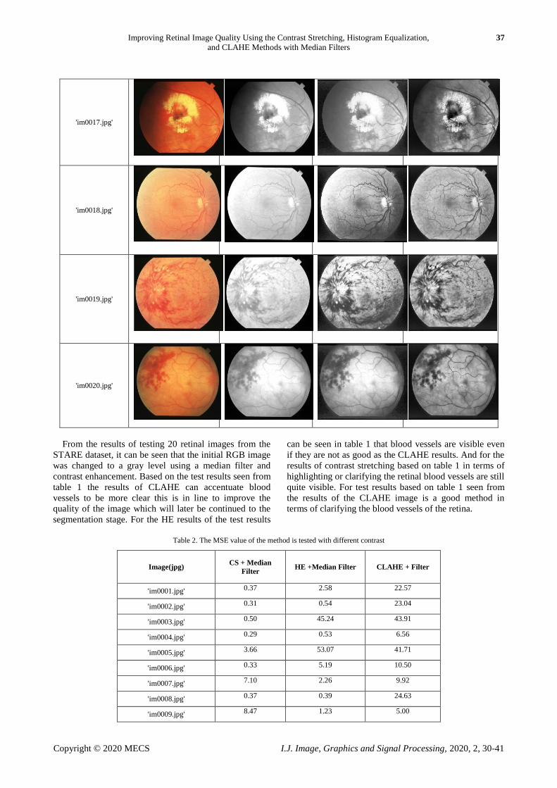
From the results of testing 20 retinal images from the
STARE dataset, it can be seen that the initial RGB image
was changed to a gray level using a median filter and
contrast enhancement. Based on the test results seen from
table 1 the results of CLAHE can accentuate blood
vessels to be more clear this is in line to improve the
quality of the image which will later be continued to the
segmentation stage. For the HE results of the test results
can be seen in table 1 that blood vessels are visible even
if they are not as good as the CLAHE results. And for the
results of contrast stretching based on table 1 in terms of
highlighting or clarifying the retinal blood vessels are still
quite visible. For test results based on table 1 seen from
the results of the CLAHE image is a good method in
terms of clarifying the blood vessels of the retina.
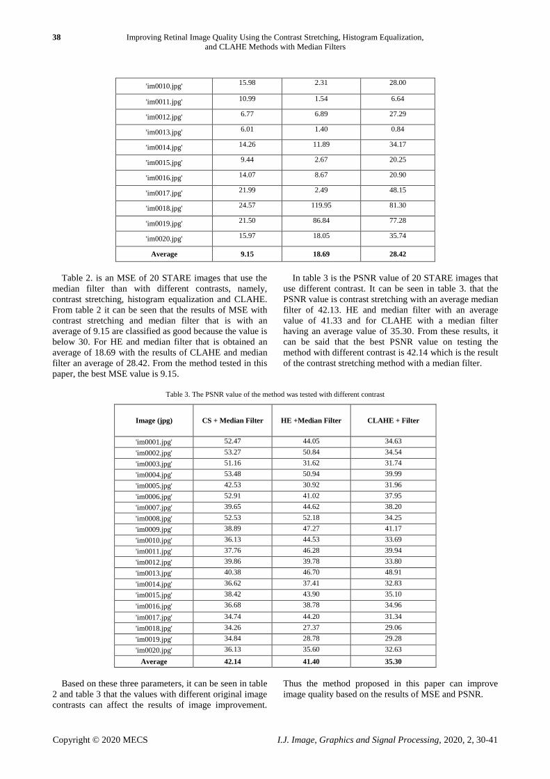
Table 2. The MSE value of the method is tested with different contrast
Image(jpg) CS + Median
Filter HE +Median Filter CLAHE + Filter
'im0001.jpg' 0.37 2.58 22.57
'im0002.jpg' 0.31 0.54 23.04
'im0003.jpg' 0.50 45.24 43.91
'im0004.jpg' 0.29 0.53 6.56
'im0005.jpg' 3.66 53.07 41.71
'im0006.jpg' 0.33 5.19 10.50
'im0007.jpg' 7.10 2.26 9.92
'im0008.jpg' 0.37 0.39 24.63
'im0009.jpg' 8.47 1.23 5.00
38 Improving Retinal Image Quality Using the Contrast Stretching, Histogram Equalization,
and CLAHE Methods with Median Filters
Copyright © 2020 MECS I.J. Image, Graphics and Signal Processing, 2020, 2, 30-41
'im0010.jpg' 15.98 2.31 28.00
'im0011.jpg' 10.99 1.54 6.64
'im0012.jpg' 6.77 6.89 27.29
'im0013.jpg' 6.01 1.40 0.84
'im0014.jpg' 14.26 11.89 34.17
'im0015.jpg' 9.44 2.67 20.25
'im0016.jpg' 14.07 8.67 20.90
'im0017.jpg' 21.99 2.49 48.15
'im0018.jpg' 24.57 119.95 81.30
'im0019.jpg' 21.50 86.84 77.28
'im0020.jpg' 15.97 18.05 35.74
Average 9.15 18.69 28.42
Table 2. is an MSE of 20 STARE images that use the
median filter than with different contrasts, namely,
contrast stretching, histogram equalization and CLAHE.
From table 2 it can be seen that the results of MSE with
contrast stretching and median filter that is with an
average of 9.15 are classified as good because the value is
below 30. For HE and median filter that is obtained an
average of 18.69 with the results of CLAHE and median
filter an average of 28.42. From the method tested in this
paper, the best MSE value is 9.15.
In table 3 is the PSNR value of 20 STARE images that
use different contrast. It can be seen in table 3. that the
PSNR value is contrast stretching with an average median
filter of 42.13. HE and median filter with an average
value of 41.33 and for CLAHE with a median filter
having an average value of 35.30. From these results, it
can be said that the best PSNR value on testing the
method with different contrast is 42.14 which is the result
of the contrast stretching method with a median filter.
Table 3. The PSNR value of the method was tested with different contrast
Image (jpg) CS + Median Filter HE +Median Filter CLAHE + Filter
'im0001.jpg' 52.47 44.05 34.63
'im0002.jpg' 53.27 50.84 34.54
'im0003.jpg' 51.16 31.62 31.74
'im0004.jpg' 53.48 50.94 39.99
'im0005.jpg' 42.53 30.92 31.96
'im0006.jpg' 52.91 41.02 37.95
'im0007.jpg' 39.65 44.62 38.20
'im0008.jpg' 52.53 52.18 34.25
'im0009.jpg' 38.89 47.27 41.17
'im0010.jpg' 36.13 44.53 33.69
'im0011.jpg' 37.76 46.28 39.94
'im0012.jpg' 39.86 39.78 33.80
'im0013.jpg' 40.38 46.70 48.91
'im0014.jpg' 36.62 37.41 32.83
'im0015.jpg' 38.42 43.90 35.10
'im0016.jpg' 36.68 38.78 34.96
'im0017.jpg' 34.74 44.20 31.34
'im0018.jpg' 34.26 27.37 29.06
'im0019.jpg' 34.84 28.78 29.28
'im0020.jpg' 36.13 35.60 32.63
Average 42.14 41.40 35.30
Based on these three parameters, it can be seen in table
2 and table 3 that the values with different original image
contrasts can affect the results of image improvement.
Thus the method proposed in this paper can improve
image quality based on the results of MSE and PSNR.

Improving Retinal Image Quality Using the Contrast Stretching, Histogram Equalization, 39
and CLAHE Methods with Median Filters
Copyright © 2020 MECS I.J. Image, Graphics and Signal Processing, 2020, 2, 30-41
Table 4. The SSIM value of the method was tested with different contrast
Image (jpg) CS + Median Filter HE +Median Filter CLAHE + Filter
'im0001.jpg' 0.93 0.81 0.86
'im0002.jpg' 0.90 0.84 0.86
'im0003.jpg' 0.89 0.77 0.82
'im0004.jpg' 0.88 0.84 0.88
'im0005.jpg' 0.91 0.78 0.79
'im0006.jpg' 0.89 0.86 0.89
'im0007.jpg' 0.85 0.79 0.89
'im0008.jpg' 0.93 0.82 0.83
'im0009.jpg' 0.83 0.71 0.86
'im0010.jpg' 0.88 0.74 0.88
'im0011.jpg' 0.84 0.67 0.87
'im0012.jpg' 0.93 0.83 0.87
'im0013.jpg' 0.78 0.72 0.90
'im0014.jpg' 0.87 0.78 0.86
'im0015.jpg' 0.87 0.77 0.86
'im0016.jpg' 0.86 0.71 0.85
'im0017.jpg' 0.88 0.76 0.86
'im0018.jpg' 0.87 0.77 0.85
'im0019.jpg' 0.86 0.71 0.78
'im0020.jpg' 0.88 0.80 0.87
Average 0.88 0.77 0.86
In table 4 the results are the SSIM values of the median
filter with three different contrasts. Of the three contrasts,
the best SSIM values are the results of HE and median
filters with an average value of 0.77. And the average
value of CS and median filter is 0.89 while the SSIM
CLAHE value and median filter average value is 0.86.
Based on the three parameters that can be seen in table 2,
table 3, and table 4 that the value with different original
image contrast can affect the results of image
improvement. Thus the method proposed in this paper
can improve image quality based on the results of MSE
and PSNR, the best value in SSIM is in the HE method
and median filter.
Table 5. Comparison of MSE and PSNR values in the method tested with the previous method to improve image quality
Method MSE PNSR SSIM
Proposed Method
(CS + Median Filter)
9.14 42.13 0.87
Proposed Method
(HE + Median Filter)
18.68 41.39 0.77
Proposed Method
(CLAHE + Median
Filter) 28.42 35.29 0.85
(S. Sahu et al,
2018) [22] - 35.37 0.96
(Zulfahmi et al,
2019) [23] 85.52 28.88 -
(B. Gupta et al,
2018) [21]
HE
- 25.79 0.56
(B. Gupta et al, 2018) [21]
CLAHE
- 9.48 0.33
Based on table 5 it can be seen that the comparison of
the results of the method being tested is better contrasted
stretching with the median filter because the values of
MSE and PSNR are better. Because the closer to zero the
MSE results of an image processing result, the better.
Whereas the PSNR value is more than 35 dB, the better
the results of image quality improvement. However, the
SSIM value proposed by S.Sahu, et al [22] is superior.
Because the SSIM value will get better if it gets closer to
zero. From the results of these comparisons, the method
being tested can match the results of the process from
previous results.
IV. CONCLUSION
Based on the method proposed in this paper using the
STARE dataset by taking retinal images in JPG format.
Of the images, image quality improvements were made
using three contrasts, contrast stretching, HE and CLAHE
filtered using a median filter. With the first comparison
using the proposed method contrast stretching and
median filter. The technique of taking maximum and
minimum values using stretchlim which was previously
the image in grayish conditions. HE and median filters
with equalization techniques on histograms whose pixel
values are at 0 to 255. The third is CLAHE and median
filters which technically limit the pixel values to the
boundary regions. The method proposed in this paper can
improve image quality based on MSE and PSNR results.
From the results of testing with three different methods
and based on three different parameters, the best MSE
and PSNR results are contrasted stretching with values of
9.14 and 42.13 dB. Thus contrast stretching is better in

40 Improving Retinal Image Quality Using the Contrast Stretching, Histogram Equalization,
and CLAHE Methods with Median Filters
Copyright © 2020 MECS I.J. Image, Graphics and Signal Processing, 2020, 2, 30-41
improving image quality. In the future, it is expected to
improve the image quality with the contrast stretching
method with fixed results as an RGB image. With the
CLAHE method in the section to clarify the blood vessels
of the retina image very well, it would be better if it
continued with image segmentation in the future.
REFERENCES
[1] M. Rajaram, “A novel approach for contrast enhancement
based on histogram equalization followed by median
filter,” ARPN J. Eng. Appl. Sci., no. September 2009,
2014.
[2] V. M. Saffarzadeh, A. Osareh, and B. Shadgar, “Vessel
Segmentation in Retinal Images Using Multi ‑ scale Line
Operator and K ‑ Means Clustering,” Dep. Comput. Eng.
Shahid Chamran Univ. Ahvaz, Khuzestan, Iran,jmss., vol.
4, no. 2, 2017.
[3] H. Aguirre-ramos, J. G. Avina-cervantes, I. Cruz-aceves, J.
Ruiz-pinales, and S. Ledesma, “Blood vessel
segmentation in retinal fundus images using Gabor filters ,
fractional derivatives , and Expectation Maximization,”
Appl. Math. Comput., vol. 339, pp. 568–587, 2018.
[4] F. Farokhian and H. Demirel, “Blood Vessels Detection
and Segmentation in Retina using Gabor Filters,” Electr.
Electron. Eng. Dep. IEEE, pp. 104–108, 2013.
[5] K. Firdausy, T. Sutikno, and E. Prasetyo, “Image
Enhancement Using Contrast Stretching On Rgb And Ihs
Digital Image,” Cent. Electr. Eng. Res. Solut. (CEERS),
ISSN, vol. 5, No.1, no. 1, pp. 45–50, 2007.
[6] Y. Zhu and C. Huang, “An Improved Median Filtering
Algorithm for Image Noise,” Phys. Procedia, vol. 25, pp.
609–616, 2012.
[7] F. A. Jassim and F. H. Altaani, “Hybridization of Otsu
Method and Median Filter for Color Image Segmentation,”
Int. J. Soft Comput. Eng. ISSN 2231-2307, vol. Volume-3,
no. 2, pp. 69–74, 2013.
[8] P. Garhwal and P. Garhwal, “A Hybrid Approach to
Image Enhancement using Contrast Stretching on Image
Sharpening and the analysis of various cases arising using
Histogram,” IEEE Int. Conf. Recent Adv. Innov. Eng., no.
3, 2014.
[9] B. Xu, Y. Zhuang, H. Tang, and L. Zhang, “Object-Based
Multilevel Contrast Stretching Method for Image
Enhancement,” IEEE Trans. Consum. Electron., vol. 56,
no. 3, pp. 1746–1754, 2010.
[10] R. K. B, H. Kabir, and S. Salekin, “Contrast Enhancement
by Top-Hat and Bottom-Hat Transform with Optimal
Structuring Element : Application to Retinal,” Dep.
Comput. Sci. Eng., pp. 533–540, 2017.
[11] M. Liao, Y. Zhao, X. Wang, and P. Dai, “Retinal vessel
enhancement based on multi-scale top-hat transformation
and histogram fitting stretching,” Opt. Laser Technol., vol.
58, pp. 56–62, 2014.
[12] P. P. Acharjya and S. Mukherjee, “Digital Image
Segmentation Using Median Filtering and Morphological
Approach,” Int. J. Adv. Res. Comput. Sci. Softw. Eng., vol.
4, no. 1, pp. 552–557, 2014.
[13] K. B. Khan, A. A. Khaliq, A. Jalil, and M. Shahid, “A
robust technique based on VLM and Frangi filter for
retinal vessel extraction and denoising,” PLoS One, vol.
13, no. 2, pp. 1–22, 2018.
[14] A. M. Reza, “Realization of the contrast limited adaptive
histogram equalization (CLAHE) for real-time image
enhancement,” J. VLSI Signal Process. Syst. Signal Image.
Video Technol., vol. 38, no. 1, pp. 35–44, 2004.
[15] H. A. Rahim, A. S. Ibrahim, W. M. D. W. Zaki, and A.
Hussain, “Methods to Enhance Digital Fundus Image for
Diabetic Retinopathy Detection,” IEEE Int. Colloq. Signal
Process. its Appl., no. Md, pp. 7–9, 2014.
[16] S. Anitha and V. Radha, “Contrast Stretching and Non
Linear Median Filters for Fabric Inspection,” Int. J.
Comput. Sci. Inf. Technol., vol. 2, no. 2, pp. 836–839,
2011.
[17] S. S. Al-amri, N. V Kalyankar, and S. D. Khamitkar,
“Linear and Non-linear Contrast Enhancement Image,”
IJCSNS Int. J. Comput. Sci. Netw. Secur. VOL.10, vol. 10,
no. 2, pp. 139–143, 2010.
[18] G. B. Iwasokun and O. C. Akinyokun, “Enhancement
Methods : A Review,” Sci. Int., vol. 4, pp. 2251–2277,
2016.
[19] J. Dash, “Retinal Blood Vessel Segmentation Using Otsu
Thresholding With Principal Component Analysis,” 2018
2nd Int. Conf. Inven. Syst. Control, no. Icisc, pp. 933–937,
2018.
[20] S. Shrestha, “I MAGE D ENOISING U SING N EW A
DAPTIVE,” Signal Image Process. An Int. J., vol. 5, no.
4, pp. 1–13, 2014.
[21] B. Gupta and M. Tiwari, “Color retinal image
enhancement using luminosity and quantile based contrast
enhancement,” PDPM Indian Inst. Inf. Technol., 2018.
[22] S. Sahu, A. Kumar, S. P. Ghrera, and M. Elhoseny, “An
approach for de-noising and contrast enhancement of
retinal fundus image using CLAHE,” Opt. Laser Technol.,
2018.
[23] Erwin, R. Zulfahmi, G. Utami, A. Harison, D. Noviyanti,
and P. Agung, “Improved Image Quality Retinal Fundus
with Contrast Limited Adaptive Histogram Equalization
and Filter Variation,” 2019 International Conference on
Informatics, Multimedia, Cyber and Information System
(ICIMCIS), vol. 2019, Jakarta, Indonesia, pp. 156–161,
2019.
Authors’ Profiles
Name :
Erwin
Affiliation:
Associate Professor at Department of Computer
Systems, Faculty of Computer Science,
Sriwijaya University, Indonesia.
Address:
Jl. Srijaya Negara, Bukit Lama, Kec. Ilir Bar. I, Kota
Palembang, Sumatera Selatan 30128, Indonesia
Brief Biographical History:
1994 Received the Bachelor degree in Mathematics from the
University of Sriwijaya, Indonesia
2002 Received the M.Sc. degrees in Actuarial from the
Bandung Institute of Technology (ITB), Bandung, Indonesia
2019 Received the Doctorate in Engineering, Faculty of
Engineering, Sriwijaya University, Indonesia
Main Works:
Current research interests are image processing and computer
vision.
Membership in Academic Societies:
International Journal of Applied Mathematics (IAENG)

Improving Retinal Image Quality Using the Contrast Stretching, Histogram Equalization, 41
and CLAHE Methods with Median Filters
Copyright © 2020 MECS I.J. Image, Graphics and Signal Processing, 2020, 2, 30-41
Institute of Electrical and Electronics Engineers (IEEE)
Name : Dwi Ratna Ningsih
Affiliation:
Student at Department of Computer Systems,
Faculty of Computer Science, Sriwijaya University, Indonesia
Address:
Jl. Tamyiz Timbangan, Indralaya, Ogan Ilir, Kota Palembang,
Sumatera Selatan 30264, Indonesia
Brief Biographical History:
2016- Pursuing bachelor's degree at Department of Computer
Systems, Faculty of Computer Science, Sriwijaya University,
Indonesia
How to cite this paper: Erwin, Dwi Ratna Ningsih, " Improving Retinal Image Quality Using the Contrast Stretching,
Histogram Equalization, and CLAHE Methods with Median Filters", International Journal of Image, Graphics and
Signal Processing(IJIGSP), Vol.12, No.2, pp. 30-41, 2020.DOI: 10.5815/ijigsp.2020.02.04