Embed Size (px)
Citation preview

Reducing Harm | Improving Healthcare | Protecting Canadians
July 2011
www.saferhealthcarenow.ca
IMPROVED CARE FOR ACUTE MYOCARDIAL INFARCTION
Getting Started KitGetting Started KitGetting Started KitGetting Started Kit

Safer Healthcare Now! Improved Care for Acute Myocardial Infarction Getting Started Kit
July 2011 2
Safer Healthcare Now!
We invite you to join Safer Healthcare Now! to help improve the safety of the Canadian healthcare system. Safer Healthcare Now! is a national program supporting Canadian healthcare organizations to improve safety through the use of quality improvement methods and the integration of evidence in practice.
To learn more about this intervention, to find out how to join Safer Healthcare Now! and to gain access to additional resources, contacts, and tools, visit our website at www.saferhealthcarenow.ca
This Getting Started Kit (GSK) has been written to help engage your interprofessional/ interdisciplinary teams in a dynamic approach for improving quality and safety while providing a basis for getting started. The Getting Started Kit represents the most current evidence, knowledge and practice, as of the date of publication and includes what has been learned since the first kits were released in 2005. We remain open to working consultatively on updating the content, as more evidence emerges, as together we make healthcare safer in Canada.
Note:
The Quebec Campaign: Together, let's improve healthcare safety! works collaboratively with Safer Healthcare Now! The Getting Started Kits for all interventions used in both Safer Healthcare Now! and the Quebec Campaign are the same and available in both French and English.
This document is in the public domain and may be used and reprinted without permission provided appropriate reference is made to Safer Healthcare Now!

Safer Healthcare Now! Improved Care for Acute Myocardial Infarction Getting Started Kit
July 2011 3
Acknowledgements
Safer Healthcare Now! and the authors of this document would like to acknowledge and thank:
The Canadian Patient Safety Institute (CPSI) is acknowledged for their financial and in-kind support of the Safer Healthcare Now! Getting Started Kits.
We also thank and acknowledge our volunteer Canadian Acute Myocardial Infarction (AMI) Faculty who significantly contributed to the work of the AMI teams and specifically for their contributions to this edition of the AMI Getting Started Kit.
A very special thank you is extended to Dr. Laurie Lambert, PhD and Dr. Clare Morrison, MD MSc. FRCPC who generously shared their expertise and provided feedback in developing the Getting Started Kit.
Thank you to the teams and organizations who work to improve STEMI care every day and to those who generously shared their ‘Canadian Success Stories’.
Safer Healthcare Now! Improved Care for Acute Myocardial Infarction Getting Started Kit
July 2011 4
Improved Care for Acute Myocardial Infarction AMI Faculty
Chantal Bellerose, DtP, MSc Conseillère en matière de sécurité et d'amélioration Soins de santé plus sécuritaires maintenant! (Québec)
Safety and Improvement Advisor, Safer Healthcare Now! (Quebec)
Dr. Sean Clarke, RN, PhD RBC Chair in Cardiovascular Nursing Research
Lawrence S. Bloomberg Faculty of Nursing, Toronto, Ontario
Dannie Currie, RN, MN, DHSA Safety Improvement Advisor, Safer Healthcare Now! Atlantic Node
Acute Myocardial Infarction Intervention Lead
Cleo Cyr, MHS, BN, RN, CCN (C) Provincial Advisor & Manager, New Brunswick Heart Centre
Cardiovascular Health & Wellness Program, Saint John, New Brunswick
Theresa Fillatre, RN, BSW, MHSA, CHE National Leader/Atlantic Canada Leader, Safer Healthcare Now!
Virginia Flintoft, BN, MSc
Project Manager, Safer Healthcare Now! Central Measurement Team University of Toronto, Toronto, Ontario
Dr. Sherry Grace, RN, PhD
Scientist, Toronto General Research Institute Associate Professor, KaHS , York University Faculty of Health, Toronto Ontario
Marie Hawkins, RN, BScN
Regional Network Director - Cardiac Services Interior Health Authority, Kelowna, British Columbia
Anne MacLaurin, RN, MN
Project Manager, Safer Healthcare Now! Canadian Patient Safety Institute
Dr. Davis Marr
Chief of Cardiology, NB Heart Centre Cardiovascular Health & Wellness Program St John, New Brunswick
Rody Pike, RN, MN
Clinical Educator Cardiology and Cardiac Catheterization Lab St John’s, Newfoundland Labrador
Safer Healthcare Now! Improved Care for Acute Myocardial Infarction Getting Started Kit
July 2011 5
Dr. Jack Tu, MD, MSc, PhD, FRCPC (Chair) Senior Scientist (Faculty), Institute for Clinical Evaluative Sciences (ICES), Toronto, Ontario,
AMI Intervention Faculty Lead
Mary Anne Waters, RN, BScN Tobacco Treatment Specialist (Mayo Clinic)
Senior Tobacco Reduction Coordinator, Interior Health Thompson Cariboo Shuswap, Kamloops, British Columbia
Dr. Randy Watson
Interventional Cardiologist, Trillium Health System, Ontario

Safer Healthcare Now! Improved Care for Acute Myocardial Infarction Getting Started Kit
July 2011 6
Table of Contents
Safer Healthcare Now! ................................................................................... 2
Acknowledgements ........................................................................................ 3
AMI Faculty ................................................................................................. 4
Table of Contents.......................................................................................... 6
How to use this Electronic Resource Toolkit ......................................................... 8
Introduction: Why is Delivering Reliable, Evidence-Based AMI Care Important? .............. 9
Opportunities for Improving Care for Acute Myocardial Infarction .............................. 9
A Canadian Success Story on Timeliness of Aspirin on Arrival ................................ 10
A Canadian Success Story for ECG within 10 minutes .......................................... 11
A Canadian Success Story for Timely Reperfusion .............................................. 13
A Canadian Success Story for Discharge Medications ........................................... 14
A Canadian Success Story for Tobacco Cessation Interventions .............................. 16
A Canadian Success Story for Cardiac Rehabilitation Referral ................................ 17
Appendices ................................................................................................ 18
Appendix A - Summary of Safer Healthcare Now! Recommendations ......................... 19
Appendix B - Quality Improvement and Improved Care for Acute Myocardial Infarction ... 20
Model for Improvement ............................................................................ 21
Appendix C - Measurement: Improved AMI Care ................................................... 27
Appendix D - Measurement Technical Descriptions ................................................ 31
1.0 Aspirin at Arrival – Technical Description ................................................. 32
2.0 Aspirin at Discharge – Technical Description ............................................. 34
3.0 Beta Blocker Prescribed at Discharge – Technical Description ........................ 36
4.0-A Thrombolytic Agent Received Within 30 Minutes of Hospital Arrival – Technical Description .............................................................................................. 38
4.0-B Primary Percutaneous Coronary Intervention (PCI) Received Within 90 Minutes of Hospital Arrival – Technical Description .......................................................... 42
5.0 ACE-Inhibitor or Angiotensin Receptor Blockers (ARB) Prescribed at Discharge – Technical Description ................................................................................. 46
6.0 Adult tobacco cessation counselling, tobacco dependence medications (nicotine replacement therapy, bupropion, varenicline, etc.) and referred to cardiac rehabilitation – Technical Description ............................................................. 49
7.0 Perfect care for AMI – Technical Description ............................................. 52
8.0 AMI Inpatient Mortality – Technical Description ......................................... 54
9.0 Statins Prescribed at Discharge – Technical Description ............................... 56

Safer Healthcare Now! Improved Care for Acute Myocardial Infarction Getting Started Kit
July 2011 7
10.0 ECG recorded within 10 minutes after hospital arrival or first medical contact - Technical Description ................................................................................. 58
11.0 Discharge Referral to Cardiac Rehabilitation - Technical Description ................ 60
Appendix E - Learning More About Cardiac Rehabilitation (CR) ................................. 63
References ................................................................................................. 67
Safer Healthcare Now! Improved Care for Acute Myocardial Infarction Getting Started Kit
July 2011 8
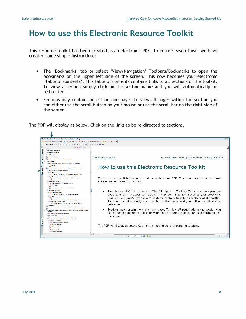
How to use this Electronic Resource Toolkit
This resource toolkit has been created as an electronic PDF. To ensure ease of use, we have created some simple instructions:
• The ‘Bookmarks’ tab or select ‘View/Navigation’ Toolbars/Bookmarks to open the bookmarks on the upper left side of the screen. This now becomes your electronic ‘Table of Contents’. This table of contents contains links to all sections of the toolkit. To view a section simply click on the section name and you will automatically be redirected.
• Sections may contain more than one page. To view all pages within the section you can either use the scroll button on your mouse or use the scroll bar on the right-side of the screen.
The PDF will display as below. Click on the links to be re-directed to sections.
Safer Healthcare Now! Improved Care for Acute Myocardial Infarction Getting Started Kit
July 2011 9
Introduction
Why is Delivering Reliable, Evidence-Based AMI Care Important?
Every year, several million people in the United States and Canada are diagnosed with an AMI and approximately one-third of these patients die during the acute phase. The American College of Cardiology (ACC), the American Heart Association (AHA), the Canadian Cardiovascular Society and the Canadian Cardiovascular Outcomes Research Team (CCORT) have worked with clinicians to develop guidelines for care based on the evidence and to promote awareness of evidenced-based care in the clinical community. When implemented in a consistent and reliable manner, these interventions have decreased AMI morbidity and mortality in hospitals. Efforts have also been made to educate the general public and emergency responders about the symptoms of AMI and the need for immediate treatment.
The Joint Commission for Accreditation of Healthcare Organizations (JCAHO) in the US and the Accreditation Canada have both identified patient safety as a key area for improvement and adoption of evidence – based practice. Health Canada has identified cardiovascular disease or heart diseases as the number one killer in Canada. It is also the most costly disease in Canada, putting the greatest burden on our national healthcare system.
Resources:
• Canadian Council on Health Services Accreditation (CCHSA). AIM: Achieving Improved Measurement. CCHSA Accreditation Program, 3rd addition, 2004.
• Health Canada: Heart and Stroke webpage: http://www.hc-sc.gc.ca/dc-ma/heart-coeur/index_e.html
• Economic Burden of Illness in Canada 1998 webpage: http://www.phac-aspc.gc.ca/publicat/ebic-femc98/
Opportunities for Improving Care for Acute Myocardial Infarction Pre-hospital and Emergency Response
1. Aspirin
Aspirin (ASA) given during the acute phase of a STEMI has been associated with decreased inpatient mortality.1 The earlier ASA is administered the better effect on patient outcomes. Research has shown a 16% reduction in 30 day mortality between ASA at two hours versus five hours post- symptom onset.2 Public education efforts and emergency responders can play an important role in ensuring the timeliness of this inexpensive and effective treatment. Emergency rooms that include aspirin in their chest pain protocols have been successful in ensuring all eligible patients receive this life saving medication.

Safer Healthcare Now! Improved Care for Acute Myocardial Infarction Getting Started Kit
July 2011 10
A Canadian Success Story on Timeliness of Aspirin on Arrival
Sir Thomas Roddick Hospital in Stephenville, Newfoundland Labrador is a 44 bed acute care centre with a comprehensive range of inpatient and outpatient services for the Bay St. George Catchment Area. The area serviced by Sir Thomas Roddick Hospital is between the Port au Port Peninsula to Gallants and south to the Heatherton to Highlands’s area serving a population of approximately 24,000. In 2010 we had 23 STEMI’s and 39 NON-STEMI’s that received care in our Emergency Department.
On the road to providing safer health care for our AMI patients, the Safer Healthcare Now! AMI intervention bundle was adopted by our site in December 2006. An AMI Team was formed with our main goals being the administration of ASA and completion of an ECG as per the AMI protocol timeframes. We developed a tracking form that is completed in the Emergency Department and is used to trigger staff to administer ASA for our chest pain classification.
Packages were put together with the Safer Healthcare Now! treatment forms which included:
� Lovenox protocol
� Thrombolytic eligibility checklist
� Guidelines for thrombolytic therapy standing orders
� Acute coronary syndrome
� Routine order sheet
� AMI discharge instruction sheet.
� Attached to this package is ASA 160 mg which has improved delivery and accessibility of medication.
Information lunch sessions were held to educate staff to AMI protocols. Further education with ambulance services led to paramedics administrating ASA prior to hospital arrival. Patient teaching and public education also enhanced early administration of ASA by patient / families. Discharge instruction sheets for AMI patients are placed on the patient’s chart. Upon discharge, the doctor completes medication checklist located on bottom of form to prevent omission of AMI Protocol Medications. Our target is to have all eligible patients receive ASA on admission and discharge. We are pleased to report that on audits done between 2006 and 2009 we had between 85% to 100% compliance with ASA being ordered on admission and discharge.
Recommendation
Based on the evidence, Safer Healthcare Now! AMI faculty recommends that all eligible patients receive ASA within 24 hours before hospital arrival or within 3 hours after hospital arrival (CCORT 2008). See AMI Measure 1

Safer Healthcare Now! Improved Care for Acute Myocardial Infarction Getting Started Kit
July 2011 11
2. ECG
ECG is a critical diagnostic step in ensuring STEMI patients receive timely and appropriate treatment. ECG within 10 minutes of hospital arrival has been identified as an important quality metric.3 When assessing for possible ACS atypical presentations such as weakness and fatigue in the elderly need to be considered. Pre-hospital care, triage, and registration processes can all impact time to ECG and therefore affect timeliness of reperfusion therapy4,5. Collaborative, patient centric, quality improvement team work can result in marked improvements in care for patients experiencing a STEMI.
A Canadian Success Story for ECG within 10 minutes
In 2010 the Sir Thomas Roddick Hospital in Stephenville, Newfoundland Labrador revised their data collection forms to include time to the first ECG time. Education sessions and participation in the Atlantic AMI Virtual Learning Collaborative improved the awareness of the need and strategies for achieving time to the first ECG within 10 minutes of hospital arrival. Our baseline showed 80% of eligible patients had ECGs within 10 minute of hospital arrival.
Nursing, medicine, ECG technology and admitting team members have made timely ECG a priority. Notices are posted in our waiting room encouraging patients to report any chest pain immediately, the registration desk is adjacent to the Triage room, patients presenting with chest pain are considered ‘stat’ for ECG and the ECGs are completed by both RNs and ECG technologists. To ensure accurate recording of time, watches and clocks are synchronized and the cardiac technologist regularly checks the time on the ECG machine.
We are collecting ECG times on all chest pain patients. The past three-month review shows 100% of eligible patients with chest pain had ECG’s completed within ten minutes from arrival to hospital.
Overall, this program has improved awareness and treatment of our AMI patients. We know every minute counts therefore we have set a new aim to have time of arrival to first ECG reduced from ten minutes to five minutes. In addition, we are working with the Regional Director of Paramedicine to have ECG capability on ambulances units through the district.
We are proud of our teams patient safety work in meeting Best Practice Standards in providing quality care to our patients.
Recommendation
Based on the evidence, Safer Healthcare Now! AMI faculty recommends that all patients presenting with possible ACS symptoms receive an ECG within 10 minutes after hospital arrival or the first medical contact, whichever occurs first. See AMI Measure 10

Safer Healthcare Now! Improved Care for Acute Myocardial Infarction Getting Started Kit
July 2011 12
Reperfusion Therapy
Optimal patient outcomes using Fibrinolytic Therapy or Primary Percutaneous Coronary Intervention are dependent on the timeliness of reperfusion of the infarct-related artery.6,7 Patient age, infarct location, symptom duration, pre-hospital care, triage level, timing of ECG, and geographical location of the patient, are some of the factors that can impact timeliness of reperfusion. Delay in reperfusion therapy is associated with increased morbidity and mortality8, 9. A significant portion of patient’s10, estimated to be more than half11, exceed recommended times to reperfusion12.
1. Primary Percutaneous Coronary Intervention
Timely Primary Percutaneous Coronary Intervention (pPCI) compared to in-hospital Fibrinolysis(FL) is associated with short term reductions of mortality, re-infarction, and stroke. PPCI efficacy is dependent upon timely delivery by experienced providers and well organized systems. PPCI is the preferred treatment for high-risk patients, patients experiencing cardiogenic shock and those with contraindications for FL.13
2. Fibrinolysis
In a large observational study, timely Fibrinolysis was shown to have similar impact to timely pPCI on patient outcomes.14 Fibrinolysis for eligible patients is generally preferred if presentation for treatment is less than 3 hours or if there is an anticipated delay of greater than 90 minutes in door to balloon time.15 16 For many, especially rural Canadians, timely Fibrinolysis can be the best treatment option. Pre hospital and in-hospital Fibrinolysis can reduce morbidity and mortality.
“Timely reperfusion therapy is the most important determinant of better outcomes for
patients suffering an ST-segment elevation myocardial infarction (STEMI). All healthcare
professionals should work together to ensure that all Canadian STEMI patients receive
reperfusion therapy in the most timely manner possible.”
-- Dr. Jack Tu, Institute for Clinical Evaluative

Safer Healthcare Now! Improved Care for Acute Myocardial Infarction Getting Started Kit
July 2011 13
A Canadian Success Story for Timely Reperfusion
In June 2007, the CSSS Pierre-Boucher and the Agence de la santé et des services sociaux de la Montérégie QC participated in the elaboration and implementation of the collaborative project Identification préhospitalière de l’infarctus du myocarde (IPIM) (Préhospital MI identification), in partnership with allies within the network, outside the network and in the private sector such as the technological solutions of Bell and Medtronic. IPIM proved that project partners were able to co-create an excellent innovation – by uniting forces and expertise, they succeeded in decreasing median delay of (Primary Percutaneous Coronary Intervention (pPCI) of the CSSS Pierre-Boucher from 71 min (2006) to 46 min (2007), a spectacular decrease of 25 min.
The IPIM project contributes to increase the speed of treatment of myocardial infarcts by permitting to ambulance technicians to:
1) do an electrocardiogram in the victim’s home; 2) transfer the reading to the emergency room using a wireless technology; 3) ensure the rapid interpretation by an emergency room physician; and 4) if confirmed, to mobilize the Cath Lab team while the patient is transported towards
the hospital.
This demonstrates that we can facilitate and make significant changes in processes that contribute to improved management of victims of infarcts by streamlining, enhancing and standardizing the work of prehospital ambulance technicians (paramedics), of emergency room physicians and of the Cath Lab team of the CSSS.
The teamwork and collaboration has lead to impressive improvements that ensure patients receive the best possible care in a timely manner thereby reducing STEMI morbidity and mortality. The work consolidated best practices, effective mechanisms, and solid team work spirit to achieve exceptional results to meet ambitious objectives. The healthcare team improved care and the health and well being of the population of the CSSS Pierre-Boucher (Population of 250 000 with characteristics of an aging population with high prevalence of risk factors and 1000 annual admissions with primary and secondary diagnosis of AMI at Hôpital Pierre-Boucher). The results of IPIM are so encouraging that the Agence de la Montérégie decided to implement its application throughout its territory.
Summary of Dr. Dave Ross information re: QC success story of improvement of timely reperfusion post AMI adapted and translated by Chantal Bellerose and Dannie Currie, SIA’s for Safer Healthcare Now! Québec and Atlantic.
Recommendation
Based on the evidence, Safer Healthcare Now! AMI faculty recommends that all eligible patients receive timely reperfusion therapy. See AMI Measure 4.0A and 4.0B
Safer Healthcare Now! Improved Care for Acute Myocardial Infarction Getting Started Kit
July 2011 14
Secondary Prevention
1. Discharge Medication
� Aspirin
� Beta Blocker
� ACE-Inhibitor or Angiotensin Receptor Blockers (ARB)
� Statin
Aspirin, beta-blockers, ACE-Inhibitor or angiotensin receptor blockers (ARB), and statins prescribed for eligible patients on discharge from hospital reduce 30 day and one year mortality rates, and rehospitalization.17 There is prescribing variation between and within provinces with a significant opportunity for many organizations to work towards optimal practice.18,19
Hospitals that have implemented a standardized process for discharge medications have achieved and sustained targets for optimal prescribing.20 Procedures that clearly identify prescribing decisions, including contraindications, can serve to trigger clinical decisions ensuring each patient receives the best possible treatment.
A Canadian Success Story for Discharge Medications
The Saint John Regional Hospital in Saint John New Brunswick initiated the Safer Healthcare Now! quality improvement initiative in 2005. Retrospective data for STEMI patients was collected and reviewed indicating a high ordering rate for discharge medications: ASA (100%), Beta Blockers (97.6%), ACE/ARB (86.9%) and Statins (86.5%) (n=95). However, data retrieval from patient’s charts was a laborious process. A data collection form was developed to initially collect data for all Safer Healthcare Now! elements with improvement seen over time in physician and staff awareness and practice. Having had success with implementing the change for STEMI patients it was determined that a more integrated process was necessary for all ACS patients.
To that end a discharge prescription form was developed and integrated into daily practice on three cardiac care departments. This triplicate form identified ASA, Beta Blockers, ACE/ARB and Statins as a standard of care for discharge medications. If the medication was not ordered a request to identify ‘why’ was indicated on the form. By integrating this simple but effective discharge form into daily practice, information is readily available on all charts and audits are performed easily. Five years later compliance to best practice has been maintained above 95% on all discharge medication elements.
Recommendation
Based on the evidence, Safer Healthcare Now! AMI faculty recommends that all eligible patients are prescribed Aspirin, Beta-blockers, ACE-Inhibitor or angiotensin receptor blockers (ARB), and statins on discharge. See AMI measures 2, 3, 5 and 9
Safer Healthcare Now! Improved Care for Acute Myocardial Infarction Getting Started Kit
July 2011 15
2. Tobacco Cessation
Smoking and exposure to second-hand smoke have many negative health effects that increase the risk of developing heart disease and stroke. Smoking contributes to the build-up of plaque in arteries, increases the risk of blood clots, reduces the oxygen in the blood, increases blood pressure and makes the heart work harder. Smoking also nearly doubles the risk of ischemic stroke.21 Cigarette smoking is the leading cause of chronic disease in Canada. It exacts a high toll on the health of Canadians and places a heavy financial burden on the health care system. Many of the negative effects of smoking on health can be reversed if smokers are able to successfully quit smoking, making it the single most powerful preventative intervention in clinical practice.22 Tobacco use presents a rare confluence of circumstances:
1. a highly significant health threat; 2. a disinclination among clinicians to intervene consistently; 3. the presence of effective interventions.
Tobacco dependence interventions, if delivered in a timely and effective manner reduce the smoker’s risk of suffering from smoking related disease. Tobacco dependence treatments are both clinically effective and highly cost effective relative to other interventions for clinical disorders.23 Advice from a health professional about quitting smoking increases quit rates by up to 30%. Seventy percent of smokers want to make a quit attempt in the next six months. Smokers who try to quit with the help of best practice counselling and cessation medications experience double or triple the success rate with quitting long term.24 Once a smoker becomes smoke-free and avoids exposure to second-hand smoke, they will immediately reduce their risk of heart attack and stroke.
• Within 48 hours, their chances of having a heart attack start to go down and their sense of smell and taste begin to improve
• Within one year, the risk of suffering a smoking-related heart attack is cut in half.
• Within 15 years, the risk of heart attack is the same as someone who never smoked at all.25
Safer Healthcare Now! Improved Care for Acute Myocardial Infarction Getting Started Kit
July 2011 16
A Canadian Success Story for Tobacco Cessation Interventions
In September of 2006, the Queensway Carleton Hospital (QCH) began working with the Ottawa Heart Institute to implement the "Ottawa Model" for Smoking Cessation, a clinical tobacco treatment program that systematically identifies all smokers admitted to hospital, provides treatment during admission, and arranges long-term follow up post-hospitalization.
Since 2006, QCH has adopted the Ottawa Model on every inpatient unit as a part of standard care and is now implementing in outpatient care clinics. In three and one-half years, frontline staff at QCH have managed to successfully identify and treat over 2100 inpatient smokers and have increased their patients' long-term quit rates by 16% (from 9% pre-Ottawa Model to 25% post-Ottawa Model). Their success and program sustainability can be attributed to several things:
1) supportive leadership - senior administrators, unit managers, a dedicated physician champion, and a strong smoking cessation task force;
2) supportive staff and a shift in cultural attitudes toward treating tobacco;
3) incorporation of ongoing staff training and orientation to the program; and
4) ongoing audit, reporting, accountability, and dissemination.
Recommendation
Based on the evidence, Safer Healthcare Now! AMI faculty recommends that all eligible patients receive tobacco cessation counselling, tobacco dependence medications (nicotine replacement therapy, bupropion, varenicline, etc.) and referred to cardiac rehabilitation on or before discharge from hospital See AMI Measure 6
3. Cardiac Rehabilitation Referral
Cardiac rehabilitation (CR) is an outpatient secondary prevention program composed of structured exercise training with comprehensive education and counselling that addresses these risks.26 Refer to Appendix C for a more detailed description of Cardiac Rehabilitation.27
CR has been shown to reduce mortality by 25%28, to reduce the need for re-hospitalization and the use of interventional procedures, and to have beneficial effects on cardiac risk factors such as systolic blood pressure and total cholesterol29, and exercise capacity30,31, all in a cost-effective manner.32 CR participation also results in significant health behaviour changes such as increased exercise33, improved diet, and smoking cessation (OR=.64).34
Despite the strong evidence supporting the benefits of Cardiac Rehabilitation only 15-30% of eligible patients are referred to CR at discharge.35 Hospitals that implement an automatic referral process at discharge are reporting an 85% or higher CR referral rate.36,37
Safer Healthcare Now! Improved Care for Acute Myocardial Infarction Getting Started Kit
July 2011 17
A Canadian Success Story for Cardiac Rehabilitation Referral
In November 2009, the Mazankowski Alberta Heart Institute and Northern Alberta Cardiac Rehabilitation Program, entered into a collaborative pilot project, RADAR (Risk Assessment in ACS patients with early Discharge and Access to Rehabilitation). The goals of the pilot project were to facilitate early access to cardiac rehabilitation while simultaneously reducing length of stay for low and moderate risk ACS patients.
Based on the GRACE risk score, a well validated calculated risk assessment tool, ACS patients are identified on admission as low or moderate risk for in-hospital death. These ACS patients are then targeted for discharge on the 3rd day following admission. Reasons for failure to discharge patients on the target date are recorded. At discharge, each patient receives an automatic referral to cardiac rehabilitation which takes place within 10 days of the discharge date.
Since implementation, 285 ACS patients have been identified as low or moderate risk for in hospital death. For this population, the mean length of stay in the acute care setting has improved from 6.96 days to 4.07 days and the mean number of days that a patient waits to access cardiac rehabilitation has improved from 43.6 days to 6.28 days.
This pilot project has been very successful and this is largely attributed to: a dedicated multidisciplinary team including physicians, front line cardiac nurses, key members from the cardiac rehabilitation team, project management and administration that supported project goals from planning through to evaluation. A detailed orientation package and process for ongoing education has provided front line staff with the tools to successfully achieve project goals. Lastly, ongoing evaluation continues to identify areas of need for improved CR access. The RADAR pilot is currently expanding to include expedited access to cardiac rehabilitation at other sites as well as the development of an Advanced Nurse Practitioner Clinic for high risk ACS patients at the Mazankowski Alberta Heart Institute.
Recommendation
Based on the evidence, Safer Healthcare Now! AMI faculty recommends that all eligible patients be referred to Cardiac Rehabilitation Services on or before discharge from hospital. See AMI Measure 11

Safer Healthcare Now! Improved Care for Acute Myocardial Infarction Getting Started Kit
July 2011 18
Appendices
Safer Healthcare Now! Improved Care for Acute Myocardial Infarction Getting Started Kit
July 2011 19
Appendix A Summary of Safer Healthcare Now! Recommendations
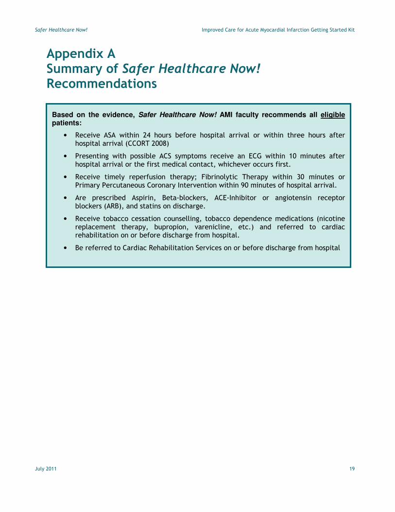
Based on the evidence, Safer Healthcare Now! AMI faculty recommends all eligible patients:
• Receive ASA within 24 hours before hospital arrival or within three hours after hospital arrival (CCORT 2008)
• Presenting with possible ACS symptoms receive an ECG within 10 minutes after hospital arrival or the first medical contact, whichever occurs first.
• Receive timely reperfusion therapy; Fibrinolytic Therapy within 30 minutes or Primary Percutaneous Coronary Intervention within 90 minutes of hospital arrival.
• Are prescribed Aspirin, Beta-blockers, ACE-Inhibitor or angiotensin receptor blockers (ARB), and statins on discharge.
• Receive tobacco cessation counselling, tobacco dependence medications (nicotine replacement therapy, bupropion, varenicline, etc.) and referred to cardiac rehabilitation on or before discharge from hospital.
• Be referred to Cardiac Rehabilitation Services on or before discharge from hospital

Safer Healthcare Now! Improved Care for Acute Myocardial Infarction Getting Started Kit
July 2011 20
Appendix B Quality Improvement and Improved Care for Acute Myocardial Infarction
*This example uses the Model for Improvement
1. Secure Senior Leadership Commitment
Improving AMI Care requires clear commitment and direction from the highest level of the organization. Aligning and locating your improvement work with the strategic goals of the organization is an important first step. Visible senior leadership support, allocation of resources, patient centeredness and accountability demonstrate commitment to improved patient care.
2. Form a Team
Including the right people on a process improvement team is critical to a successful improvement effort. Many organizations are multi site and require an organizing structure that guides the overall implementation of AMI care. Consider examining your existing teams and how they might support or provide leadership in improving AMI care. Some organizations may have different teams (e.g., a management team to guide the process and provide support; a frontline team to implement and refine the process.)
At the clinical level you will require an implementation team that is focused on improving care at the facility level. The AMI bundle is design to address emergency care, inpatient treatment, discharge and rehabilitation. Teams should establish baseline measurement and focus their energy on addressing their most significant practice gaps.
A team for improving AMI care may include some or all of the following: • Senior Administrative leadership (executive sponsor)
• Chief of Cardiology
• Chief of Emergency Medicine
• Family Practice / Internal Medicine Physicians
• Client, family, caregiver
• Cardiac Care Nurses (frontline)
• Cardiology Technologist
• Paramedic
• Nursing Clinical Coordinator or Educator
• Case Manager
• Pharmacy Representative
• Quality Improvement and decision support personnel
• Cath Lab Representative
Safer Healthcare Now! Improved Care for Acute Myocardial Infarction Getting Started Kit
July 2011 21
3. Use the Model for Improvement to Accelerate Change
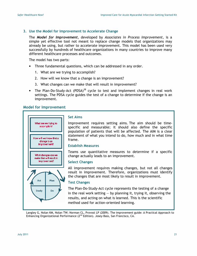
The Model for Improvement, developed by Associates in Process Improvement, is a simple yet effective tool not meant to replace change models that organizations may already be using, but rather to accelerate improvement. This model has been used very successfully by hundreds of healthcare organizations in many countries to improve many different healthcare processes and outcomes.
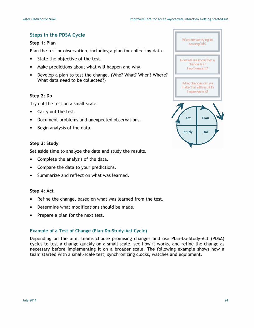
The model has two parts:
• Three fundamental questions, which can be addressed in any order.
1. What are we trying to accomplish?
2. How will we know that a change is an improvement?
3. What changes can we make that will result in improvement?
• The Plan-Do-Study-Act (PDSA)38 cycle to test and implement changes in real work settings. The PDSA cycle guides the test of a change to determine if the change is an improvement.
Model for Improvement
Set Aims
Improvement requires setting aims. The aim should be time-specific and measurable; it should also define the specific population of patients that will be affected. The AIM is a clear statement of what you intend to do, how much and in what time frame.
Establish Measures
Teams use quantitative measures to determine if a specific change actually leads to an improvement.
Select Changes
All improvement requires making changes, but not all changes result in improvement. Therefore, organizations must identify the changes that are most likely to result in improvement.
Test Changes
The Plan-Do-Study-Act cycle represents the testing of a change
in the real work setting — by planning it, trying it, observing the
results, and acting on what is learned. This is the scientific
method used for action-oriented learning.
Langley G, Nolan KM, Nolan TW; Norman CL, Provost LP (2009). The improvement guide: A Practical Approach to Enhancing Organizational Performance (2nd Edition). Josey-Bass, San Francisco, CA.

Safer Healthcare Now! Improved Care for Acute Myocardial Infarction Getting Started Kit
July 2011 22
A. Set Aims (Goals and Objectives)
Improvement requires setting aims. An organization will not improve without a clear and firm intention to do so. Agreeing on the aim is crucial; so is allocating the people and resources necessary to accomplish the aim.
Setting an aim can assist teams to focus on and build consensus about what they are hoping to achieve. The aim should be time-specific, measurable and define the specific population of clients who will be affected.
The following are examples of AIMS:
• Reduce inpatient AMI mortality by 25% by implementing all evidence-based care components by December 31, 2012.
• 100% of eligible patients will receive thrombolytic agents within or before 30 minutes of hospital arrival by January 15, 2012.
• 100% of eligible patients will receive tobacco cessation interventions before discharge by June 30, 2011.
As teams work on different aspects along the patient care process, the aims should be specific to what they are hoping to achieve in a given time.
B. Establish Measures
Measurement is a critical part of testing and implementing changes; measures tell a team whether the changes they are making actually lead to improvement.
Measurement for improvement starts with collecting baseline data to allow a comparison between current performance and optimal or preferred performance in relation to the elements of care being measured. Baseline measurement should facilitate local engagement and identify opportunities for improvement. Ongoing measurement allows the team and organization to track, trend and monitor progress over time.
C. Select Changes
While all changes do not lead to improvement, all improvement requires change. The ability to develop, test and implement changes is essential for any individual, group, or organization that wants to continuously improve. Change concepts can be used to generate. There are many kinds of changes that will lead to improvement, but these specific changes are developed from a limited number of change concepts.
Change concepts are categories of “general ideas that have proven merit with sound scientific and logical foundations.”39 Change concepts are intended to stimulate specific local ideas for changes intended to lead to improvement. The specific ideas for change are then tested using small rapid cycle tests of change, or PDSAs. Within your clinical area, you work with your team to Plan tests of change, Do small tests,
Safer Healthcare Now! Improved Care for Acute Myocardial Infarction Getting Started Kit
July 2011 23
Study the results, and make decisions to Act. You may act by adopting, adjusting or abandoning the change. The cycle repeats itself until you implement changes that allow you to reach and maintain your desired aim or goal.
D. Test Changes
Creatively combining change concepts with knowledge about specific clinical work and care environment can help generate ideas for tests of change. After generating ideas, run Plan-Do-Study-Act (PDSA) cycles to test a change or group of changes on a small scale to see if they result in improvement. If they do, expand the tests and gradually incorporate larger and larger samples until you are confident that the changes should be adopted more widely.
Reasons to Test Changes
• To adapt and customize general principles and evidence to the local practice context.
• To increase your belief that the change will result in improvement.
• To decide which of several proposed changes will lead to the desired improvement.
• To evaluate how much improvement can be expected from the change.
• To decide whether the proposed change will work in the actual environment of interest.
• To decide which combinations of changes will have the desired effects on the important measures of quality.
• To evaluate costs, social impact, and side effects from a proposed change.
• To minimize resistance upon implementation.
Safer Healthcare Now! Improved Care for Acute Myocardial Infarction Getting Started Kit
July 2011 24
Steps in the PDSA Cycle
Step 1: Plan
Plan the test or observation, including a plan for collecting data.
• State the objective of the test.
• Make predictions about what will happen and why.
• Develop a plan to test the change. (Who? What? When? Where? What data need to be collected?)
Step 2: Do
Try out the test on a small scale.
• Carry out the test.
• Document problems and unexpected observations.
• Begin analysis of the data.
Step 3: Study
Set aside time to analyze the data and study the results.
• Complete the analysis of the data.
• Compare the data to your predictions.
• Summarize and reflect on what was learned.
Step 4: Act
• Refine the change, based on what was learned from the test.
• Determine what modifications should be made.
• Prepare a plan for the next test.
Example of a Test of Change (Plan-Do-Study-Act Cycle)
Depending on the aim, teams choose promising changes and use Plan-Do-Study-Act (PDSA) cycles to test a change quickly on a small scale, see how it works, and refine the change as necessary before implementing it on a broader scale. The following example shows how a team started with a small-scale test; synchronizing clocks, watches and equipment.

Safer Healthcare Now! Improved Care for Acute Myocardial Infarction Getting Started Kit
July 2011 25
PLAN:
• What change idea are you willing to test by next Tuesday? o Test Synchronizing clocks, watches and equipment (individual, departmental,
and equipment)
• What do you expect (predict) will happen? o Equipment, watches and clocks will need to be adjusted. o The protocol will need refinements o The huddle will identify new change ideas for testing
• How will you know your change is leading to an improvement? (small measure) o Time needs adjustment to be synchronized o Protocol tested did (not) account for all times i.e. mobile equipment off unit o During Huddle staff ID changes
• Who will be involved? o Mary RN; Jane Team Lead; Susan Unit Manager; John RN
• When specifically do you plan to carry out the test? o Monday at 0830
• Where will it take place? o ED
• How will it be done? o Identify time ‘data points’ in patient flow thru ED o Audit current clocks and equipment for current time o Audit a sample staff watch time o Design and test a protocol for daily synchronization of clocks, watches and
equipment o Arrange a huddle o Complete the “testing” worksheet
ACT
• Will you adopt, adapt or abandon the change? o Designate one clock and time for synchronization
• How does this connect with your next test of change? o All ED staff, ECG technicians and physicians will be asked to set their time in
sync with the clock in the ED trauma room
DO
• Describe what actually happened: o Most clocks and watches were easily reset but equipment posed more of a
challenge because some were off unit at the time.
STUDY
• What did you learn from this test? o 3 of 5 clocks; 4 of 6 watches; and every piece of equipment needed the time
adjusted
Safer Healthcare Now! Improved Care for Acute Myocardial Infarction Getting Started Kit
July 2011 26
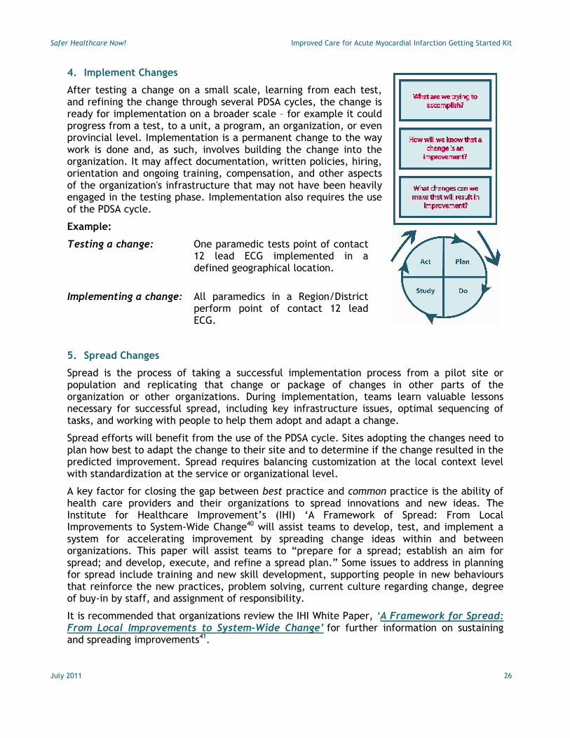
4. Implement Changes
After testing a change on a small scale, learning from each test, and refining the change through several PDSA cycles, the change is ready for implementation on a broader scale – for example it could progress from a test, to a unit, a program, an organization, or even provincial level. Implementation is a permanent change to the way work is done and, as such, involves building the change into the organization. It may affect documentation, written policies, hiring, orientation and ongoing training, compensation, and other aspects of the organization's infrastructure that may not have been heavily engaged in the testing phase. Implementation also requires the use of the PDSA cycle.
Example:
Testing a change: One paramedic tests point of contact 12 lead ECG implemented in a defined geographical location.
Implementing a change: All paramedics in a Region/District perform point of contact 12 lead ECG.
5. Spread Changes
Spread is the process of taking a successful implementation process from a pilot site or population and replicating that change or package of changes in other parts of the organization or other organizations. During implementation, teams learn valuable lessons necessary for successful spread, including key infrastructure issues, optimal sequencing of tasks, and working with people to help them adopt and adapt a change.
Spread efforts will benefit from the use of the PDSA cycle. Sites adopting the changes need to plan how best to adapt the change to their site and to determine if the change resulted in the predicted improvement. Spread requires balancing customization at the local context level with standardization at the service or organizational level.
A key factor for closing the gap between best practice and common practice is the ability of health care providers and their organizations to spread innovations and new ideas. The Institute for Healthcare Improvement’s (IHI) ‘A Framework of Spread: From Local Improvements to System-Wide Change40 will assist teams to develop, test, and implement a system for accelerating improvement by spreading change ideas within and between organizations. This paper will assist teams to “prepare for a spread; establish an aim for spread; and develop, execute, and refine a spread plan.” Some issues to address in planning for spread include training and new skill development, supporting people in new behaviours that reinforce the new practices, problem solving, current culture regarding change, degree of buy-in by staff, and assignment of responsibility.
It is recommended that organizations review the IHI White Paper, ‘A Framework for Spread: From Local Improvements to System-Wide Change’ for further information on sustaining and spreading improvements41.

Safer Healthcare Now! Improved Care for Acute Myocardial Infarction Getting Started Kit
July 2011 27
Appendix C Measurement: Improved AMI Care
Measuring Performance for Improvement
Measuring quality using a consistent set of measures evaluates the improvement strategy, identifies positive or negative effects on the organization and helps secure clinical, managerial and senior administrative support. Measuring is the only way to know if the actions you take actually lead to an improvement. Measure on an ongoing basis:
• Start by collecting baseline data prior to the implementation of the AMI intervention measures. All new teams should submit their data directly to the Safer Healthcare Now! Central Measurement Team using the new web-based Patient Safety Metric System. Go to the Safer Healthcare Now! home page www.saferhealthcarenow.ca , and click on the Measurement button “Submit Your Data”. After submitting data you will be able to generate reports for monitoring your own performance against your goal and comparing it to other teams. Go to the ‘Support’ tab on the Patient Safety Metrics website to learn more and access the user manual or contact your Safety and Improvement Advisor for assistance.
• If you are currently submitting your data using excel worksheets all your data has been transferred to the Patient Safety Metric System and you should contact the Central Measurement Team or your SIA for directions on how to enter your data directly on line. Data submitted on excel worksheets will not be accepted after December 2011.
• Remember to report your data to your team and key stakeholders within your organization.
• Using your baseline data decide with your improvement team the aspects of AMI care that will be the focus of your improvement work. Measurement data should also be presented to the organization’s senior leadership and clinicians monthly at baseline, during early implementation stages, and less frequently during the full implementation stage, for monitoring improvement.
• You may decide to use all measures, focus on initial care, in hospital care or discharge depending on your baseline assessment and available resources. Measure frequently, using small samples over time to trend your progress toward established goals.
• If measurement does not reflect improvement, your team should investigate the reason why (e.g., processes which are not working, non-compliance to these processes and/or existence of barriers which prevent the process from working effectively).
Collecting Baseline Data
Collecting baseline data is important when initiating any quality improvement activity to determine the effectiveness of the improvement. This means collecting measurement data using current processes before the introduction of changes.

Safer Healthcare Now! Improved Care for Acute Myocardial Infarction Getting Started Kit
July 2011 28
When to Collect Data
Measurement data should be collected monthly to provide opportunities for timely analysis in order to identify areas for improvement. Efforts should be made to integrate measurement into routine clinical documentation that can easily extract for ongoing monitoring and feedback.
Tools and Approaches to Data Collection
The selection or development of tools for data collection will depend on available resources. The organization needs to determine a way to capture data, aggregate it for data submission and trending, analyse and interpret the data, and display and share your results.
Sample tools include available on the AMI Community of Practice (CoP) include:
• Moncton data collection sheet following the patient from initial contact to discharge.
• Cape Breton Regional Hospitals excel tally sheet and graphs.
Visit the AMI Community of Practice (CoP) for these and other tools, tips and forms. Click here http://tools.patientsafetyinstitute.ca/Communities/ami/default.aspx
Measurement Data Collection Strategies, Tips and Tools
Safer Healthcare Now! recommends that before your facility, team or unit begins implementing the intervention, you obtain baseline data on each care component of the bundle. Baseline data will give you a sense of where you are starting from, and what some of the potential areas of focus are for your facility or team. We suggest that you take a “snapshot” of three months or more, or whatever is practical and feasible for your organization.
For ongoing measurement, Safer Healthcare Now! recommends calculating each of the AMI measures on a monthly or quarterly basis, to enable facilities to track the progress of their quality improvement initiatives. The following suggestions consider the number of eligible patients treated at your hospital.
1. Select up to 15 consecutive eligible AMI patients each month if AMI volume at your institution makes this possible; /or
2. Select a minimum of 5 consecutive eligible AMI patients each quarter if AMI volume at you institution makes this possible; /or
3. Submit all eligible AMI patients each quarter.
Safer Healthcare Now! Improved Care for Acute Myocardial Infarction Getting Started Kit
July 2011 29
Concurrent data collection while patients are still in the hospital is the preferred process for measurement and quality improvement.
This strategy allows for the identification of real time improvement opportunities so that mitigation can occur before discharge. Hospitals collecting data have found that the process works best if documentation of the care components is integrated into clinical documentation.). Use of clinical orders sets for admission and discharge that allow for selection of appropriate care components and rationale for contraindications allow the patients treatment to be easily documented and communicated. Standardizing clinical documentation can also serve as a trigger to consider various treatment options during the patients care episode. Examples of forms can be located on the AMI Community of Practice at http://tools.patientsafetyinstitute.ca/Communities/ami/Lists/Clinical%20Paths%20%20Order%20Sets/AllItems.aspx
Retrospective chart review
Appropriate patients can be identified using the required data elements in administrative data and health records. Detailed descriptions of the patient population appropriate for each of the indicators follows. A hospital information system (e.g. ADT, abstracting, etc.) may be able to identify the patients from all discharges by sorting based on these elements. Another alternative is to work with the coding or health records department to identify the patients at the time of coding and prepare a list or set aside records for review. After the patients have been identified, manual review of the medical record will be required to look for documentation that each appropriate intervention was either provided or contraindicated. If documentation for either cannot be found, the measure should be considered as not being met.
CIHI project field
Several CIHI databases, including DAD and NACRS, have “project” fields. These fields allow each hospital to collect additional information related to a patient’s hospitalization. Hospitals may opt to enter the required AMI data in a pre-defined project field in the CIHI abstract. This approach:
• May reduce the data collection burden, as several data elements required for the measures (e.g. patient age and diagnosis, transfer in or out status, deaths, discharged against medical advice, etc.) are already documented in the abstract
• Builds on an existing process for multi-site data collection
• May prove to be a cost-effective and timely alternative to chart reviews
Coding guidelines for the CIHI project fields are available. Organizations are encouraged to consult with their CIHI field office for assistance with application of the guidelines.
Those opting for concurrent data collection (whether or not they use the CIHI project field) will need to check that they are collecting data on all appropriate patients. This group can be determined after discharge and coding, using an algorithm. Any cases missed and not identified until after discharge will require a manual retrospective review.

Safer Healthcare Now! Improved Care for Acute Myocardial Infarction Getting Started Kit
July 2011 30
Track Measures over Time
Improvement takes place over time. Determining if improvement has really occurred and if it is a lasting effect requires observing patterns over time. Run charts show data over time and are one of the single most important tools in performance improvement. Using run charts has a variety of benefits:
• They help improvement teams formulate aims by depicting how well (or poorly) a team is performing relative to a specific process.
• They help in determining when changes are truly improvements by displaying a pattern of data that you can observe and monitor as you make changes.
• They give direction as you work on improvement and information about the value of particular changes.
• Staff in your Node or at the Central Measurement Team will be pleased to work with you on getting your measurement working for you and your team.
• Your Region’s contact information is available at: http://www.saferhealthcarenow.ca/EN/about/WhoWeAre/Pages/default.aspx
• Staff of the Central Measurement Team can be contacted by email at [email protected]
Safer Healthcare Now! Improved Care for Acute Myocardial Infarction Getting Started Kit
July 2011 31
Appendix D Measurement Technical Descriptions All measurement worksheets for AMI are located at: www.saferhealthcarenow.ca/EN/Interventions/ami/Pages/measurement.aspx
Technical Description of the Measurement Worksheets:
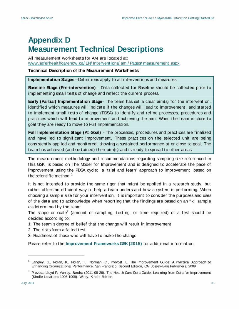
Implementation Stages – Definitions apply to all interventions and measures
Baseline Stage (Pre-intervention) - Data collected for Baseline should be collected prior to implementing small tests of change and reflect the current process.
Early (Partial) Implementation Stage- The team has set a clear aim(s) for the intervention, identified which measures will indicate if the changes will lead to improvement, and started to implement small tests of change (PDSA) to identify and refine processes, procedures and practices which will lead to improvement and achieving the aim. When the team is close to goal they are ready to move to Full Implementation.
Full Implementation Stage (At Goal) - The processes, procedures and practices are finalized and have led to significant improvement. These practices on the selected unit are being consistently applied and monitored, showing a sustained performance at or close to goal. The team has achieved (and sustained) their aim(s) and is ready to spread to other areas.
The measurement methodology and recommendations regarding sampling size referenced in this GSK, is based on The Model for Improvement and is designed to accelerate the pace of improvement using the PDSA cycle; a "trial and learn" approach to improvement based on the scientific method.1
It is not intended to provide the same rigor that might be applied in a research study, but rather offers an efficient way to help a team understand how a system is performing. When choosing a sample size for your intervention, it is important to consider the purposes and uses of the data and to acknowledge when reporting that the findings are based on an “x” sample as determined by the team. The scope or scale2 (amount of sampling, testing, or time required) of a test should be decided according to: 1. The team’s degree of belief that the change will result in improvement 2. The risks from a failed test 3. Readiness of those who will have to make the change
Please refer to the Improvement Frameworks GSK (2015) for additional information.
1 Langley, G., Nolan, K., Nolan, T., Norman, C., Provost, L. The Improvement Guide: A Practical Approach to
Enhancing Organizational Performance. San Francisco, Second Edition, CA. Jossey-Bass Publishers. 2009 2 Provost, Lloyd P; Murray, Sandra (2011-08-26). The Health Care Data Guide: Learning from Data for Improvement
(Kindle Locations 1906-1909). Wiley. Kindle Edition
Safer Healthcare Now! Improved Care for Acute Myocardial Infarction Getting Started Kit
July 2011 32
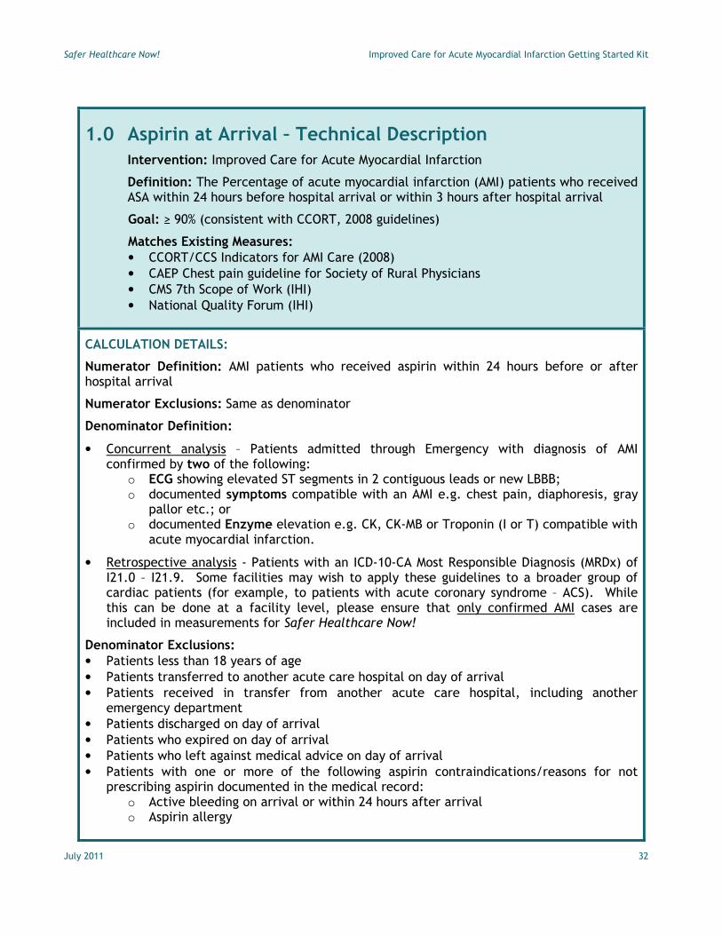
1.0 Aspirin at Arrival – Technical Description
Intervention: Improved Care for Acute Myocardial Infarction
Definition: The Percentage of acute myocardial infarction (AMI) patients who received ASA within 24 hours before hospital arrival or within 3 hours after hospital arrival
Goal: ≥ 90% (consistent with CCORT, 2008 guidelines)
Matches Existing Measures: • CCORT/CCS Indicators for AMI Care (2008) • CAEP Chest pain guideline for Society of Rural Physicians • CMS 7th Scope of Work (IHI) • National Quality Forum (IHI)
CALCULATION DETAILS:
Numerator Definition: AMI patients who received aspirin within 24 hours before or after hospital arrival
Numerator Exclusions: Same as denominator
Denominator Definition:
• Concurrent analysis – Patients admitted through Emergency with diagnosis of AMI confirmed by two of the following:
o ECG showing elevated ST segments in 2 contiguous leads or new LBBB; o documented symptoms compatible with an AMI e.g. chest pain, diaphoresis, gray
pallor etc.; or o documented Enzyme elevation e.g. CK, CK-MB or Troponin (I or T) compatible with
acute myocardial infarction.
• Retrospective analysis - Patients with an ICD-10-CA Most Responsible Diagnosis (MRDx) of I21.0 – I21.9. Some facilities may wish to apply these guidelines to a broader group of cardiac patients (for example, to patients with acute coronary syndrome – ACS). While this can be done at a facility level, please ensure that only confirmed AMI cases are included in measurements for Safer Healthcare Now!
Denominator Exclusions:
• Patients less than 18 years of age • Patients transferred to another acute care hospital on day of arrival • Patients received in transfer from another acute care hospital, including another
emergency department • Patients discharged on day of arrival • Patients who expired on day of arrival • Patients who left against medical advice on day of arrival • Patients with one or more of the following aspirin contraindications/reasons for not
prescribing aspirin documented in the medical record: o Active bleeding on arrival or within 24 hours after arrival o Aspirin allergy

Safer Healthcare Now! Improved Care for Acute Myocardial Infarction Getting Started Kit
July 2011 33
o Coumadin/warfarin as pre-arrival medication
o Other reasons documented by a physician, nurse practitioner, or physician assistant for not giving aspirin within 24 hours before or after hospital arrival
Measurement Period Length and Sample Size:
• Concurrent Sampling: Select 15 consecutive AMI patients as defined above, each month if AMI volume at your institution makes this possible. If volume allows, Safer Healthcare Now! recommends obtaining a weekly sample of patients that meet the concurrent or retrospective definitions for AMI. Rotate the day and time of weekly sampling strategy and be sure to include weekends and nights. This is an on-going weekly measure. Hospitals with a lower AMI volume may have to select the sample over a 3-month period, reporting quarterly.
• Retrospective Sampling: With the support of your Health Records Department staff select a sample of 15 consecutive AMI patients discharged within this month with an ICD-10-CA MRDx of I21.0 – I21.9. Patient volume, coding practices and policies regarding completing charts may make it difficult to select this sample from this month making it necessary to select the sample over a 3-month period and reporting quarterly.
Definition of Terms:
• Hospital Arrival: The earliest documented date the patient arrived at the hospital; this may differ from the admission time
• AMI Patients: • Patients identified retrospectively who at discharge had an ICD-10-CA Most
Responsible Diagnosis (MRD) of I21.0 – I21.9.
• Patients identified concurrently who are admitted through Emergency with diagnosis of AMI confirmed by two of the following: o ECG showing elevated ST segments in 2 contiguous leads or new LBBB; o documented symptoms compatible with an AMI e.g. chest pain, diaphoresis, gray
pallor etc.; or o documented Enzyme elevation e.g. CK, CK-MB or Troponin (I or T) compatible with
acute myocardial infarction.
Calculate as:
Numerator / Denominator; as a percentage of AMI patients who received aspirin within 24 hours before or 3 hours after hospital arrival.
Comments:
• Do not double count the patient in this measure. If the patient received ASA within 24 hours prior to arrival and within 24 hours following arrival (s)he meets the standard of care and is counted once only.

Safer Healthcare Now! Improved Care for Acute Myocardial Infarction Getting Started Kit
July 2011 34
2.0 Aspirin at Discharge – Technical Description
Intervention: Improved Care for Acute Myocardial Infarction
Definition: The percentage of acute myocardial infarction (AMI) patients who are prescribed aspirin at hospital discharge
Goal: ≥ 90% (consistent with CCORT, 2008 guidelines)
Matches Existing Measures: • CCORT/CCS Indicators for AMI Care (2008) • JCAHO Core Measure AMI-1 (IHI) • CMS 7th Scope of Work (IHI) • National Quality Forum (IHI)
CALCULATION DETAILS:
Numerator Definition: AMI patients who are prescribed aspirin at hospital discharge
Numerator Exclusions: Same as denominator
Denominator Definition:
• Concurrent analysis – Patients admitted through Emergency with diagnosis of AMI confirmed by two of the following:
o ECG showing elevated ST segments in 2 contiguous leads or new LBBB; o documented symptoms compatible with an AMI e.g. chest pain, diaphoresis, gray
pallor etc.; or o documented Enzyme elevation e.g. CK, CK-MB or Troponin (I or T) compatible with
acute myocardial infarction.
• Retrospective analysis - Patients with an ICD-10-CA Most Responsible Diagnosis (MRDx) of I21.0 – I21.9. Some facilities may wish to apply these guidelines to a broader group of cardiac patients (for example, to patients with acute coronary syndrome – ACS). While this can be done at a facility level, please ensure that only confirmed AMI cases are included in measurements for Safer Healthcare Now!
Denominator Exclusions:
• Patients less than 18 years of age • Patients transferred to another acute care hospital • Patients who expired • Patients who left against medical advice • Patients with one or more of the following aspirin contraindications/reasons for not
prescribing aspirin documented in the medical record: o Active bleeding o Aspirin allergy o Coumadin/warfarin prescribed at discharge o Other reasons documented by a physician, nurse practitioner, or physician
assistant for not giving aspirin at discharge
Safer Healthcare Now! Improved Care for Acute Myocardial Infarction Getting Started Kit
July 2011 35
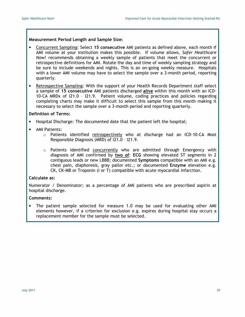
Measurement Period Length and Sample Size:
• Concurrent Sampling: Select 15 consecutive AMI patients as defined above, each month if AMI volume at your institution makes this possible. If volume allows, Safer Healthcare Now! recommends obtaining a weekly sample of patients that meet the concurrent or retrospective definitions for AMI. Rotate the day and time of weekly sampling strategy and be sure to include weekends and nights. This is an on-going weekly measure. Hospitals with a lower AMI volume may have to select the sample over a 3-month period, reporting quarterly.
• Retrospective Sampling: With the support of your Health Records Department staff select a sample of 15 consecutive AMI patients discharged alive within this month with an ICD-10-CA MRDx of I21.0 – I21.9. Patient volume, coding practices and policies regarding completing charts may make it difficult to select this sample from this month making it necessary to select the sample over a 3-month period and reporting quarterly.
Definition of Terms:
• Hospital Discharge: The documented date that the patient left the hospital;
• AMI Patients: o Patients identified retrospectively who at discharge had an ICD-10-CA Most
Responsible Diagnosis (MRD) of I21.0 – I21.9.
o Patients identified concurrently who are admitted through Emergency with diagnosis of AMI confirmed by two of: ECG showing elevated ST segments in 2 contiguous leads or new LBBB; documented Symptoms compatible with an AMI e.g. chest pain, diaphoresis, gray pallor etc.; or documented Enzyme elevation e.g. CK, CK-MB or Troponin (I or T) compatible with acute myocardial infarction.
Calculate as:
Numerator / Denominator; as a percentage of AMI patients who are prescribed aspirin at hospital discharge.
Comments:
• The patient sample selected for measure 1.0 may be used for evaluating other AMI elements however, if a criterion for exclusion e.g. expires during hospital stay occurs a replacement member for the sample must be selected.

Safer Healthcare Now! Improved Care for Acute Myocardial Infarction Getting Started Kit
July 2011 36
3.0 Beta Blocker Prescribed at Discharge – Technical Description
Intervention: Improved Care for Acute Myocardial Infarction
Definition: The percentage of acute myocardial infarction (AMI) patients who are prescribed a beta blocker at hospital discharge
Goal: ≥ 90% (consistent with CCORT, 2008 guidelines)
Matches Existing Measures: • CCORT/CCS Indicators for AMI Care (2003) • CMS 7th Scope of Work (IHI) • National Quality Forum (IHI)
CALCULATION DETAILS:
Numerator Definition: AMI patients who are prescribed a beta blocker at hospital discharge
Numerator Exclusions: Same as denominator
Denominator Definition:
• Concurrent analysis – Patients admitted through Emergency with diagnosis of AMI confirmed by two of the following:
o ECG showing elevated ST segments in 2 contiguous leads or new LBBB; o documented symptoms compatible with an AMI e.g. chest pain, diaphoresis, gray
pallor etc.; or o documented Enzyme elevation e.g. CK, CK-MB or Troponin (I or T) compatible with
acute myocardial infarction.
• Retrospective analysis - Patients with an ICD-10-CA Most Responsible Diagnosis (MRDx) of I21.0 – I21.9. Some facilities may wish to apply these guidelines to a broader group of cardiac patients (for example, to patients with acute coronary syndrome – ACS). While this can be done at a facility level, please ensure that only confirmed AMI cases are included in measurements for Safer Healthcare Now!
Denominator Exclusions:
• Patients less than 18 years of age • Patients transferred to another acute care hospital • Patients who expired • Patients who left against medical advice • Patients with one or more of the following beta blocker contraindications/reasons for not
prescribing beta blocker documented in the medical record: o Beta blocker allergy o Bradycardia (heart rate less than 60 bpm) on day of discharge or day prior to
discharge while not on a beta blocker o Second or third degree heart block on ECG on arrival or during hospital stay and
does not have a pacemaker o Systolic blood pressure less than 90 mm Hg on day of discharge or day prior to
discharge while not on a beta blocker
Safer Healthcare Now! Improved Care for Acute Myocardial Infarction Getting Started Kit
July 2011 37
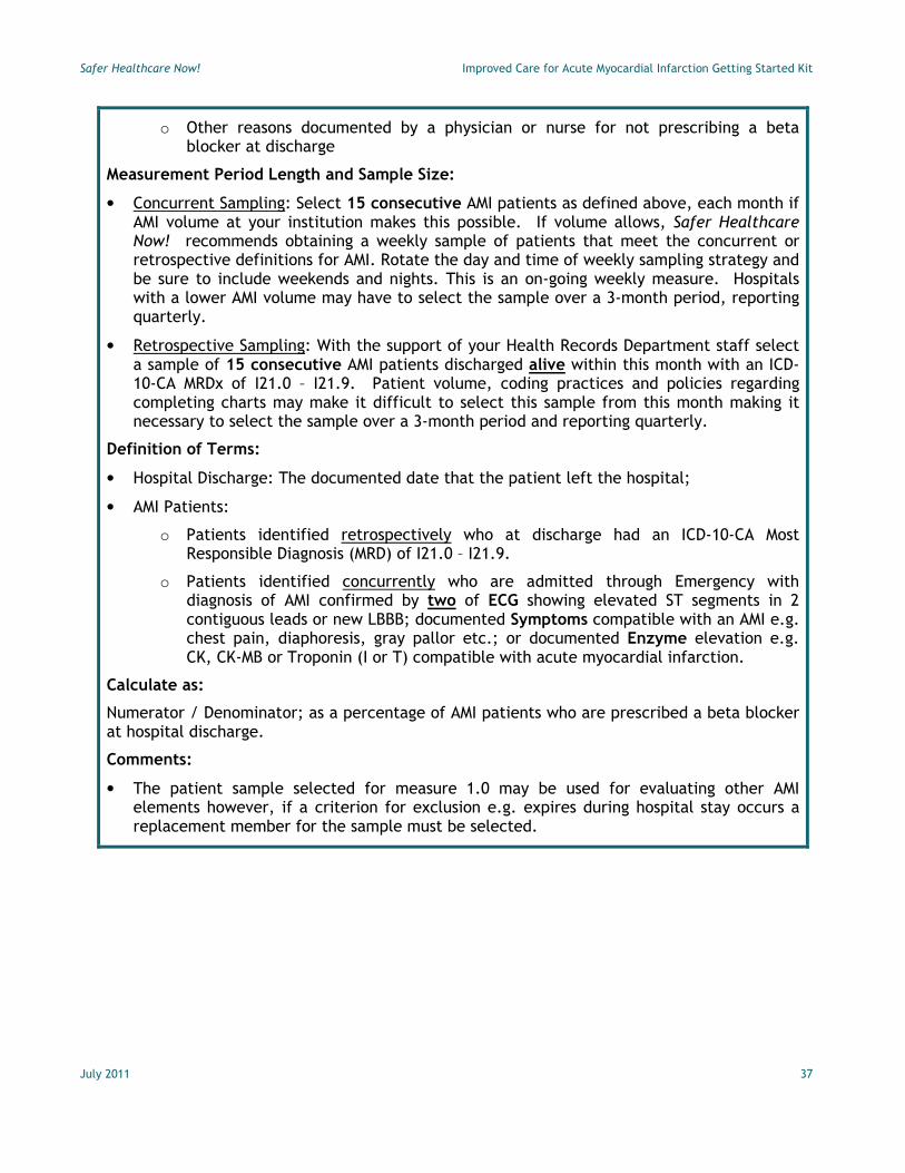
o Other reasons documented by a physician or nurse for not prescribing a beta blocker at discharge
Measurement Period Length and Sample Size:
• Concurrent Sampling: Select 15 consecutive AMI patients as defined above, each month if AMI volume at your institution makes this possible. If volume allows, Safer Healthcare Now! recommends obtaining a weekly sample of patients that meet the concurrent or retrospective definitions for AMI. Rotate the day and time of weekly sampling strategy and be sure to include weekends and nights. This is an on-going weekly measure. Hospitals with a lower AMI volume may have to select the sample over a 3-month period, reporting quarterly.
• Retrospective Sampling: With the support of your Health Records Department staff select a sample of 15 consecutive AMI patients discharged alive within this month with an ICD-10-CA MRDx of I21.0 – I21.9. Patient volume, coding practices and policies regarding completing charts may make it difficult to select this sample from this month making it necessary to select the sample over a 3-month period and reporting quarterly.
Definition of Terms:
• Hospital Discharge: The documented date that the patient left the hospital;
• AMI Patients:
o Patients identified retrospectively who at discharge had an ICD-10-CA Most Responsible Diagnosis (MRD) of I21.0 – I21.9.
o Patients identified concurrently who are admitted through Emergency with diagnosis of AMI confirmed by two of ECG showing elevated ST segments in 2 contiguous leads or new LBBB; documented Symptoms compatible with an AMI e.g. chest pain, diaphoresis, gray pallor etc.; or documented Enzyme elevation e.g. CK, CK-MB or Troponin (I or T) compatible with acute myocardial infarction.
Calculate as:
Numerator / Denominator; as a percentage of AMI patients who are prescribed a beta blocker at hospital discharge.
Comments:
• The patient sample selected for measure 1.0 may be used for evaluating other AMI elements however, if a criterion for exclusion e.g. expires during hospital stay occurs a replacement member for the sample must be selected.

Safer Healthcare Now! Improved Care for Acute Myocardial Infarction Getting Started Kit
July 2011 38
4.0-A Thrombolytic Agent Received Within 30 Minutes of Hospital Arrival – Technical Description
Intervention: Improved Care for Acute Myocardial Infarction
Definition: The percentage of acute myocardial infarction (AMI) patients (STEMI or new LBBB only) receiving thrombolytic therapy during the hospital stay and having a time from hospital arrival to thrombolysis of 30 minutes or less.
Goal: ≥ 90% (consistent with CCORT, 2008 guidelines)
Matches Existing Measures:
• CCORT/CCS Indicators for AMI Care (2008)
• CAEP Chest pain guideline for Society of Rural Physicians
• JCAHO Core Measures AMI – 7a (IHI)
• CMS 7th Scope of Work (IHI)
• National Quality Forum (IHI)
CALCULATION DETAILS:
Numerator Definition: AMI patients with ST elevation (STEMI) or new LBBB on ECG who received thrombolytic therapy whose time from hospital arrival to thrombolysis is 30 minutes or less.
Numerator Exclusions: Same as denominator
Denominator Definition:
• Concurrent analysis: o Patients admitted through Emergency with diagnosis of AMI and, o ECG showing elevated ST segments (STEMI) in 2 contiguous leads or new LBBB and, o Symptoms compatible with an AMI e.g. chest pain, diaphoresis, gray pallor etc.; or
• Retrospective analysis: o Patients with an ICD-10-CA Most Responsible Diagnosis (MRDx) of I21.0 – I21.3; I21.9
and, o ST elevation (STEMI) or new LBBB on ECG and, o Received thrombolytic therapy (CCI Code 1.ZZ.35.HA-1C) o Some facilities may wish to apply these guidelines to a broader group of cardiac
patients (for example, to patients with acute coronary syndrome – ACS). While this can be done at a facility level, please ensure that only confirmed AMI cases are included in measurements for Safer Healthcare Now!
o Enzyme elevation e.g. CK, CK-MB or Troponin (I or T) compatible with acute myocardial infarction and,
o Received thrombolytic therapy
Denominator Exclusions:
• Patients with NSTEMI, non-Q wave or subendocardial MIs
• Patients less than 18 years of age
• Patients transferred in from another acute care hospital including another emergency department

Safer Healthcare Now! Improved Care for Acute Myocardial Infarction Getting Started Kit
July 2011 39
Measurement Period Length and Sample Size:
• Concurrent Sampling: Select 15 consecutive AMI patients as defined above, each month if AMI volume at your institution makes this possible. Be sure to select patients who demonstrate ST elevation (STEMI) or new LBBB on the earliest ECG and either symptoms or enzyme elevation. If volume allows, Safer Healthcare Now! recommends obtaining a weekly sample of patients that meet the concurrent or retrospective definitions for AMI. Rotate the day and time of weekly sampling strategy and be sure to include weekends and nights. This is an on-going weekly measure. Hospitals with a lower AMI volume may have to select the sample over a 3-month period, reporting quarterly. If using the original monthly AMI sample selected you may have to over-sample for thrombolytic patients in order to obtain an adequate sample – this is a decision should be made by your AMI team.
• Retrospective Sampling: With the support of your Health Records Department staff select a sample of 15 consecutive AMI patients discharged within this month with an ICD-10-CA MRDx of I21.0 – I21.3; I21.9 and received thrombolytic therapy and had ST elevation (STEMI) or new LBBB (ICD-10-CA code I44.4-I44.7) on the earliest ECG. It may be necessary to select more than 15 patient charts which meet the coding and thrombolysis criteria in order to fulfill the ECG criterion for inclusion. Patient volume, coding practices and policies regarding completing charts may make it difficult to select this sample from this month making it necessary to select the sample over a 3-month period and reporting quarterly. If using the original monthly AMI sample selected you may have to over-sample for thrombolytic patients in order to obtain an adequate sample – this is a decision should be made by your AMI team.
Definition of Terms:
• AMI Patients with ST elevation (STEMI) or new LBBB on ECG who received thrombolytic therapy - Patients discharged with all of the following:
o Patients identified retrospectively who at discharge had an ICD-10-CA Most Responsible Diagnosis (MRD) of I21.0 – I21.3; I21.9 and,
o ST Segment elevation or new LBBB on the ECG performed closest to hospital arrival, and,
o Thrombolytic therapy within 6 hours after hospital arrival (CCI Code 1.ZZ.35.HA-C1)
• AMI Patients with ST elevation (STEMI) or new LBBB on ECG who received thrombolytic therapy - Patients discharged with all of the following:
o Patients identified concurrently who are admitted through Emergency with a diagnosis of AMI (STEMI) and
o ST Segment elevation or new LBBB on the ECG performed closest to hospital arrival, and,
o Symptoms compatible with AMI or Enzyme elevation o Thrombolytic therapy within 6 hours after hospital arrival
• Arrival: The earliest documented time the patient arrived at the hospital; this may differ from the admission time
• ST segment elevation or new LBBB on ECG - see “Comments” below

Safer Healthcare Now! Improved Care for Acute Myocardial Infarction Getting Started Kit
July 2011 40
Calculate as:
Numerator / Denominator; as a percentage of AMI patients receiving a thrombolytic agent who received it within 30 minutes of arrival.
Comments:
Use the following rules for abstraction of ST segment elevation or new LBBB on ECG:
• Use the 12-lead ECG performed closest to the time of hospital arrival, whether prior to or after hospital arrival (e.g., 12-lead ECG done in the ambulance 10 minutes before hospital arrival and a second one done in the ED 30 minutes after arrival – use the ECG done in the ambulance). If there is no interpretation available from the 12-lead ECG performed closest to the time of hospital, select “No”. Do not use an interpretation from another ECG performed that may be available.
• Do NOT use ECGs done more than 1 hour prior to hospital arrival.
• This information must be taken from the interpretation. An ECG interpretation is defined as:
o A 12-lead ECG report in which the name or initials of the physician or nurse who reviewed the ECG is signed, stamped, or typed on the report, or
o Physician or nurse notation of ECG findings in another source (e.g., progress notes).
• Interpretations must be taken directly from documentation of ECG findings. Do not measure ST segments or attempt to identify a new LBBB on the ECG tracing.
• If the ECG report is not specifically labelled “12-lead”, infer that it was 12-lead if lead markings (i.e., I, II, III, aVR, aVL, aVF, V1, V2, V3, V4, V5, V6) are noted on the report.
• If the physician/nurse references ECG findings but does not specify the ECG was 12-lead, infer that it was 12-lead, unless documentation indicates otherwise.
• If unable to determine which 12-lead ECG was done closest to arrival (e.g., one ECG does not have a time, and it cannot be determined whether it is closer to hospital arrival than another ECG which does have a time), or if the time between the pre-arrival and post-arrival ECG is the same (e.g., both were done15 minutes from hospital arrival time), include the patient if any of these ECGs have ST segment elevation or new LBBB documented on the interpretation.
• If the location of an MI is documented and it is described as acute/evolving, or an acute/evolving MI is described as “STEMI” or “transmural” or “Q wave”, the presumption is being made that it is an ST elevation MI.
• Do not consider “subendocardial” an MI “location” (e.g. “acute subendocardial MI” should be excluded). (Exclude ICD-10-CA code I21.40 – I21.49)
• Consider “infarct” synonymous with myocardial infarction (e.g., “acute inferior infarct”) should be included.
• MIs MUST be described as acute or evolving (in addition to documentation of location or description of MI as “transmural” or “Q wave”). Do NOT include MIs specified as old or previously seen, where the age is documented as undetermined (e.g., “inferior MI age undetermined”, “Extensive anterior infarct, age indeterminate”, “anterolateral MI on or before 09-01-2004”), or where age is not addressed in any manner (e.g., “Q wave MI”).

Safer Healthcare Now! Improved Care for Acute Myocardial Infarction Getting Started Kit
July 2011 41
“New” should not be considered synonymous with “acute”. “Evolving” should be considered synonymous with “acute”.
• Exclude the patient when both an inclusion and exclusion are documented in reference to the same ECG, or documentation is otherwise conflicting. Consider documentation as conflicting if there is documentation of both an included term and excluded term (per inclusion/exclusion lists or Notes for Abstraction) or documentation of an included term with additional documentation, which clearly contradicts the inclusion term. Examples:
o Signed ECG report lists “new LBBB” and “non Q wave MI” o The ER physician reports “ST elevation” on the initial ECG, while the attending
cardiologist interprets this same ECG as “No ST elevation” o Signed ECG report notes ST segment = .05mV, which the physician labels “ST
elevation”
• If there is documentation of an included term and documentation of a finding which is not addressed in the inclusion/exclusion lists or Notes for Abstraction, this should NOT be considered conflicting documentation. In the following examples, “Yes” should be selected:
o Signed ECG report notes “probable lateral injury”, while the physician’s progress note states “ST elevation present”
o Findings of “posterior AMI” and “ST depression” are noted on the signed ECG report
• LBBBs described as old should not be included. An old LBBB pattern obscures the ability of the ECG to develop recognizable ST elevation, impairing diagnosis of acute MI. Under this uncertainty, these cases should be treated with reperfusion.
• The term “ST abnormality” should not be considered synonymous with “ST elevation”.
Use the following rules for abstraction of thrombolytic agent administration:
• Patients arriving in the Emergency Department via ambulance with thrombolytic therapy infusing at the time of arrival are to be included.
• Patients arriving in the Emergency Department via ambulance and thrombolytic therapy were infused during transport but completed prior to the time of hospital arrival are to be excluded.
• Patients suffering an AMI post admission (i.e. on nursing unit) and receiving thrombolysis are to be excluded.
• Thrombolysis is not indicated in NON-STEMI or Sub-endocardial MIs
• By the time you have completed applying all exclusions (e.g. transfers in, transfers out) to your monthly AMI patient sample which include both STEMIs and Non-STEMIs you may not have a sufficient number of STEMI patients eligible for inclusion in measure 4A or B.
• You should consider selecting a different patient sample for measure 4A and 4B limiting the sample to the STEMI infarcts only.

Safer Healthcare Now! Improved Care for Acute Myocardial Infarction Getting Started Kit
July 2011 42
4.0-B Primary Percutaneous Coronary Intervention (PCI) Received Within 90 Minutes of Hospital Arrival – Technical Description
Intervention: Improved Care for Acute Myocardial Infarction
Definition: The percentage of acute myocardial infarction (AMI) patients (STEMI or new LBBB only) who received Primary Percutaneous Coronary Intervention (PCI) within 90 minutes or less from hospital arrival to PCI
Goal: ≥ 90% (consistent with CCORT, 2008 guidelines)
Matches Existing Measures: (None)
CALCULATION DETAILS:
Numerator Definition: AMI patients with ST elevation (STEMI) or new LBBB on ECG who received Primary PCI whose time from hospital arrival to PCI was 90 minutes or less.
Numerator Exclusions: Same as denominator
Denominator Definition:
• Concurrent analysis: o Patients admitted through Emergency with diagnosis of AMI and, o ECG showing elevated ST segments (STEMI) in 2 contiguous leads or new LBBB and, o Symptoms compatible with an AMI e.g. chest pain, diaphoresis, gray pallor etc.; or o Enzyme elevation e.g. CK, CK-MB or Troponin (I or T) compatible with acute
myocardial infarction and, o Received a Primary PCI
• Retrospective analysis: o Patients with an ICD-10-CA Most Responsible Diagnosis (MRDx) of I21.0 – I21.3; I21.9
and, o ST elevation (STEMI) or new LBBB (ICD-10-CA code I44.4-I44.7) on ECG and, o Received a Primary PCI (CCI code 1.IJ.50)
Some facilities may wish to apply these guidelines to a broader group of cardiac patients (for example, to patients with acute coronary syndrome – ACS). While this can be done at a facility level, please ensure that only confirmed AMI cases are included in measurements for Safer Healthcare Now!
Denominator Exclusions:
• Patients with NSTEMI, non Q wave or subendocardial MIs
• Patients less than 18 years of age
• Patients transferred in from another acute care hospital including another emergency department
• Patients who received a thrombolytic agent administration

Safer Healthcare Now! Improved Care for Acute Myocardial Infarction Getting Started Kit
July 2011 43
Measurement Period Length and Sample Size:
• Concurrent Sampling: Select 15 consecutive AMI patients as defined above, each month if AMI volume at your institution makes this possible. Be sure to select patients who demonstrate ST elevation (STEMI) or new LBBB on the earliest ECG and either symptoms or enzyme elevation. If volume allows, Safer Healthcare Now! recommends obtaining a weekly sample of patients that meet the concurrent or retrospective definitions for AMI. Rotate the day and time of weekly sampling strategy and be sure to include weekends and nights. This is an on-going weekly measure. Hospitals with a lower AMI volume may have to select the sample over a 3-month period, reporting quarterly. If using the original monthly AMI sample selected you may have to over sample for Primary PCI patients in order to obtain an adequate sample – this is a decision should be made by your AMI team.
• Retrospective Sampling: With the support of your Health Records Department staff select a sample of 15 consecutive AMI patients discharged within this month with an ICD-10-CA MRDx of I21.0 – I21.3; I21.9 and received a Primary PCI (CCI code 1.IJ.50) and had ST elevation or new LBBB (ICD-10-CA code I44.4-I44.7) on ECG. It may be necessary to select more than 15 patient charts, which meet the coding, and primary PCI criteria in order to fulfill the ECG criterion for inclusion. Patient volume, coding practices and policies regarding completing charts may make it difficult to select this sample from this month making it necessary to select the sample over a 3-month period and reporting quarterly. If using the original monthly AMI sample selected you may have to over-sample for Primary PCI patients in order to obtain an adequate sample – this is a decision should be made by your AMI team.
Definition of Terms:
• AMI Patients with ST elevation (STEMI) or new LBBB on ECG who received thrombolytic - Patients discharged with all of the following:
o Patients identified retrospectively who at discharge had an ICD-10-CA Most Responsible Diagnosis (MRD) of I21.0 – I21.3; I21.9 and,
o Primary PCI (CCI Code 1.IJ.50.^^) Dilation, coronary arteries) and, o ST Segment elevation or new LBBB (ICD-10-CA code I44.4-I44.7) on the ECG
performed closest to hospital arrival, and, o Primary PCI performed within 24 hours after hospital arrival (CCI code 1.IJ.50)
• AMI Patients with ST elevation (STEMI) or new LBBB on ECG who received thrombolytic - Patients discharged with all of the following:
o Patients identified concurrently who are admitted through Emergency with a diagnosis of AMI and
o ST Segment elevation (STEMI) or new LBBB on the ECG performed closest to hospital arrival, and,
o Symptoms compatible with AMI or Enzyme elevation o PCI performed within 24 hours after hospital arrival
• Arrival: The earliest documented time the patient arrived at the hospital; this may differ from the admission time
ST segment elevation (STEMI) or new LBBB (ICD-10-CA code I44.4-I44.7) on ECG - see “Comments” below

Safer Healthcare Now! Improved Care for Acute Myocardial Infarction Getting Started Kit
July 2011 44
Calculate as:
Numerator / Denominator; as a percentage of AMI patients receiving a Primary PCI who received it within 90 minutes of arrival.
Comments:
Use the following rules for abstraction of ST segment elevation or new LBBB on ECG:
• Use the 12-lead ECG performed closest to the time of hospital arrival, whether prior to or after hospital arrival (e.g., 12-lead ECG done in the ambulance 10 minutes before hospital arrival and a second one done in the ED 30 minutes after arrival – use the ECG done in the ambulance). If there is no interpretation available from the 12-lead ECG performed closest to the time of hospital, select “No”. Do not use an interpretation from another ECG performed that may be available.
• Do NOT use ECGs done more than 1 hour prior to hospital arrival.
• This information must be taken from the interpretation. An ECG interpretation is defined as:
o A 12-lead ECG report in which the name or initials of the physician or nurse who reviewed the ECG is signed, stamped, or typed on the report, or
o Physician or nurse notation of ECG findings in another source (e.g., progress notes).
• Interpretations must be taken directly from documentation of ECG findings. Do not measure ST segments or attempt to identify a new LBBB on the ECG tracing.
• If the ECG report is not specifically labelled “12-lead”, infer that it was 12-lead if lead markings (i.e., I, II, III, aVR, aVL, aVF, V1, V2, V3, V4, V5, V6) are noted on the report.
• If the physician or nurse references ECG findings but does not specify the ECG was 12-lead, infer that it was 12-lead, unless documentation indicates otherwise.
• If unable to determine which 12-lead ECG was done closest to arrival (e.g., one ECG does not have a time, and it cannot be determined whether it is closer to hospital arrival than another ECG which does have a time), or if the time between the pre-arrival and post-arrival ECG is the same (e.g., both were done15 minutes from hospital arrival time), include the patient any of these ECGs have ST segment elevation or new LBBB documented on the interpretation.
• If the location of an MI is documented and it is described as acute/evolving, or an acute/evolving MI is described as “STEMI” or “transmural” or “Q wave”, the presumption is being made that it is an ST elevation MI.
• Do not consider “subendocardial” an MI “location” (e.g. “acute subendocardial MI” should be excluded). (Exclude ICD-10-CA code I21.40 – I21.49)
• Consider “infarct” synonymous with myocardial infarction (e.g., “acute inferior infarct”) should be included.
• MIs MUST be described as acute or evolving (in addition to documentation of location or description of MI as “transmural” or “Q wave”). DO NOT include MIs specified as old or previously seen, where the age is documented as undetermined (e.g., “inferior MI age

Safer Healthcare Now! Improved Care for Acute Myocardial Infarction Getting Started Kit
July 2011 45
undetermined”, “Extensive anterior infarct, age indeterminate”, “anterolateral MI on or before 09-01-2004”), or where age is not addressed in any manner (e.g., “Q wave MI”).
“New” should not be considered synonymous with “acute”. “Evolving” should be considered synonymous with “acute”.
• Exclude the patient when the PCI is performed as a “salvage” procedure.
• Exclude the patient when both an inclusion and exclusion are documented in reference to the same ECG, or documentation is otherwise conflicting. Consider documentation as conflicting if there is documentation of both an included term and excluded term (per inclusion/exclusion lists or Notes for Abstraction) or documentation of an included term with additional documentation, which clearly contradicts the inclusion term. Examples:
o Signed ECG report lists “LBBB” and “non Q wave MI” o The ER physician reports “ST elevation” on the initial ECG, while the attending
cardiologist interprets this same ECG as “No ST elevation” o Signed ECG report notes ST segment = .05mV, which the physician labels “ST
elevation”
• If there is documentation of an included term and documentation of a finding which is not addressed in the inclusion/exclusion lists or Notes for Abstraction, this should NOT be considered conflicting documentation. In the following examples, “Yes” should be selected:
o Signed ECG report notes “probable lateral injury”, while the physician’s progress note states “ST elevation present”
o Findings of “posterior AMI” and “ST depression” are noted on the signed ECG report
• LBBBs described as old should be included. An old LBBB pattern obscures the ability of the ECG to develop recognizable ST elevation, impairing diagnosis of acute MI. Under this uncertainty, these cases should be treated with reperfusion.
• The term “ST abnormality” should not be considered synonymous with “ST elevation”.

Safer Healthcare Now! Improved Care for Acute Myocardial Infarction Getting Started Kit
July 2011 46
5.0 ACE-Inhibitor or Angiotensin Receptor Blockers (ARB) Prescribed at Discharge – Technical Description
Intervention: Improved Care for Acute Myocardial Infarction
Definition: The percentage of acute myocardial infarction (AMI) patients with Q-wave infarction, hypertension, diabetes or left ventricular systolic dysfunction who were prescribed an ACE-inhibitor or Angiotensin Receptor Blockers (ARB) at discharge from hospital.
Goal: ≥ 90% (consistent with CCORT, 2008 guidelines)
Matches Existing Measures:
• CCORT/CCS Indicators for AMI Care (2008)
CALCULATION DETAILS:
Numerator Definition: AMI patients with Q-wave infarction, hypertension, diabetes or left ventricular systolic dysfunction who are prescribed an ACE-inhibitor or angiotensin receptor blockers (ARB) at hospital discharge.
Numerator Exclusions: Same as denominator
Denominator Definition:
• Concurrent analysis – Patients admitted through Emergency with diagnosis of AMI confirmed by two of ECG showing elevated ST segments in 2 contiguous leads or new LBBB; documented symptoms compatible with an AMI e.g. chest pain, diaphoresis, gray pallor etc.; or documented Enzyme elevation e.g. CK, CK-MB or Troponin (I or T) compatible with acute myocardial infarction and chart documentation of with Q-wave infarction, hypertension, diabetes or left ventricular systolic dysfunction
• Chart documentation of left ventricular systolic dysfunction ICD-10-CA code I51.8 or I50 with evidence of heart failure.
• Retrospective analysis - Patients with an ICD-10-CA Most Responsible Diagnosis (MRDx) of I21.0 – I21.9. Some facilities may wish to apply these guidelines to a broader group of cardiac patients (for example, to patients with acute coronary syndrome – ACS). While this can be done at a facility level, please ensure that only confirmed AMI cases are included in measurements for Safer Healthcare Now!
Denominator Exclusions:
• Patients less than 18 years of age
• Patients transferred to another acute care hospital
• Patients who expired
• Patients who left against medical advice
• Patients with a chart documentation of participation in clinical trials testing alternatives to ACE-Is or ARBs as first-line therapy

Safer Healthcare Now! Improved Care for Acute Myocardial Infarction Getting Started Kit
July 2011 47
• Patients with one or more of the following ACE-Inhibitors contraindications/reasons for not prescribing ACE Inhibitor or ARB documented in the medical record:
o Moderate or severe aortic stenosis o Allergy or intolerance to ACE inhibitors or ARBs o Severe renal dysfunction (i.e., peak or last prehospital discharge serum creatinine
level >200 µmol/L) o Systolic blood pressure <100 mmHg at discharge o Bilateral renal artery stenosis o Hyperkalemia (ie, peak or last prehospital discharge K+ >5.5 mmol/L) o Physician documented reason for nonuse of ACE inhibitor at discharge (eg, patient
refusal, symptomatic hypotension)
Measurement Period Length and Sample Size:
• Concurrent Sampling: Select 15 consecutive AMI patients as defined above, each month if AMI volume at your institution makes this possible. If volume allows, Safer Healthcare Now! recommends obtaining a weekly sample of patients that meet the concurrent or retrospective definitions for AMI. Rotate the day and time of weekly sampling strategy and be sure to include weekends and nights. This is an on-going weekly measure. Hospitals with a lower AMI volume may have to select the sample over a three-month period, reporting quarterly.
• Retrospective Sampling: With the support of your Health Records Department staff select a sample of 15 consecutive AMI patients discharged alive within this month with an ICD-10-CA MRDx of I21.0 – I21.9 and ICD-10-CA code for Q-wave infarction, hypertension, diabetes or left ventricular systolic dysfunction. Patient volume, coding practices and policies regarding completing charts may make it difficult to select this sample from this month making it necessary to select the sample over a 3-month period and reporting quarterly.
Definition of Terms:
• Hospital Discharge: The documented date that the patient left the hospital;
• AMI Patients:
o Patients identified retrospectively who at discharge had an ICD-10-CA Most Responsible Diagnosis (MRD) of I21.0 – I21.9 and chart documentation of left ventricular systolic dysfunction ICD-10-CA code for Q-wave infarction, hypertension, diabetes or left ventricular systolic dysfunction
o Patients identified concurrently who are admitted through Emergency with diagnosis of AMI confirmed by two of ECG showing elevated ST segments in 2 contiguous leads or new LBBB; documented Symptoms compatible with an AMI e.g. chest pain, diaphoresis, gray pallor etc.; or documented Enzyme elevation e.g. CK, CK-MB or Troponin (I or T) compatible with acute myocardial infarction and chart documentation of Q-wave infarction, hypertension, diabetes or left ventricular systolic dysfunction
• Pathological Q waves occur if they are 25% or more of the height of the partner R wave and/or they are greater than 0.04 seconds in width - one small square - and greater than 2mm (two small squares) in depth. Although not always evident in the early stages of and AMI they usually confirm the event has occurred.

Safer Healthcare Now! Improved Care for Acute Myocardial Infarction Getting Started Kit
July 2011 48
• Hypertension is defined as having one or more of a number of criteria including: a diagnosis of hypertension recorded by the physician; antihypertensive medications prescribed; blood pressure >140/90mmHg or > 130/80 in patients with diabetes or renal disease and >125/75 in those with proteinuria.
• Diabetes is defined as having one or more of a number of criteria including a diagnosis of diabetes recorded by a physician; a fasting plasma glucose of >7.0 mmol/l or two-hour glucose level of >11.1 mmol/l following an oral glucose tolerance test.
• Left Ventricular Systolic Dysfunction (LVSD): A left ventricular ejection fraction (LVEF) less than 40%, or a narrative of left ventricular function consistent with moderate or severe systolic dysfunction. ICD-10-CA code I51.8
Calculate as:
Numerator / Denominator; as a percentage of AMI patients who are prescribed an ACE Inhibitor or ARB at hospital discharge.
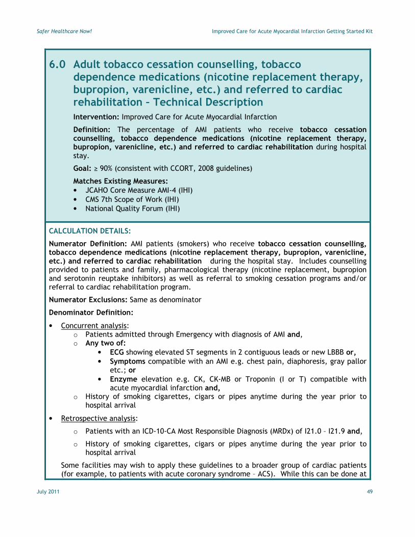
Safer Healthcare Now! Improved Care for Acute Myocardial Infarction Getting Started Kit
July 2011 49
6.0 Adult tobacco cessation counselling, tobacco dependence medications (nicotine replacement therapy, bupropion, varenicline, etc.) and referred to cardiac rehabilitation – Technical Description
Intervention: Improved Care for Acute Myocardial Infarction
Definition: The percentage of AMI patients who receive tobacco cessation counselling, tobacco dependence medications (nicotine replacement therapy, bupropion, varenicline, etc.) and referred to cardiac rehabilitation during hospital stay.
Goal: ≥ 90% (consistent with CCORT, 2008 guidelines)
Matches Existing Measures: • JCAHO Core Measure AMI-4 (IHI) • CMS 7th Scope of Work (IHI) • National Quality Forum (IHI)
CALCULATION DETAILS:
Numerator Definition: AMI patients (smokers) who receive tobacco cessation counselling, tobacco dependence medications (nicotine replacement therapy, bupropion, varenicline, etc.) and referred to cardiac rehabilitation during the hospital stay. Includes counselling provided to patients and family, pharmacological therapy (nicotine replacement, bupropion and serotonin reuptake inhibitors) as well as referral to smoking cessation programs and/or referral to cardiac rehabilitation program.
Numerator Exclusions: Same as denominator
Denominator Definition:
• Concurrent analysis: o Patients admitted through Emergency with diagnosis of AMI and, o Any two of:
• ECG showing elevated ST segments in 2 contiguous leads or new LBBB or, • Symptoms compatible with an AMI e.g. chest pain, diaphoresis, gray pallor
etc.; or • Enzyme elevation e.g. CK, CK-MB or Troponin (I or T) compatible with
acute myocardial infarction and, o History of smoking cigarettes, cigars or pipes anytime during the year prior to
hospital arrival
• Retrospective analysis:
o Patients with an ICD-10-CA Most Responsible Diagnosis (MRDx) of I21.0 – I21.9 and,
o History of smoking cigarettes, cigars or pipes anytime during the year prior to hospital arrival
Some facilities may wish to apply these guidelines to a broader group of cardiac patients (for example, to patients with acute coronary syndrome – ACS). While this can be done at

Safer Healthcare Now! Improved Care for Acute Myocardial Infarction Getting Started Kit
July 2011 50
a facility level, please ensure that only confirmed AMI cases are included in measurements for Safer Healthcare Now!
Denominator Exclusions:
• Patients less than 18 years of age
• Patients transferred out to another acute care hospital
• Patients who expired
• Patients who left against medical advice
Measurement Period Length and Sample Size:
• Concurrent Sampling: Select 15 consecutive AMI patients as defined above, each month if AMI volume at your institution makes this possible. Be sure to select patients who have a history of smoking cigarettes anytime during this year. Clearly established criteria for what constitutes counselling for smoking cessation and/or pharmacologic intervention may be concurrently validated against the chart, Kardex, and discussions with the staff. If volume allows, Safer Healthcare Now! recommends obtaining a weekly sample of patients that meet the concurrent or retrospective definitions for AMI. Rotate the day and time of weekly sampling strategy and be sure to include weekends and nights. This is an on-going weekly measure. Hospitals with a lower AMI volume may have to select the sample over a 3-month period, reporting quarterly.
• Retrospective Sampling: With the support of your Health Records Department staff select a sample of 15 consecutive AMI patients discharged alive within this month with an ICD-10-CA MRDx of I21.0 – I21.9 and had a history of smoking cigarettes anytime during the previous year. It may be necessary to select more than 15 patient charts which meet the coding criteria in order to fulfill the smoking criterion for inclusion. Furthermore, the AMI Team should clearly specify the documentation required for smoking counselling and/or nicotine replacement in order meet the criterion (see below). Patient volume, coding practices and policies regarding completing charts may make it difficult to select this sample from this month making it necessary to select the sample over a 3-month period and reporting quarterly.
Definition of Terms:
• AMI Patients:
o Patients identified retrospectively who at discharge had an ICD-10-CA Most Responsible Diagnosis (MRD) of I21.0 – I21.9.
o Patients identified concurrently who are admitted through Emergency with diagnosis of AMI confirmed by two of ECG showing elevated ST segments in 2 contiguous leads or new LBBB; documented Symptoms compatible with an AMI e.g. chest pain, diaphoresis, gray pallor etc.; or documented Enzyme elevation e.g. CK, CK-MB or Troponin (I or T) compatible with acute myocardial infarction.
• Smoking: history of smoking cigarettes, cigars or pipes anytime during the year prior to hospital arrival
• Pharmacologic Intervention: nicotine replacement therapy, serotonin reuptake inhibitors or other drug therapy for withdrawal.

Safer Healthcare Now! Improved Care for Acute Myocardial Infarction Getting Started Kit
July 2011 51
• Smoking cessation counselling: Involves a recognized program that may include patient video, information brochure or referral to formal program e.g. the Lung Association. Smoking cessation counselling is a component of cardiac rehabilitation. At discharge it is recommended that all AMI patients be referred to a multidisciplinary cardiac rehabilitation program or an appropriate medical ambulatory clinic which integrates cardiac rehabilitation care if available.
Calculate as:
Numerator / Denominator; as a percentage of AMI patients with a history of smoking within this year who received counselling for smoking cessation and/or pharmacologic therapy during the hospital stay.

Safer Healthcare Now! Improved Care for Acute Myocardial Infarction Getting Started Kit
July 2011 52
7.0 Perfect care for AMI – Technical Description
Intervention: Improved Care for Acute Myocardial Infarction
Definition: The percentage of acute myocardial infarction (AMI) patients who received all appropriate AMI evidence-based elements.
Goal: 95% or more of all AMI receive all elements of the AMI Bundle in the absence of contraindications.
Matches Existing Measures: none
CALCULATION DETAILS:
Numerator Definition: AMI patients who either received all of the following elements or had documentation of a contraindication to a medication, transferred in or out of the healthcare organization etc. The AMI elements for perfect care are:
• Early administration of aspirin
• Aspirin at discharge
• Beta Blocker prescribed at discharge
• Timely administration of thrombolytics or percutaneous coronary intervention (PCI)
• ACE-inhibitor or angiotensin receptor blockers (ARB) at discharge
• Smoking cessation counselling and or Pharmacologic therapy
• Statins at discharge
• ECG obtained within 10 minutes of hospital arrival
• Discharge Referral to cardiac Rehabilitation
Numerator Exclusions: Same as denominator
Denominator Definition:
• Concurrent analysis – Patients admitted through Emergency with diagnosis of AMI confirmed by two of ECG showing elevated ST segments in 2 contiguous leads or new LBBB; documented symptoms compatible with an AMI e.g. chest pain, diaphoresis, gray pallor etc.; or documented Enzyme elevation e.g. CK, CK-MB or Troponin (I or T) compatible with acute myocardial infarction.
• Retrospective analysis - Patients with an ICD-10-CA Most Responsible Diagnosis (MRDx) of I21.0 – I21.9. Some facilities may wish to apply these guidelines to a broader group of cardiac patients (for example, to patients with acute coronary syndrome – ACS). While this can be done at a facility level, please ensure that only confirmed AMI cases are included in measurements for Safer Healthcare Now!
Denominator Exclusions:
• Patients less than 18 years of age
Safer Healthcare Now! Improved Care for Acute Myocardial Infarction Getting Started Kit
July 2011 53
• Death in emergency department
• Transferred in or out and not returned to the hospital of origin within 24 hours unless there is an agreement between hospitals or within regions regarding which hospital will count these patients
Measurement Period Length and Sample Size:
• Concurrent Sampling: Select 15 consecutive AMI patients as defined above, each month if AMI volume at your institution makes this possible. If volume allows Safer Healthcare Now! recommends obtaining a weekly sample of patients that meet the concurrent or retrospective definitions for AMI. Rotate the day and time of weekly sampling strategy and be sure to include weekends and nights. This is an on-going weekly measure. Hospitals with a lower AMI volume may have to select the sample over a 3-month period, reporting quarterly.
• Retrospective Sampling: With the support of your Health Records Department staff select a sample of 15 consecutive AMI patients discharged within this month with an ICD-10-CA MRDx of I21.0 – I21.9. Patient volume, coding practices and policies regarding completing charts may make it difficult to select this sample from this month making it necessary to select the sample over a 3-month period and reporting quarterly.
Definition of Terms:
• AMI Patients:
o Patients identified concurrently who are admitted through Emergency with diagnosis of AMI confirmed by two of ECG showing elevated ST segments in 2 contiguous leads or new LBBB; documented Symptoms compatible with an AMI e.g. chest pain, diaphoresis, gray pallor etc.; or documented Enzyme elevation e.g. CK, CK-MB or Troponin (I or T) compatible with acute myocardial infarction.
o Patients identified retrospectively who at discharge had an ICD-10-CA Most Responsible Diagnosis (MRD) of I21.0 – I21.9.
Calculate as:
Numerator / Denominator; as a percentage of AMI patients receiving perfect care – all components provided (see below) or documented as contraindicated.
Comments:
Perfect care for AMI is defined as the provision of all 9 of the key elements, or documentation of clear contraindication. Patients are only counted as having received “perfect care” if all interventions are documented as given in appropriate time frames, or that clear contraindications existed. If documentation for any one item is missing, the patient is not considered as having received “perfect care”. When reporting this as a percentage, only those patients with documentation of perfect care are counted in the numerator. All patients with contraindications to a medication, transferred in or out of the healthcare organization etc. are considered to have met the inclusion criteria for Perfect Care.
Note: Transfer patients should still receive all of the key elements for AMI care. Both the sending and receiving hospitals should work together to ensure that this occurs.

Safer Healthcare Now! Improved Care for Acute Myocardial Infarction Getting Started Kit
July 2011 54
8.0 AMI Inpatient Mortality – Technical Description
Intervention: Improved Care for Acute Myocardial Infarction
Definition: The percentage of acute myocardial infarction (AMI) patients who died during hospital stay.
Goal: Decrease by 25%.
Matches Existing Measures: none
CALCULATION DETAILS:
Numerator Definition: AMI patients who died during the hospital stay.
Numerator Exclusions: Same as denominator
Denominator Definition:
• Retrospective analysis - Patients with an ICD-10-CA Most Responsible Diagnosis (MRDx) of I21.0 – I21.9.
Denominator Exclusions:
• Patients less than 18 years of age
• Patients transferred in from another acute care hospital, including another emergency department.
• Patients transferred out to another acute care hospital Patients less than 18 years of age
• Death in emergency department Measurement Period Length and Sample Size:
• Concurrent Sampling for this measure is challenging.
• Retrospective Sampling: With the support of your Health Records Department staff select a sample of all AMI patients discharged within this month with an ICD-10-CA MRDx of I21.0 – I21.9. Patient volume, coding practices and policies regarding completing charts may make it difficult to select this sample from this month making it necessary to select the sample over a 3-month period and reporting quarterly.
Definition of Terms:
• AMI Patients:
o Patients identified retrospectively who at discharge had an ICD-10-CA Most Responsible Diagnosis (MRD) of I21.0 – I21.9.
Calculate as:
Numerator / Denominator; as a percentage of AMI mortality.

Safer Healthcare Now! Improved Care for Acute Myocardial Infarction Getting Started Kit
July 2011 55
Comments:
Use the analytical capacity and support of your Health Records Department to determine the AMI admission and mortality rate for the preceding month. Although CIHI may be able to calculate this measure on your behalf coding practice and data submission may delay this process.
This is a useful measure to teams which are interested in monitoring their own performance improvement against baseline in a timely fashion.
The AMI faculty recommends you monitor your AMI inpatient mortality every month.
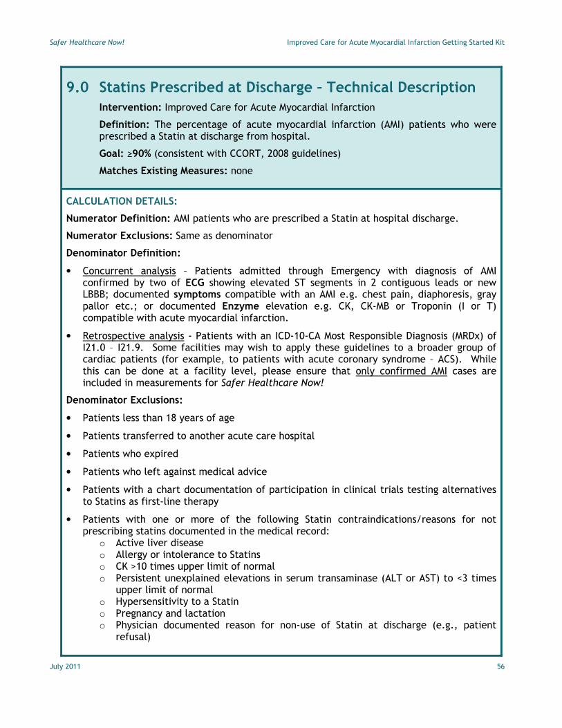
Safer Healthcare Now! Improved Care for Acute Myocardial Infarction Getting Started Kit
July 2011 56
9.0 Statins Prescribed at Discharge – Technical Description
Intervention: Improved Care for Acute Myocardial Infarction
Definition: The percentage of acute myocardial infarction (AMI) patients who were prescribed a Statin at discharge from hospital.
Goal: ≥90% (consistent with CCORT, 2008 guidelines)
Matches Existing Measures: none
CALCULATION DETAILS:
Numerator Definition: AMI patients who are prescribed a Statin at hospital discharge.
Numerator Exclusions: Same as denominator
Denominator Definition:
• Concurrent analysis – Patients admitted through Emergency with diagnosis of AMI confirmed by two of ECG showing elevated ST segments in 2 contiguous leads or new LBBB; documented symptoms compatible with an AMI e.g. chest pain, diaphoresis, gray pallor etc.; or documented Enzyme elevation e.g. CK, CK-MB or Troponin (I or T) compatible with acute myocardial infarction.
• Retrospective analysis - Patients with an ICD-10-CA Most Responsible Diagnosis (MRDx) of I21.0 – I21.9. Some facilities may wish to apply these guidelines to a broader group of cardiac patients (for example, to patients with acute coronary syndrome – ACS). While this can be done at a facility level, please ensure that only confirmed AMI cases are included in measurements for Safer Healthcare Now!
Denominator Exclusions:
• Patients less than 18 years of age
• Patients transferred to another acute care hospital
• Patients who expired
• Patients who left against medical advice
• Patients with a chart documentation of participation in clinical trials testing alternatives to Statins as first-line therapy
• Patients with one or more of the following Statin contraindications/reasons for not prescribing statins documented in the medical record:
o Active liver disease o Allergy or intolerance to Statins o CK >10 times upper limit of normal o Persistent unexplained elevations in serum transaminase (ALT or AST) to <3 times
upper limit of normal o Hypersensitivity to a Statin o Pregnancy and lactation o Physician documented reason for non-use of Statin at discharge (e.g., patient
refusal)

Safer Healthcare Now! Improved Care for Acute Myocardial Infarction Getting Started Kit
July 2011 57
Measurement Period Length and Sample Size:
• Concurrent Sampling: Select 15 consecutive AMI patients as defined above, each month if AMI volume at your institution makes this possible. If volume allows, Safer Healthcare Now! recommends obtaining a weekly sample of patients that meet the concurrent or retrospective definitions for AMI. Rotate the day and time of weekly sampling strategy and be sure to include weekends and nights. This is an on-going weekly measure. Hospitals with a lower AMI volume may have to select the sample over a 3-month period, reporting quarterly.
• Retrospective Sampling: With the support of your Health Records Department staff select a sample of 15 consecutive AMI patients discharged alive within this month with an ICD-10-CA MRDx of I21.0 – I21.9. Patient volume, coding practices and policies regarding completing charts may make it difficult to select this sample from this month making it necessary to select the sample over a 3-month period and reporting quarterly.
Definition of Terms:
• Hospital Discharge: The documented date that the patient left the hospital;
• AMI Patients:
o Patients identified retrospectively who at discharge had an ICD-10-CA Most Responsible Diagnosis (MRD) of I21.0 – I21.9.
o Patients identified concurrently who are admitted through Emergency with diagnosis of AMI confirmed by two of ECG showing elevated ST segments in 2 contiguous leads or new LBBB; documented Symptoms compatible with an AMI e.g. chest pain, diaphoresis, gray pallor etc.; or documented Enzyme elevation e.g. CK, CK-MB or Troponin (I or T) compatible with acute myocardial.
Calculate as:
Numerator / Denominator; as a percentage of AMI patients who are prescribed a Statin at hospital discharge.
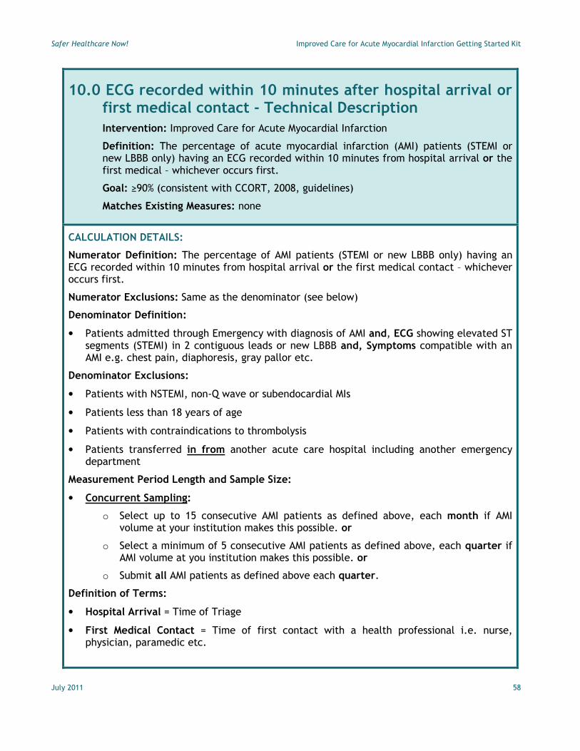
Safer Healthcare Now! Improved Care for Acute Myocardial Infarction Getting Started Kit
July 2011 58
10.0 ECG recorded within 10 minutes after hospital arrival or first medical contact - Technical Description
Intervention: Improved Care for Acute Myocardial Infarction
Definition: The percentage of acute myocardial infarction (AMI) patients (STEMI or new LBBB only) having an ECG recorded within 10 minutes from hospital arrival or the first medical – whichever occurs first.
Goal: ≥90% (consistent with CCORT, 2008, guidelines)
Matches Existing Measures: none
CALCULATION DETAILS:
Numerator Definition: The percentage of AMI patients (STEMI or new LBBB only) having an ECG recorded within 10 minutes from hospital arrival or the first medical contact – whichever occurs first.
Numerator Exclusions: Same as the denominator (see below)
Denominator Definition:
• Patients admitted through Emergency with diagnosis of AMI and, ECG showing elevated ST segments (STEMI) in 2 contiguous leads or new LBBB and, Symptoms compatible with an AMI e.g. chest pain, diaphoresis, gray pallor etc.
Denominator Exclusions:
• Patients with NSTEMI, non-Q wave or subendocardial MIs
• Patients less than 18 years of age
• Patients with contraindications to thrombolysis
• Patients transferred in from another acute care hospital including another emergency department
Measurement Period Length and Sample Size:
• Concurrent Sampling:
o Select up to 15 consecutive AMI patients as defined above, each month if AMI volume at your institution makes this possible. or
o Select a minimum of 5 consecutive AMI patients as defined above, each quarter if AMI volume at you institution makes this possible. or
o Submit all AMI patients as defined above each quarter.
Definition of Terms:
• Hospital Arrival = Time of Triage
• First Medical Contact = Time of first contact with a health professional i.e. nurse, physician, paramedic etc.
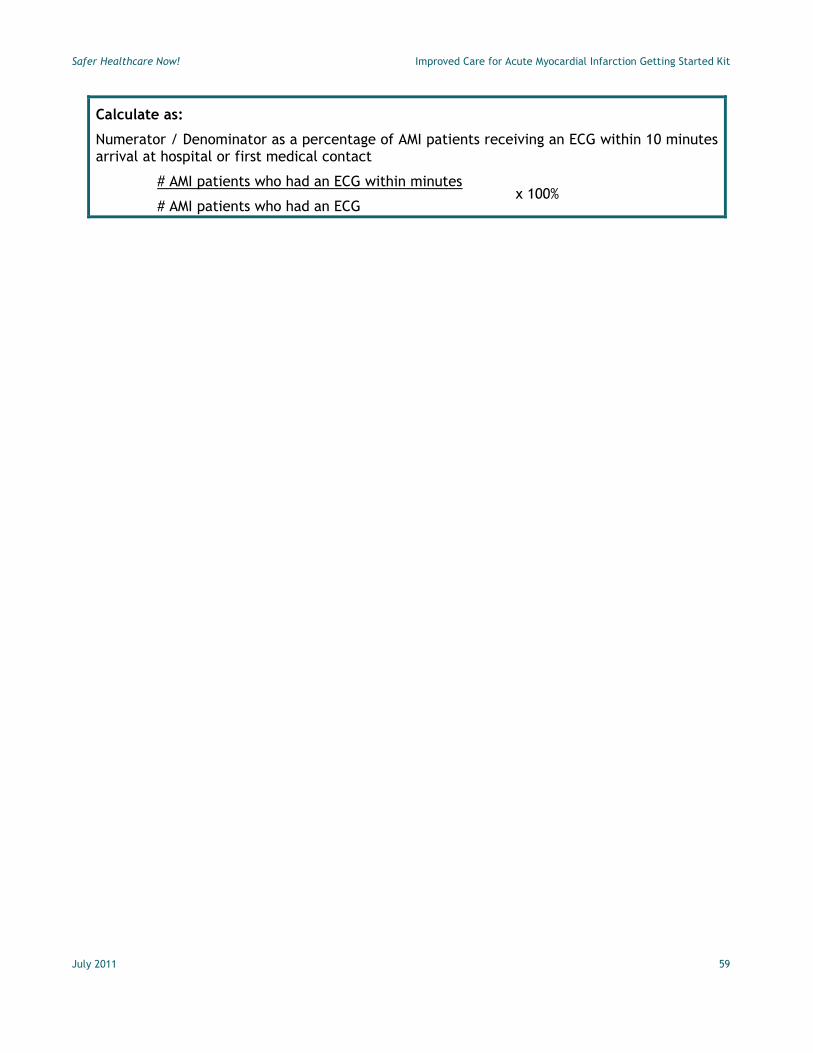
Safer Healthcare Now! Improved Care for Acute Myocardial Infarction Getting Started Kit
July 2011 59
Calculate as:
Numerator / Denominator as a percentage of AMI patients receiving an ECG within 10 minutes arrival at hospital or first medical contact
# AMI patients who had an ECG within minutes
# AMI patients who had an ECG x 100%

Safer Healthcare Now! Improved Care for Acute Myocardial Infarction Getting Started Kit
July 2011 60
11.0 Discharge Referral to Cardiac Rehabilitation - Technical Description
Intervention: Improved Care for Acute Myocardial Infarction
Definition: The percentage of eligible AMI patients referred to cardiac rehabilitation programs at discharge.
Goal: ≥90% (consistent with CCORT, 2008, guidelines)
Matches Existing Measures: • Canadian Association of Cardiac Rehabilitation (CACR) 2009 • AHA Get with the Guidelines • AACVPR/ACC/AHA Performance Measures for Cardiac Rehabilitation
CALCULATION DETAILS:
Numerator Definition: The number of eligible acute myocardial infarction (AMI) patients who are referred to cardiac rehabilitation at the time of hospital discharge.
Numerator Exclusions: Same as the denominator
Denominator Definition:
• Patients admitted through Emergency Departments with diagnosis of AMI and,
• Any two of:
o ECG showing elevated ST segments in 2 contiguous leads or new LBBB or,
o Symptoms compatible with an AMI e.g. chest pain, diaphoresis, gray pallor etc.; or
• Enzyme elevation e.g. CK, CK-MB or Troponin (I or T) compatible with acute myocardial infarction and,
Some facilities may wish to apply these guidelines to a broader group of cardiac patients (for example, to patients with acute coronary syndrome (ACS), chronic stable angina, or post procedure such as percutaneous coronary intervention, coronary artery bypass surgery, cardiac valve surgery, cardiac transplantation or cardiac resynchronization therapy). While this can be done at a facility level, please ensure that only confirmed AMI cases are included in measurements for Safer Healthcare Now!
Denominator Exclusions:
• Patients less than 18 years of age
• Patients transferred out to another acute care hospital and are not transferred back within 24hrs
• Patients who expired
• Patients who left against medical advice
• Patient refused referral

Safer Healthcare Now! Improved Care for Acute Myocardial Infarction Getting Started Kit
July 2011 61
• Nondysphoric psychiatric conditions i.e. advanced stage dementia
Measurement Period Length and Sample Size:
• Concurrent Sampling: Select 15 consecutive AMI patients as defined above, each month if AMI volume at your institution makes this possible. If volume allows, Safer Healthcare Now! recommends obtaining a weekly sample of patients that meet the definitions. Rotate the day and time of weekly sampling to include weekends and nights. Hospitals with an AMI volume lower than 6 PER MONTH are encouraged to report on all patients quarterly.
Calculate as:
Numerator / Denominator; calculate as a percentage of AMI patients referred for cardiac rehabilitation on discharge.
Definition of terms:
1. Cardiac Rehabilitation: The Canadian Association of Cardiac Rehabilitation (CACR) has defined CR as "the enhancement and maintenance of cardiovascular health through individualized programs designed to optimize physical, psychological, social, vocational, and emotional status. This process includes the facilitation and delivery of secondary prevention through risk factor identification and modification in an effort to prevent disease progression and the recurrence of cardiac events" (Stone & Arthur, 2004: p.2).
2. Cardiac Rehabilitation Program: The Canadian Association of Cardiac Rehabilitation defines a program as being hospital and/or community based or a satellite of either. All programs must have:
o Appropriate medical assessment
o A multidisciplinary team of health care professionals, including a physician
o A core element of exercise
o The ability to provide and/or access approved exercise testing procedures
o Client and family education
o Structured heart hazard identification and behaviour modification
3. Program Models for Cardiac Rehabilitation
Note – it is highly recommended that Chapter 11 of the 2009 Canadian Guidelines for Cardiac Rehabilitation and Cardiovascular Disease Prevention, 3rd Ed. be reviewed for in-depth descriptions of the models for cardiac rehabilitation. Stone, J. A. & Arthur, H. M. (Eds), (2009). Canadian Guidelines for Cardiac Rehabilitation and Cardiovascular Disease Prevention, 3rd Ed. www.cacr.ca
Synopsis:
o Service delivery models within cardiac rehabilitation include inpatient, outpatient, facility-based, home-based and alternatives such as internet-based or web-based (and telehealth) interdisciplinary programs for patients in remote areas or areas where cardiac rehabilitation services are not available or where patients prefer.
Safer Healthcare Now! Improved Care for Acute Myocardial Infarction Getting Started Kit
July 2011 62
o CR programs should be patient and outcome focused, utilizing institution-based exercise, home-based exercise or a combination of both, depending upon individual patient needs and program resources.
o Given the large service delivery gap and wide geographic spread of populations affected by heart disease in Canada, alternate models including case management and chronic disease management for delivery of CR services in combination with enhanced on-site programs must be explored, developed and implemented. CR services may need to be integrated with existing CDM programs or ambulatory clinics and augmented by case management, home exercise and self-care.
4. Discharge Referral to Cardiac Rehabilitation The implementation of standing referral orders to cardiac rehabilitation based on eligible diagnoses supported by clinical practice guidelines (Fischer, 2008). A referral is defined as an official communication between the healthcare provider and the patient to recommend and carry out a referral order to an early outpatient program. This includes the provision of all necessary information to the patient that will allow the patient to enroll in an early outpatient CR program. This also includes a communication between the healthcare provider or healthcare system and the CR program that includes the patient’s referral information for the program. A hospital discharge summary or office note MAY potentially be formatted to include the necessary patient information to communicate to the CR program (the patient’s cardiovascular history, testing, and treatments, for instance). All communication must maintain appropriate confidentiality as outlined by the 2004 Personal Health Information Protection Act (PHIPA). (AACVPR/ACC/AHA 2007)
5. AMI Patients:
• Patients identified concurrently who are admitted through Emergency with diagnosis of AMI confirmed by two of ECG showing elevated ST segments in 2 contiguous leads or new LBBB; documented Symptoms compatible with an AMI e.g. chest pain, diaphoresis, gray pallor etc.; or documented Enzyme elevation e.g. CK, CK-MB or Troponin (I or T) compatible with acute myocardial infarction.
• Patients identified retrospectively must have an ICD-10-CA Most Responsible Diagnosis (MRD) of I21.0 – I21.9.
Safer Healthcare Now! Improved Care for Acute Myocardial Infarction Getting Started Kit
July 2011 63
Appendix E Learning More About Cardiac Rehabilitation (CR)
Cardiac Rehabilitation is a multidisciplinary and multidimensional process for cardiovascular chronic disease management.
Canadian Association of Cardiac Rehabilitation (CACR)
• For more information about CR or to obtain a copy of current guidelines, contact the Canadian Association of Cardiac Rehabilitation at www.cacr.ca
• To locate programs in your geographical area contact [email protected]
• To become a CACR member contact [email protected]
Cardiac Rehabilitation Core Components for Program Delivery
The following table from the 2009 Canadian Guidelines for Cardiac Rehabilitation and Cardiovascular Disease Prevention (3rd Ed) describes the core components for delivery of cardiac rehabilitation programs.

Safer Healthcare Now! Improved Care for Acute Myocardial Infarction Getting Started Kit
July 2011 64
Table 11-1: Updated Core Components of Cardiac Rehabilitation42
Core Component Component Elements
Systematic patient referral processes
• Automated referral
• Negative option (must decline rather than request cardiac rehabilitation)
Patient assessments: • History and physical examination
• Risk stratification
• Exercise stress testing
• Risk factor assessment
• Psychosocial assessment
• Nutritional assessment
Health behaviour interventions and risk factor modification:
• Nutritional counselling
• Lipid management
• Hypertension management
• Smoking cessation
• Weight management
• Diabetes management
• Adherence to appropriate pharmacotherapy
• Psychosocial management
• Physical activity counselling
Adaptations of program models to improve accessibility especially for underserved populations
• Home-based exercise, program supervised
• Home-based exercise with web-based, program supervision
• Hybrid programs, both home and program based exercise
Development of self-management techniques based around individualized assessment, problem-solving, goal-setting and follow up (See Chapter 6)
• Problem-solving. Patients should learn how to define their disease-related problems and how to generate practical solutions to daily problems faced as a result of chronic illness.
• Decision-making. Cardiac rehabilitation counsellors should assist patients in acquiring the necessary health-related information sufficient to enable effective decision-making about health-related problems and changes in their disease condition.
• Resource utilization. Patients should be assisted in finding and utilizing multiple resources, many of which may be commonplace or community-based

Safer Healthcare Now! Improved Care for Acute Myocardial Infarction Getting Started Kit
July 2011 65
Table 11-1: Updated Core Components of Cardiac Rehabilitation42
Core Component Component Elements
• Partnership formation. Patients should learn how to form productive partnerships with health care providers such as physicians, nurses and cardiac rehabilitation counsellors. This will enhance patients’ abilities to report changes in their disease condition while facilitating informed decisions regarding their disease treatment.
• Action planning. It is vitally important for patients themselves to take action in solution implementation and the development of self-efficacy. This may involve making an action plan or goal-setting, typically very short-term and specific, which is realistic or achievable, and that evokes a reasonably high feeling of confidence that it can be accomplished. This skill is similar to the SMART (Specific, Measurable, Attainable, Realistic and Time-defined) goal-setting plan.
• Self-tailoring. Patients should be encouraged to self-tailor health-enhancing programs or activities such as exercise or dietary change, based upon the previously defined skills.
Exercise training • Aerobic training
• Strength (resistance) training
• Flexibility training
Leisure time activities • Daily, moderate physical exertion in sustainable and enjoyable activities
• Step counting with pedometers
Outcomes assessment programs and performance measures:
• Clinical outcomes
• Health outcomes
• Educational outcomes
• Behavioural outcomes
• Service outcomes
Continuous quality improvement programs
• Quality can be improved by eliminating defects in the process and adding features that better meet patients ’ needs or preferences;
• The patient is central to every process and processes are improved to meet the patient's needs reliably and efficiently;

Safer Healthcare Now! Improved Care for Acute Myocardial Infarction Getting Started Kit
July 2011 66
Table 11-1: Updated Core Components of Cardiac Rehabilitation42
Core Component Component Elements
• The main source of quality defects is problems in the process. Preventing defects in the process saves resources;
• Focus on the most important processes to improve – use statistical thinking and tools to identify desired performance levels, measure current performance, interpret it and take action when necessary;
• Involve every worker in quality improvement. Use new structures such as teams and quality councils to advise and plan quality improvement strategies.;
• Set high standards for performance; go for being the best.
Continuous professional development programs
• Identify formal and informal continuous learning processes that update relevant knowledge, skills and attitudes.
• Maintain professional continuing education credits
• Obtain ACLS certification and re-certification
• Obtain CPR certification and re-certification
• Engage in: Employee mentorship of students and other professionals, Journal Club In-services, ACSM/CSEP certification, annual professional society meetings

Safer Healthcare Now! Improved Care for Acute Myocardial Infarction Getting Started Kit
July 2011 67
References 1 Gale CP; Manda SOM; Batin PD; Weston CF; Birkhead JS; Hall AS. Predictors of in-hospital mortality for patients admitted with ST-elevation myocardial infarction: a real-world study using the Myocardial Infarction National Audit Project, Heart (HEART), 2008 Nov; 94(11): 1407-12 (33 ref)
2 Coronary angioplasty versus fibrinolytic therapy in acute myocardial infarction... ISIS-2 (Second International Study of Infarct Survival) Collaborative Group. Randomised trial of intravenous streptokinase, oral aspirin, both, or neither among 17 187 cases of suspected acute myocardial infarction: ISIS-2. Lancet 1988;2:349-60.
3 Phelan MP, Glauser J, Smith E, Martin C, Schrump S, Mahone P, Peacock WF, Improving emergency department door-to-electrocardiogram time in ST segment elevation myocardial infarction, Critical Pathways In Cardiology [Crit Pathw Cardiol], ISSN: 1535-2811, 2009 Sep; Vol. 8 (3), pp. 119-21
4 Schull MJ; Vaillancourt S; Donovan L; Boothroyd LJ; Andrusiek D; Trickett J; Sookram S; Travers A; Vermeulen MJ; Tu JV, Underuse of prehospital strategies to reduce time to reperfusion for ST-elevation myocardial infarction patients in 5 Canadian provinces, CAN J EMERG MED 2009 Nov;11(6):570
5 Atzema CL, Austin PC, Tu JV, Schull MJ, Emergency department triage of acute myocardial infarction patients and the effect on outcomes. Annals Of Emergency Medicine [Ann Emerg Med], ISSN: 1097-6760, 2009 Jun; Vol. 53 (6), pp. 736-45
6 Levy, A.R., Terashima, M., and Travers, A. (2010) ‘Should geographic analyses guide the creation of regionalized care models for ST-segment elevation myocardial infarction?’ Open medicine; 1(1):E22
7 Lambert, L., Brown, K., Brophy, J., Rodes-Cabau, J., and Bogaty, P. (2010). “Association between timeliness of reperfusion therapy and clinical outcomes in ST-elevation myocardial infarction’ JAMA, 2010, Vol 303(21), pp 2148-2155.
8 Lambert, L., Brown, K., Brophy, J., Rodes-Cabau, J., and Bogaty, P. (2010). “Association between timeliness of reperfusion therapy and clinical outcomes in ST-elevation myocardial infarction’ JAMA, 2010, Vol 303(21), pp 2148-2155.
9 Nallamothu B; Fox KA; Kennelly BM; Van de Werf F; Gore JM; Steg PG; Granger CB; Dabbous OH; Kline-Rogers E; Eagle KA. ( 2007). Relationship of treatment delays and mortality in patients undergoing fibrinolysis and primary percutaneous coronary intervention. The Global Registry of Acute Coronary Events. Heart, Dec; 93(12): 1552-5
10 Lambert, L., Brown, K., Brophy, J., Rodes-Cabau, J., and Bogaty, P. (2010). “Association between timeliness of reperfusion therapy and clinical outcomes in ST-elevation myocardial infarction’ JAMA, 2010, Vol 303(21), pp 2148-2155.
11 Atzema CL, Austin PC, Tu JV, Schull MJ, (2009).Emergency department triage of acute myocardial infarction patients and the effect on outcomes. Annals Of Emergency Medicine [Ann Emerg Med], ISSN: 1097-6760, Jun; Vol. 53 (6), pp. 736-45
12 Tu, J. V., Khalid, L., Donovan, L. R., Ko, D. T., (2008). Indicators of quality of care for patients with acute myocardial infarction. Canadian Cardiovascular Outcomes Research Team / Canadian Cardiovascular Society Acute Myocardial Infarction Quality Indicator Panel. CMAJ, Oct 21: 179(9).
13 Agence d’évaluation des technologies et des modes d’intervention en santé (2008). Comparison of Efficacy,Effectiveness and Safety of Fibrinolysis and Primary Percutaneous Coronary Intervention (PPCI) forST-segment Elevation Myocardial Infarction (STEMI)
14 Lambert, L., Brown, K., Brophy, J., Rodes-Cabau, J., and Bogaty, P. (2010). “Association between timeliness of reperfusion therapy and clinical outcomes in ST-elevation myocardial infarction’ JAMA, 2010, Vol 303(21), pp 2148-2155.

Safer Healthcare Now! Improved Care for Acute Myocardial Infarction Getting Started Kit
July 2011 68
15 Antman EM, Anbe DT, Armstrong PW, et al. (2004). ACC/AHA guidelines for the management of patients with ST-elevation myocardial infarction – executive summary. A report of the American College of Cardiology/American Heart Association Task Force on Practice Guidelines (Writing to revise the 1999 guidelines for the management of patients with acute myocardial infarction). Journal of the American College of Cardiologists, 44(3),671-719.
16 Antman EM, Anbe DT, Armstrong PW, et al. (2004). ACC/AHA guidelines for the management of patients with ST-elevation myocardial infarction--executive summary: a report of the American College of Cardiology/American Heart Association Task Force on Practice Guidelines (Writing Committee to Revise the 1999 Guidelines for the Management of Patients With Acute Myocardial Infarction). Circulation, 110(5),588-636.
17 Björck L; Wallentin L; Stenestrand U; Lappas G; Rosengren A. Medication in Relation to ST-Segment Elevation Myocardial Infarction in Patients With a First Myocardial Infarction: Swedish Register of Information and Knowledge About Swedish Heart Intensive Care Admissions (RIKS-HIA). Archives Of Internal Medicine [Arch Intern Med] 2010 Aug 9; Vol. 170 (15), pp. 1375-81.
18 Haddad H, Searles G, Gillis A. The management of patients who have suffered an acute myocardial infarction in a tertiary centre. Canadian Journal of Cardiology. 2001 Feb; 17(2):179-83.
19 Pilote L, Beck CA, Karp I et al. CORPORATE NAME: Canadian Cardiovascular Outcomes Research Team. Secondary prevention after acute myocardial infarction in four Canadian provinces, 1997-2000. Can J Cardiol 2004; 20(1):61-7.
20 Lappe JM, Muhlestein JB, Lappe DL, et al. Improvements in 1-year cardiovascular clinical outcomes associated with a hospital-based discharge medication program. Ann Intern Med. 2004;141:446-453.
21 US Department of Health and Human Services, Public Health Service. Treating Tobacco Use and Dependence, Clinical Practice Guideline, 2008 Update
22 Ottawa Model for Smoking Cessation; http://www.ottawamodel.ca/en_about.php; accessed June 30, 2010
23 Heart and Stroke foundation; http://www.heartandstroke.com
24 Ottawa Model for Smoking Cessation; http://www.ottawamodel.ca/en_about.php; accessed June 30, 2010
25 US Department of Health and Human Services, Public Health Service. Treating Tobacco Use and Dependence, Clinical Practice Guideline, 2008 Update
26 Canadian Association of Cardiac Rehabilitation. Canadian Guidelines for Cardiac Rehabilitation and Cardiavascular Disease Prevention: Enhancing the science, refining the art. 2004.
27 Stone, J. A. & Arthur, H. M. (Eds), (2009). Canadian Guidelines for Cardiac Rehabilitation and Cardiovascular Disease Prevention, 3rd Ed. www.cacr.ca
28 Taylor RS, Brown A, Ebrahim S, Jolliffe J, Noorani H, Rees K, et al. Exercise-based rehabilitation for patients with coronary heart disease: systematic review and meta-analysis of randomized controlled trials. Am.J.Med. 2004 May 15;116(10):682-692.
29 Stone, J. A. & Arthur, H. M. (Eds), (2009). Canadian Guidelines for Cardiac Rehabilitation and Cardiovascular Disease Prevention, 3rd Ed. www.cacr.ca
30 Maines TY, Lavie CJ, Milani RV, Cassidy MM, Gilliland YE, Murgo JP. Effects of cardiac rehabilitation and exercise programs on exercise capacity, coronary risk factors, behavior, and quality of life in patients with coronary artery disease. South.Med.J. 1997 Jan;90(1):43-49.
Safer Healthcare Now! Improved Care for Acute Myocardial Infarction Getting Started Kit
July 2011 69
31 Vanhees L, Fagard R, Thijs L, Amery A. Prognostic value of training-induced change in peak exercise capacity in patients with myocardial infarcts and patients with coronary bypass surgery. Am.J.Cardiol. 1995 Nov 15;76(14):1014-1019.
32 Brown A, Taylor R, Noorani H, Stone J, Skidmore B. Exercise-based cardiac rehabilitation programs for coronary artery disease : A systematic clinical and economic review.
33 Grace SL, Grewal K, Arthur HM, Abramson BL, Stewart DE. A prospective controlled multi-site study of psychosocial and behavioral change following women's cardiac rehabilitation participation. Journal of Women's Health 2008 In press;17(2):241-248.
34 Stone, J. A. & Arthur, H. M. (Eds), (2009). Canadian Guidelines for Cardiac Rehabilitation and Cardiovascular Disease Prevention, 3rd Ed. www.cacr.ca
35 Suaya JA, Shepard DS, Normand SL, Ades PA, Prottas J, Stason WB. Use of cardiac rehabilitation by Medicare beneficiaries after myocardial infarction or coronary bypass surgery. Circulation. 2007;116(15):1653-1662.
36 Grace, S.L., Russell, K.*, Reid, R., Oh, P., Anand, S., Rush, J., Williamson, K., Gupta, M., Alter, D.A., & Stewart D.E. (in press). Effect of cardiac rehabilitation referral strategies on utilization rates: A prospective, controlled study. Archives of Internal Medicine.
37 Gravely-Witte, S.*, Leung, Y.W.*, Nariani, R., Tamim, H., Oh, P., Chan, V., & Grace, S.L. (2010). Effects of cardiac rehabilitation referral strategies on referral and enrollment rates. Nature Reviews: Cardiology; 7, 87-96. February. doi:10.1038/nrcardio.2009.223
38 Langley G; Nolan KM, Nolan TW, Norman CL, Provost LP. The Improvement Guide: A Practical Approach to Enhancing Organizational Performance.
39 Institute for Healthcare Improvement. Accessed at www.ihi.org on April 26, 2007.
40 Massoud MR, Nielsen GA, Nolan K, Schall MW, Sevin C. A Framework for Spread: From Local Improvements to System-Wide Change. IHI Innovation Series white paper. Cambridge, MA: Institute for Healthcare Improvement; 2006. (Available on www.IHI.org)
41 Massoud MR, Nielsen GA, Nolan K, Schall MW, Sevin C. A Framework for Spread: From Local Improvements to System-Wide Change. IHI Innovation Series white paper. Cambridge, MA: Institute for Healthcare Improvement; 2006. (Available on www.IHI.org)
42 Stone, J. A. & Arthur, H. M. (Eds), (2009). Canadian Guidelines for Cardiac Rehabilitation and Cardiovascular Disease Prevention, 3rd Ed. www.cacr.ca