Embed Size (px)
Citation preview

Importance of the test medium for the release kinetics of a somatostatin
analogue from PLGA microspheres
María J. Blanco-Príetoa, Kamel Besseghir b, Piero Orsolini c, Frédéric Heimgartner c,
Christine Deuschel b, Hans P. Merklea, Hô Nam-Trând and Bruno Gander a*
a Department of Pharmacy, ETH Zürich, 8057 Zürich, Switzerland
b Debiopharm S. A., Rue des Terraux 17, 1000 Lausanne, Switzerland
c Debio Recherche Pharmaceutique, S. A., Route du Levant 146, 1920 Martigny,
Switzerland
d Institute of Analytical Pharmacy, University of Lausanne, 1015 Dorigny, Switzerland
a*To whom correspondence should be addressed:
Tel: +41 1 6356012
Fax: +41 1 6356881
Email: [email protected]
2
Abstract
The determination of in vitro release kinetics of peptides from poly(D,L-
lactide-co-glyolide) (PLGA) microspheres generally requires optimization of the test
conditions for a given formulation. This is particularly important when in vitro/in vivo
correlation should be determined. Here, the somatostatin analogue vapreotide
pamoate, an octapeptide, was microencapsulated into PLGA 50:50 by spray-drying.
The solubility of this peptide and its in vitro release kinetics from the microspheres
was studied in various test media. The solubility of vapreotide pamoate was approx.
20-40 μg/mL in 67 mM phosphate buffer saline (PBS) of pH 7.4, but increased to
approx. 500-1000 μg/mL at a pH of 3.5. At low pH, the solubility increased with the
buffer concentration (1-66 mM). Very importantly, proteins (aqueous BSA solution or
human serum) appeared to solubilize the peptide pamoate, resulting in solubilities
ranging from 900 to 6100 μg/mL. The release rate was also greatly affected by the
medium composition. Typically, in PBS of pH 7.4, only 33±1% of the peptide were
released within 4 days, whereas 53±2% and 61±0.9% were released in 1% BSA-
solution and serum, respectively. The type of medium was found critical for the
estimation of the in vivo release. The in vivo release kinetics of vapreotide pamoate
from PLGA microspheres following administration to rats were qualitatively in good
agreement with those obtained in vitro using serum as release medium. Finally,
sterilization by γ-irradiation had only a minor effect on the in vivo pharmacokinetics.
Keywords: Somatostatin analogue - Microencapsulation - Solubility - Release kinetics
- Microspheres - PLGA - Stability

3
1. Introduction
Peptide drugs are excellent candidates for microencapsulation into
biodegradable injectable microspheres made of poly(D,L-lactide) (PLA) or poly(D,L-
lactide-co-glycolide) (PLGA). Entrapment in PLA/PLGA microspheres can protect the
peptide from proteolysis and prolong its release and bioavailability. Various peptide
drugs have been encapsulated in this type of delivery system including thyrotropin
releasing hormone (Hashimoto et al., 1993; Miyamoto et al., 1993), several LH-RH
analogues (Ogawa et al., 1989; Okada et al., 1988, 1989; Stoeckemann and
Sandow, 1993), octreotide (Bodmer et al., 1992) or cholecystokinin agonist (Blanco-
Príeto et al., 1996, 1997).
Most of the available reports describe highly water soluble forms of peptides,
such as base and acetate. Depending on the drug, polymer type and test conditions,
the in vitro peptide drug release from PLA/PLGA microspheres is generally prolonged
over a few weeks and follows either a continuous or pulsatile pattern (Bodmer et al.,
1992; Thomasin et al., 1996). While the effect of the polymer characteristics, e.g.
molecular weight, and copolymer composition, on the prolonged release has been
quite well investigated (Kissel et al., 1991; Bodmer et al., 1992; Heya et al., 1991),
the importance of the in vitro release conditions seems to be less well defined.
Generally, microspheres are incubated in a certain volume of release medium
consisting of phosphate buffer saline (PBS), a preservative and, optionally, a
surfactant. The test is then performed in glass or plastic tubes or flasks at 37 °C
under some agitation. Alternative experimental set-ups have been studied for a non-
peptide drug and shown to affect the release kinetics for a given formulation (Conti et
al., 1995).
For peptide and protein drugs, the composition of the release medium is
most important (Bodmer et al., 1992; Park et al., 1995; Johansen et al., 1998; Yang
4
and Cleland, 1997). For octreotide, the release slowed down with increasing ionic
strength (Bodmer et al., 1992). For BSA, 20% of the dose were released after a few
days in phosphate buffer saline, whereas 80% of the dose were released after six
hours in small intestinal fluid (Russel-Jones and Jeffery, 1994). Another issue is the
formation of acidic polymer degradation products leading to a pH-change of the
medium during release studies (Park et al., 1995). Continuous removal of these
acidic moieties by dialysis would be desirable, but is technically cumbersome (Park et
al., 1995). On the other hand, the commonly used technique of replacing the release
medium at the sampling time points or upon pH-drop appears rather accidental.
Thus, a practical and well validated method of maintaining the pH does not seem to
be available.
Besides the release kinetics, the stability of the released material in the
medium is also most important. Most peptides and proteins are not stable in buffer
media at 37 °C. Chemical degradation (cleavage, oxidation, reduction, etc.) and
physical changes (conformational, aggregation, adsorption on surfaces) have been
reported (Park et al., 1995; Morlock et al., 1997; Johansen et al., 1998). Considering
all these aspects it becomes clear that in vitro release tests are complex experiments
which require careful attention for each individual formulation. This may be even
more critical for peptide salts of poor water solubility, such as peptide pamoate or
tannate.
The goal of the present work was to determine an appropriate in vitro release
test medium for microencapsulated vapreotide pamoate, a poorly water soluble
somatostatin analogue. The pharmacological importance of this drug has been
recently summarized by Rothen-Weinhold et al. (1997). Therapeutically, it is of
interest for the treatment of, among others, acromegaly and neuroendocrine tumours.

5
Irradiated and non-irradiated microparticles were injected intramuscularly in rats and
the plasma levels were assessed.
2. Materials and methods
2.1 Materials
Poly(d,l-lactide-co-glycolide) (PLGA 50:50) with free carboxyl end groups
was purchased from Boehringer Ingelheim (Ingelheim, Germany; RG 502H, inherent
viscosity of 0.2 dl/g). The somatostatin analogue vapreotide pamoate
(D-Phe–Cys–Tyr–D-Trp–Lys–Val–Cys–Trp-NH2) pamoate
was synthesized by Novabiochem, Laufingen, Switzerland. Bovine serum albumin
(BSA) and the organic analytical grade solvents ethyl formate (EF), dichloromethane
(DCM), nitromethane (NMet), nitroethane (NEt), n-propanol (ProOH) and
dimethylsulfoxid (DMSO) were from Fluka, Buchs, Switzerland. Foetal bovine serum
was obtained from Gibco BRL (Basel, Switzerland) and human serum albumin
(Albumin 20% SRK) was from the ZLB Zentrallaboratorium, Bern, Switzerland.
2.2 Peptide solubility
The solubility of the vapreotide pamoate was studied in different media: (i)
phosphate buffer saline (PBS) of different pH and molarities, and with or without 1, 5
and 10% BSA or 1, 4 and 10% HSA; (ii) in serum (S), serum ultrafiltrates (SU) (cut-
off 5’000 and 100’000 Da), and mixtures thereof at S:SU ratios of 100:0, 10:90, 1:99
and 0:100. We used serum ultrafiltrate to mimic the intersticial environment. The
solubility was determined by incubating an excess of peptide in 4 ml of test medium
at room temperature for 8, 24 and 48 h. The dispersions were magnetically stirred.
The samples were centrifuged for 10 minutes at 10,000 g and the solubilized peptide
was assayed by HPLC, as specified below.

6
2.3 Peptide stability
Peptide stability was determined in 67 mM PBS of pH 7.4 and in serum upon
incubation at 37 °C. Periodically (1, 3, 5, 7, 10 and 14 days), aliquots were taken and
the amount of intact peptide was assayed by HPLC using the procedure described
below.
2.4 HPLC-method for peptide assay
The intact peptide was analyzed by HPLC (Column Licrospher® RP-18, 4 x
250 mm, Merck, Darmstadt, Germany). The elution phase consisted of a gradient of
A (triethylammonium phosphate buffer of pH 2.3 (TEAP) and B (acetonitrile/TEAP pH
2.3, 60/40), with B increasing from 30 to 80% (v/v) within 25 min. Detection was at
215 nm.
2.5 Microencapsulation
The peptide was either dispersed in a 5% (w/w) polymer solution in DCM or
EF, or dissolved in the solvent mixtures described in Table 2. The suspension or
solution containing the polymer and drug was spray-dried (nozzle of 0.7 mm
diameter) in a laboratory spray-dryer (Mini Spray-Dryer 190, Büchi, CH-Flawil) at a
rate of 3 ml/min. The process was run under a flow of pressurized air of 450 Nl/h, a
flow of drying air of 40 m3/h, and inlet and outlet temperatures of 50 and 40 °C,
respectively. The obtained microspheres were washed with a 0.1% (w/w) poloxamer
188 solution and distilled water, and collected on a 0.2 μm cellulose acetate filter.
After drying under vacuum at room temperature for 24 h, the microspheres were
redispersed in hexane to break up any aggregated particles, and dried again under

7
vacuum for 12 h. A fraction of the preparation was sterilized by γ-irradation (60Co
source, 0.797 kGy/h) with 25 kGy.
2.6 Microspheres characterization
The morphology and size of the microspheres were analyzed by light
microscopy (Wild, Heerbrugg, Switzerland) and laser light diffraction (Mastersizer®,
Malvern, U.K.). The drug content in the microparticles was determined by first
dissolving 10 mg of microspheres in 3 ml of acetonitrile, then 2 ml of chloroform were
added to the suspension and the peptide extracted three times with 2 ml of
triethylamino phosphate buffer of pH 2.3. The peptide was assayed in the aqueous
phase by HPLC, as described above.
2.7 In vitro release
The in vitro drug release was determined by dispersing a precisely weighed
amount of microspheres in 4.0 ml of either isoosmolar PBS (pH 7.4, 67 mM
phosphate), aqueous solutions of 1% (w/w) BSA, or serum. All test media were
preserved with 0.02% (w/w) of thiomersal. Incubation took place in rotating vials at 37
°C. At regular intervals, the samples were centrifuged, and the amount of peptide
remaining in the microspheres was determined by HPLC, as specified before. The
HPLC assay detected the intact unreleased peptide.
2.8 In vivo study
One particular microsphere preparation (the formulation prepared with ethyl
formate alone, wherein the peptide was suspended) was tested in rats (male
Sprague-Dawley rats weighing 380-400 g, from C.C.R.J., Les Genest St. Isle,
8
France.) Rats were housed in groups of four in a well-ventilated environment under
controlled temperature (22±1°C) and humidity (50±5%) with food and water made
available ad libitum. The animals were taken care of in accordance with the UFR des
Sciences Pharmaceutiques et Biologiques Local Ethical Comittee and the NIH
Guidelines for the Care and Use of Laboratory Animals (1985).
Twelve animals were divided into two groups. One group received
intramuscularly γ-radiation sterilized microspheres, and the other group non-sterilized
microspheres. The dose of peptide per rat was 1.5 mg (amount calculated as
vapreotide base). At different time intervals, 1.5 ml (0, 1 and 6 hours) or 2.5 ml (2, 4,
7, 14, 21 and 28 days) of blood were collected. Vapreotide plasma levels were
assessed by RIA (Manson-Garcia et al., 1988).
3. Results and discussion
3.1 Peptide solubility
The solubility of vapreotide pamoate in PBS increased with time (8, 24, 48 h)
and reached an apparent equilibrium after 48 h. This time dependency was
particularly pronounced at low pH (3.5 versus 5.5. and 7.4). At the lower pH-values,
the vapreotide pamoate was substantially more soluble (Fig. 1), which must be
ascribed to an increased dissociation of the peptide salt. At pH 3.5, the solubility also
increased with the molarity of the medium (Fig. 1). The solubility of vapreotide
pamoate was greatly enhanced in protein containing media such as aqueous BSA
solutions and serum, whereas in serum ultrafiltrates it was comparable to that in PBS
at pH 7.4 (Tab. 1, Fig. 1). In the protein containing media, maximum solubility was
observed already after 8 or 24 h, wherefore the amount of peptide in solution
decreased gradually. Thus, in the protein containing media, no equilibrium was
observed. This decrease in peptide concentration over time was due to peptide
9
degradation, which was significant after 8 hours (serum) or 48 hours (BSA-medium)
already (see below). The data also reveal that the vapreotide solubility depended on
the protein concentration in the test solutions, with the solubility increasing from
approx. 900 to 6100 mg ml-1 when the BSA concentration was lifted from 1 to 10%.
Similar results were obtained with HSA (data not shown). These findings may be
partly explained by the binding in vitro of the peptide to plasma proteins. Tipically,
vapreotide binds 80% to albumin (mole % of bound peptide to total peptide) and 1%
to α-glycoproteins. Further, we assume that albumin has solubilized the peptide.
Such a solubilizing effect of albumin has already been reported for an aldose
reductase inhibitor, for which the solubility increased about 17- to 57-fold in the
presence of 6x10⁻⁴ M (approx. 4%, w/w) HSA, as compared to buffer solutions or
pure water (Kurono et al., 1987).
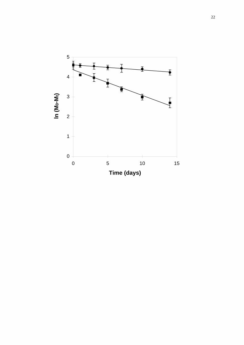
3.2 Peptide degradation
In vitro degradation of vapreotide pamoate at 37 °C over prolonged periods
of time was quite important in both PBS (31% degraded within 14 days) and serum
(85% degraded within 14 days) (Fig. 2). The increased degradation in the protein
containing media can be attributed to the higher solubility of the peptide in these
media. If the degradation kinetics was fitted to a first order reaction, the rate
constants were 2.52 • 10-2 d-1 (r2 = 0.971) in PBS and 12.87 • 10-2 d-1 (r2 = 0.959) in
serum. Fits were exclusively for the purpose of determining an order of magnitude of
the degradation rates. As various unidentified degradation steps were expected to
occur, the first order model may not be adequate. Particularly in serum, a second
order was actually better (k = 4.01 • 10-3 ml mg-1 d-1; r2 = 0.981). The occurence of
various degradation steps and products was revealed by HPLC-chromatograms. The
retention time of intact vapreotide was 17.7 min. In PBS, the first degradation

10
products appeared on day 3 of incubation with retention times of 14.3 and 17.1 min.
On day 5 of incubation, two additional peaks appeared with retention times of 9.4 and
20.9 min. No attempts were made here to identify these degradation products. On
the other hand, degradation products were not distinguishable in serum, because of
the great number of ghost peaks from the serum itself.
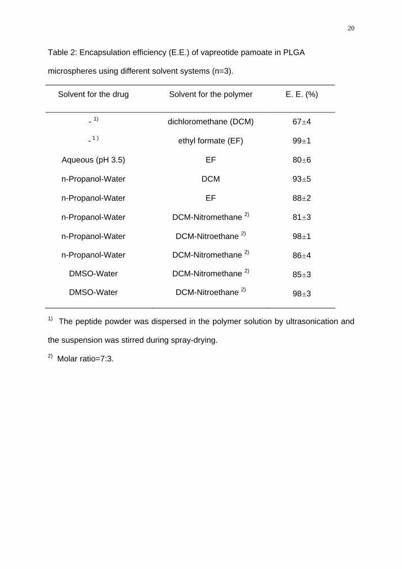
3.3 Microsphere characteristics
The microspheres had a regular spherical morphology. The particle sizes had
a unimodal distribution with diameters between 1 and 12 μm and a mean diameter of
3.5 μm. Several microsphere formulations were prepared from different solvents for
the peptide and polymer (Table 2). The solvents were chosen on the basis of
thermodynamic interaction data (Blanco-Príeto et al., 1998). When the peptide
powder was suspended in the polymer solution rather than dissolved in a second
solvent system, the encapsulation efficiencies were 67 and 99% with
dichloromethane and ethyl formate being used as polymer solvents, respectively.
This result might be ascribed to a higher affinity of the peptide salt to
dichloromethane as compared with ethyl formate. Due to the possible higher affinity,
some drug is transported to the particle surface upon solvent evaporation during
spray-drying. This surface located drug was then eliminated during the washing
process of the powder. When the peptide was dissolved in a acidic aqueous solution
(pH 3.5) and this solution dispersed in a polymer solution made with ethyl formate,
the encapsulation efficiency was significantly lower (80%) than when the drug powder
was suspended in a corresponding polymer solution (99%). Further, when propanol-
water (molar fraction of 0.2) or dimethylsulfoxide-water (molar fraction of 0.4) mixture
where used to solubilize the peptide, highest encapsulation efficiency (98%) were
observed with the dichloromethane-nitroethane mixture (molar ratio of 7:3) as

11
polymer solvents. Significantly lower encapsulation were generally attained (81-93%),
when either dichloromethane, ethyl formate or the mixture dichloromethane-
nitromethane were used as polymer solvents. Generally, these differences may be
explained by the low polymer-solvent interaction energy of the dichloromethane-
nitroethane system, as described recently (Blanco-Príeto et al., 1998). The
formulation prepared with the solvent ethyl formate alone wherein the peptide was
suspended, was used for all further experiments.
3.4 In vitro release kinetics
The in vitro release of vapreotide pamoate from the microspheres depended
greatly on the type of release medium (Fig. 3). In PBS of pH 7.4, only 33±1% of the
peptide were released within 4 days, whereas 53±2% and 61±0.9% were released in
1% BSA-solution and serum, respectively (Fig. 3). The faster release in the latter two
media may be ascribed to the much higher solubility of the peptide in these media
(Table 1). In serum a total of 67±0.7% were released within 8 days. In PBS and
aqueous solution of 1% BSA, after the initial burst a total amount of 74±1.2 and
46±1%, respectively were released after 8 days. These continuous release profiles
contrast with the often observed pulsatile pattern reported for peptide drugs (Bodmer
et al., 1992; Lawter et al., 1987; Sanders et al., 1984).
3.5 In vivo study
After intramuscular injection of γ-irradiated and non-irradiated microspheres
in rats (the formulation prepared with the solvent ethyl formate alone wherein the
peptide was suspended), the vapreotide plasma levels showed an initial peak
followed by a plateau at approx. 10 ng/ml over one week (Fig. 4). After one week, the
plasma level dropped off and reached undetectable levels after a second week.

12
Although the maintenance of therapeutic plasma levels (1 ng/ml) over approx. 12
days may have clinical importance, clinical testing of the present formulation cannot
be envisaged because of the high burst release, potencially including toxic effects.
Sterilization by γ-irradiation had only a minor effect on the pharmacokinetic profiles.
Further, the in vitro release of the peptide from irradiated and non-irradiated
microspheres showed no significant difference (data not shown); this is in good
agreement with the marginal (approx. 5%) molecular weight loss of the polymer used
after γ-sterilization (Rothen-Weinhold et al., 1997). These in vivo results were
qualitatively in good agreement with those of the in vitro release in serum. Data from
in vitro release studies may not always give an insight into the performance of a
dosage form in vivo. However, selection of appropriate release test media and
conditions should provide meaningful in vitro release data for further development
and improvements of dosage form design. Gido et al., (1993) have found a better
correlation using diluted plasma as dissolution medium as compared to phosphate
buffer pH 7.4 for microspheres embedding doxepin. With thyrotropin releasing
hormone (TRH), a relatively good agreement was found between in vitro and in vivo
release from PLGA75:25, when the release medium was a 0.033 M citrate-phosphate
buffer of pH 7 (Heya et al., 1994). In the present case, vapreotide pamoate may
diffuse rapidly through the endothelium of capillaries and enter very quickly the
systemic circulation. Therefore, we used serum as release medium to simulate a
physiological environment. The improved agreement between in vitro and in vivo
results, when using serum as release medium may be due to the high solubility of
vapreotide in serum and binding to plasma proteins. For vapreotide one may
conclude that the proper choice of the in vitro release test medium is very important
for the estimation of in vivo release. This aspect has to be tested with every drug
encapsulated in microspheres.

13
4. Conclusion
The peptide vapreotide pamoate was very efficiently microencapsulated (up
to 99%) into end-groups uncapped PLGA 50:50 by spray-drying. The differences in
release profiles observed in different media underline the importance of testing the in
vitro release in an appropriate fluid simulating in vivo conditions. Further experiments
are under way to reduce the burst and extend the release of RC-160 from
microparticles over one month.
14
References
Blanco-Príeto, M.J., Leo, E., Delie, F., Gulik, A., Couvreur, P., Fattal, E., 1996. Study
of the influence of several stabilizing agents on the entrapment and in vitro release
of pBC 264 from poly(lactide-co-glycolide) microspheres prepared by a W/O/W
solvent evaporation method. Pharm. Res. 13, 1127-1129.
Blanco-Príeto, M.J., Fattal, E., Gulik, A., Dedieu, J. C., Roques, B. P., Couvreur, P.,
1997. Characterization and morphological analysis of a cholecystokinin derivative
peptide-loaded poly(lactide-co-glycolide) microspheres prepared by a water-in-oil-
in-water emulsion solvent evaporation method. J. Control. Release 43, 81-87.
Blanco-Príeto, M.J., Nam-Trân, Hô, Ngûyen, V.P., Orsolini, P., Besseghir, K., Merkle,
H.P., Gander, B., 1998. Thermodynamic parameters for microencapsulation of an
octapeptide into PLA/PLGA microspheres. Proc. 2nd World Meeting APGI/APV,
521-522.
Bodmer, D., Kissel, T., Traechslin, E., 1992. Factors influencing the release of
peptides and proteins from biodegradable depot systems. J. Control. Release 21,
129-138.
Conti, B., Genta, I., Giunchedi, P., Modena, T., 1995. Testing of in vitro dissolution
behaviour of microparticulate drug delivery systems. Drug Dev. Ind. Pharm. 21,
1223-1233.
Gido, C., Langguth, P., Mutschler, E., 1994. Predictions of in vivo plasma
concentrations from in vitro release kinetics: Application to doxepin parenteral (i.
m.) suspensions in lipophilic vehicles in dogs. Pharm. Res. 11, 800-808.
Hashimoto, T., Wada, T., Fukuda, N., Nagaoka, A., 1993. Effect of thyrotropin-
releasing hormone on pentobarbitone-induced sleep in rats: continous treatment
with a sustained release injectable formulation. J. Pharm. Pharmacol. 45, 94-97.

15
Heya, T., Okada, H., Ogawa, Y., Toguchi, H., 1991. Factors influencing the profils of
TRH release from copoly(D,L-lactic/glycolic acid) microspheres. Int. J. Pharm. 72,
199-205.
Heya, T., Okada, H., Ogawa, Y., Toguchi, H., 1994. In vitro and in vivo evaluation of
thyrotrophin releasing hormone release from copoly(dl-lactic/glycolic acid)
microspheres. J. Pharm. Sci. 83, (5), 636-640.
Johansen, P., Corradin, G., Merkle, H.P., Gander, B., 1998. Release of tetanus
toxoid from adjuvants and PLGA microspheres: How experimental set-up and
surface adsorption fool the pattern. J. Control. Release, In Press.
Kissel, T., Brich, Z., Bantle, S., Lancranjan, I., Nimmerfall, F., Vit, P., 1991.
Parenteral depot-systems on the basis of biodegradable polyesters. J. Control.
Release. 16, 27-42.
Kurono, Y., Furukawa, A., Takesue, Y., Li, F., Ikeda, K., 1987. Photo-stabilization
and solubilization of an aldose reducttase inhibitor, (E)-3-Carboxymethyl-5-[(2E)-
methyl-3-phenylpropenylidene]rhodanine (ONO-2235), by human serum albumin.
Chem. Pharm. Bull. 37, (7), 3045-3048.
Lawter, J.R., Brizzolara, N.S., Lanzilotti, M.G., Morton, G.O., 1987. Drug release from
poly(glycolid-co-lactide) microcapsules. Proc. Intern. Symp. Controlled Release
Bioact. Mater. 14, 99-100.
Manson-Garcia, M., Vaccarella, M., Horvath, J., Redding, T. W., Groot, K., Orsolini,
P., Schally, A., 1988. Radio-immunoassay for octapeptide analogs of
somatostatin: Measurement of serum levels after administration of long-acting
microcapsule formulations. Proc. Natl. Acad. Sci. USA 85, 5688-5692.
16
Miyamoto, M., Hirai, K., Takahashi, H., Kato, K., Nishiyama, M., okada, H., Nagaoka,
A., 1993. Effect of sustained release formulation of thyrotropin-releasing hormone
on learning impairment caused by scopolamine and AF64A in rodents. Eur. J.
Pharmacol. 238, 181-189.
Morlock, M., Koll, H., Winter, G., Kissel, T., 1997. Microencapsulation of rh-
erytropoietin, using biodegradable poly(D,L-lactide-co-glycolide): protein stability
and the effects of stabilizing excipients. Eur. J. Pharm. Biopharm. 43, 29-36.
Ogawa, Y., Okada, H., Heya, T. Shimamoto, T., 1989. Controlled release of LHRH
agonist, leuprolide acetate, from microcapsules; serum drug level profiles and
pharmacological effects in animals. J. Pharm. Pharmacol. 41, 439-444.
Okada, H., Heya, T., Ogawa, Y., Shimamoto, T., 1988. One-month release injectable
microcapsules of a luteinizing hormone-releasing hormone agonist (leuprolide
acetate) for treating experimental endometriosis in rats. J. Pharm. Exp. Ther. 244,
744-749.
Okada, H., Heya, T., Igari, Y., Ogawa, Y., Toguchi, H., Shimamoto, T., 1989. One-
month release injectable microspheres of leuprolide acetate inhibit steroidogenesis
and genital organ growth in rats. Int. J. Pharm. 54, 231-239.
Park, T.G., Lu, W., Crotts, G., 1995. Importance of in vitro experimental conditions on
protein release kinetics, stability and polymer degradation in protein encapsulated
poly(D,L-lactic acid-co-glycolic acid) microspheres. J. Control. Release 33, 211-
222.
Rothen-Weinhold, A., Besseghir, K., Gurny, R., 1997. Analysis of the influence of
polymer characteristics and core loading on the in vivo release of a somatostatin
analogue. Eur. J. Pharm. Biopharm. 5, 303-313.

17
Russel-Jones, G.J., Jeffery, H., 1994. In vitro characterization of biodegradable
microparticles for oral antigen delivery. Proc. Int. Symp. Control. Release Bioact.
Mater. 21, 873-874.
Sanders, L.M., Kent, J.S., McRae, G.I., Vickery, B.H., Tice, T.R., Lewis, D.H., 1984.
Controlled release of luteinizing hormone-releasing hormone analogue from
poly(d,l-lactide-co-glycolide) microspheres. J. Pharm. Sci., 73, 1294-1297.
Stoeckemann, K., Sandow, J., 1993. Effects of the luteinizing-hormone-releasing
hormone (LHRH) antagonist ramorelix (Hoe013) and the LHRH agonist buserelin
on dimethylbenz(a)anthracene-induced mammary carcinoma: Studies with slow-
release formulations. J. Cancer Res. Clin. Oncol. 119, 457-462.
Thomasin, C., Corradin, G., Men, Y., Merkle, H.P., Gander, B., 1996. Tetanus toxoid
and synthetic malaria antigen containing poly(lactide)/poly(lactide-co-glycolide)
microspheres: importance of polymer degradation and antigen release for immune
response. J. Control. Release 41, 131-145.
Yang, J., Cleland, J.L., 1997. Factor affecting the in vitro release of recombinant
human interferon-γ (rhINF-γ) from PLGA microspheres. J. Pharm. Sci. 86, 980-
914.

18
Figure legends
Figure 1. Solubility of vapreotide pamoate in PBS at various pH and molarities: 1mM
( ), 10 mM (n), 66 mM (s) (t=24 hours).
Figure 2. First order degradation kinetics of vapreotide pamoate in serum (n) and
PBS (l), at 37°C.
Figure 3. In vitro release kinetics of vapreotide pamoate from PLGA microspheres in
BSA 1% ( ), serum (n) and PBS (s).
Figure 4. In vivo release kinetics profiles of RC-160 following i.m. administration of γ-
irradiated microspheres ( ) or non-irradiated microspheres (n) in rats (1.5 mg
equivalent peptide).

19
Table 1: Solubility of RC-160 in serum (S), serum ultrafiltrates (SU), and aqueous
BSA-solutions (n=3).
Medium Time (h) Solubility (μg/ml)
serum (S)
8 24 48
920±45
745±37
594±20
Serum ultrafiltrate (SU)1)
8 24 48
42±5
30±3
27±4
S:SU (10:90)
8 24 48
103±11
82±10
71±10
S:SU (1:99)
8 24 48
32±3
28±2
24±3
BSA, 1%
8 24 48
394±59
903±40
520±39
BSA, 5%
8 24 48
2736±59
3131±53
2698±88
BSA, 10%
8 24 48
5524±330
6128±450
3185±172
1) Cut-off of the membrane was 5 kDa; a 100 kDa cut-off membrane was also used,
but the resulting peptide solubility in the ultrafiltrate was not significantly different.
20
Table 2: Encapsulation efficiency (E.E.) of vapreotide pamoate in PLGA
microspheres using different solvent systems (n=3).
Solvent for the drug Solvent for the polymer E. E. (%)
- 1) dichloromethane (DCM) 67±4
- 1 ) ethyl formate (EF) 99±1
Aqueous (pH 3.5) EF 80±6
n-Propanol-Water DCM 93±5
n-Propanol-Water EF 88±2
n-Propanol-Water DCM-Nitromethane 2) 81±3
n-Propanol-Water
n-Propanol-Water
DMSO-Water
DMSO-Water
DCM-Nitroethane 2)
DCM-Nitromethane 2)
DCM-Nitromethane 2)
DCM-Nitroethane 2)
98±1
86±4
85±3
98±3
1) The peptide powder was dispersed in the polymer solution by ultrasonication and
the suspension was stirred during spray-drying.
2) Molar ratio=7:3.

21
0
200
400
600
800
1000
1200
2 3 4 5 6 7 8
pH
Solu
bilit
y of
RC
-160
( μg/
mL)
22
0
1
2
3
4
5
0 5 10 15
Time (days)
ln (M
0-M
t)

23
0
10
20
30
40
50
60
70
80
0 5 10 15
Time (days)
RC
-160
rele
ased
(%)

24
0
20
40
60
80
100
120
140
0 5 10 15 20
Time (days)
RC
-160
ng/
ml


![Thermodynamic analysis and isothermal bainitic ...Thermodynamic analysis and isothermal bainitic transformation kinetics in lean medium-Mn… 1713 13 researchofNavarro-Lopezetal.[15],theauthorsshowed](https://img.dokumen.tips/doc/110x75/60cef82ff3529d050f7f528c/thermodynamic-analysis-and-isothermal-bainitic-thermodynamic-analysis-and-isothermal.jpg)


