Embed Size (px)
Citation preview
O
Iirfl
JM
O(
a
A
R
A
A
K
K
A
L
A
L
a2�
h2
r e v b r a s o r t o p . 2 0 1 4;4 9(6):625–629
www.rbo.org .br
riginal Article
mportance of anatomically locating thenfrapatellar branch of the saphenous nerve ineconstructing the anterior cruciate ligament usingexor tendons�,��
ulio Cesar Gali ∗, André Franca Resina, Gabriel Pedro, Ildefonso Angelo Mora Neto,arco Antonio Pires Almagro, Phelipe Augusto Cintra da Silva, Edie Benedito Caetano
rthopedics and Traumatology Service, School of Medical Sciences and Health of Sorocaba, Pontifical Catholic University of São PauloPUC-SP), Sorocaba, SP, Brazil
r t i c l e i n f o
rticle history:
eceived 8 October 2013
ccepted 11 October 2013
vailable online 27 October 2014
eywords:
nee
nterior cruciate
igament/innervation
nterior cruciate
igament/surgery
a b s t r a c t
Objective: To describe the path of the infrapatellar branch of the saphenous nerve (IBSN)
using the medial joint line, anterior tibial tuberosity (ATT), tibial collateral ligament and a
horizontal line parallel to the medial joint line that passes over the ATT, as reference points,
in order to help surgeons to diminish the likelihood of injuring this nerve branch during
reconstruction of the anterior cruciate ligament (ACL) using flexor tendons.
Methods: Ten frozen knees that originated from amputations were examined. Through
anatomical dissection performed with the specimens flexed, we sought to find the IBSN,
from its most medial and proximal portion to its most lateral and distal portion. Follow-
ing this, the anatomical specimens were photographed and, using the ImageJ software, we
determined the distance from the IBSN to the medial joint line and to a lower horizontal
line going through the ATT and parallel to the first line. We also measured the angle of the
direction of the path of the nerve branch in relation to this lower line.
Results: The mean angle of the path of the nerve branch in relation to the lower horizon-
tal line was 17.50 ± 6.17◦. The mean distance from the IBSN to the medial joint line was
2.61 ± 0.59 cm and from the IBSN to the lower horizontal line, 1.44 ± 0.51 cm.
Conclusion: The IBSN was found in all the knees studied. In three knees, we found a second
branch proximal to the first one. The direction of its path was always from proximal and
medial to distal and lateral. The IBSN was always proximal and medial to the ATT and distal
to the medial joint line. The medial angle between its direction and a horizontal line going
through the ATT was 17.50 ± 6.17◦.
© 2014 Sociedade Brasileira de Ortopedia e Traumatologia. Published by Elsevier Editora
Ltda. All rights reserved.
� Please cite this article as: Gali JC, Resina AF, Pedro G, Neto IAM, Almagro MAP, da Silva PAC, Caetano EB. Importância da localizacãonatômica do ramo infrapatelar do nervo safeno na reconstrucão do ligamento cruzado anterior com tendões flexores. Rev Bras Ortop.014;49:625–629.� Work developed in the School of Medical Sciences and Health of Sorocaba, PUC-SP, Sorocaba, SP, Brazil.∗ Corresponding author.
E-mail: [email protected] (J.C. Gali).ttp://dx.doi.org/10.1016/j.rboe.2013.10.004255-4971/© 2014 Sociedade Brasileira de Ortopedia e Traumatologia. Published by Elsevier Editora Ltda. All rights reserved.
626 r e v b r a s o r t o p . 2 0 1 4;4 9(6):625–629
Importância da localizacão anatômica do ramo infrapatelar do nervosafeno na reconstrucão do ligamento cruzado anterior com tendõesflexores
Palavras-chave:
Joelho
Ligamento cruzado
anterior/inervacão
Ligamento cruzado
anterior/cirurgiar
r e s u m o
Objetivo: Descrever o trajeto do ramo infrapatelar do nervo safeno (RIPNS) com o uso da linha
articular medial, da tuberosidade anterior da tíbia (TAT), do ligamento colateral tibial e de
uma linha horizontal, paralela à linha articular medial e que passa sobre a TAT, como pontos
de referência, a fim de poder auxiliar os cirurgiões a diminuir a probabilidade de lesão desse
ramo nervoso na reconstrucão do ligamento cruzado anterior (LCA) com tendões flexores.
Métodos: Foram examinados 10 joelhos congelados, originados de amputacões. Na
dissecacão anatômica, feita com as pecas flexionadas, procuramos encontrar o RIPNS, desde
a sua porcão mais medial e proximal até sua porcão mais lateral e distal. Em seguida, as
pecas anatômicas foram fotografadas e, com o programa ImageJ, determinamos a distância
do RIPNS até a linha articular medial e até uma linha horizontal inferior, que passa pela TAT
e é paralela à primeira. Medimos, também, o ângulo da direcão do trajeto do ramo nervoso
em relacão a essa linha horizontal inferior.
Resultados: O ângulo médio do trajeto do ramo nervoso, em relacão à linha horizontal infe-
rior, foi de 17,50◦ ± 6,17◦. A distância média do RIPNS até a linha articular medial foi de
2,61 ± 0,59 cm e até a linha horizontal inferior, de 1,44 ± 0,51 cm.
Conclusão: O RIPNS foi encontrado em todos os joelhos estudados; em três, encontramos
um segundo ramo, proximal ao primeiro. A direcão de seu trajeto foi sempre de proximal e
medial para distal e lateral. O RIPNS esteve sempre proximal e medial à TAT e distal à linha
articular medial. A angulacão média de sua direcão, em relacão a uma linha horizontal que
passa pela TAT, foi de 17,50◦ ± 6,17◦.
© 2014 Sociedade Brasileira de Ortopedia e Traumatologia. Publicado por Elsevier
Editora Ltda. Todos os direitos reservados.
Introduction
Surgical reconstruction of the anterior cruciate ligament (ACL)is a very frequently performed procedure. It has been esti-mated that 100,000 of these procedures are performed in theUnited States every year and that this is the sixth commonestorthopedic surgical procedure in that country.1
Use of grafts from the tendons of the gracilis and semi-tendinosus muscles for surgical reconstruction of the ACL isincreasingly common, because these grafts withstand highloads before failure; their cross-sectional area is large; theypass easily though the tunnels; they only need a small inci-sion; they present low postoperative morbidity; and give riseto lower morbidity at the donor site.2
However, because of their anatomical location, there is apotential risk of injury to the infrapatellar branch of the saphe-nous nerve (IPBSN) during harvesting of autologous tendonsfrom the gracilis and semitendinosus muscles.3–6
In the literature, the percentage occurrence of iatrogeniclesions of the IPBSN during reconstruction of the ACL usingflexor tendons ranges from 14.9% to 77%.5–10
The orientation of the surgical incision for harvesting thetendons may, theoretically, influence the risk of injury to
11 12
the IPBSN. Tifford et al. reported that vertical incisionsare perpendicular to the nerve trunk and put the IPBSN atrisk. According to Sabat and Kumar,13 vertical incisions havegreater incidence of injuries to the IPBSN, with persistenthyperesthesia, extensive areas of sensory loss and worse sub-jective results. Several authors have recommended that thisincision should preferentially be oblique.3,4,6,8,9,11,13
The aim of our study was to describe the path of the IPBSNin the region of flex or tendon harvesting, in order to provideinformation on where this branch is commonly encounteredand thus to diminish the chances of iatrogenic injuries.
Materials and methods
Ten frozen knees originating from amputations were dissec-ted. Six were from men and four from women. Six were rightknees and four were left knees. The patients’ ages ranged from28 to 72 years, with a mean of 41.
We removed the skin from the proximal and medial thirdsof the lower leg and from the distal and medial thirds of thethigh. We then carefully searched for the IPBSN from its mostmedial and proximal portion to its most lateral and distalportion. The dissection was performed with the specimensflexed.
After isolating the IPBSN, we photographed each specimenusing a Nikon D 3100 digital camera. The images obtained wereevaluated using the ImageJ software.
On each photo, we outlined a rectangle with its sides drawnas follows: a medial vertical line that went along the lateraledge of the tibial collateral ligament; an upper horizontal linethat went along the medial joint line; a lateral vertical line
r e v b r a s o r t o p . 2 0 1 4;4 9(6):625–629 627
A B
1
α
2
3
4
x
y
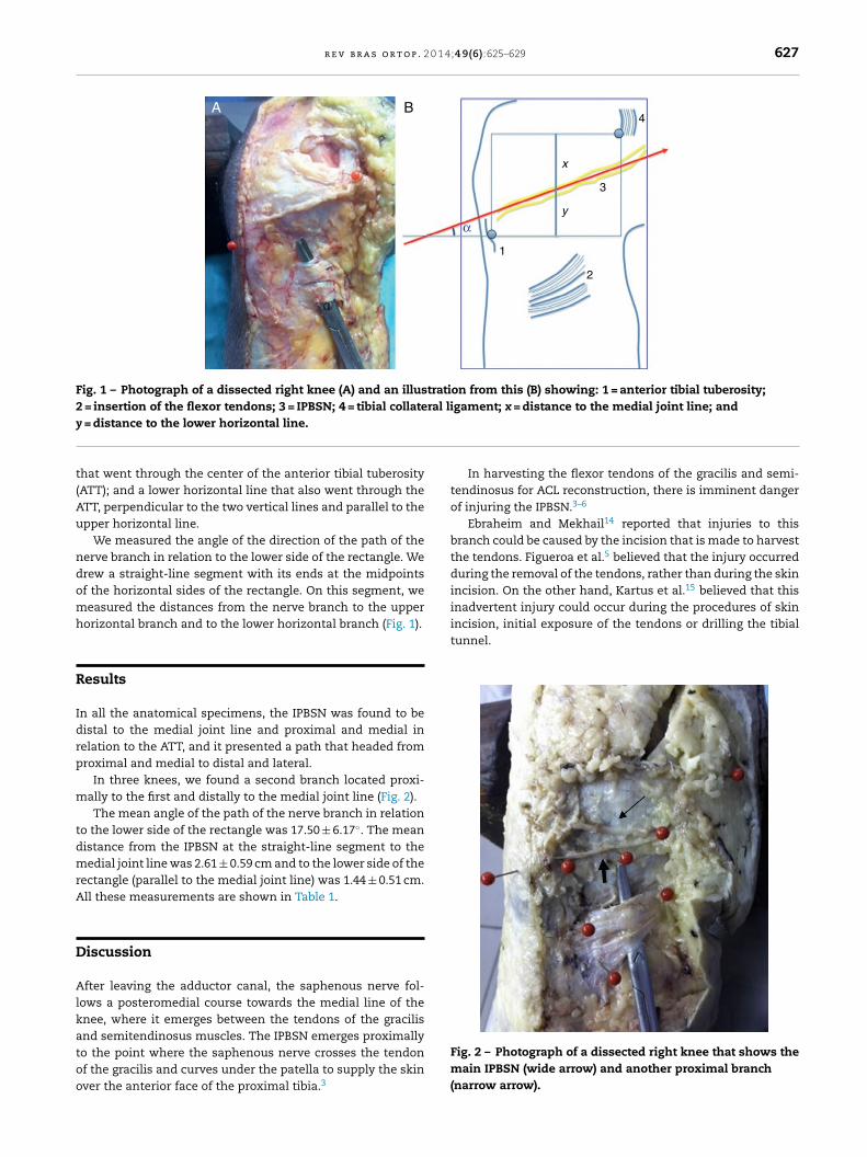
Fig. 1 – Photograph of a dissected right knee (A) and an illustration from this (B) showing: 1 = anterior tibial tuberosity;2 = insertion of the flexor tendons; 3 = IPBSN; 4 = tibial collateral ligament; x = distance to the medial joint line; andy
t(Au
ndomh
R
Idrp
m
tdmrA
D
Alkatoo
incision, initial exposure of the tendons or drilling the tibialtunnel.
= distance to the lower horizontal line.
hat went through the center of the anterior tibial tuberosityATT); and a lower horizontal line that also went through theTT, perpendicular to the two vertical lines and parallel to thepper horizontal line.
We measured the angle of the direction of the path of theerve branch in relation to the lower side of the rectangle. Werew a straight-line segment with its ends at the midpointsf the horizontal sides of the rectangle. On this segment, weeasured the distances from the nerve branch to the upper
orizontal branch and to the lower horizontal branch (Fig. 1).
esults
n all the anatomical specimens, the IPBSN was found to beistal to the medial joint line and proximal and medial inelation to the ATT, and it presented a path that headed fromroximal and medial to distal and lateral.
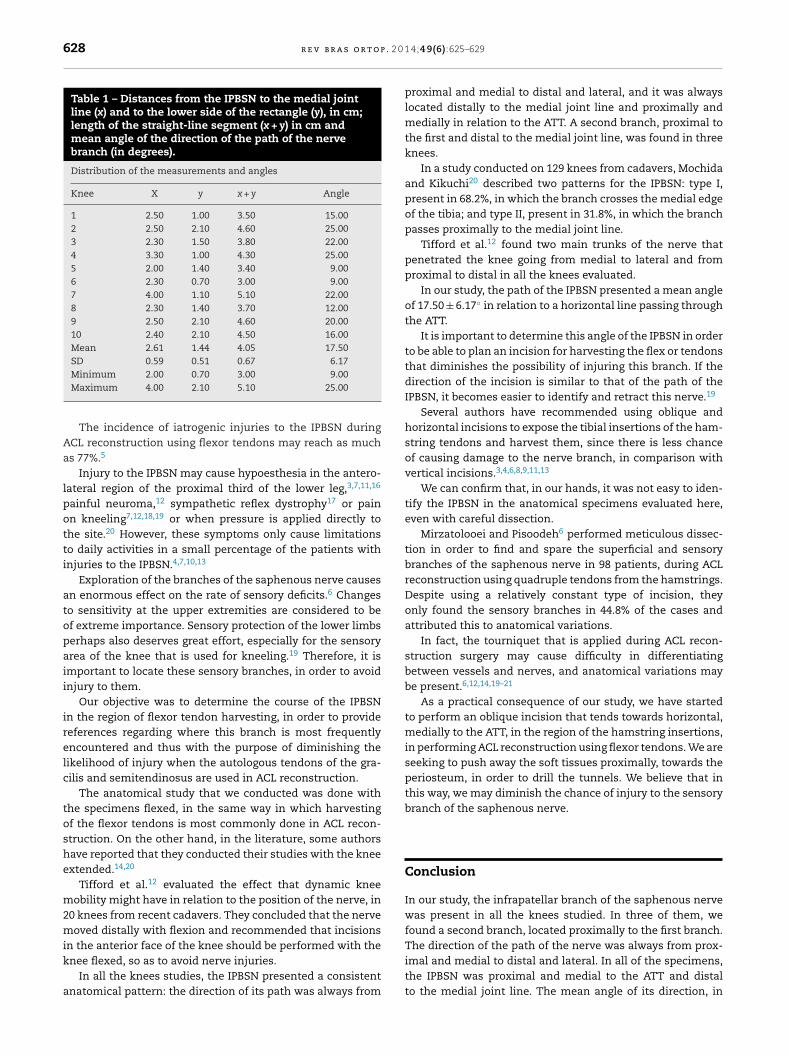
In three knees, we found a second branch located proxi-ally to the first and distally to the medial joint line (Fig. 2).The mean angle of the path of the nerve branch in relation
o the lower side of the rectangle was 17.50 ± 6.17◦. The meanistance from the IPBSN at the straight-line segment to theedial joint line was 2.61 ± 0.59 cm and to the lower side of the
ectangle (parallel to the medial joint line) was 1.44 ± 0.51 cm.ll these measurements are shown in Table 1.
iscussion
fter leaving the adductor canal, the saphenous nerve fol-ows a posteromedial course towards the medial line of thenee, where it emerges between the tendons of the gracilis
nd semitendinosus muscles. The IPBSN emerges proximallyo the point where the saphenous nerve crosses the tendonf the gracilis and curves under the patella to supply the skinver the anterior face of the proximal tibia.3In harvesting the flexor tendons of the gracilis and semi-tendinosus for ACL reconstruction, there is imminent dangerof injuring the IPBSN.3–6
Ebraheim and Mekhail14 reported that injuries to thisbranch could be caused by the incision that is made to harvestthe tendons. Figueroa et al.5 believed that the injury occurredduring the removal of the tendons, rather than during the skinincision. On the other hand, Kartus et al.15 believed that thisinadvertent injury could occur during the procedures of skin
Fig. 2 – Photograph of a dissected right knee that shows themain IPBSN (wide arrow) and another proximal branch(narrow arrow).
628 r e v b r a s o r t o p . 2 0
Table 1 – Distances from the IPBSN to the medial jointline (x) and to the lower side of the rectangle (y), in cm;length of the straight-line segment (x + y) in cm andmean angle of the direction of the path of the nervebranch (in degrees).
Distribution of the measurements and angles
Knee X y x + y Angle
1 2.50 1.00 3.50 15.002 2.50 2.10 4.60 25.003 2.30 1.50 3.80 22.004 3.30 1.00 4.30 25.005 2.00 1.40 3.40 9.006 2.30 0.70 3.00 9.007 4.00 1.10 5.10 22.008 2.30 1.40 3.70 12.009 2.50 2.10 4.60 20.0010 2.40 2.10 4.50 16.00Mean 2.61 1.44 4.05 17.50SD 0.59 0.51 0.67 6.17
The direction of the path of the nerve was always from prox-
Minimum 2.00 0.70 3.00 9.00Maximum 4.00 2.10 5.10 25.00
The incidence of iatrogenic injuries to the IPBSN duringACL reconstruction using flexor tendons may reach as muchas 77%.5
Injury to the IPBSN may cause hypoesthesia in the antero-lateral region of the proximal third of the lower leg,3,7,11,16
painful neuroma,12 sympathetic reflex dystrophy17 or painon kneeling7,12,18,19 or when pressure is applied directly tothe site.20 However, these symptoms only cause limitationsto daily activities in a small percentage of the patients withinjuries to the IPBSN.4,7,10,13
Exploration of the branches of the saphenous nerve causesan enormous effect on the rate of sensory deficits.6 Changesto sensitivity at the upper extremities are considered to beof extreme importance. Sensory protection of the lower limbsperhaps also deserves great effort, especially for the sensoryarea of the knee that is used for kneeling.19 Therefore, it isimportant to locate these sensory branches, in order to avoidinjury to them.
Our objective was to determine the course of the IPBSNin the region of flexor tendon harvesting, in order to providereferences regarding where this branch is most frequentlyencountered and thus with the purpose of diminishing thelikelihood of injury when the autologous tendons of the gra-cilis and semitendinosus are used in ACL reconstruction.
The anatomical study that we conducted was done withthe specimens flexed, in the same way in which harvestingof the flexor tendons is most commonly done in ACL recon-struction. On the other hand, in the literature, some authorshave reported that they conducted their studies with the kneeextended.14,20
Tifford et al.12 evaluated the effect that dynamic kneemobility might have in relation to the position of the nerve, in20 knees from recent cadavers. They concluded that the nervemoved distally with flexion and recommended that incisionsin the anterior face of the knee should be performed with the
knee flexed, so as to avoid nerve injuries.In all the knees studies, the IPBSN presented a consistentanatomical pattern: the direction of its path was always from
1 4;4 9(6):625–629
proximal and medial to distal and lateral, and it was alwayslocated distally to the medial joint line and proximally andmedially in relation to the ATT. A second branch, proximal tothe first and distal to the medial joint line, was found in threeknees.
In a study conducted on 129 knees from cadavers, Mochidaand Kikuchi20 described two patterns for the IPBSN: type I,present in 68.2%, in which the branch crosses the medial edgeof the tibia; and type II, present in 31.8%, in which the branchpasses proximally to the medial joint line.
Tifford et al.12 found two main trunks of the nerve thatpenetrated the knee going from medial to lateral and fromproximal to distal in all the knees evaluated.
In our study, the path of the IPBSN presented a mean angleof 17.50 ± 6.17◦ in relation to a horizontal line passing throughthe ATT.
It is important to determine this angle of the IPBSN in orderto be able to plan an incision for harvesting the flex or tendonsthat diminishes the possibility of injuring this branch. If thedirection of the incision is similar to that of the path of theIPBSN, it becomes easier to identify and retract this nerve.19
Several authors have recommended using oblique andhorizontal incisions to expose the tibial insertions of the ham-string tendons and harvest them, since there is less chanceof causing damage to the nerve branch, in comparison withvertical incisions.3,4,6,8,9,11,13
We can confirm that, in our hands, it was not easy to iden-tify the IPBSN in the anatomical specimens evaluated here,even with careful dissection.
Mirzatolooei and Pisoodeh6 performed meticulous dissec-tion in order to find and spare the superficial and sensorybranches of the saphenous nerve in 98 patients, during ACLreconstruction using quadruple tendons from the hamstrings.Despite using a relatively constant type of incision, theyonly found the sensory branches in 44.8% of the cases andattributed this to anatomical variations.
In fact, the tourniquet that is applied during ACL recon-struction surgery may cause difficulty in differentiatingbetween vessels and nerves, and anatomical variations maybe present.6,12,14,19–21
As a practical consequence of our study, we have startedto perform an oblique incision that tends towards horizontal,medially to the ATT, in the region of the hamstring insertions,in performing ACL reconstruction using flexor tendons. We areseeking to push away the soft tissues proximally, towards theperiosteum, in order to drill the tunnels. We believe that inthis way, we may diminish the chance of injury to the sensorybranch of the saphenous nerve.
Conclusion
In our study, the infrapatellar branch of the saphenous nervewas present in all the knees studied. In three of them, wefound a second branch, located proximally to the first branch.
imal and medial to distal and lateral. In all of the specimens,the IPBSN was proximal and medial to the ATT and distalto the medial joint line. The mean angle of its direction, in
0 1 4
r1
C
T
r
1
1
1
1
1
1
1
1
1
1
2
r e v b r a s o r t o p . 2
elation to a horizontal line passing through the ATT was7.50 ± 6.17 degrees.
onflicts of interest
he authors declare no conflicts of interest.
e f e r e n c e s
1. Brown CH Jr, Carson EW. Revision anterior cruciate ligamentsurgery. Clin Sports Med. 1999;18(1):109–71.
2. Bartlett RJ, Clatworthy MG, Nguyen TN. Graft selection inreconstruction of the anterior cruciate ligament. J Bone JointSurg Br. 2001;83(5):625–34.
3. Pagnani MJ, Warner JJ, O’Brien SJ, Warren RF. Anatomicconsiderations in harvesting the semitendinosus and gracilistendons and a technique of harvest. Am J Sports Med.1993;21(4):565–71.
4. Boon JM, Van Wyk MJ, Jordaan D. A safe area and angle forharvesting autogenous tendons for anterior cruciate ligamentreconstruction. Surg Radiol Anat. 2004;26(3):167–71.
5. Figueroa D, Calvo R, Vaisman A, Campero M, Moraga C. Injuryto the infrapatellar branch of the saphenousnerve in ACLreconstruction with the hamstrings technique: clinical andelectrophysiological study. Knee. 2008;15(5):360–3.
6. Mirzatolooei F, Pisoodeh K. Impact of exploration of sensorybranches of saphenous nerve in anterior cruciate ligamentreconstructive surgery. Arch Iran Med. 2012;15(4):219–22.
7. Spicer DD, Blagg SE, Unwin AJ, Allum RL. Anterior kneesymptoms after four-strand hamstring tendon anteriorcruciate ligament reconstruction. Knee Surg SportsTraumatol Arthrosc. 2000;8(5):286–9.
8. Mochizuki T, Muneta T, Yagishita K, Shinomiya K, Sekiya I.Skin sensory change after arthroscopically-assisted anteriorcruciate ligament reconstruction using medial hamstringtendons with a vertical incision. Knee Surg Sports TraumatolArthrosc. 2004;12(3):198–202.
9. Papastergiou SG, Voulgaropoulos H, Mikalef P, Ziogas E,Pappis G, Giannakopoulos I. Injuries to the infrapatellar
branch(es) of the saphenousnerve in anterior cruciateligament reconstruction with four-strand hamstring tendonautograft: vertical versus horizontal incision for harvest.Knee Surg Sports Traumatol Arthrosc. 2006;14(8):789–93.2
;4 9(6):625–629 629
0. Sanders B, Rolf R, McClelland W, Xerogeanes J. Prevalence ofsaphenous nerve injury after autogenous hamstring harvest:an anatomic and clinical study of sartorial branch injury.Arthroscopy. 2007;23(9):956–63.
1. Kjaergaard J, Faunø LZ, Faunø P. Sensibility loss after ACLreconstruction with hamstring graft. Int J Sports Med.2008;29(6):507–11.
2. Tifford CD, Spero L, Luke T, Plancher KD. The relationship ofthe infrapatellar branches of the saphenous nerve toarthroscopy portals and incisions for anterior cruciateligament surgery. An anatomic study. Am J Sports Med.2000;28(4):562–7.
3. Sabat D, Kumar V. Nerve injury during hamstring graftharvest: a prospective comparative study of three differentincisions. Knee Surg Sports Traumatol Arthrosc.2013;21(9):2089–95.
4. Ebraheim NA, Mekhail AO. The infrapatellar branch of thesaphenous nerve: an anatomic study. J Orthop Trauma.1997;11(3):195–9.
5. Kartus J, Movin T, Karlsson J. Donor-site morbidity andanterior knee problems after anterior cruciate ligamentreconstruction using autografts. Arthroscopy.2001;17(9):971–80.
6. Kartus J, Magnusson L, Stener S, Brandsson S, Eriksson BI,Karlsson J. Complications following arthroscopic anteriorcruciate ligament reconstruction. A 2–5-year follow-up of 604patients with special emphasis on anterior knee pain. KneeSurg Sports Traumatol Arthrosc. 1999;7(1):2–8.
7. Poehling GG, Pollock FE Jr, Koman LA. Reflex sympatheticdystrophy of the knee after sensory nerve injury. Arthroscopy.1988;4(1):31–5.
8. Corry IS, Webb JM, Clingeleffer AJ, Pinczewski LA.Arthroscopic reconstruction of the anterior cruciate ligament.A comparison of patellar tendon autograft and four-strandhamstring tendon autograft. Am J Sports Med.1999;27(4):444–54.
9. Hunter LY, Louis DS, Ricciardi JR, O’Connor GA. Thesaphenous nerve: its course and importance in medialarthrotomy. Am J Sports Med. 1979;7(4):227–30.
0. Mochida H, Kikuchi S. Injury to infrapatellar branch of
saphenous nerve in arthroscopic knee surgery. Clin OrthopRelat Res. 1995;320:88–94.1. Arthornthurasook A, Gaew-Im K. Study of the infrapatellarnerve. Am J Sports Med. 1988;16(1):57–9.