Embed Size (px)
Citation preview
nature biotechnology VOLUME 28 NUMBER 8 AUGUST 2010 863
l e t t e r s
Recombinant glycoprotein therapeutics produced in nonhuman mammalian cell lines and/or with animal serum are often modified with the nonhuman sialic acid N-glycolylneuraminic acid (Neu5Gc; refs. 1,2). This documented contamination has generally been ignored in drug development because healthy individuals were not thought to react to Neu5Gc (ref. 2). However, recent findings indicate that all humans have Neu5Gc-specific antibodies, sometimes at high levels3,4. Working with two monoclonal antibodies in clinical use, we demonstrate the presence of covalently bound Neu5Gc in cetuximab (Erbitux) but not panitumumab (Vectibix). Anti-Neu5Gc antibodies from healthy humans interact with cetuximab in a Neu5Gc-specific manner and generate immune complexes in vitro. Mice with a human-like defect in Neu5Gc synthesis generate antibodies to Neu5Gc after injection with cetuximab, and circulating anti-Neu5Gc antibodies can promote drug clearance. Finally, we show that the Neu5Gc content of cultured human and nonhuman cell lines and their secreted glycoproteins can be reduced by adding a human sialic acid to the culture medium. Our findings may be relevant to improving the half-life, efficacy and immunogenicity of glycoprotein therapeutics.
Therapeutic glycoproteins, including antibodies, growth factors, cytokines, hormones and clotting factors, generate sales with annual double-digit growth rates5. They must often be produced in mammalian expression systems because of the crucial influence of the location, number and structure of N-glycans on their yields, bioactivity, solubi-lity, stability against proteolysis, immunogenicity and rate of clearance from the bloodstream6–8.
Two differences between the protein glycosylation apparatus of humans and rodents account for major potential differences between the N-glycans on glycoproteins made in cultured human cells and those made using rodent cell lines. First, humans cannot synthesize a terminal Galα1-3Gal motif (known as alpha-Gal) on N-glycans. As a consequence, they express antibodies against this structure9. Second, unlike other mammals, humans cannot biosynthesize the sialic acid Neu5Gc because the human gene CMAH, encoding CMP-N-acetyl-neuraminic acid hydroxylase, the enzyme responsible for producing CMP-Neu5Gc from CMP-N-acetylneuraminic acid (CMP-Neu5Ac), is irreversibly mutated10. The use of cultured human cells to address
this issue is not a solution, as Neu5Gc can be taken up from animal products present in the culture medium and then metabolically incor-porated into secreted glycoproteins11.
Owing largely to limitations of the assays originally used to detect anti-Neu5Gc antibodies, including the fact that only a small number of possible Neu5Gc-containing epitopes were tested, healthy humans were long believed to show no immune reaction to Neu5Gc (ref. 2). Subsequent reports that all humans possess anti-Neu5Gc antibodies3, sometimes at high levels, approaching 0.1–0.2% of circulating IgG3,4, have led to re-evaluation of the potential significance of Neu5Gc contamination7,8. Especially in light of trends toward administering increasingly higher amounts of certain biotherapeutics over longer periods of time, some biopharmaceutical companies are exploring steps to reduce levels of Neu5Gc in their products12.
Given that they are produced using nonhuman cell lines, animal serum or serum-derived factors, or a combination of these, it is likely that most recombinant therapeutic glycoproteins carry some Neu5Gc. However, given the diversity of products and production protocols, it is difficult to make generalizations. Thus, we chose to compare two US Food and Drug Administration (FDA)-approved monoclonal antibodies with the same therapeutic target, the EGF receptor. The first, Erbitux (cetuximab, obtained from the University of California, San Diego Pharmacy), is a chimeric antibody produced in mouse myeloma cells13,14. The second, Vectibix (panitumumab, obtained from Amgen), is a fully human antibody produced in Chinese hamster ovary (CHO) cells15. The samples studied were preparations that would normally be administered to patients.
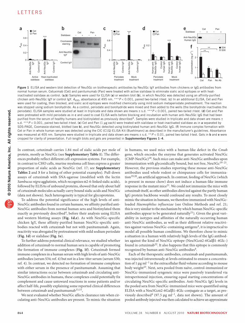
We first performed enzyme-linked immunosorbent assays (ELISAs) using an affinity-purified polyclonal chicken Neu5Gc-specific anti-body preparation that is highly monospecific for Neu5Gc (ref. 16, alongside a nonreactive control IgY). Bound Neu5Gc was easily detectable on cetuximab but not on panitumumab (Fig. 1a). Sialidase pretreatment abolished binding, confirming specificity. Western blot analysis also showed sialidase-sensitive anti-Neu5Gc IgY reactivity on the heavy chains of cetuximab but not those of panitumumab (Fig. 1b). The specificity of anti-Neu5Gc IgY binding was reaffirmed by pretreatment with mild sodium periodate under conditions that selectively cleave sialic acid side chains (Fig. 1c) and abolish reactivity of such antibodies3,16. Finally, we quantified sialic acids on the thera-peutic antibodies, as described in Online Methods. Panitumumab carries 0.22 mol of sialic acids per mole of protein, with <0.1% Neu5Gc.
Implications of the presence of N-glycolylneuraminic acid in recombinant therapeutic glycoproteinsDarius Ghaderi1,2, Rachel E Taylor1, Vered Padler-Karavani1, Sandra Diaz1 & Ajit Varki1
1Glycobiology Research and Training Center, Departments of Medicine and Cellular & Molecular Medicine, University of California, San Diego, La Jolla, California, USA. 2Present address: Sialix, Inc., Vista, California, USA. Correspondence should be addressed to A.V. ([email protected]).
Received 7 August 2009; accepted 24 May 2010; published online 25 July 2010; doi:10.1038/nbt.1651
© 2
010
Nat
ure
Am
eric
a, In
c. A
ll ri
gh
ts r
eser
ved
.
864 VOLUME 28 NUMBER 8 AUGUST 2010 nature biotechnology
l e t t e r s
In contrast, cetuximab carries 1.84 mol of sialic acids per mole of protein, mostly as Neu5Gc (see Supplementary Table 1). The differ-ences probably reflect different cell-expression systems. For example, in contrast to CHO cells, murine myeloma cell lines express a greater proportion of sialic acids as Neu5Gc (ref. 17; see Supplementary Tables 2 and 3 for a listing of other potential examples). Pull-down assays of cetuximab with SNA-agarose (modified with the lectin Sambucus nigra agglutinin, which recognizes α2-6-linked sialic acids), followed by ELISAs of unbound proteins, showed that only about half of cetuximab molecules actually carry bound sialic acids and Neu5Gc (data not shown). Such heterogeneity is typical for glycoproteins.
To address the potential significance of the high levels of anti-Neu5Gc antibodies found in certain humans, we affinity purified anti-Neu5Gc antibodies from normal human sera and biotinylated them exactly as previously described4, before their analysis using ELISA and western blotting assays (Fig. 1d,e). As with Neu5Gc-specific chicken IgY, these affinity-purified human Neu5Gc-specific anti-bodies reacted with cetuximab but not with panitumumab. Again, reactivity was abrogated by pretreatment with mild sodium periodate (Fig. 1d) or sialidase (Fig. 1e).
To further address potential clinical relevance, we studied whether addition of cetuximab to normal human sera is capable of promoting the formation of immune complexes (Fig. 1f). Cetuximab formed immune complexes in a human serum with high levels of anti-Neu5Gc antibodies (serum S34; ref. 4) but not in a low-titer serum (serum S30; ref. 4). In contrast, we detected no formation of immune complexes with either serum in the presence of panitumumab. Assuming that similar interactions occur between cetuximab and circulating anti-Neu5Gc antibodies in humans, these complexes could potentially fix complement and cause untoward reactions in some patients and/or affect half-life, possibly explaining some reported clinical differences between cetuximab and panitumumab13,15,
We next evaluated whether Neu5Gc affects clearance rate when cir-culating anti-Neu5Gc antibodies are present. To mimic the situation
in humans, we used mice with a human-like defect in the Cmah gene, which encodes the enzyme that generates activated Neu5Gc (CMP-Neu5Gc)18. Such mice can make anti-Neu5Gc antibodies upon immunization with glycosidically bound, but not free, Neu5Gc19–21. However, the previous studies reporting these mouse anti-Neu5Gc antibodies used whole rodent or chimpanzee cells for immuniza-tion19,20, an artificial approach. In contrast, feeding of Neu5Gc (which is present in mouse chow) does not induce a human-like immune response in the mutant mice21. We could not immunize the mice with cetuximab itself, as other antibodies directed against the partly human IgG protein backbone would confound any results. To most closely mimic the situation in humans, we therefore immunized with Neu5Gc-loaded Haemophilus influenzae (see Online Methods and ref. 21; this is very similar to the mechanism by which human Neu5Gc-specific antibodies appear to be generated naturally21). Given the great vari-ability in isotypes and affinities of the naturally occurring human anti-Neu5Gc antibodies, as well as their different relative reactivi-ties against various Neu5Gc-containing antigens4, it is impractical to model all possible human conditions. We therefore chose to mimic a situation in a human with relatively high levels of the IgG antibod-ies against the kind of Neu5Gc epitope (Neu5Gcα2-6Galβ1-4Glc-) found in cetuximab22. It also happens that this epitope is commonly recognized by human anti-Neu5Gc antibodies4.
Each of the therapeutic antibodies, cetuximab and panitumumab, was injected intravenously at levels estimated to ensure a concentra-tion of 1 μg ml−1 in the extracellular fluid volume according to mouse body weight23. Next, sera pooled from naïve, control-immunized or Neu5Gc-immunized syngeneic mice were passively transferred via intraperitoneal injection, ensuring equal starting concentrations of circulating Neu5Gc-specific antibodies. Anti-Neu5Gc IgG levels in the pooled sera from Neu5Gc-immunized mice were quantified using ELISA with a Neu5Gcα2-6Galβ1-4Glc-conjugate as a target, as pre-viously described4 (97.5 μg ml−1, data not shown). The amount of pooled antibody injected was then calculated to achieve an approximate
0.6
a d e
f
b c
0.8
0.4
02
1
0
CetPan
CetPan
A49
5
A49
5
A49
5
Con
cent
ratio
n (n
g µl
–1)
0.4
0.2
Ant
i-Neu
5Gc
IgY
Hum
an a
nti-
Neu
5Gc
IgG
Hum
an a
nti-
Neu
5Gc
IgG
Con
trol
hum
an Ig
G
Periodatetreatment
Mocktreatment
Con
trol
hum
an Ig
G
Cet
***
Pan
Cet Pan
Cet Pan No Ab
S34S30**
**
***
***
Ant
i-Neu
5Gc
IgY
Con
trol
IgY
Activesialidase
Heat-inactivatedsialidase
Coomassiestaining
Anti-Neu5GcIgY
Anti-Neu5GcHuman IgG
Sialidase: – + – +
Control IgY
Sialidase:Cet
– + – +Pan
Con
trol
IgY
Ant
i-Neu
5Gc
IgY
Ant
i-Neu
5Gc
IgY
Con
trol
IgY
Mocktreatment
Periodatetreatment
Con
trol
IgY
0
0.2
0
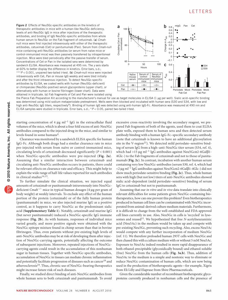
Figure 1 ELISA and western blot detection of Neu5Gc on biotherapeutic antibodies by Neu5Gc IgY antibodies from chickens or IgG antibodies from normal human serum. Cetuximab (Cet) and panitumumab (Pan) were treated with active sialidase to eliminate sialic acid epitopes or with heat-inactivated sialidase as control. (a,b) Samples were used for ELISA (a) or western blot (b), in which Neu5Gc was detected using an affinity-purified chicken anti-Neu5Gc IgY or control IgY. A495, absorbance at 495 nm. ***P < 0.001, paired two-tailed t-test. (c) In an additional ELISA, Cet and Pan were used for coating, then blocked, and sialic acid epitopes were modified chemically using mild sodium metaperiodate pretreatment. The reaction was stopped using sodium borohydride. As a control, periodate and borohydride were mixed and then added to the wells (the borohydride inactivates the periodate). ELISA samples were studied at least in triplicate and data shown are means ± s.d. ***P < 0.001, paired two-tailed t-test. (d) Cet and Pan were pretreated with mild periodate as in c and used to coat ELISA wells before blocking and incubation with human anti-Neu5Gc IgG that had been purified from the serum of healthy humans and biotinylated as previously described4. Samples were studied in triplicate and data shown are means ± s.d. ***P < 0.001, paired two-tailed t-test. (e) Cet and Pan (1 μg each) were treated with sialidase or heat-inactivated sialidase as in a separated by SDS-PAGE, Coomassie-stained, blotted (see b), and Neu5Gc detected using biotinylated human anti-Neu5Gc IgG. (f) Immune complex formation with Cet or Pan in whole human serum was detected using the CIC (C1Q) ELISA Kit (Buehlmann) as described in the manufacturer’s guidelines. Absorbance was measured at 405 nm. Samples were studied in triplicate and data shown are means ± s.d. **P < 0.01, paired two-tailed t-test. Gels in b and e were cropped for clarity of presentation. Full-length blots and gels are presented in Supplementary Figures 1–4.
© 2
010
Nat
ure
Am
eric
a, In
c. A
ll ri
gh
ts r
eser
ved
.
nature biotechnology VOLUME 28 NUMBER 8 AUGUST 2010 865
l e t t e r s
starting concentration of 4 μg ml−1 IgG in the extracellular fluid volumes of the mice, which is about a four fold excess of anti-Neu5Gc antibodies compared to the injected drug in the mice, and similar to levels found in some humans4.
Clearance was monitored by a sandwich ELISA specific for human IgG-Fc. Although both drugs had a similar clearance rate in mice pre-injected with serum from naïve or control-immunized mice, circulating levels of cetuximab decreased significantly (P < 0.001) when Neu5Gc-specific antibodies were pre-injected (Fig. 2a). Assuming that a similar interaction between cetuximab and circulating anti-Neu5Gc antibodies occurs in patients, there could be relevant effects on clearance rate and efficacy. This might help to explain the wide range of half-life values reported for such antibodies in clinical studies14,15.
To further simulate the clinical situation, we injected equal amounts of cetuximab or panitumumab intravenously into Neu5Gc-deficient Cmah−/− mice in typical human dosages (4 μg per gram of body weight) at weekly intervals. To exclude any effect of the human portion of the protein (cetuximab) or of the fully human protein (panitumumab) in mice, we also injected murine IgG as a positive control, as it happens to carry Neu5Gc as the predominant sialic acid (Supplementary Table 1). Notably, cetuximab and murine IgG (but never panitumumab) induced a Neu5Gc-specific IgG immune response (Fig. 2b). As with humans, responses of individual mice varied greatly, and more positive signals were obtained with the Neu5Gc epitope mixture found in chimp serum than that in bovine fibrinogen. Thus, even patients without pre-existing high levels of anti-Neu5Gc antibodies may be at risk of developing them after injec-tion of Neu5Gc-carrying agents, potentially affecting the outcome of subsequent injections. Moreover, repeated injections of Neu5Gc-carrying agents could result in the accumulation of this nonhuman sugar in human tissues. Together with Neu5Gc-specific antibodies, accumulation of Neu5Gc in tissues can mediate chronic inflammation and potentially facilitate progression of diseases such as cancer19 and atherosclerosis24. Thus, chronic use of Neu5Gc-bearing therapeutics might increase future risk of such diseases.
Finally, we studied direct binding of anti-Neu5Gc antibodies from whole human sera to both cetuximab and panitumumab. To avoid
excessive cross-reactivity involving the secondary reagent, we pre-pared Fab fragments of both of the agents, used them to coat ELISA plate wells, exposed them to human sera and then detected serum antibody binding with a human IgG-Fc–specific secondary antibody (note that cetuximab is known to have an additional glycosylation site in the V-region21). We detected mild periodate–sensitive bind-ing of serum IgG from a high–anti-Neu5Gc titer serum (S34, ref. 4), which had >15 μg ml−1 IgG antibodies against Neu5Gcα2-6Galβ1-4Glc-) to the Fab fragments of cetuximab and not to those of panitu-mumab (Fig. 2c). In contrast, incubation with another human serum containing very low Neu5Gc-antibodies (serum S30, ref. 4, which had <2 μg ml−1 IgG antibodies against Neu5Gcα2-6Galβ1-4Glc-) did not show much periodate-sensitive binding (Fig. 2c). Thus, whole human sera with high (but not low) titers of anti-Neu5Gc antibodies showed sialic acid–dependent (mild periodate–sensitive) binding of serum IgG to cetuximab but not to panitumumab.
Assuming that our in vitro and in vivo data translate into clinically relevant difficulties for some patients given Neu5Gc-containing bio-therapeutics, how can one prevent this problem? Even biotherapeutics produced in human cell lines can be contaminated with Neu5Gc incor-porated from animal-derived culture medium materials. Furthermore, it is difficult to change from the well-established and FDA-approved cell lines currently in use. Also, Neu5Gc in cells is ‘recycled’ in lyso-somes and reused11. We hypothesized that free N-acetylneuraminic acid (Neu5Ac) in the medium would be taken up and compete with pre-existing Neu5Gc, preventing such recycling. Also, excess Neu5Ac would compete with any further incorporation of medium Neu5Gc (ref. 11). We therefore preloaded human 293T cells with Neu5Gc and then chased this with a culture medium with or without 5 mM Neu5Ac. Exposure to Neu5Ac indeed resulted in more rapid disappearance of both ethanol-precipitable (glycosidically bound) and ethanol-soluble (free) Neu5Gc from the human cells (Fig. 3a,b). Thus, addition of Neu5Ac to the medium is a simple and nontoxic way to eliminate or reduce Neu5Gc contamination of human cells, which are now being used in the production of biotherapeutic agents—for example, Xigris from Eli Lilly and Elaprase from Shire Pharmaceuticals.
Given the considerable number of recombinant biotherapeutic glyco-proteins currently produced in nonhuman cells and the presence of
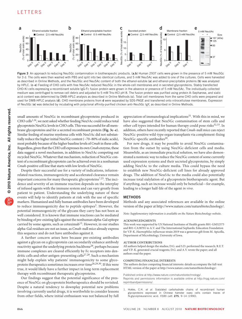
Figure 2 Effects of Neu5Gc-specific antibodies on the kinetics of therapeutic antibodies in mice with a human-like Neu5Gc deficiency, levels of anti-Neu5Gc IgG in mice after injections of the therapeutic antibodies, and binding of IgG Neu5Gc-specific antibodies from whole human serum to Neu5Gc on the Fab fragment of cetuximab. (a) Cmah-null mice were first injected intravenously with either of the therapeutic antibodies, cetuximab (Cet) or panitumumab (Pan). Serum from Cmah-null mice containing anti-Neu5Gc antibodies (or serum from naïve mice or control-immunized mice) was then passively transferred by intraperitoneal injection. Mice were bled periodically after the passive transfer of serum. Concentrations of Cet or Pan in the isolated sera were determined by sandwich ELISA. Absorbance was measured at 495 nm. The y axis starts at 60% to better display the difference in kinetics. Error bars, s.d.; ***P < 0.001, unpaired two-tailed t-test. (b) Cmah-null mice were injected intravenously with Cet, Pan or mouse IgG weekly and were bled initially and after the third intravenous injection. To detect Neu5Gc-specific antibodies by ELISA, we coated wells with human (Neu5Gc-deficient) or chimpanzee (Neu5Gc-positive) serum glycoproteins (upper chart), or alternatively with human or bovine fibrinogen (lower chart). Data were obtained in triplicate. (c) Fab fragments of Cet and Pan were isolated using the Pierce Fab Preparation Kit according to the manufacturer’s manual for use as target molecules in ELISA (1 μg per well). Sialic acid–specific binding was determined using mild sodium metaperiodate pretreatment. Wells were then blocked and incubated with human sera (S30 and S34, with low and high anti-Neu5Gc IgG titers, respectively4). Binding of human IgG was detected using anti-human IgG-Fc. Absorbance was measured at 490 nm and ELISA samples were studied in triplicate. Error bars, s.d.; *P < 0.05, paired two-tailed t-test.
0.8 mlgG Pan Cet
0.60.40.2
0
Cha
nge
in A
495
A49
5
–0.20.3
0.2
0.1
0
S34S30
–0.1
PeriodateFab Cet Fab Pan
Mock Periodate Mock
0.6 *10050Hours
*** ***
60
80
Circ
ulat
ing
Cet
or
Pan
(nor
mal
ized
)
100a b
c0
0.4NaÏvemouseserum
Cet
Pan
Serum ofcontrol-
immunizedmice
Serum ofNeu5Gc-
immunizedmice
0.2
0
© 2
010
Nat
ure
Am
eric
a, In
c. A
ll ri
gh
ts r
eser
ved
.
866 VOLUME 28 NUMBER 8 AUGUST 2010 nature biotechnology
l e t t e r s
small amounts of Neu5Gc in recombinant glycoproteins produced in CHO cells1,16, we next asked whether feeding Neu5Ac could reduce total glycoprotein Neu5Gc levels in CHO cells. This was successful for all mem-brane glycoproteins and for a secreted recombinant protein (Fig. 3c–e). Similar feeding of murine myeloma cells with Neu5Ac did not substan-tially reduce the higher initial Neu5Gc content (~70–80% of sialic acids), most probably because of the higher baseline levels of Cmah in these cells. Regardless, given that the CHO cell expresses its own Cmah enzyme, these data suggest a novel mechanism, in addition to Neu5Ac competing out recycled Neu5Gc. Whatever that mechanism, reduction of Neu5Gc con-tent of a recombinant glycoprotein can be achieved even in a nonhuman Cmah-positive cell line that starts with low levels of Neu5Gc.
Despite their successful use for a variety of indications, infusion-related reactions, immunogenicity and accelerated clearance remain important concerns for many therapeutic glycoproteins7,25. The inci-dence and severity of an immune reaction depends on the interplay of infused agents with the immune system and can vary greatly from patient to patient. Understanding the underlying nature of these events will help to identify patients at risk with the use of specific markers. Humanized and fully human antibodies have been developed to reduce immunogenicity due to peptide epitopes5. However, the potential immunogenicity of the glycans they carry has not been as well considered. It is known that immune reactions can be mediated by binding of pre-existing IgEs against the nonhuman alpha-Gal epitope carried by some agents, such as cetuximab13. However, in our studies alpha-Gal residues are not an issue, as Cmah-null mice already express this sequence and do not have antibodies against it.
A further concern arises here because pre-existing antibodies against a glycan on a glycoprotein can secondarily enhance antibody reactivity against the underlying protein backbone26, perhaps because immune complexes are cleared efficiently by Fc receptors into den-dritic cells and other antigen-presenting cells27,28. Such a mechanism might help explain why patients’ immunogenicity to some glyco-protein therapeutics sometimes increases over time26,29,30. If this were true, it would likely have a further impact in long-term replacement therapy with recombinant therapeutic glycoproteins.
Our findings suggest that the potential significance of the pres-ence of Neu5Gc on glycoprotein biotherapeutics should be revisited. Despite a natural tendency to downplay potential new problems involving currently useful drugs, it is worthwhile to consider lessons from other fields, where initial enthusiasm was not balanced by full
appreciation of immunological implications31. With this in mind, we have also suggested that Neu5Gc contamination of stem cells and other cell types intended for human therapy could pose risks32,33. In addition, others have recently reported that Cmah-null mice can reject Neu5Gc-positive wild-type organ transplants via complement-fixing Neu5Gc-specific antibodies20.
For new drugs, it may be possible to avoid Neu5Gc contamina-tion from the outset by using Neu5Gc-deficient cells and media. Meanwhile, as an immediate practical solution, we have also demon-strated a nontoxic way to reduce the Neu5Gc content of some currently used expression systems and their secreted glycoproteins, by simply adding Neu5Ac to the culture media. This could bypass the need to establish new Neu5Gc-deficient cell lines for already approved drugs. The addition of Neu5Ac to the media could also potentially increase total sialylation of a glycoprotein biotherapeutic agent. But if anything, such an increase would only be beneficial—for example, leading to a longer half-life of the agent in vivo.
METHOdSMethods and any associated references are available in the online version of the paper at http://www.nature.com/naturebiotechnology/.
Note: Supplementary information is available on the Nature Biotechnology website.
AcKnowlEDGmEnTSThis work was supported by US National Institutes of Health grants R01-GM32373 and R01-CA38701 to A.V. and The International Sephardic Education Foundation for V.P.-K. Haemophilus influenzae strain 2019 was a generous gift from M. Apicella, Department of Microbiology, University of Iowa.
AUTHoR conTRIBUTIonSAll authors helped design the studies; D.G. and S.D. performed the research; R.E.T. and V.P.-K. generated crucial reagents; D.G. and A.V. wrote the paper; and all authors read the paper.
comPETInG FInAncIAl InTERESTSThe authors declare competing financial interests: details accompany the full-text HTML version of the paper at http://www.nature.com/naturebiotechnology/.
Published online at http://www.nature.com/naturebiotechnology/. reprints and permissions information is available online at http://npg.nature.com/reprintsandpermissions/.
1. Hokke, C.H. et al. Sialylated carbohydrate chains of recombinant human glycoproteins expressed in Chinese hamster ovary cells contain traces of N-glycolylneuraminic acid. FEBS Lett. 275, 9–14 (1990).
80
a b c d eEthanol-soluble fraction Secreted protein
Control 5 mM Neu5Ac Control 5 mM Neu5Ac Control 5 mM Neu5AcControl 5 mM Neu5Ac
Ethanol-precipitable fraction 5 mM Neu5AcSize– +
Neu
5Gc
(% o
f tot
al s
ialic
aci
ds)
Neu
5Gc
(% o
f tot
al s
ialic
aci
ds)
Neu
5Gc
(% o
f tot
al s
ialic
aci
ds)
Neu
5Gc
(% o
f tot
al s
ialic
aci
ds)
60 15
10
5
0
10
Membrane fraction
5
0
40
Day 0
Day 1
Day 2
Day 3
Day 4
Day 5
Day 3
Day 5
Day 7
Day 3
Day 5
Day 7
Day 0
Day 1
Day 2
Day 3
Day 4
Day 5
197 (kDa)125
83
3720
0
80
60
40
20
0
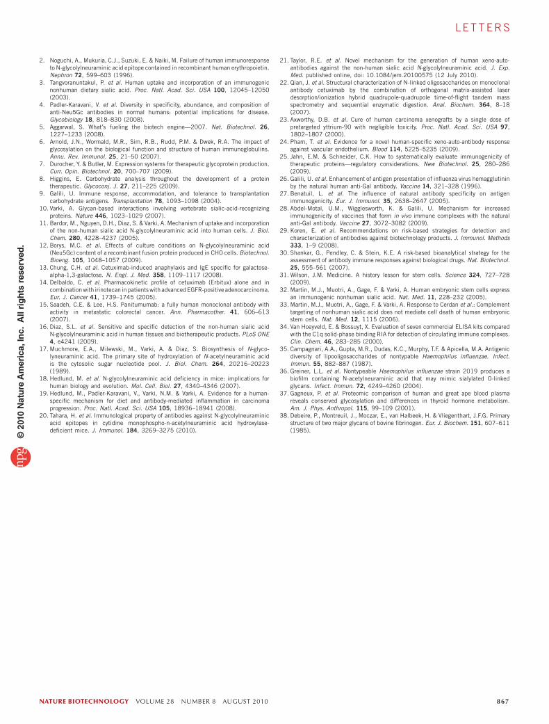
Figure 3 An approach to reducing Neu5Gc contamination in biotherapeutic products. (a,b) Human 293T cells were grown in the presence of 5 mM Neu5Gc for 3 d. The cells were then washed with PBS and split into two identical cultures, and 5 mM Neu5Ac was added to one of the cultures. Cells were harvested as described in Online Methods, and the Neu5Gc and Neu5Ac content of both the ethanol-soluble (a) and ethanol-precipitable proteins (b) was analyzed by HPLC. (c–e) Feeding of CHO cells with free Neu5Ac reduced Neu5Gc in the whole-cell membranes and in secreted glycoproteins. Stably transfected CHO-KI cells expressing a recombinant soluble IgG-Fc fusion protein were grown in the absence or presence of 5 mM Neu5Ac. The individually collected medium was centrifuged to remove cell debris and adjusted to 5 mM Tris-HCl pH 8. The fusion protein was purified using protein A–Sepharose, and sialic acid content was determined by DMB-HPLC analysis as described in Online Methods (c). Total cell membranes from the same CHO cells were prepared and used for DMB-HPLC analysis (d). CHO membrane proteins from d were separated by SDS-PAGE and transferred onto nitrocellulose membranes. Expression of Neu5Gc (e) was detected by incubating with polyclonal affinity-purified chicken anti-Neu5Gc IgY, as described in Online Methods.
© 2
010
Nat
ure
Am
eric
a, In
c. A
ll ri
gh
ts r
eser
ved
.
nature biotechnology VOLUME 28 NUMBER 8 AUGUST 2010 867
l e t t e r s
2. Noguchi, A., Mukuria, C.J., Suzuki, E. & Naiki, M. Failure of human immunoresponse to N-glycolylneuraminic acid epitope contained in recombinant human erythropoietin. Nephron 72, 599–603 (1996).
3. Tangvoranuntakul, P. et al. Human uptake and incorporation of an immunogenic nonhuman dietary sialic acid. Proc. Natl. Acad. Sci. USA 100, 12045–12050 (2003).
4. Padler-Karavani, V. et al. Diversity in specificity, abundance, and composition of anti-Neu5Gc antibodies in normal humans: potential implications for disease. Glycobiology 18, 818–830 (2008).
5. Aggarwal, S. What′s fueling the biotech engine—2007. Nat. Biotechnol. 26, 1227–1233 (2008).
6. Arnold, J.N., Wormald, M.R., Sim, R.B., Rudd, P.M. & Dwek, R.A. The impact of glycosylation on the biological function and structure of human immunoglobulins. Annu. Rev. Immunol. 25, 21–50 (2007).
7. Durocher, Y. & Butler, M. Expression systems for therapeutic glycoprotein production. Curr. Opin. Biotechnol. 20, 700–707 (2009).
8. Higgins, E. Carbohydrate analysis throughout the development of a protein therapeutic. Glycoconj. J. 27, 211–225 (2009).
9. Galili, U. Immune response, accommodation, and tolerance to transplantation carbohydrate antigens. Transplantation 78, 1093–1098 (2004).
10. Varki, A. Glycan-based interactions involving vertebrate sialic-acid-recognizing proteins. Nature 446, 1023–1029 (2007).
11. Bardor, M., Nguyen, D.H., Diaz, S. & Varki, A. Mechanism of uptake and incorporation of the non-human sialic acid N-glycolylneuraminic acid into human cells. J. Biol. Chem. 280, 4228–4237 (2005).
12. Borys, M.C. et al. Effects of culture conditions on N-glycolylneuraminic acid (Neu5Gc) content of a recombinant fusion protein produced in CHO cells. Biotechnol. Bioeng. 105, 1048–1057 (2009).
13. Chung, C.H. et al. Cetuximab-induced anaphylaxis and IgE specific for galactose-alpha-1,3-galactose. N. Engl. J. Med. 358, 1109–1117 (2008).
14. Delbaldo, C. et al. Pharmacokinetic profile of cetuximab (Erbitux) alone and in combination with irinotecan in patients with advanced EGFR-positive adenocarcinoma. Eur. J. Cancer 41, 1739–1745 (2005).
15. Saadeh, C.E. & Lee, H.S. Panitumumab: a fully human monoclonal antibody with activity in metastatic colorectal cancer. Ann. Pharmacother. 41, 606–613 (2007).
16. Diaz, S.L. et al. Sensitive and specific detection of the non-human sialic acid N-glycolylneuraminic acid in human tissues and biotherapeutic products. PLoS ONE 4, e4241 (2009).
17. Muchmore, E.A., Milewski, M., Varki, A. & Diaz, S. Biosynthesis of N-glyco-lyneuraminic acid. The primary site of hydroxylation of N-acetylneuraminic acid is the cytosolic sugar nucleotide pool. J. Biol. Chem. 264, 20216–20223 (1989).
18. Hedlund, M. et al. N-glycolylneuraminic acid deficiency in mice: implications for human biology and evolution. Mol. Cell. Biol. 27, 4340–4346 (2007).
19. Hedlund, M., Padler-Karavani, V., Varki, N.M. & Varki, A. Evidence for a human-specific mechanism for diet and antibody-mediated inflammation in carcinoma progression. Proc. Natl. Acad. Sci. USA 105, 18936–18941 (2008).
20. Tahara, H. et al. Immunological property of antibodies against N-glycolylneuraminic acid epitopes in cytidine monophospho-n-acetylneuraminic acid hydroxylase-deficient mice. J. Immunol. 184, 3269–3275 (2010).
21. Taylor, R.E. et al. Novel mechanism for the generation of human xeno-auto-antibodies against the non-human sialic acid N-glycolylneuraminic acid. J. Exp. Med. published online, doi: 10.1084/jem.20100575 (12 July 2010).
22. Qian, J. et al. Structural characterization of N-linked oligosaccharides on monoclonal antibody cetuximab by the combination of orthogonal matrix-assisted laser desorption/ionization hybrid quadrupole-quadrupole time-of-flight tandem mass spectrometry and sequential enzymatic digestion. Anal. Biochem. 364, 8–18 (2007).
23. Axworthy, D.B. et al. Cure of human carcinoma xenografts by a single dose of pretargeted yttrium-90 with negligible toxicity. Proc. Natl. Acad. Sci. USA 97, 1802–1807 (2000).
24. Pham, T. et al. Evidence for a novel human-specific xeno-auto-antibody response against vascular endothelium. Blood 114, 5225–5235 (2009).
25. Jahn, E.M. & Schneider, C.K. How to systematically evaluate immunogenicity of therapeutic proteins—regulatory considerations. New Biotechnol. 25, 280–286 (2009).
26. Galili, U. et al. Enhancement of antigen presentation of influenza virus hemagglutinin by the natural human anti-Gal antibody. Vaccine 14, 321–328 (1996).
27. Benatuil, L. et al. The influence of natural antibody specificity on antigen immunogenicity. Eur. J. Immunol. 35, 2638–2647 (2005).
28. Abdel-Motal, U.M., Wigglesworth, K. & Galili, U. Mechanism for increased immunogenicity of vaccines that form in vivo immune complexes with the natural anti-Gal antibody. Vaccine 27, 3072–3082 (2009).
29. Koren, E. et al. Recommendations on risk-based strategies for detection and characterization of antibodies against biotechnology products. J. Immunol. Methods 333, 1–9 (2008).
30. Shankar, G., Pendley, C. & Stein, K.E. A risk-based bioanalytical strategy for the assessment of antibody immune responses against biological drugs. Nat. Biotechnol. 25, 555–561 (2007).
31. Wilson, J.M. Medicine. A history lesson for stem cells. Science 324, 727–728 (2009).
32. Martin, M.J., Muotri, A., Gage, F. & Varki, A. Human embryonic stem cells express an immunogenic nonhuman sialic acid. Nat. Med. 11, 228–232 (2005).
33. Martin, M.J., Muotri, A., Gage, F. & Varki, A. Response to Cerdan et al.: Complement targeting of nonhuman sialic acid does not mediate cell death of human embryonic stem cells. Nat. Med. 12, 1115 (2006).
34. Van Hoeyveld, E. & Bossuyt, X. Evaluation of seven commercial ELISA kits compared with the C1q solid-phase binding RIA for detection of circulating immune complexes. Clin. Chem. 46, 283–285 (2000).
35. Campagnari, A.A., Gupta, M.R., Dudas, K.C., Murphy, T.F. & Apicella, M.A. Antigenic diversity of lipooligosaccharides of nontypable Haemophilus influenzae. Infect. Immun. 55, 882–887 (1987).
36. Greiner, L.L. et al. Nontypeable Haemophilus influenzae strain 2019 produces a biofilm containing N-acetylneuraminic acid that may mimic sialylated O-linked glycans. Infect. Immun. 72, 4249–4260 (2004).
37. Gagneux, P. et al. Proteomic comparison of human and great ape blood plasma reveals conserved glycosylation and differences in thyroid hormone metabolism. Am. J. Phys. Anthropol. 115, 99–109 (2001).
38. Debeire, P., Montreuil, J., Moczar, E., van Halbeek, H. & Vliegenthart, J.F.G. Primary structure of two major glycans of bovine fibrinogen. Eur. J. Biochem. 151, 607–611 (1985).
© 2
010
Nat
ure
Am
eric
a, In
c. A
ll ri
gh
ts r
eser
ved
.

nature biotechnology doi:10.1038/nbt.1651
ONLINE METHOdSMice. The Cmah-null mice used for this study have been described previously18 and were backcrossed to C57Bl/6 mice for over ten generations. All experi-ments were approved by the University of California, San Diego Institutional Review Board committee responsible for approving animal experiments.
Sialidase treatment of therapeutic antibodies. One milligram each of cetuxi-mab or panitumumab (obtained from the University of California, San Diego pharmacy or the manufacturer) were treated with 50 mU of active or heat-inactivated Arthrobacter ureafaciens sialidase (EY Laboratories) in 100 mM sodium acetate pH 5.5, at 37 °C for 24 h. Samples were used for ELISA or western blots.
Periodate treatment of therapeutic antibodies on ELISA plate. Untreated cetuximab and panitumumab (1 μg per well) were used for coating, then blocked with PBST for 2 h and incubated with freshly made 2 mM sodium metaperiodate in PBS for 20 min at 4 °C in the dark. The reaction was stopped by addition of 200 mM sodium borohydride to a final concentration of 20 mM. As a control, periodate and borohydride were premixed and then added to the wells (the borohydride inactivates the periodate). To remove resulting borates, wells were then washed three times with 100 mM sodium acetate with 100 mM NaCl pH 5.5 before further analysis.
ELISA detection of Neu5Gc on therapeutic antibodies. For the ELISA, wells were coated with 1 μg of cetuximab or panitumumab (either before sialidase treatment or after periodate treatment), blocked with TBST for 2 h and then incubated with affinity-purified chicken anti-Neu5Gc IgY or control IgY for 1 h (1:20,000 in TBST). Binding of IgY was detected using horseradish peroxidase (HRP)-conjugated donkey anti-chicken IgY (1:50,000 in TBST) and developed with O-phenylenediamine in citrate-phosphate buffer, pH 5.5, with absorbance measured at 495 nm. ELISA samples were studied at least in triplicate. Similar to the ELISA with the anti-Neu5Gc chicken IgY, human anti-Neu5Gc IgG that had been purified from the serum of healthy humans and biotinylated (exactly as described in ref. 4) was also used as the primary antibody (1:100 in TBST). Binding of the human antibodies to the therapeutic antibodies was detected using HRP-conjugated streptavidin (1:10,000) followed by development as described above. Samples were studied in triplicate.
Western blot detection of Neu5Gc on therapeutic antibodies. For western blot detection, cetuximab or panitumumab (1 μg per lane) was separated by 12.5% SDS-PAGE and Coomassie-stained or blotted on nitrocellulose membranes. Blotted membranes were blocked with TBST containing 0.5% cold-water fish-skin gelatin overnight at 4 °C and subsequently incubated with affinity-purified chicken anti-Neu5Gc IgY for 4 h at room temperature (1:100,000 in TBST). Binding of the chicken anti-Neu5Gc IgY was detected using HRP-conjugated donkey anti-chicken IgY for 1 h (1:50,000 in TBST), followed by incubation with SuperSignal West Pico Substrate (Pierce) as per the manufacturer’s recommendation, exposure to X-ray film and development of the film. Similar to the western blot with the chicken anti-Neu5Gc IgY, purified biotinylated human anti-Neu5Gc IgG was also used as the primary antibody (1:100 in TBST). Binding of the human antibodies to the therapeutic antibodies was detected using HRP-conjugated streptavidin (1:10,000 in TBST) followed by development as described above.
CIC-C1q binding assay. Immune complex formation was detected using the CIC (C1Q) ELISA Kit (Buehlmann) as described in the manufacturer’s guidelines34. Briefly, 100 μl of human serum with low or high anti-Neu5Gc (S30 and S34, respectively4) was incubated with 40 μg of cetuximab or pani-tumumab for 14 h at 4 °C. We applied 1:50 dilutions of the mix to human C1q–coated ELISA wells and incubated for 1 h at 25 °C. Binding was detected using alkaline phosphatase–conjugated protein A. After another washing step, the enzyme substrate (para-nitrophenylphosphate) was added, followed by a stopping step. The absorbance was measured at 405 nm. Samples were studied in triplicate.
Generation of murine Neu5Gc-specific antibodies. Haemophilus influenzae strain 2019 (ref. 35) was a generous gift from M. Apicella, Department of
Microbiology, University of Iowa. Bacteria were grown to mid log phase in sialic acid–free media36 with or without addition of 1 mM Neu5Gc21, heat-killed and injected intraperitoneally (200 μl of culture at an absorbance of 600 nm of 0.4) into Cmah-null mice.
Effects of anti-Neu5Gc antibodies on in vivo kinetics of therapeutic anti-bodies. Cetuximab or panitumumab in PBS (0.24 μg per gram mouse body weight) were injected intravenously, and 14 h later, mouse serum pooled from syngeneic Cmah-null mice containing anti-Neu5Gc antibodies (or pooled serum from syngeneic naïve or control-immunized mice) was passively trans-ferred via intraperitoneal injection into syngeneic Cmah-null mice that were prescreened for the absence of pre-existing antibodies against human IgG. Mice were bled 0, 2, 8, 32, 56 and 80 h after the passive transfer of mouse serum. For quantification of therapeutic antibody concentrations in the sera, wells of ELISA plates were coated with 1 μg of anti-human IgG (Biorad), then blocked with TBST for 2 h and incubated with 1:500 dilutions of the sera in each well. Captured therapeutic antibodies were detected by HRP-conjugated anti-human Fc (Jackson; 1:10,000), with development by O-phenylenediamine in citrate-phosphate buffer, pH 5.5, and absorbance measured at 495 nm (n = 5 for injections of both control sera groups; n = 10 for injections of anti-Neu5Gc serum groups).
Quantification of Neu5Gc-specific IgG antibodies in Neu5Gc-immunized mice. A Neu5Gcα2-6Galβ1-4Glc-conjugate4 (1 μg per well) and serial dilu-tions of mouse IgG as standards (0.625–20 ng per well) were used for coating overnight, then blocked with PBST for 2 h and incubated with pooled serum from Neu5Gc-immunized mice (1:250 dilution) for 2 h at 25 °C. Binding of mouse IgG was detected using HRP-conjugated goat anti-mouse IgG-Fc (Jackson; 1:10,000 in PBST) and developed with O-phenylenediamine in citrate-phosphate buffer, pH 5.5, with absorbance measured at 490 nm. ELISA samples were studied in triplicate.
Levels of anti-Neu5Gc IgG after injections of the antibodies. Cmah-null mice were injected intravenously with 4 μg antibody per gram of mouse body weight in PBS weekly for 3 weeks. Mice were bled initially, and again 1 week after the third intravenous injection. Wells of ELISA plates were coated with 1:1,000 dilutions of human (Neu5Gc-deficient) or chimpanzee (Neu5Gc-positive) serum glycoproteins (note that the only major difference between human and chimp serum glycosylation is the absence or presence of Neu5Gc; ref. 37). Alternatively, wells were coated with human or bovine fibrinogen, which carry Neu5Ac or Neu5Gc on otherwise identical N-glycans38. Wells were then blocked with TBST for 2 h followed by incubation with 1:100 dilutions of the mouse sera. Binding of the mouse antibodies was detected using HRP-conjugated goat anti-mouse IgG Fc fragment (1:10,000 in TBST). Neu5Gc-specific binding (change in absorbance at 495 nm) was determined by subtracting the background signal of the wells coated with human serum or human fibrinogen (no Neu5Gc) from the signal of chimpanzee serum–coated or bovine fibrinogen–coated wells (containing Neu5Gc). Data were obtained in triplicate (n = 5 for injection of mouse IgG; n = 4 for injection of pani-tumumab; n = 6 for injection of cetuximab ).
An approach to reduce Neu5Gc contamination in biotherapeutic products. Human 293T kidney cells were grown in DME supplemented with 10% (vol/vol) fetal calf serum. Cells were lifted from the culture plate using 20 mM EDTA in PBS and allowed to grow to 50% confluence. At this point, buffered 100 mM Neu5Gc was added to the culture in duplicate for a final 5 mM concentration, and the cells were grown in this supplemented media for 3 d. At the end of this Neu5Gc pulse, the cells were once again lifted using 20 mM EDTA in PBS, pelleted, washed once with PBS to remove any excess Neu5Gc and then suspended in 30 ml of growth medium. We added 5 ml of this cell suspension to each of five P-100 dishes. We immediately harvested the last aliquot of cell suspension, at time 0, by pelleting the cells, washing once with PBS, suspend-ing them in 1 ml of PBS and transferring them to a 1.5-ml microcentrifuge tube. The cells were repelleted and frozen until all time points were collected. Buffered 100 mM Neu5Ac was added to each of the other five plates for the ‘Neu5Ac chase’ and an equivalent amount of media added to the ‘minus chase’ samples. We harvested cells at days 1, 2, 3, 4 and 5 by scraping them into the
© 2
010
Nat
ure
Am
eric
a, In
c. A
ll ri
gh
ts r
eser
ved
.
nature biotechnologydoi:10.1038/nbt.1651
culture media, collecting by pelleting, washing once with PBS, transferring them to a 1.5-ml microcentrifuge tube, pelleting and freezing the cell pellet. At the end of the 5 d of chase, all collected cell pellets were homogenized in 300 μl of ice-cold 20 mM potassium phosphate pH 7 using a 3- to 20-s burst with a Fisher Sonicator. We precipitated glycoconjugate-bound sialic acids by adding 700 μl of 100% ice-cold ethanol (final 70% (vol/vol) correct ethanol) and incubating at −20 °C overnight. The samples were spun at 20,000g for 15 min and the supernatants transferred to clean tubes and dried on a speed vac. The precipitated glycoconjugates and dried ethanol supernatants were each suspended in 100 μl of 20 mM potassium phosphate pH 7 by sonication. Sialic acids were released from both fractions by acid hydrolysis with 2 M acetic
acid (final) and incubation at 80 °C for 3 h. Samples were passed through a Microcon-10 filter and the filtrate derivatized with DMB (1,2-diamino-4, 5-methylenedioxybenzene) reagent for analysis of sialic acids by HPLC.
A similar approach was taken with CHO cells stably expressing a Siglec-Fc protein in the medium, except that the Neu5Gc pulse was omitted and the secreted glycoproteins were captured on protein A–Sepharose beads. The cells were also processed similarly, except that total cell membranes were pelleted by centrifugation. The sialic acid content of the secreted proteins and cell membranes was determined by acid hydrolysis, DMB derivatization and HPLC. The cell membranes were also studied by western blotting with the chicken anti-Neu5Gc IgY, as described above.
© 2
010
Nat
ure
Am
eric
a, In
c. A
ll ri
gh
ts r
eser
ved
.