Embed Size (px)
Citation preview

6/24/2014
1
Implementation in Clinical Practice:
Challenges in Developing Economies
Umesh Mahantshetty, DMRT, MD, DNB (RT)Department of Radiation Oncology
Tata Memorial Hospital
Mumbai, India
Disclosure
Umesh Mahantshetty, DMRT, MD, DNB (RT), does not have any financial relationships or products or devices with any commercial interest related to the content of this activity of any amount during the past 12 months.

6/24/2014
2
Tata Memorial Hospital
ACTRECTata Memorial Centre
ACTREC
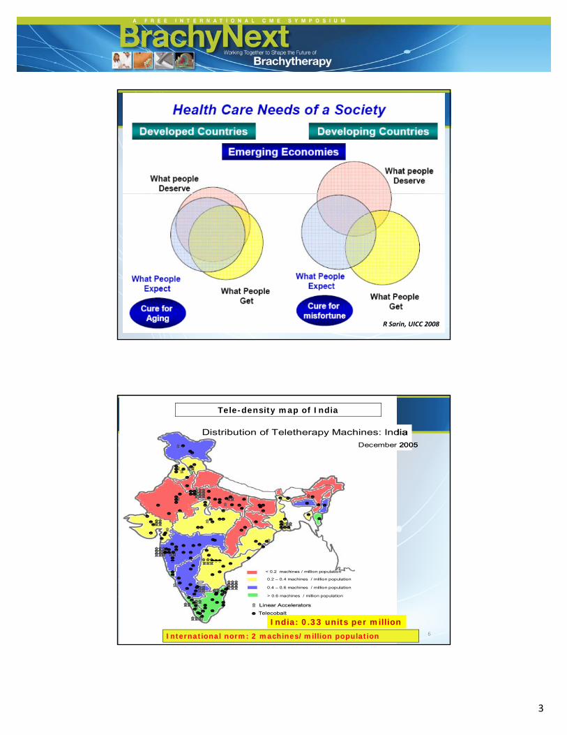
Cancer Care Disparity: A Harsh Reality
6/24/2014
3
R Sarin, UICC 2008
۩۩
۩۩۩۩۩۩۩۩
Distribution of Teletherapy Machines: India
December 2005
☻
☻
☻
☻☻
☻☻
☻
☻☻
۩۩
۩۩۩۩۩۩۩۩۩۩۩۩
Distribution of Teletherapy Machines: India
December 2005
☻
☻
☻
☻☻
☻☻
☻
☻☻
Tele-density map of India
۩۩۩۩۩۩۩۩ ۩۩۩۩
۩۩
۩۩۩۩۩۩۩۩
۩۩۩۩
۩۩۩۩۩۩ ۩۩۩۩۩۩ ۩۩۩۩۩۩
۩۩۩۩۩۩ ۩۩۩۩۩۩ ۩۩۩۩۩۩
۩۩
۩۩
۩۩۩۩۩۩۩۩
۩۩۩۩
۩۩۩۩
☻☻
☻
☻
☻
☻
☻
☻
☻
☻☻
☻ ☻☻
☻
☻
☻
☻☻
☻
☻
☻
☻
☻
☻
☻
☻
☻
☻
☻
☻
☻
☻
☻
☻☻
☻☻
☻
☻
☻ ☻☻☻
☻☻
☻
☻☻☻
☻☻☻
☻☻☻☻
☻
☻☻
☻☻
☻
☻
☻☻
☻☻
☻☻
☻☻
☻ ☻☻
☻☻
☻☻
☻
☻
☻☻☻
☻☻
☻☻
☻
☻
۩۩۩۩۩۩۩۩ ۩۩۩۩۩۩۩۩۩۩۩۩ ۩۩۩۩
۩۩
۩۩۩۩۩۩۩۩۩۩۩۩۩۩۩۩
۩۩۩۩۩۩۩۩
۩۩۩۩۩۩ ۩۩۩۩۩۩ ۩۩۩۩۩۩۩۩۩۩۩۩ ۩۩۩۩۩۩ ۩۩۩۩۩۩
۩۩۩۩۩۩ ۩۩۩۩۩۩ ۩۩۩۩۩۩۩۩۩۩۩۩ ۩۩۩۩۩۩ ۩۩۩۩۩۩
۩۩
۩۩
۩۩۩۩۩۩۩۩۩۩۩۩۩۩
۩۩۩۩۩۩۩۩
۩۩۩۩
☻☻
☻
☻
☻
☻
☻
☻
☻
☻☻
☻ ☻☻
☻
☻
☻
☻☻
☻
☻
☻
☻
☻
☻
☻
☻
☻
☻
☻
☻
☻
☻
☻
☻☻
☻☻
☻
☻
☻ ☻☻☻
☻☻
☻
☻☻☻
☻☻☻
☻☻☻☻
☻
☻☻
☻☻
☻
☻
☻☻
☻☻
☻☻
☻☻
☻ ☻☻
☻☻
☻☻
☻
☻
☻☻☻
☻☻
☻☻
☻
☻
6
< 0.2 machines / million population
0.2 – 0.4 machines / million population
0.4 – 0.6 machines / million population
> 0.6 machines / million population
۩۩۩۩۩۩۩۩ ۩۩۩۩
۩۩۩۩۩۩ ۩۩۩۩۩۩ ۩۩۩۩۩۩
۩۩۩۩۩۩
۩۩
۩۩۩۩۩۩
۩۩۩۩
۩۩۩۩
۩۩۩۩۩۩۩۩
۩۩ Linear AcceleratorsLinear Accelerators
☻ TelecobaltTelecobalt
☻
☻☻
☻
☻☻
☻
☻
☻
☻
☻
☻☻
☻☻
☻☻
☻☻
☻
☻
☻
☻
☻ ☻☻
☻
☻☻
☻☻
☻☻☻☻☻
☻
☻☻☻
☻
☻
☻
☻
< 0.2 machines / million population
0.2 – 0.4 machines / million population
0.4 – 0.6 machines / million population
> 0.6 machines / million population
۩۩۩۩۩۩۩۩ ۩۩۩۩۩۩۩۩۩۩۩۩ ۩۩۩۩
۩۩۩۩۩۩ ۩۩۩۩۩۩ ۩۩۩۩۩۩۩۩۩۩۩۩ ۩۩۩۩۩۩ ۩۩۩۩۩۩
۩۩۩۩۩۩۩۩۩۩۩۩
۩۩
۩۩۩۩۩۩۩۩۩۩
۩۩۩۩۩۩۩۩
۩۩۩۩۩۩۩۩
۩۩۩۩۩۩۩۩۩۩۩۩۩۩۩۩
۩۩ Linear AcceleratorsLinear Accelerators
☻ TelecobaltTelecobalt
☻
☻☻
☻
☻☻
☻
☻
☻
☻
☻
☻☻
☻☻
☻☻
☻☻
☻
☻
☻
☻
☻ ☻☻
☻
☻☻
☻☻
☻☻☻☻☻
☻
☻☻☻
☻
☻
☻
☻
International norm: 2 machines/million population
India: 0.33 units per million

6/24/2014
4
Radiation therapy facilities in India
AERB; January 2014
• RT Centres 357
• Tele - cobalt 231
• Medical LINACCyberknife
TomotherapyGammaknife
286050307
• HDR Brachy 250
y
Atomic Energy Regulatory Board, India, Newsletter 2013
• HDR Brachy 250
• LDR Manual 25
• CT Simulator 120
• RT Simulator 160
Estimated number of new cases of Ca. Cervix1990 - 2050
1
1.2
n
0
0.2
0.4
0.6
0.8
1
Nu
mb
ers
in m
illio
n
World 0 37 0 45 0 58 0 7 0 98
1990 2000 2010 2020 2050
Source : Ferlay J, Parikh D.M. & Pisani P IARC, Lyon Globocan 2002
World 0.37 0.45 0.58 0.7 0.98
Developed Countries 0.07 0.089 0.089 0.092 0.094
Developing Countries 0.29 0.37 0.51 0.64 0.97
India 0.09 0.11 0.16 0.2 0.33

6/24/2014
5
Five Most Common Cancers in Indian WomenEstimated No. of New Cases & Deaths (in Thousands): Year 2005*
71127Cervix uteri
15
16
52
25
24
27
100
23
Ovary
Oesophagus
Oral cavity
Breast
Deaths New cases
5 most common cancers account for almost two‐thirds
of total cases and deaths due to cancer in Indian women
* estimated
0 20 40 60 80 100 120 140
Down the DecadesCancer Cervix: Tata Memorial Hospital 1941–2000
1663518000
4679
9055
13662
1663515080
6000
8000
10000
12000
14000
16000
2102
4679
0
2000
4000
1941-50 '51-60 '61-70 '71-80 '81-90 '91-00

6/24/2014
6
Down Staging of Carcinoma Cervix
63.3
68.565.8
70
1985
Tata Memorial Hospital Cancer Registry
1985–2002
28.2 28
35.2
54.151.7
30
40
50
601985
1989
1993
1997
20002002
44.5
28.8
10 9.1
11.8
6.19.5
17.119.7
7.710.6
0
3.6
0
10
20
Stage I Stage II Stage III Stage IV
5.2
9.1
• Applicator development: Intracavitary (IC), Interstitial (IS) & IC+IS
Advances in Gynecological BrachytherapyTata Memorial Hospital Experience
• Incorporation of newer imaging modalities: CT, MR, US, etc.
• Advances in treatment planning systems
• Image‐/volume‐based brachytherapy
6/24/2014
7
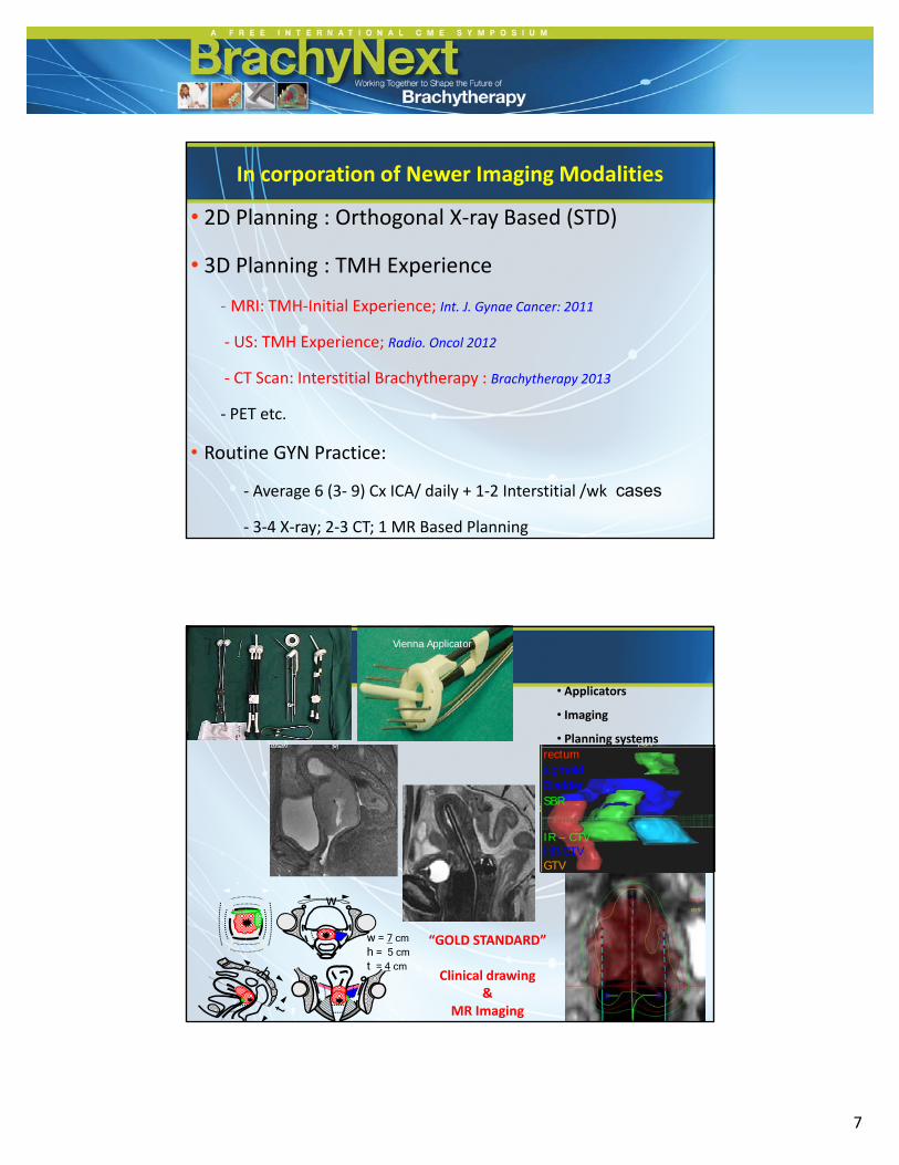
In corporation of Newer Imaging Modalities
• 2D Planning : Orthogonal X‐ray Based (STD)
• 3D Planning : TMH Experienceg p
‐ MRI: TMH‐Initial Experience; Int. J. Gynae Cancer: 2011
‐ US: TMH Experience; Radio. Oncol 2012
‐ CT Scan: Interstitial Brachytherapy : Brachytherapy 2013
‐ PET etc.
• Routine GYN Practice:
‐ Average 6 (3‐ 9) Cx ICA/ daily + 1‐2 Interstitial /wk cases
‐ 3‐4 X‐ray; 2‐3 CT; 1 MR Based Planning
Vienna Applicator
• Applicators
• Imaging
• Planning systems rectum
IR – CTVHR-CTVGTV
rectumsigmoidBladderSBR
w = 7 cmh = 5 cmt = 4 cm
w
“GOLD STANDARD”
Clinical drawing &
MR Imaging
6/24/2014
8
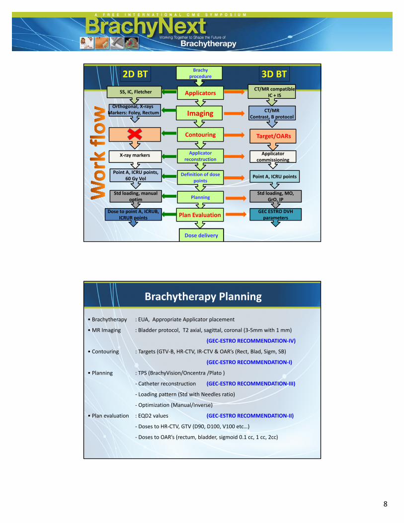
Brachyprocedure
SS, IC, Fletcher
Orthogonal, X‐raysMarkers: Foley, Rectum
CT/MR compatibleIC + IS
Imaging CT/MRContrast, B protocol
Applicators
2D BT 3D BT
X‐ray markers
Target/OARsContouring
Applicator reconstruction
Definition of dose points
Applicator commissioning
Point A, ICRU points, 60 Gy Vol Point A, ICRU points
po ts
PlanningStd loading, manual
optimStd loading, MO,
GrO, IP
Plan EvaluationDose to point A, ICRUB,
ICRUR pointsGEC ESTRO DVH parameters
Dose delivery
Brachytherapy Planning
• Brachytherapy : EUA, Appropriate Applicator placement
• MR Imaging : Bladder protocol, T2 axial, sagittal, coronal (3‐5mm with 1 mm)
(GEC ESTRO RECOMMENDATION IV)(GEC‐ESTRO RECOMMENDATION‐IV)
• Contouring : Targets (GTV‐B, HR‐CTV, IR‐CTV & OAR’s (Rect, Blad, Sigm, SB)
(GEC‐ESTRO RECOMMENDATION‐I)
• Planning : TPS (BrachyVision/Oncentra /Plato )
‐ Catheter reconstruction (GEC‐ESTRO RECOMMENDATION‐III)
‐ Loading pattern (Std with Needles ratio)
‐ Optimization (Manual/Inverse)
• Plan evaluation : EQD2 values (GEC‐ESTRO RECOMMENDATION‐II)
‐ Doses to HR‐CTV, GTV (D90, D100, V100 etc…)
‐ Doses to OAR’s (rectum, bladder, sigmoid 0.1 cc, 1 cc, 2cc)
6/24/2014
9
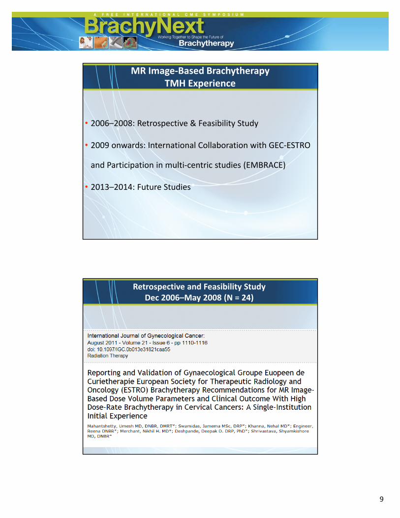
MR Image‐Based BrachytherapyTMH Experience
• 2006 2008 Retrospective & Feasibility Study• 2006–2008: Retrospective & Feasibility Study
• 2009 onwards: International Collaboration with GEC‐ESTRO
and Participation in multi‐centric studies (EMBRACE)
• 2013–2014: Future Studies
Retrospective and Feasibility StudyDec 2006–May 2008 (N = 24)

6/24/2014
10
Vienna ICIJROBP2005
Vienna IC/ISIJROBP2005
BrabandereRO 2008
Lindegaard IJROBP2008
Chargari IJROBP 2008
TMH studyIJGC 2011
HRCTV
Vol in cc 34 +/- 17 44 +/- 27 48+/-19 34+/- 12 36.3±35 45.2 + 15.8
D100 66 +/- 7 70 +/- 6 64+/-6 76 +/- 7 61.66±7 53.9 + 6.5
D90 87 +/-10 96 +/- 12 79+/-7 91 +/- 10 74.85±10 70.3 + 10.6
Avg. Pt A 89 +/- 8 93 +/- 9 79+/-5 92 +/- 9 71.4±6 73.4 + 4.5
BladderBladder
Vol in cc -- -- -- 80.3 (20.3-235)
ICRU Bmax 75 +/-16 73 +/- 19 74+/-15 67 +/- 31 63.7±9 80.4 + 34.4
D0.1cc 121+/-25 113+/- 30 100+/-12 86 +/- 45 87.6±12 136.0 + 54.7
D2cc 83 +/-9 83 +/-14 82+/-6 73 +/- 16 71.7±6 91.4 + 24.6
Rectum
Vol cc -- -- -- 33.4 (11-64.6)
ICRU Rmax 69 +/- 13 71 +/- 13 66+/-9 71 +/- 5 67.3±8 63.5 + 8.1
D0.1cc 77 +/- 10 77 +/- 9 64+/-5 71 +/- 10 70.6±11 67.2 + 9.9
D2cc 64+/- 6 66 +/- 6 66+/-9 61 +/- 6 67.3±8 57.9 + 7.7
Sigmoid
Vol cc -- -- -- 49.0 (14.5-97.5)
D0.1cc 79 +/- 12 84 +/- 14 82+/-13 79 +/- 13 72.7±18 101.9 + 45.2
D2cc 63 +/- 7 67 +/- 7 68+/-7 69 +/- 9 60.6±6 74.4 + 19.6
Dosimetric Outcome Mahantshetty et al, IJGC Aug. 2011
Stage
TMH Data (Dec 2006 –May 2008) (N = 24)
Median Follow‐up: 18 (12–40) months
Clinical Outcome
Stage
I B2 / IIAN=2
IIBN=10
IIIBN=12
TotalN=24
Local -- 2* 1# 3
Pelvic Node -- -- 1 1
Dist. metastasis -- 1 1
Total 2 3 5Total -- 2 3 5
* Point A: 79 Gy and HR‐CTV D90 doses: 56.5, 67 Gy;
# Point A: 70 Gy and HR‐CTV D90 doses: 65 Gy;
Late sequelae: 1 pt with proctosigmoiditis
(0.1 and 2cc: R 46 & 64; S: 140 & 260 Gy)
Mahantshetty et al, Clin. Oncol. 2011 ; IJGC Aug. 2011
6/24/2014
11
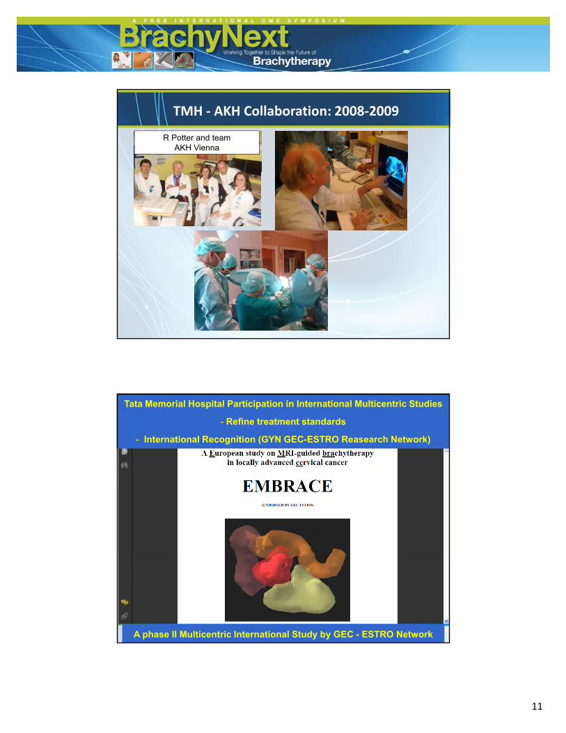
R Potter and team AKH Vienna
TMH ‐ AKH Collaboration: 2008‐2009
Tata Memorial Hospital Participation in International Multicentric Studies
- Refine treatment standards
- International Recognition (GYN GEC-ESTRO Reasearch Network)
A phase II Multicentric International Study by GEC - ESTRO Network
6/24/2014
12
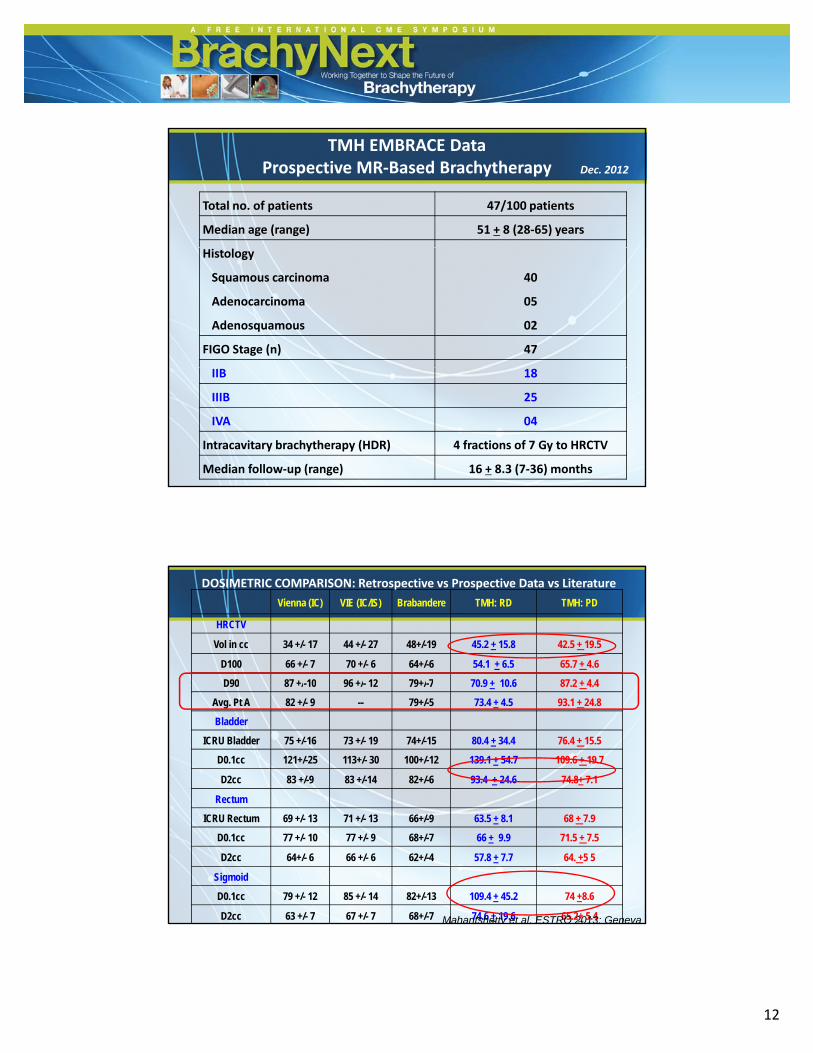
Total no. of patients 47/100 patients
Median age (range) 51 + 8 (28‐65) years
TMH EMBRACE DataProspective MR‐Based Brachytherapy Dec. 2012
Histology
Squamous carcinoma
Adenocarcinoma
Adenosquamous
40
05
02
FIGO Stage (n) 47
IIB 18
IIIB 25
IVA 04
Intracavitary brachytherapy (HDR) 4 fractions of 7 Gy to HRCTV
Median follow‐up (range) 16 + 8.3 (7‐36) months
Vienna (IC) VIE (IC/IS) Brabandere TMH: RD TMH: PD
HRCTV
Vol in cc 34 +/- 17 44 +/- 27 48+/-19 45.2 + 15.8 42.5 + 19.5
D100 66 +/- 7 70 +/- 6 64+/-6 54.1 + 6.5 65.7 + 4.6
D90 87 +/-10 96 +/- 12 79+/-7 70 9 + 10 6 87 2 + 4 4
DOSIMETRIC COMPARISON: Retrospective vs Prospective Data vs Literature
D90 87 +/ 10 96 +/ 12 79+/ 7 70.9 + 10.6 87.2 + 4.4
Avg. Pt A 82 +/- 9 -- 79+/-5 73.4 + 4.5 93.1 + 24.8
Bladder
ICRU Bladder 75 +/-16 73 +/- 19 74+/-15 80.4 + 34.4 76.4 + 15.5
D0.1cc 121+/-25 113+/- 30 100+/-12 139.1 + 54.7 109.6 + 19.7
D2cc 83 +/-9 83 +/-14 82+/-6 93.4 + 24.6 74.8+ 7.1
Rectum
ICRU Rectum 69 +/- 13 71 +/- 13 66+/-9 63.5 + 8.1 68 + 7.9
D0.1cc 77 +/- 10 77 +/- 9 68+/-7 66 + 9.9 71.5 + 7.5
D2cc 64+/- 6 66 +/- 6 62+/-4 57.8 + 7.7 64. +5 5
Sigmoid
D0.1cc 79 +/- 12 85 +/- 14 82+/-13 109.4 + 45.2 74 +8.6
D2cc 63 +/- 7 67 +/- 7 68+/-7 74.6 + 19.6 65.2+ 5.4Mahantshetty et al, ESTRO 2013; Geneva
6/24/2014
13
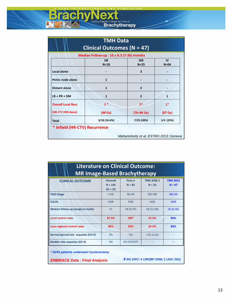
IIBN=18
IIIBN=25
IVN=04
L l l 3
TMH DataClinical Outcomes (N = 47)
Median Follow‐up : 16 + 8.3 (7‐36) months
Local alone ‐ 3 ‐
Pelvic node alone 1 ‐ ‐
Distant alone 1 2 ‐
LR + PR + DM 1 2 1
Overall Local Recc 1 * 5* 1*Overall Local Recc
(HR‐CTV D90 doses)
1
(88 Gy)
5
(76–86 Gy)
1
(87 Gy)
Total 3/18 (16.6%) 7/25 (28%) 1/4 (25%)
* Infield (HR‐CTV) Recurrence
Mahantshetty et al, ESTRO 2013; Geneva
CLINICAL OUTCOME Vienna#
N = 145
(IC + IS)
Paris ♦
N = 45
TMH 2006 $
N = 24
TMH 2012
N = 47
FIGO Stage I‐IVA IB‐IVA IB2‐IIIB IIB‐IVA
Literature on Clinical Outcome: MR Image‐Based Brachytherapy
ICA/IS HDR PDR HDR HDR
Median follow‐up (range) in moths 51 26 (9‐47) 18 (12‐40) 16 (6‐36)
Local control rates 87.5% 100* 91.5% 85%
Loco‐regional control rates 86% 95% 87.5% 83%
Rectal/sigmoid late sequelae (G3‐4) 4% 5% <1% so far ‐‐
Bladder late sequelae (G3‐4) 4% 6% (2%VVF) ‐‐ ‐‐‐
* 26/45 patients underwent hysterectomy
# RO 2007; ♦ IJROBP 2008; $ IJGC 2011EMBRACE Data : Final Analysis

6/24/2014
14
Background
2D BrachyRobust data
3D BrachyData emerging
2D 2D + CT 3D + CTLocal control rates:
IIB 75% 85% 96% ~ 11%IIIB 55% 65% 86% ~ 21%
LEVEL I EVIDENCE: 2013–2014Conventional X‐ray Based 2D vs 3D MR Image‐Based Brachytherapy in Cervical
Cancers: A PHASE III RANDOMIZED CONTROL TRIAL (MULTICENTRIC)
Standard Arm: Conventional 2D X‐ray based Study Arm: 3D MR Image based
• Primary Endpoints:
1. Absolute benefit in local control rates by 10% for FIGO IIB and IIIB in study arm as
shown in table below (defined a priori)
2. Non‐inferiority for Grade 3/4 late toxicities: 480 pts in each arm
FIGO Stage Expected benefit Sample size Total
Alpha error: 0.05; Power: 80% 2 sided tailed test
• Secondary Endpoints: ‐ Disease free and overall survivals
IIB 85–95% (10% benefit ) 160 pts each arm 350*
IIIB 65–75% (10% benefit) 326 pts each arm 680*
* Additional 30‐50 pts for 10% lost to follow – up / violations
PI: TMH

6/24/2014
15
• Ultrasound‐guided insertion of central
tandem
T d l h
US in Cx Brachytherapy
• Tandem length • Retroverted uterus• False passage
• Ultrasound‐based planning
• Uterine wall thickness• Bladder points• Rectal points• Rectal points
• Drawbacks
• Coronal imaging not available• Posterior uterine surface not visible well

6/24/2014
16
Extrapolation of USG Contour Over MRI
USG contour
Mahantshetty et al. Rad. Oncol. 2012
USG and MRI Correlation (TMH Data)
• 32 Applications with MRI‐Compatible Applicator
• Anterior Reference Points: 96%
• Posterior Reference Points: 72%
• Magnitude of Variation (>15%): <8%• Magnitude of Variation (>15%): <8%
Significant Correlation between the USG and MRI Reference Points
Suggest: Use of USG for ICA Planning (21/2 D Planning)
Mahantshetty et al. Rad. Oncol. 2012
6/24/2014
17
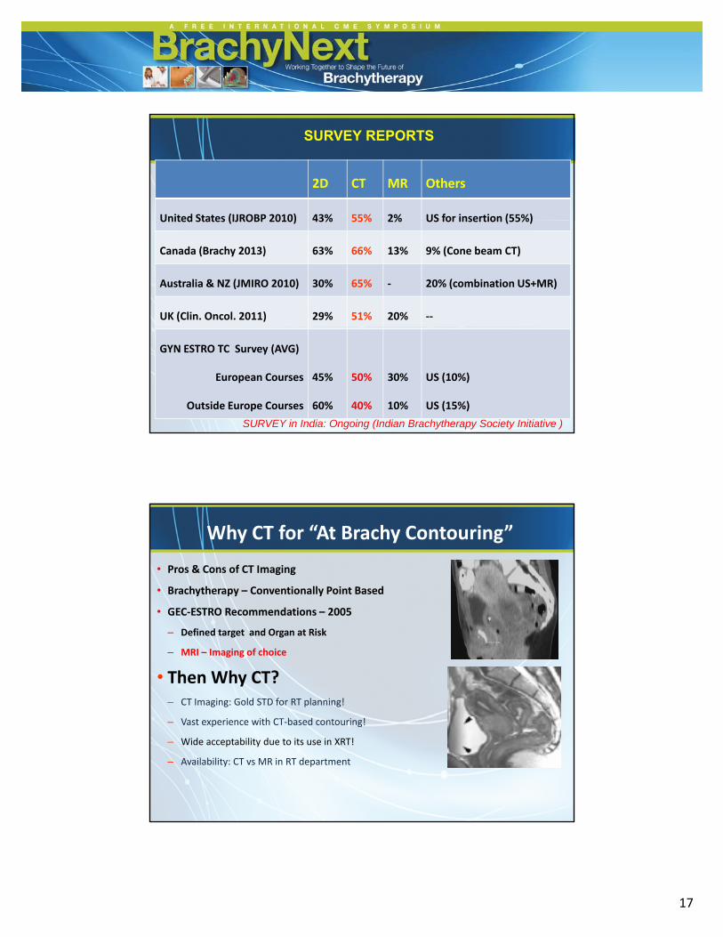
2D CT MR Others
United States (IJROBP 2010) 43% 55% 2% US for insertion (55%)
SURVEY REPORTS
United States (IJROBP 2010) 43% 55% 2% US for insertion (55%)
Canada (Brachy 2013) 63% 66% 13% 9% (Cone beam CT)
Australia & NZ (JMIRO 2010) 30% 65% ‐ 20% (combination US+MR)
UK (Clin. Oncol. 2011) 29% 51% 20% ‐‐
GYN ESTRO TC Survey (AVG)
European Courses
Outside Europe Courses
45%
60%
50%
40%
30%
10%
US (10%)
US (15%)
SURVEY in India: Ongoing (Indian Brachytherapy Society Initiative )
Why CT for “At Brachy Contouring”
• Pros & Cons of CT Imaging
• Brachytherapy – Conventionally Point Based
GEC ESTRO R d ti 2005• GEC‐ESTRO Recommendations – 2005
– Defined target and Organ at Risk
– MRI – Imaging of choice
• Then Why CT?– CT Imaging: Gold STD for RT planning!
– Vast experience with CT‐based contouring!
– Wide acceptability due to its use in XRT!
– Availability: CT vs MR in RT department

6/24/2014
18
Delineation of Target on CT
• Experience of MR‐Based Approach: Mandatory
• CT Imaging Protocol: IV Contrast, bladder filling…
• Target at brachytherapyTarget at brachytherapy– GTV: poor visualization of residual tumor on CT
– HRCTV: Clinical Drawing at Diagnosis and Brachy + CT imaging findings
– IRCTV: margins to HR‐CTV
• HR‐CTV: Practical & feasible contour possible on CT Imaging
• Defined conceptually as p y– GTV‐B + Whole of Cervix
– With presumed extensions at brachy in:
• Parametrium
• Endocervical
• Vagina

6/24/2014
19
Ongoing Study at TMH: CT vs MR‐Based HR‐CTV Contouring
00.51
1.52
2.53
3.51
2
3
4
59
10
11
12
Series1
Series2
MR
CT
5
6
7
8
9
1
1.5
2
2.5
31
2
311
12
0
0.5
4
5
6
7
8
9
10Series1
Series2
MR
CT

6/24/2014
20
SUMMARY AND CONCLUSIONS
• Brachytherapy Set-ups: Steady Increase in facilities
• Image Based Brachytherapy: Implementation logistics still evolving
• Educational Programs: Workshops, Demo’s more in developing world
• MR Image Based Brachytherapy Implementation in our setting: Challenge
• Alternate Imaging Modalities: Ongoing Research in our Settings
• Randomized trial: Poses a Bigger Challenge?
• Use of US: Potential in our settings, ongoing feasibility studies
Tata Memorial Hospital Complex,
Mumbai, India
Dr Umesh Mahantshetty
e-mail: [email protected]
Acknowledgements:• Departments of Radiation Oncology & Medical Physics• Department of Radio‐diagnosis • GYN Disease Management Group TMC• GYN GEC–ESTRO Research Network