Embed Size (px)
Citation preview
Implants and Periodontal Disease Implants and Periodontal Disease Aftercare, maintenance and treatment of Aftercare, maintenance and treatment of
PeriPeri--implantitisimplantitis
Robert Robert OrettiOretti
WexhamWexham Park 13Park 13thth July 2012July 2012

Pentangle Dental TransformationsPentangle Dental Transformations
Implant referral centre established in 2006www.pentangledental.co.uk

Pentangle Dental TransformationsPentangle Dental Transformations

Pentangle Dental TransformationsPentangle Dental Transformations
We are 95% referral-based specialist practice.We have 150 referring dentists.We receive 50 referrals a month .85% of our work involves dental implants.15% are referred for other treatments –mainly an aesthetic improvement involving ortho/restorative.
5% are ‘word of mouth’ self referred.


The Right TeamThe Right Team


PeriPeri--MucositisMucositis and and PeriPeri--ImplantitisImplantitis
••PrevalencePrevalence
••Differential DiagnosisDifferential Diagnosis
••AetiologyAetiology
••Confounding (risk) FactorsConfounding (risk) Factors
••IdentificationIdentification
••PreventionPrevention

7 year review

18 months later…..are the tissues healthy ?4 year reviewEarly bone loss with no gingival inflammation ?

••PrevalencePrevalencePrevalence and risk variables for peri-implant diseaseS. D. Ferreira G. L. M SilvaJournal of Clinical Periodontology 2006
212 subjects (smokers were excluded) with a total of 578 implantswere evaluated three and a half years later…..
73.5% of subjects presented with peri-implant BOP
64.6% with peri-implant mucositis
8.9% with peri-implantitis

Extent of peri-implantitis-associated bone lossTord BerglundhJournal of Clinical Periodontology 2009
1070 implants examined at the ten year mark.(No note was made of the incidence of peri-mucositis)The study was to examine pathological bone loss 419 of the 1070 implants exhibited peri-implantitis-associated bone loss……....40%
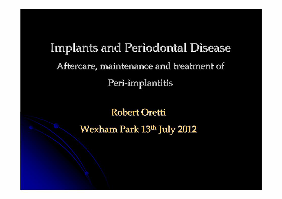
Peri-implant diseases : Consensus Report of the Sixth European Workshop on PeriodontologyJan Lindhe Joerg Meyle
Journal of Clinical Periodontology 2008
One study included a review of 3,413 implants assessed at the five year mark……..
Peri-implant mucositis occurred in about 80% of subjects restored with implants.
Peri-implantitis occurred in between 28% and 56%

PeriPeri--implant diseases are defined as an infection of the implant diseases are defined as an infection of the tissues around a functioning implant.tissues around a functioning implant.
Risk factors are :
poor oral hygieneSub-gingival dental plaquea history of periodontitissmokingdiabetes. alcohol consumption
……..and time

‘’Triggering factors for peri-implantitis are generally gathered under four categories: Lesions of peri-implant attachment (poor oral hygiene)Presence of aggressive bacteria (subgingival dental plaque)Excessive mechanical stress (functional overloading)Corrosion (early or late implant contamination)If only one of these factors would start a chain reaction leading to lesions, then the other factors may combine to worsen the condition. In other words, peri-implantitis is a general term dependent on a synergy of several factors, irrespective of the precise reason for the first triggering of symptoms.’’Tomas Albrektsson 2009

PeriPeri--MucositisMucositis and and PeriPeri--ImplantitisImplantitis
••PrevalencePrevalence
••Differential DiagnosisDifferential Diagnosis
••AetiologyAetiology
••Confounding (risk) FactorsConfounding (risk) Factors
••IdentificationIdentification
••PreventionPrevention

Bone Loss around implantsBone Loss around implantsCauses of early bone loss…..
•Bone trauma during implant placement
Over-heating.
Excessive bone compression.
Excessive bone distraction (ridge splitting).

Bone Loss around implantsBone Loss around implantsCauses of early bone loss…..
•Bone trauma during implant placement
• Inadequate residual bone thickness after implant placement.


Bone Loss around implantsBone Loss around implantsCauses of early bone loss…..
•Bone trauma during implant placement
• Inadequate residual bone thickness after implant placement.
•Excess cement pushed down around implant head.
•Aggressive peri-implantitis ?


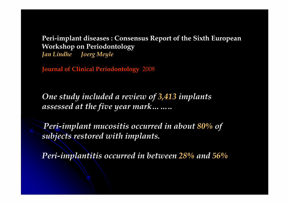
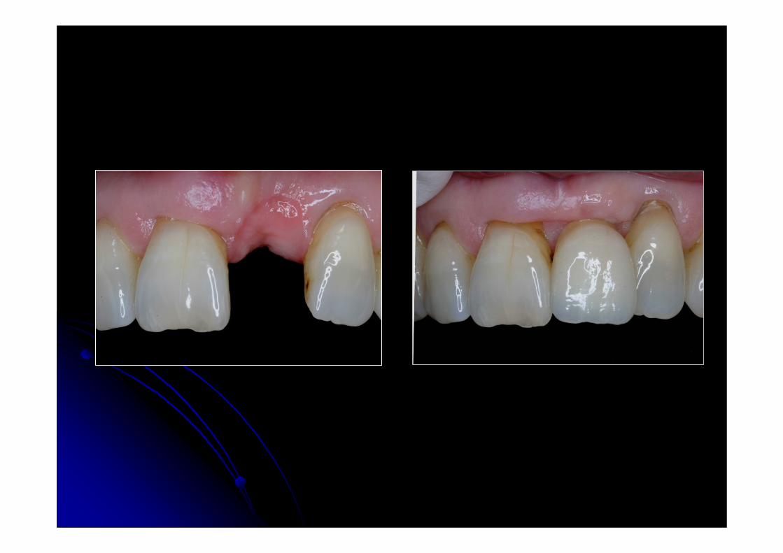
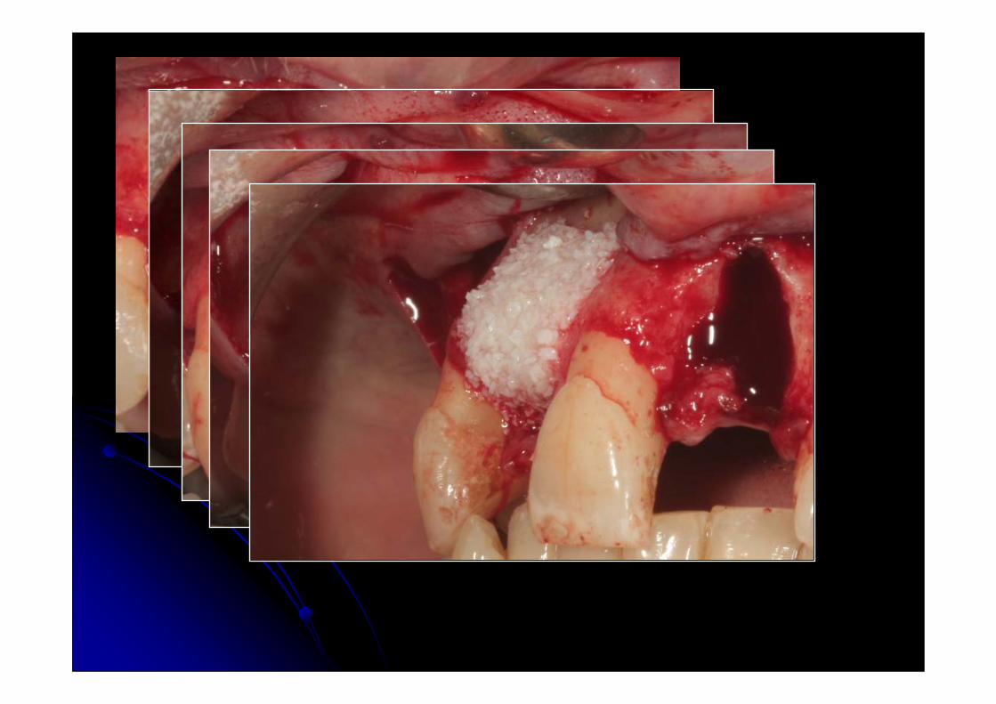
Cementation issuesCementation issues
8mm pocket distally
Who is liable for the new implant later on ?

Another case exampleAnother case example
Healed implant after 8 weeks At one year recall – cement
and bone loss seen

One solution One solution –– set abutment margins at, or above, set abutment margins at, or above, gingival level to control flow of cement excessgingival level to control flow of cement excess
Cementation issues

Sometimes bone may be hard and Sometimes bone may be hard and relatively relatively avascularavascular
35 year old man
Lower first molar site
Extremely hard cortical bone, avascular
CT scan gives a Hounsfield unit density score of 1464
(Type 1 bone about 1000)

Surgical trauma was induced within the bone crest, visible at 8 weeks.
Crestal bone loss resulted.

60 year old woman wishes to dispense with single 60 year old woman wishes to dispense with single tooth denturetooth denture

Aggressive Aggressive periodontitisperiodontitis ????
Allergic to titanium ?


3 month rev


Bone Loss around implantsBone Loss around implantsCauses of Late bone loss…..
•Non passivity of fit of supra-structure.
• Peri-implantitis.
•Occlusal trauma ?
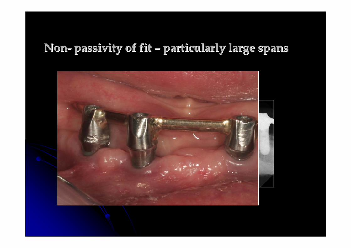
NonNon-- passivity of fit passivity of fit –– particularly large spansparticularly large spans

PeriPeri--MucositisMucositis and and PeriPeri--ImplantitisImplantitis
••PrevalencePrevalence
•Differential Diagnosis
••Aetiology Aetiology -- BacterialBacterial
••Confounding (risk) FactorsConfounding (risk) Factors
••IdentificationIdentification
••PreventionPrevention

What is What is PeriPeri--implantitisimplantitis??
An inflammatory process affecting the tissues around an osseo-integrated implant in function and resulting in the loss of supporting bone.The aetiology is of a bacterial nature with characteristics similar to periodontitis.Pain is not normally a feature and frequently the patient is unaware of any problems.You have to have started with a peri mucositis– although this key indicator is difficult to diagnose.Peri-implantitis is more aggressive and progresses faster than periodontitis.
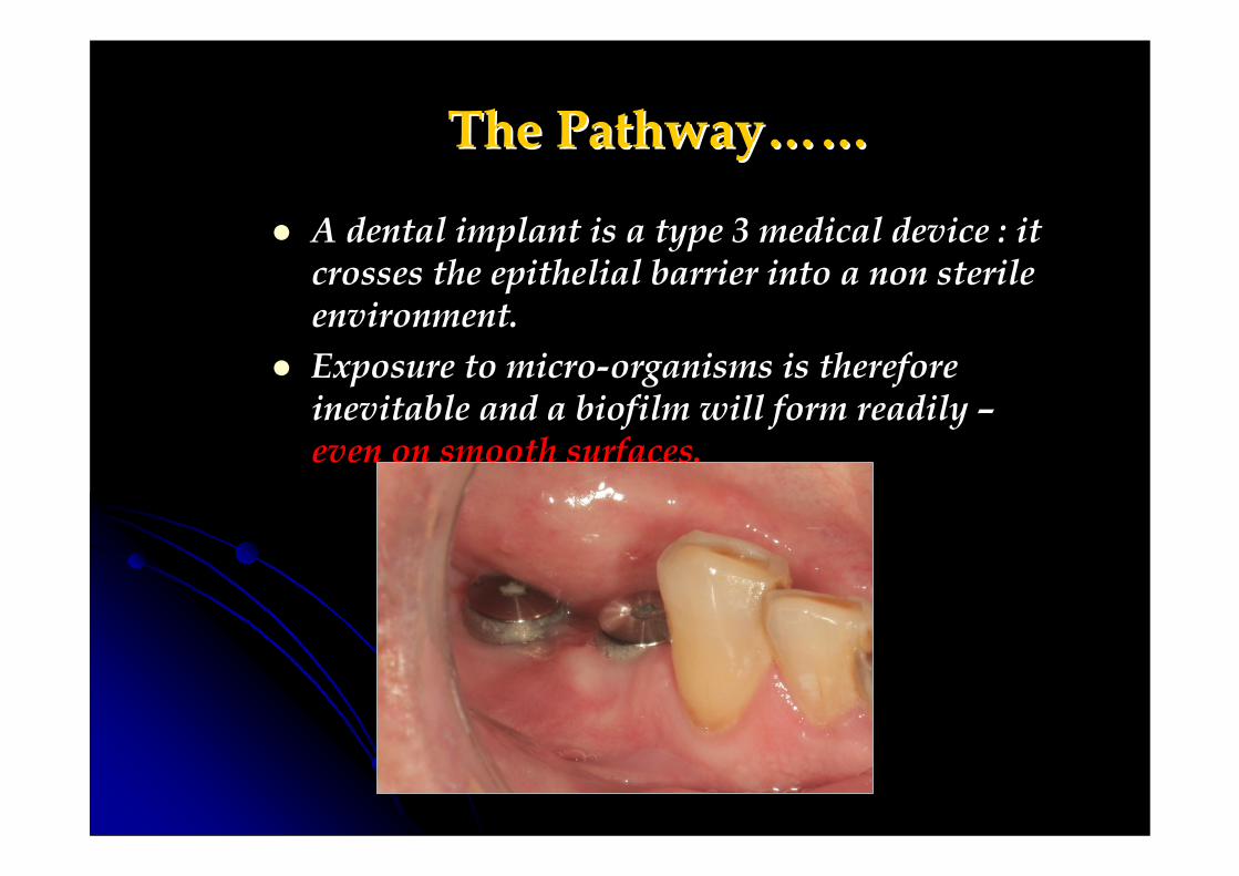
The PathwayThe Pathway…………
A dental implant is a type 3 medical device : it crosses the epithelial barrier into a non sterile environment.Exposure to micro-organisms is therefore inevitable and a biofilm will form readily –even on smooth surfaces.

The PathwayThe Pathway…………A dental implant is a type 3 medical device : it crosses the epithelial barrier into a non sterile environment.Exposure to micro-organisms is therefore inevitable and a biofilm will form readily –even on smooth surfaces. Colonisation – proliferation – increased oxygen demand – anaerobic proliferation – host response elicited – mucositis.
Mucositis is an opportunistic infection borne out of a change in the environment and results in an inflammatory lesion which resides in the mucosa only

BLEEDING ON PROBING
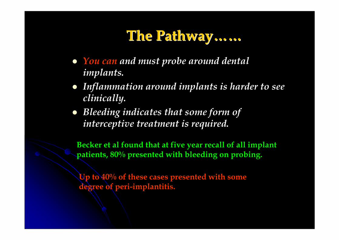
The PathwayThe Pathway…………You can and must probe around dental implants.Inflammation around implants is harder to see clinically.Bleeding indicates that some form of interceptive treatment is required.
Becker et al found that at five year recall of all implant patients, 80% presented with bleeding on probing.
Up to 40% of these cases presented with some degree of peri-implantitis.
PeriPeri--MucositisMucositis and and PeriPeri--ImplantitisImplantitis
••PrevalencePrevalence
••Differential DiagnosisDifferential Diagnosis
••Aetiology Aetiology -- BacterialBacterial
••Confounding (risk) FactorsConfounding (risk) Factors
••IdentificationIdentification
••PreventionPrevention

Diabetes mellitus is a chronic metabolic disorder that affects more than 11% of the adult population. This prevalence represents a 28% increase in the number of patients with diabetes since 2005 (CDC 2005, 2008). Current projections of diabetes incidence suggest that as much as 33% of the population may be diagnosed with diabetes by 2050, with type 2 diabetes mellitus accounting for 90–95% of all diabetes patients (Boyle et al. 2010). Worldwide, over 150 million people were estimated as having diabetes in the year 1980, and that number had grown to over 350 million by 2008 (Danaei et al. 2011). Taken together, these trends highlight the urgency for a better understanding of diabetes as well as for improving the care of patients with diabetes.
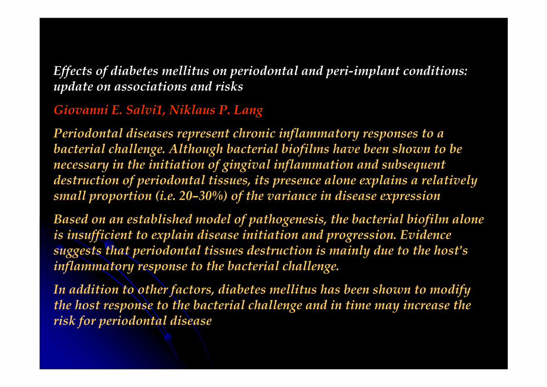
Effects of diabetes mellitus on periodontal and peri-implant conditions: update on associations and risks
Giovanni E. Salvi1, Niklaus P. Lang
Periodontal diseases represent chronic inflammatory responses to a bacterial challenge. Although bacterial biofilms have been shown to be necessary in the initiation of gingival inflammation and subsequent destruction of periodontal tissues, its presence alone explains a relatively small proportion (i.e. 20–30%) of the variance in disease expression
Based on an established model of pathogenesis, the bacterial biofilm alone is insufficient to explain disease initiation and progression. Evidence suggests that periodontal tissues destruction is mainly due to the host's inflammatory response to the bacterial challenge.
In addition to other factors, diabetes mellitus has been shown to modify the host response to the bacterial challenge and in time may increase the risk for periodontal disease

The findings indicated that diabetes did not represent an absolute contraindication for implant placement, provided that there was good glycaemic control. However, implants in diabetic subjects were significantly associated with an increased risk for peri-implantitis compared with implants placed in non-diabetic subjects (Ferreira et al. 2006).
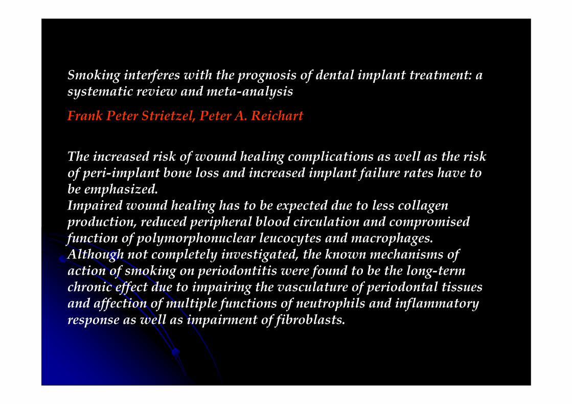
Smoking interferes with the prognosis of dental implant treatment: a systematic review and meta-analysis
Frank Peter Strietzel, Peter A. Reichart
The increased risk of wound healing complications as well as the risk of peri-implant bone loss and increased implant failure rates have to be emphasized. Impaired wound healing has to be expected due to less collagen production, reduced peripheral blood circulation and compromisedfunction of polymorphonuclear leucocytes and macrophages.Although not completely investigated, the known mechanisms of action of smoking on periodontitis were found to be the long-term chronic effect due to impairing the vasculature of periodontal tissues and affection of multiple functions of neutrophils and inflammatory response as well as impairment of fibroblasts.

Smoking and Implant FailureSmoking and Implant FailureSanchezSanchez--Perez.APerez.A et Al. 2007 et Al. 2007 –– A 5 year studyA 5 year study
NonNon--smokers had a success rate of 98.6%smokers had a success rate of 98.6%Light Smokers (<10 per day) had 10% increase Light Smokers (<10 per day) had 10% increase risk of failurerisk of failureMedium Smokers (<20 per day) had 20Medium Smokers (<20 per day) had 20--30% 30% increased riskincreased riskHeavy smokers (>20 per day) had 30% Heavy smokers (>20 per day) had 30% increased riskincreased risk
Taken as an average figure (people lie !), the Taken as an average figure (people lie !), the overall success rate for smokers in this study overall success rate for smokers in this study was 84.2%was 84.2%

•Very poor oral hygiene was highly associated with the presence of peri-implantitis.
•Individuals with periodontitis were more likely to develop peri-implant inflammatory lesions - the presence of residual periodontal pockets represent niches of infection for adjacent implants.
•Presence of periodontitis and/or smoking and/or diabetes were statistically associated with increased risk of peri-implantitis.
•Additional variables were the time elapsed since implant placement and the frequency of visits for maintenance care.


PeriPeri--MucositisMucositis and and periperi--ImplantitisImplantitis
••PrevalencePrevalence
••Differential DiagnosisDifferential Diagnosis
••Aetiology Aetiology -- BacterialBacterial
••Confounding (risk) FactorsConfounding (risk) Factors
••IdentificationIdentification
••PreventionPrevention
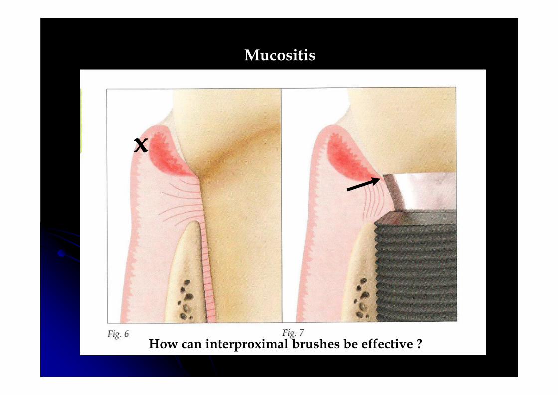
Mucositis
x
How can interproximal brushes be effective ?

Peri-implantitis

•Histologically there is a 3–4 mm width of biological soft tissue coverage over the implant-supporting bone.
•While the outer zone of the connective tissue is well vascularized with fibres running in different directions, the inner zone is poorly vascularizedand consists of collagen fibres running close to the implant surface in a parallel direction.
•The peri-implant mucosa is therefore recognized as scar tissue, exhibiting an impaired resistance to bacterial colonization……………. and probing

•The gingival tissues around all implant restorations should be checked for health by probing …..irregardless of the outward appearance.
•Probing the tissues is the first procedure to determine health or otherwise. Bleeding on probing (BOP) is always present with peri-implant disease.
•Probing around implants using a moderate force (0.5N) will cause damage to the peri-implant mucosal seal and will penetrate to the peri-implant marginal bone – particularly if the tissues are inflamed.
•A light probing force of 0.25 N will not cause damage to the peri-implant tissues and complete reformation of the mucosal seal after 5 days will occur.
•In health, you will not be able to penetrate more than 2mm and there will be no BOP. If the probing depth is 3-4mm (maximum) with BOP then the diagnosis is peri-mucositis……….and ??

•Other clinical signs could include reddened or oedematous tissues, recession or frank suppuration.
•X-rays may need to be taken........how often ?
•Reference to baseline measurements are essential to determine progress of peri-implant disease.
•Once peri-mucositis is present, it is common to see it again on recall. Commonly, the BOP and the probing depth has increased

PeriPeri--MucositisMucositis and and PeriPeri--ImplantitisImplantitis
••PrevalencePrevalence
••Differential DiagnosisDifferential Diagnosis
••Aetiology Aetiology -- BacterialBacterial
••Confounding (risk) FactorsConfounding (risk) Factors
••IdentificationIdentification
••PreventionPrevention

•Firstly gently probe with very light pressure – do not force the probe –and measure the depth and then remove.
•If there is BOP after probe removal - but you are no more than 4mm within the tissues – this is mucositis.
•This is the precursor to peri-implantitis and this is the only stage that is reversible.
•Explain the aetiology (biofilm build up) to the patient and the mandatory requirement for the patient to clean subgingivally and disrupt the biofilm.
•Persistent biofilm presence and BOP will lead to bone loss probably within five years.
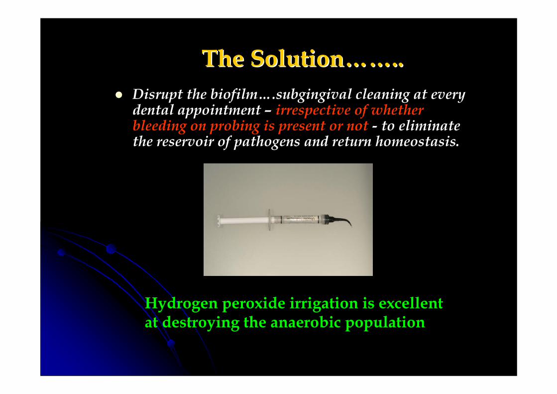
The SolutionThe Solution…………....Disrupt the biofilm….subgingival cleaning at every dental appointment – irrespective of whether bleeding on probing is present or not - to eliminate the reservoir of pathogens and return homeostasis.
Hydrogen peroxide irrigation is excellent at destroying the anaerobic population

The SolutionThe Solution…………....
The patient must be instructed in the daily use of aggressive crevice cleaning with small brushes, ‘sharpened’ end tufted brushes, interproximal brushes and ‘rough’ floss.

The SolutionThe Solution…………....
You should clean the submucosal area using these oral hygiene aids to remove the biofilmand to teach the patient the correct homecare regime.This may be all that is required at this stage without the need for further instrumentation.

The SolutionThe Solution…………....However, can you use hand scalers and
ultra-sonics ?Standard stainless steel hygienist instruments cannot be used asthey will scratch and damage the titanium - any metal-to-metal contact will lead to a roughened implant surface and an environment that may allow the tenacious adherence of a bacterial biofilm to reform.
However, the use of ultrasonics is recognised as a superior technique for biofilm disruption due to cavitation and acoustic microstreaming
Plastic coated or titanium ultrasonic tips are recommended.
The SolutionThe Solution…………....Patients are not very good at cleaning subgingivally and it is a skill they must acquire.no amount of instrumentation will resolve peri-implant inflammation (or periodontitis) if the oral hygiene remains poor and bleeding scores remain high.After cleaning and irrigating the subginival area, the patient should go away attempting to keep the area clean and inspection should be carried out 6 months later (dentist or hygienist)The biofilm will return but no bleeding on probing indicates homeostasis.Continued BOP requires revalidation of the above and introduction of local antimicrobials – dentomycin ?As soon as probing is +4mm or x-rays confirm bone loss then we are now beyond simple management

PeriPeri--implantitisimplantitis
Mild, moderate and advanced forms of peri-implantitisoccur in 28% - 56% of implant patients.History of previous periodontitis - even in previously treated patients.Poor oral hygiene.Smokers and poorly controlled diabetics.Implants in place longer than seven years.Patients will exhibit more peri-implantitis the longer they have had their implantsMore likely now that all implants have rough rather than machined surfacesPoor prosthetic design.

Histopathology of Histopathology of PeriPeri--implantitisimplantitis
X-rays are required to confirm if bone loss has occurred – the key indicator of peri-implantitis.Vertical destruction of the crestal bone – often saucer shaped.
Mild peri-implantitis : up to 2mm bone loss.Moderate peri-implantitis : up to 4mm bone loss.Advanced peri-implantitis : over 5mm bone loss.Suppuration may be present in all three types.

C.I.S.T.C.I.S.T.Cumulative Interceptive Supportive TherapyCumulative Interceptive Supportive Therapy
CleaningIrrigationSterilisationTreatment
C.I.S.T.C.I.S.T.Cumulative Interceptive Supportive TherapyCumulative Interceptive Supportive Therapy
CleaningThe disturbance and removal of the bacterial biofilm in the peri-implant pocket - with instrumentation, polishing and thorough re-instruction of OH.
Definitive treatment for non-mucositis, mucositis
and phase I of treatment for mild/moderate peri-implantitis.
The role of the GDPThe role of the GDP’’s practice ??s practice ??

C.I.S.T.C.I.S.T.Cumulative Interceptive Supportive TherapyCumulative Interceptive Supportive Therapy
IrrigationThe disturbance and removal of the bacterial biofilm in the peri-implant pocket.
Saline, Chlorhexidine, Hydrogen peroxide, Tetracycline solution
Definitive treatment for mucositis and phase II of treatment for mild/moderate peri-implantitis
UltrasonicsUltrasonics –– inform patients that gingival recession may inform patients that gingival recession may occuroccur

Pus Pus exudateexudate is suggestive of an aggressively is suggestive of an aggressively destructive processdestructive process
These implants were extracted

C.I.S.T.C.I.S.T.Cumulative Interceptive Supportive Therapy
SterilisationSterilisationTo decimate the bacterial challenge.Infection control with Dentomycin/Actisite local anti-
microbial delivery systems.500mg Amoxicillin and 400mg Metronidazole TDS for
seven days.Laser Therapy
Phase III of treatment for mild/moderate peri-implantitis: Decontamination and conditioning of the implant surface.

C.I.S.T.C.I.S.T.Cumulative Interceptive Supportive Therapy
TreatmentBone loss of 4mm or higher from the implant head.
Rough surface implants cannot be cleaned effectively with antimicrobials –Ultrasonics, diamond burs
Require visual inspection - raise a flap
Phase IV of treatment for moderate/advanced peri-implantitis


What is good for initial clot and bone matrix aggregation is also good for future bacterial biofilm adhesion!

Treatment aims: Treatment aims: PeriPeri--implantitisimplantitis
Remove all granulation tissue‘deep clean’ implant surface with micro-abrasion and soak in ‘chemicals’ and/or antimicrobials.Polish the implant surface clean ??Resective or regenerative surgery, or explantation
Dependant on whether the site can be adequately maintained by oral hygiene measures
Aesthetics ??

Peri-implant diseases – Consensus report of the sixth European workshop on Periodontology Lindhe. J 2008
Despite the number of differing treatment approaches utilised :Non-surgical debridement with or without the use of local and/or systemicantibiotics
Laser treatment
Access flaps combined with antimicrobial therapy and regenerative procedures
None of the above treatment procedures provided evidence corroborating their long term predictability.
Access flaps with systemic antibiotics demonstrated a 58% resolution at 5 year follow up. Leonhardt A 2003
Successful management of Successful management of periperi--implantitisimplantitis with a with a regenerative approach regenerative approach FroumFroum 20122012
Anti- infective regime approach :
Surgical access for debridement and decontamination
Detoxification of the implant surface
Guided bone regeneration and re-osseointegration
Average bone gain was 3.5mm – at 3 to 7.5 year follow up
Difficult to achieve more ?
The non-surgical treatment of peri-implantitis, including a mechanical treatment alone or combined with antiseptics or antibiotics can improve clinical parameters in the short term but residual defects may still persist.
Surgical treatment such as guided bone regeneration may result in a gain of clinical attachment level and bone reconstruction in the long term.
The limited effect of laser-assisted therapy needs to be further evaluated.
The concept of prevention based on early detection and regular maintenance plays a principal role in reducing the occurrence of peri-implantitis.

OH MeasuresOH Measures
Fine tufts and Fine tufts and ‘‘roughrough’’ floss floss subgingivallysubgingivally..Dipped in Dipped in CorsodylCorsodyl gelgelSyringe gel into crevicesSyringe gel into crevicesPatient to continue at homePatient to continue at home
Recommendation is three monthly hygiene maintenance …..for life


Care and Maintenance of the patientCare and Maintenance of the patientOverviewOverview
Like all expensive things, implants need to be looked after.Maintenance of oral hygiene and monitoring is essential.The assumption that implants are indestructible and further care is not required must be removed from the patient’s mind.Implant/crowns should be monitored just like natural teeth – every six months.X-rays should be taken annually at first and then every three years.Hygienist appointments will be dependant on the patients presentation but will probably be bi-annual.For the healthy implant, customised end-tufted brushes and sub-gingival flossing are the most useful for gum crevice cleaning.Implant gum crevices are deeper – approx 4mm
4 yr review
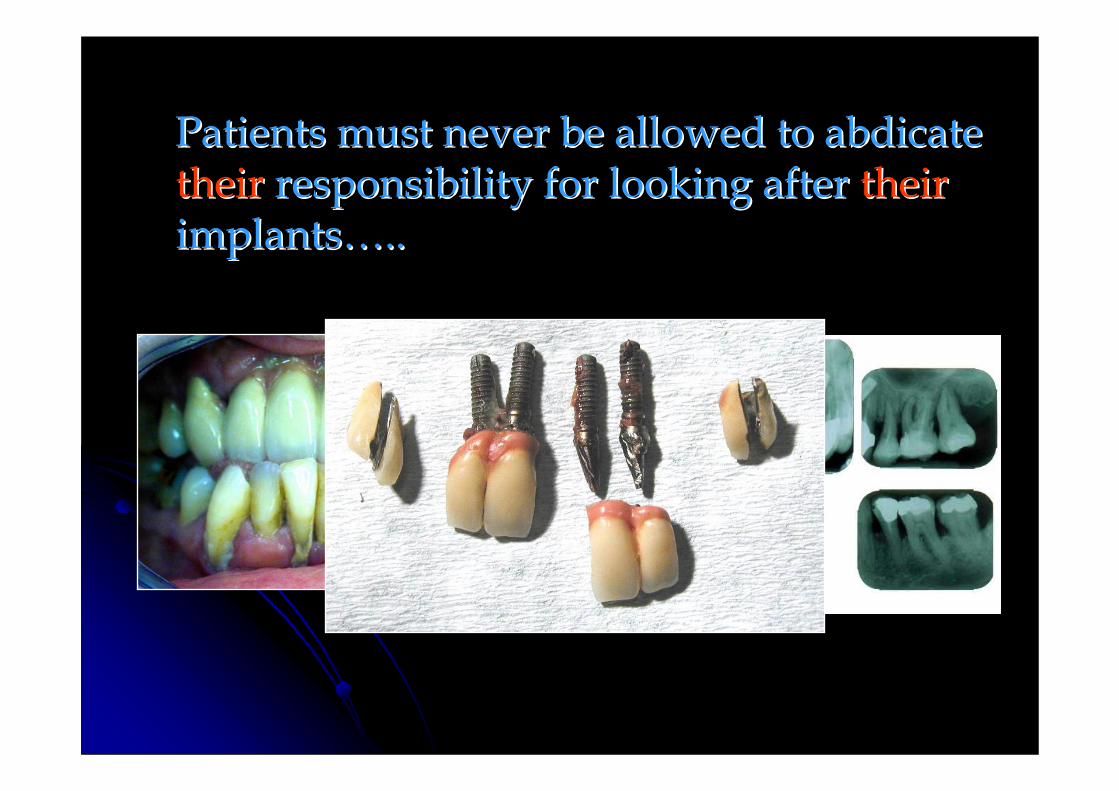
Patients must never be allowed to abdicate Patients must never be allowed to abdicate theirtheir responsibility for looking after responsibility for looking after theirtheirimplantsimplants……....