Embed Size (px)
Citation preview
RESEARCH Open Access
Impact of investigational microbiotatherapeutic RBX2660 on the gutmicrobiome and resistome revealed by aplacebo-controlled clinical trialSuryang Kwak1,2† , JooHee Choi1†, Tiffany Hink3, Kimberly A. Reske3, Kenneth Blount4, Courtney Jones4,Margaret H. Bost3, Xiaoqing Sun1,2, Carey-Ann D. Burnham2,3,6, Erik R. Dubberke3*, Gautam Dantas1,2,5,6* and for theCDC Prevention Epicenter Program
Abstract
Background: Intestinal microbiota restoration can be achieved by complementing a subject’s perturbed microbiotawith that of a healthy donor. Recurrent Clostridioides difficile infection (rCDI) is one key application of suchtreatment. Another emerging application of interest is reducing antibiotic-resistant genes (ARGs) and organisms(AROs). In this study, we investigated fecal specimens from a multicenter, randomized, double-blind, placebo-controlled phase 2b study of microbiota-based investigational drug RBX2660. Patients were administered eitherplacebo, 1 dose of RBX2660 and 1 placebo, or 2 doses of RBX2660 via enema and longitudinally tracked forchanges in their microbiome and antibiotic resistome.
Results: All patients exhibited significant recovery of gut microbiome diversity and a decrease of ARG relativeabundance during the first 7 days post-treatment. However, the microbiome and resistome shifts toward averageconfigurations from unperturbed individuals were more significant and longer-lasting in RBX2660 recipientscompared to placebo. We quantified microbiome and resistome modification by RBX2660 using a novel“transplantation index” metric. We identified taxonomic and metabolic features distinguishing the baselinemicrobiome of non-transplanted patients and taxa specifically enriched during the process of transplantation. Weelucidated the correlation between resistome and taxonomic transplantations and post-treatment dynamics ofpatient-specific and RBX2660-specific ARGs. Whole genome sequencing of AROs cultured from RBX2660 productand patient samples indicate ARO eradication in patients via RBX2660 administration, but also, to a lesser extent,introduction of RBX2660-derived AROs.
(Continued on next page)
© The Author(s). 2020 Open Access This article is licensed under a Creative Commons Attribution 4.0 International License,which permits use, sharing, adaptation, distribution and reproduction in any medium or format, as long as you giveappropriate credit to the original author(s) and the source, provide a link to the Creative Commons licence, and indicate ifchanges were made. The images or other third party material in this article are included in the article's Creative Commonslicence, unless indicated otherwise in a credit line to the material. If material is not included in the article's Creative Commonslicence and your intended use is not permitted by statutory regulation or exceeds the permitted use, you will need to obtainpermission directly from the copyright holder. To view a copy of this licence, visit http://creativecommons.org/licenses/by/4.0/.The Creative Commons Public Domain Dedication waiver (http://creativecommons.org/publicdomain/zero/1.0/) applies to thedata made available in this article, unless otherwise stated in a credit line to the data.
* Correspondence: [email protected]; [email protected]†Suryang Kwak and JooHee Choi contributed equally to this work.3Department of Medicine, Division of Infectious Diseases, WashingtonUniversity School of Medicine in St. Louis, St. Louis, MO 63110, USA1The Edison Family Center for Genome Sciences & Systems Biology,Washington University School of Medicine in St. Louis, St. Louis, MO 63110,USAFull list of author information is available at the end of the article
Kwak et al. Microbiome (2020) 8:125 https://doi.org/10.1186/s40168-020-00907-9
(Continued from previous page)
Conclusions: Through shotgun metagenomic sequencing, we elucidated the effects of RBX2660 in the microbiomeand resistome. Antibiotic discontinuation alone resulted in significant recovery of gut microbial diversity andreduced ARG relative abundance, but RBX2660 administration more rapidly and completely changed thecomposition of patients’ microbiome, resistome, and ARO colonization by transplanting RBX2660 microbiota intothe recipients. Although ARGs and AROs were transmitted through RBX2660, the resistome post-RBX2660 moreclosely resembled that of the administered product—a proxy for the donor—than an antibiotic perturbed state.
Trial registration: ClinicalTrials.gov, NCT02299570. Registered 19 November 2014
Keywords: Microbiota-based therapy, Placebo, Microbiome, Resistome, Clostridioides difficile infection, Antibiotic-resistant organisms
BackgroundIntestinal microbiota restoration by microbiota-basedtherapy, such as fecal microbiota transplantation (FMT)from healthy donors to patients, has been applied as atreatment for disorders caused by intestinal dysbiosis [1].As the contributions of the gut microbiota to the hostimmune system, energy metabolism, and central nervoussystem have been uncovered, the range of potential ap-plications of intestinal microbiota restoration therapy isexpanding to various disorders, such as inflammatorybowel disease [2], functional gastrointestinal disorders[3], metabolic syndrome [4, 5], and neuropsychiatric dis-orders [6, 7]. Accordingly, studies for understanding andrefining the action of intestinal microbiota restorationtherapies are being actively conducted [8].Clostridioides difficile infection (CDI) is one area
where intestinal microbiota restoration therapy has beenapplied successfully. Although oral administration of an-tibiotics is the standard first-line therapy for CDI, antibi-otics perturb the commensal gut microbiota anddecrease colonization resistance against other pathogens[9, 10]. Approximately 15 to 30% of CDI patients there-fore experience recurrent CDI (rCDI) resulting from ei-ther a relapse of the previous CDI or reinfection [11].Moreover, antibiotic therapies during CDI treatmentmay promote the expansion of antibiotic-resistant or-ganisms (AROs) such as vancomycin-resistant Entero-cocci (VRE) [12, 13]. On the other hand, intestinalmicrobiota restoration has shown to be effective for CDItreatment as well as the restoration of colonization re-sistance against C. difficile and AROs [14, 15]. Indeed,intestinal microbiota restoration has become a com-monly performed investigational therapy for rCDI withdecent success rates [8, 16–19].However, due to the transmissive nature of the treat-
ment, microbiota restoration therapy may communicatenot only desirable but also undesirable factors derivedfrom donors. For instance, the transmission ofantibiotic-resistant genes (ARGs) and AROs derivedfrom donor samples is a potential risk of fecal trans-plantation [20, 21]. AROs are responsible for increasing
infection cases each year, and more than 35,000 patientsdied as a result of ARO infections in the United Statesin 2017 [22]. Recently, two cases of bacteremia causedby extended-spectrum beta-lactamase (ESBL)-producingEscherichia coli in patients after FMT from the samedonor sample have been reported, resulting in the deathof one of the patients [21]. Moreover, the disseminationof ARGs and pathogenic AROs in patients hampers ef-fective medical care of infections and results in longerhospitalization and higher medical expenditures [23].Still, multiple studies report efficient reduction of ARGsand decolonization of AROs through microbiota trans-plantation [24, 25].In the current study, we explored the effect of a
microbiota-based investigational drug RBX2660, a sus-pension of healthy donor microbiota [26–29], on the in-testinal microbiome and resistome of recipients treatedfor rCDI. In an international, multicenter, randomized,and blinded phase 2b study, rCDI patients received ei-ther placebo (control group), one dose, or two doses ofRBX2660 (Fig. 1), with more patients being recurrence-free after either RBX2660 regimen than placebo [26].Through shotgun metagenomic sequencing, we demon-strate considerable shifts of taxonomic and resistomestructures common to both placebo- and RBX2660-treated patients likely from discontinuation of antibiotics,particularly during the first week after treatment. By con-trolling for placebo effects, we could also distinguish taxo-nomic and resistome changes specific to RBX2660treatment. Furthermore, we identified discriminative fea-tures strongly correlated with microbiota transplant anddemonstrated an overall decrease in AROs as well asintroduction of a few AROs by RBX2660.
ResultsStudy cohorts and sample collectionAll donors of RBX2660 microbiota completed a compre-hensive initial health and lifestyle questionnaire. Theirblood and fecal samples were tested for immunodeficiencyviruses, C. difficile toxin, and pathogens including AROssuch as VRE and methicillin-resistant Staphylococcus
Kwak et al. Microbiome (2020) 8:125 Page 2 of 16

Fig. 1 (See legend on next page.)
Kwak et al. Microbiome (2020) 8:125 Page 3 of 16
aureus before enrollment into the donor program [27, 28].Fecal specimens from a total of 66 patients and their cor-responding RBX2660 products were collected during amulticenter, randomized, blinded, and placebo-controlledphase 2b study for the treatment of rCDI (Fig. 1) [26].Ninety-four percent of all patients (62/66) had receivedvancomycin, with the remainder receiving metronidazoleor fidaxomicin prior to study drug (Fig. 1). Twenty-onepatients received 2 doses of placebo (14 females, 9 CDI re-currence, median age 63 years), 22 patients received 1dose of RBX2660 and 1 dose of placebo sequentially (15females, 5 CDI recurrence, median age 63 years), and 23patients received 2 doses of RBX2660 (15 females, 8 CDIrecurrence, median age 68 years) [26]. Each RBX2660 dosederives from a single donor, and RBX2660 dose selectionwas not constrained to ensure a single donor was repre-sented in patients that received two RBX2660 doses (Sup-plementary Table 1). The first dose of study drug(RBX2660 or placebo) was administered 24–48 h followingcompletion of antibiotic treatment for CDI, and the secondtreatment was administered 7 ± 3 days later (Fig. 1). Pa-tients who experienced a new rCDI episode within 60 daysafter the first dose (9 placebo recipients, 5 single RBX2660recipients, 8 double RBX2660 recipients) were moved toopen-label treatment and received two additional doses ofrandomized RBX2660 (Fig. 1). Patient fecal specimens werecollected at selected time points from baseline (day 0)through 365 days after the first dose. AROs from each fecalsample were isolated on selective media plates (the“Methods” section, Supplementary Table 2).
RBX2660 shifted taxonomic structures of patients’intestinal microbiome in a dose-dependent mannerrCDI patients had significantly lower alpha diversity(Shannon diversity) than RBX2660 products beforethe treatment (Fig. 2a) as previously described with16S sequencing [29]. Following study drug administra-tion, the alpha diversity of all rCDI patients’ micro-biota increased to near-RBX2660 levels regardless ofthe treatment group, with the steepest increase duringthe first week (Fig. 2b). The largest taxonomic struc-tural shift also occurred during the first week in alltreatment groups (Fig. S1 and S2).Bray-Curtis dissimilarities between recipient and cor-
responding RBX2660 product were calculated to assessthe level of taxonomic transformation toward that ofRBX2660. For placebo recipients, the dissimilarity wasmeasured from a pseudo-donor (DS00) profile calculated
from the average species-level taxonomic profile of allRBX2660 products in this study (Fig. 2c). The meanBray-Curtis dissimilarity of DS00 from RBX2660 prod-ucts was 0.4926, which was lower than the inter-RBX2660 Bray-Curtis distance of 0.6274. Consideringthe thorough inspection criteria for donors of RBX2660products, we defined RBX2660 microbiomes as “unper-turbed” gut microbiomes. Bray-Curtis dissimilarities be-tween patients and RBX2660 demonstrate that RBX2660administration effectively changed recipients’ micro-biome structure toward unperturbed configurations at alarger magnitude and for a longer duration as comparedto placebo (Kruskal-Wallis test, P = 0.043 at day 30, P =0.028 at day 60, Fig. 2d). These microbiome shifts byRBX2660 were not sensitive to the kind of antibiotic ad-ministered prior to RBX2660 (Fig. S3).We further compared the original Bray-Curtis dissimi-
larities between patients and respective RBX2660 (DR) todissimilarities between patients and other randomRBX2660 (DO). RBX2660 recipients still exhibited lowerDOs than those of placebo recipients in dose-dependentmanner (Fig. S4), indicating that RBX2660 shifted pa-tients’ gut microbiomes toward an unperturbed micro-biome more actively than placebo. In addition,significantly lower DRs than DOs of double-dose recipi-ents after the RBX2660 administration demonstrateddose-dependent and specific shifts toward correspondingRBX2660 (Fig. S4). Principal coordinates analysis (PCoA)and PERMANOVA for patients and RBX2660 also indi-cated that placebo recipients did exhibit taxonomicstructural shifts toward RBX2660, but they were not asdramatic as those of double RBX2660 dose recipients to-ward the first dose RBX2660 (Fig. 2e).When comparing groups based on rCDI treatment
success, treatment-failure patients (who experienced anew rCDI episode within 60 days post-treatment) andtreatment-success patients did not exhibit significant dif-ferences (Fig. S5a–c). This is likely due to limited num-ber of treatment-failure samples after baseline, aspatients were omitted from the current blinded study forthe standard-of-care treatment at failure determination.Thus, we performed general linear model-based multi-variate statistical analyses of patients’ baseline metagen-omes using MaAsLin2 [30] to identify baseline featurescorrelated to rCDI prevention success or failure. Klebsi-ella pneumoniae was the only species whose relativeabundance was significantly associated with treatmentfailure in all patients (Fig. S5d). When patients were
(See figure on previous page.)Fig. 1 Study design for the use of RBX2660 to prevent recurrent Clostridioides difficile infection (rCDI). Total of 66 patients with a history of rCDIwere treated with RBX2660 in a randomized and blinded manner. Placebo (white triangle) and RBX2660 (brown triangle) were administered andfecal samples (black circle) were collected at the indicated time points. Patients who were declared a new episode of rCDI within 60 days (whitesquare) were moved to open-label treatment
Kwak et al. Microbiome (2020) 8:125 Page 4 of 16

grouped by RBX2660 dose, the model identified K. pneu-moniae as the only potential failure-associated featureagain from placebo recipients (Fig. S5e) but did not fromRBX2660 recipients.
RBX2660 transplanted taxonomic structures to patientsTo quantify and compare patients’ levels of change inmicrobiome composition, we calculated a transplant-ation index quantifying the extent of microbiome con-vergence toward corresponding RBX2660 product. Thisindex was defined as the change in Bray-Curtis distancesbetween baseline (DistanceBL) and selected time point(DistanceT), scaled by the distance from RBX2660 atbaseline: (DistanceBL − DistanceT)/DistanceBL. DS00 was
used for placebo recipients, who were then used todetermine taxonomic transplantation success. To valid-ate the transplantation index as a metric for quantifyingmicrobiome shifts by RBX2660, we also calculatedpseudo transplantation indices using dissimilarities be-tween patients and random, non-corresponding RBX2660products and compared them with the original transplant-ation indices. The dose-dependent increase in pseudo in-dices (Fig. S6) is additional evidence that RBX2660 shiftedpatients’ intestinal microbiome toward the unperturbedmicrobiome of RBX2660. Some of the pseudo indiceswere lower than zero, indicating that the transplantationindex well reflects individual directionality of recipient’smicrobiome shift toward respective RBX2660 (Fig. S6).
Fig. 2 RBX2660 shifted taxonomic structures of the gut microbiome of recipients toward a healthy state. a RBX2660 products exhibitedsignificantly higher alpha diversity than patient samples before treatment (Wilcoxon signed-rank test) based on the metagenomic taxonomicprofiling data. b Alpha diversity of all patients including placebo recipients increased similarly after treatment. Changes in alpha diversity weresignificant for the first week after treatment, but there was no statistically significant difference among treatment groups (Kruskal-Wallis test). cPrincipal coordinates analysis (PCoA) showed a species-level clustering of RBX2660 (white) and pseudo-donor sample DS00 (yellow) distinct frompatient baseline samples (violet). d Bray-Curtis distance between taxonomic structures of patients and corresponding RBX2660. D1 and D2indicate the first dose and the second dose, respectively. DS00 was used for calculating the Bray-Curtis distance of placebo recipients. Thedecrease in Bray-Curtis distances was steepest during the first week after treatment (black, Wilcoxon signed-rank test). RBX2660 recipients showeda more dynamic decrease in Bray-Curtis distances than placebo recipients by day 60 (red, Kruskal-Wallis test). *P ≤ 0.05, **P ≤ 0.01, ***P ≤ 0.001,****P ≤ 0.0001. e Upper panels: PCoA describing the direction of changes in taxonomic structures of RBX2660 recipients. Corresponding RBX2660products and all placebo recipients were included. Lower panels: adjusted P values of PERMANOVA and relevant pairwise comparisons (Pillai-Bartlett non-parametric trace and Benjamini-Hochberg FDR correction). P values of comparisons between placebo and RBX2660 recipients (redasterisks, left y-axis), placebo recipients and RBX2660 (circle, right y-axis), single-dose recipients and RBX2660 (triangle, right y-axis), and double-dose recipients and RBX2660 (square, right y-axis) of PCoA plots were presented in corresponding lower panels
Kwak et al. Microbiome (2020) 8:125 Page 5 of 16
Statistically significant differences between the originaland pseudo transplantation indices of double-dose recipi-ents, but not single dose (Fig. S6), connoted that double-dose administration allows more RBX2660-specific micro-biome shift than single dose.RBX2660 recipients were categorized as transplanted
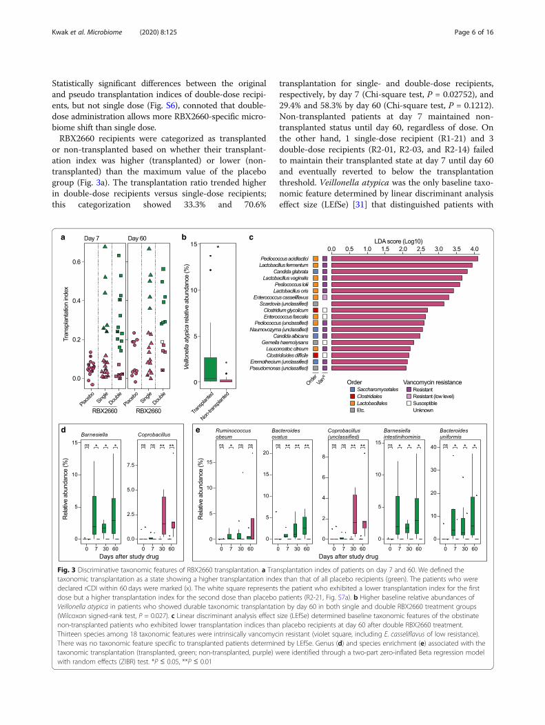
or non-transplanted based on whether their transplant-ation index was higher (transplanted) or lower (non-transplanted) than the maximum value of the placebogroup (Fig. 3a). The transplantation ratio trended higherin double-dose recipients versus single-dose recipients;this categorization showed 33.3% and 70.6%
transplantation for single- and double-dose recipients,respectively, by day 7 (Chi-square test, P = 0.02752), and29.4% and 58.3% by day 60 (Chi-square test, P = 0.1212).Non-transplanted patients at day 7 maintained non-transplanted status until day 60, regardless of dose. Onthe other hand, 1 single-dose recipient (R1-21) and 3double-dose recipients (R2-01, R2-03, and R2-14) failedto maintain their transplanted state at day 7 until day 60and eventually reverted to below the transplantationthreshold. Veillonella atypica was the only baseline taxo-nomic feature determined by linear discriminant analysiseffect size (LEfSe) [31] that distinguished patients with
Fig. 3 Discriminative taxonomic features of RBX2660 transplantation. a Transplantation index of patients on day 7 and 60. We defined thetaxonomic transplantation as a state showing a higher transplantation index than that of all placebo recipients (green). The patients who weredeclared rCDI within 60 days were marked (x). The white square represents the patient who exhibited a lower transplantation index for the firstdose but a higher transplantation index for the second dose than placebo patients (R2-21, Fig. S7a). b Higher baseline relative abundances ofVeillonella atypica in patients who showed durable taxonomic transplantation by day 60 in both single and double RBX2660 treatment groups(Wilcoxon signed-rank test, P = 0.027). c Linear discriminant analysis effect size (LEfSe) determined baseline taxonomic features of the obstinatenon-transplanted patients who exhibited lower transplantation indices than placebo recipients at day 60 after double RBX2660 treatment.Thirteen species among 18 taxonomic features were intrinsically vancomycin resistant (violet square, including E. casseliflavus of low resistance).There was no taxonomic feature specific to transplanted patients determined by LEfSe. Genus (d) and species enrichment (e) associated with thetaxonomic transplantation (transplanted, green; non-transplanted, purple) were identified through a two-part zero-inflated Beta regression modelwith random effects (ZIBR) test. *P ≤ 0.05, **P ≤ 0.01
Kwak et al. Microbiome (2020) 8:125 Page 6 of 16
successful microbiome transplantation by day 60 fromnon-transplanted patients in both single and doubleRBX2660 treatment arms (Fig. 3b).Although double RBX2660 dosage led to more effect-
ive transplantation of RBX2660 microbiome structure,there were 4 double-dose recipients (R2-01, R2-02, R2-03, R2-14) who showed lower transplantation indicesthan placebo recipients at day 60 (Fig. 3a and S7a). Allof the 4 patients received vancomycin prior to RBX2660administration (Fig. 1). We determined 18 taxa (Fig. 3c)and 21 functions (Fig. S7b) as features specificallyexplaining the baseline microbiome of these 4 patientsby comparing with other double-dose recipients thatshowed durable taxonomic transplantation by day 60using LEfSe [31]. Of these, 4 taxonomic features werefungi, which are intrinsically vancomycin insensitive, and7 functional features of eukaryote-specific metabolicpathways (Fig. 3c and S6b). We further investigated thepredicted vancomycin insensitivity of other taxonomicfeatures and found 8 additional intrinsicallyvancomycin-resistant bacteria including Pediococcusstrains [32–34] and Lactobacillus and Leuconostocstrains [35–37] as well as gram-negative and fungalstrains. Enterococcus casseliflavus, which has low levelresistance to vancomycin, was also identified [38]. Fourtaxa (Clostridium glycolicum [39], Gemella haemolysans[40], E. faecalis [41], and C. difficile [42]) are predictedto be vancomycin susceptible. Compared to the trans-planted patients, the 4 non-transplanted patients did notexhibit any other distinctive taxonomic characteristics interms of alpha diversity and composition of Bacteroi-detes, Firmicutes, and Proteobacteria phyla (Fig. S7c–g).Beyond baseline features, we further investigated which
taxa were enriched during the process of transplantation.Through a two-part zero-inflated beta regression modelwith random effects (ZIBR) test [43], we investigated asubset of 12 patients (R1-02, R1-03, R1-09, R1-14, R1-21,R2-05, R2-06, R2-10, R2-11, R2-12, R2-13, and R2-20)matched for 4 different time points: baseline, day 7, 30,and 60. ZIBR models a taxon’s presence and absence (lo-gistic component) as well as its non-zero abundance (Betacomponent), while incorporating patient and time as ran-dom variables (random intercepts). Only two genera, Bar-nesiella and Coprobacillus, are significantly correlatedwith the taxonomic transplantation. Barnesiella is signifi-cantly overrepresented in the transplanted patients asearly on as day 7, while Coprobacillus is overrepresentedin non-transplanted patients at days 30 and 60 (Fig. 3d).At the species level, ZIBR models identified Barnesiellaintestinihominis, Coprobacillus (unclassified), Bacteroidesovatus, Bacteroides uniformis, Ruminococcus obeum, andAkkermansia muciniphila (Fig. 3e, A. muciniphila wasomitted because its time point comparisons were not sta-tistically significant in the actual data). Barnesiella
intestinihominis and unclassified Coprobacillus speciesfollowed near-identical patterns from the genus-level ana-lysis due to single species being identified from eachgenus.
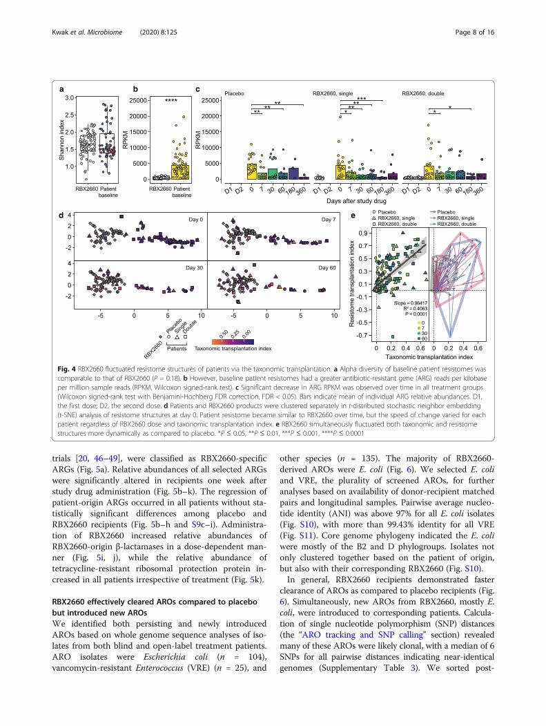
Resistome regression significantly correlated withtransplantation indexPrior to treatment, rCDI patients showed a similar resis-tome alpha diversity (Wilcoxon signed-rank test, P =0.18, Fig. 4a) when ARGs were grouped into ARG fam-ilies based on the organizational structure in CARD [44].However, the relative abundance of total ARGs was sig-nificantly higher in the patients than RBX2660 (Wil-coxon signed-rank test, P < 0.0001, Fig. 4b). It decreasedover time in all treatment arms including the placebogroup (Fig. 4c). Patients’ resistome composition was dis-tinct from RBX2660 products, but the antibiotic treat-ment prior to study drug administration did not lead tonoticeable difference in resistome (Fig. S8a–c). Specific-ally, major facilitator superfamily (MFS) and resistance-nodulation-cell division (RND) efflux pumps were themajor ARG families present in rCDI patients before thetreatment, whereas CfxA beta-lactamase, tetracycline-resistant ribosomal protection proteins, and Erm 23SrRNA methyltransferases were representative of theRBX2660 resistome (Fig. Se).We tracked individual changes in resistome compos-
ition of each patient for 60 days using t-distributed sto-chastic neighbor embedding (t-SNE) analysis [45] andresistome transplantation indices defined analogously tothe microbiome transplantation index. rCDI patientsshowed distinctive resistome compositions as comparedto those of RBX2660 prior to the treatment, but overtime their resistome compositions converged to becomesimilar to RBX2660 (Fig. 4d). The speed of resistometransformation toward RBX2660-like structures variedby patient. The convergence toward RBX2660 resistomestructure showed strong correlation to the taxonomictransplantation irrespective of treatment arm (R2 =0.406, P < 0.0001, Fig. 4e). RBX2660 administration ledto higher taxonomic and resistome transplantation indi-ces than the placebo (Fig. 4e).To identify features distinguishing patient and
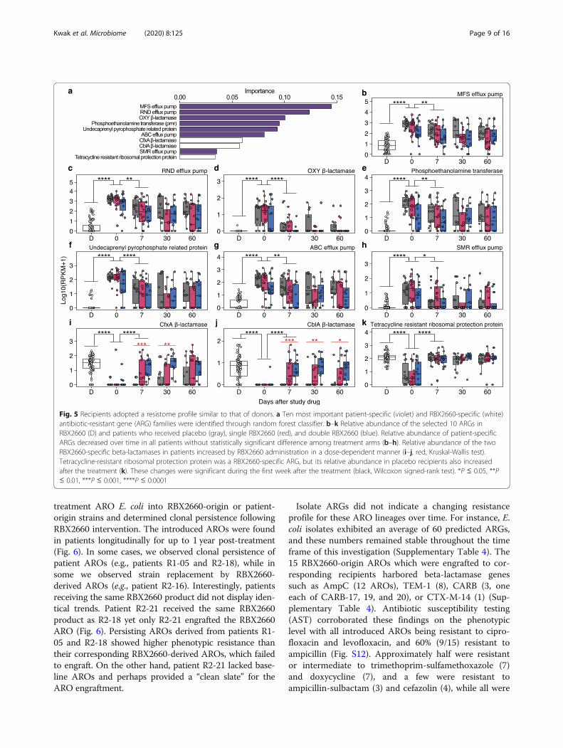
RBX2660 resistomes, we used a random forest classifier(Fig. S9a–b). Of the top 10 features of importance, 7ARGs, namely MFS efflux pump, RND efflux pump,OXY β-lactamase, Pmr phosphoethanolamine transfer-ase, undecaprenyl pyrophosphate related proteins, ATP-binding cassette (ABC) efflux pump, small multidrug re-sistance (SMR) efflux pump, and tetracycline-resistantribosomal protein, were specific to patients’ baselineresistomes. Class A β-lactamases (CfxA and CblA) and atetracycline-resistance protein, which are frequentlyidentified in healthy populations or donor stools in FMT
Kwak et al. Microbiome (2020) 8:125 Page 7 of 16
trials [20, 46–49], were classified as RBX2660-specificARGs (Fig. 5a). Relative abundances of all selected ARGswere significantly altered in recipients one week afterstudy drug administration (Fig. 5b–k). The regression ofpatient-origin ARGs occurred in all patients without sta-tistically significant differences among placebo andRBX2660 recipients (Fig. 5b–h and S9c–i). Administra-tion of RBX2660 increased relative abundances ofRBX2660-origin β-lactamases in a dose-dependent man-ner (Fig. 5i, j), while the relative abundance oftetracycline-resistant ribosomal protection protein in-creased in all patients irrespective of treatment (Fig. 5k).
RBX2660 effectively cleared AROs compared to placebobut introduced new AROsWe identified both persisting and newly introducedAROs based on whole genome sequence analyses of iso-lates from both blind and open-label treatment patients.ARO isolates were Escherichia coli (n = 104),vancomycin-resistant Enterococcus (VRE) (n = 25), and
other species (n = 135). The majority of RBX2660-derived AROs were E. coli (Fig. 6). We selected E. coliand VRE, the plurality of screened AROs, for furtheranalyses based on availability of donor-recipient matchedpairs and longitudinal samples. Pairwise average nucleo-tide identity (ANI) was above 97% for all E. coli isolates(Fig. S10), with more than 99.43% identity for all VRE(Fig. S11). Core genome phylogeny indicated the E. coliwere mostly of the B2 and D phylogroups. Isolates notonly clustered together based on the patient of origin,but also with their corresponding RBX2660 (Fig. S10).In general, RBX2660 recipients demonstrated faster
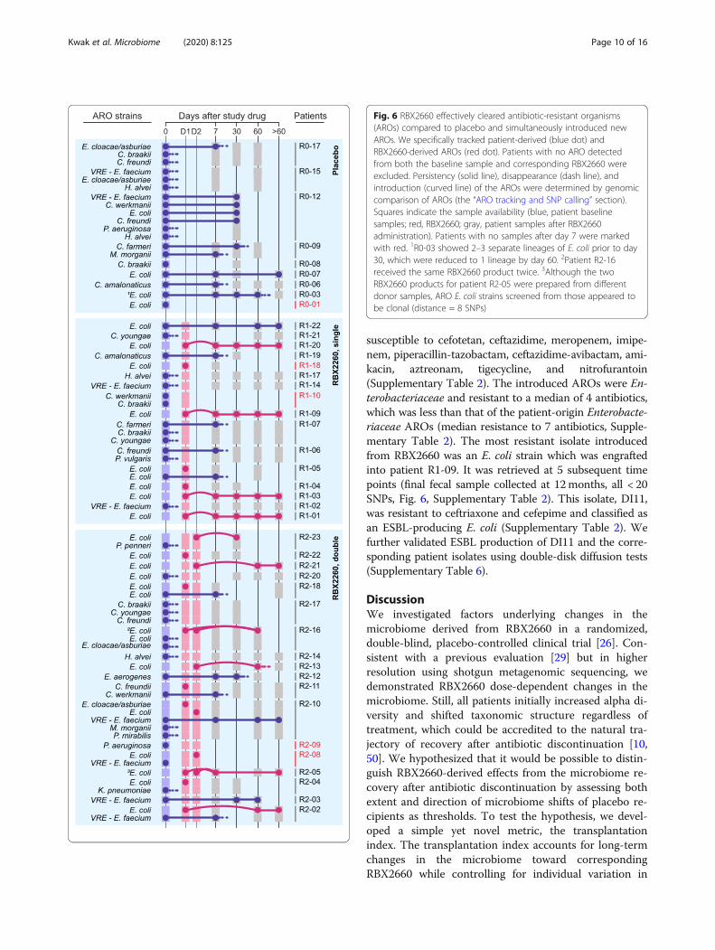
clearance of AROs as compared to placebo recipients (Fig.6). Simultaneously, new AROs from RBX2660, mostly E.coli, were introduced to corresponding patients. Calcula-tion of single nucleotide polymorphism (SNP) distances(the “ARO tracking and SNP calling” section) revealedmany of these AROs were likely clonal, with a median of 6SNPs for all pairwise distances indicating near-identicalgenomes (Supplementary Table 3). We sorted post-
Fig. 4 RBX2660 fluctuated resistome structures of patients via the taxonomic transplantation. a Alpha diversity of baseline patient resistomes wascomparable to that of RBX2660 (P = 0.18). b However, baseline patient resistomes had a greater antibiotic-resistant gene (ARG) reads per kilobaseper million sample reads (RPKM, Wilcoxon signed-rank test). c Significant decrease in ARG RPKM was observed over time in all treatment groups(Wilcoxon signed-rank test with Benjamini-Hochberg FDR correction, FDR < 0.05). Bars indicate mean of individual ARG relative abundances. D1,the first dose; D2, the second dose. d Patients and RBX2660 products were clustered separately in t-distributed stochastic neighbor embedding(t-SNE) analysis of resistome structures at day 0. Patient resistome became similar to RBX2660 over time, but the speed of change varied for eachpatient regardless of RBX2660 dose and taxonomic transplantation index. e RBX2660 simultaneously fluctuated both taxonomic and resistomestructures more dynamically as compared to placebo. *P ≤ 0.05, **P ≤ 0.01, ***P ≤ 0.001, ****P ≤ 0.0001
Kwak et al. Microbiome (2020) 8:125 Page 8 of 16
treatment ARO E. coli into RBX2660-origin or patient-origin strains and determined clonal persistence followingRBX2660 intervention. The introduced AROs were foundin patients longitudinally for up to 1 year post-treatment(Fig. 6). In some cases, we observed clonal persistence ofpatient AROs (e.g., patients R1-05 and R2-18), while insome we observed strain replacement by RBX2660-derived AROs (e.g., patient R2-16). Interestingly, patientsreceiving the same RBX2660 product did not display iden-tical trends. Patient R2-21 received the same RBX2660product as R2-18 yet only R2-21 engrafted the RBX2660ARO (Fig. 6). Persisting AROs derived from patients R1-05 and R2-18 showed higher phenotypic resistance thantheir corresponding RBX2660-derived AROs, which failedto engraft. On the other hand, patient R2-21 lacked base-line AROs and perhaps provided a “clean slate” for theARO engraftment.
Isolate ARGs did not indicate a changing resistanceprofile for these ARO lineages over time. For instance, E.coli isolates exhibited an average of 60 predicted ARGs,and these numbers remained stable throughout the timeframe of this investigation (Supplementary Table 4). The15 RBX2660-origin AROs which were engrafted to cor-responding recipients harbored beta-lactamase genessuch as AmpC (12 AROs), TEM-1 (8), CARB (3, oneeach of CARB-17, 19, and 20), or CTX-M-14 (1) (Sup-plementary Table 4). Antibiotic susceptibility testing(AST) corroborated these findings on the phenotypiclevel with all introduced AROs being resistant to cipro-floxacin and levofloxacin, and 60% (9/15) resistant toampicillin (Fig. S12). Approximately half were resistantor intermediate to trimethoprim-sulfamethoxazole (7)and doxycycline (7), and a few were resistant toampicillin-sulbactam (3) and cefazolin (4), while all were
Fig. 5 Recipients adopted a resistome profile similar to that of donors. a Ten most important patient-specific (violet) and RBX2660-specific (white)antibiotic-resistant gene (ARG) families were identified through random forest classifier. b–k Relative abundance of the selected 10 ARGs inRBX2660 (D) and patients who received placebo (gray), single RBX2660 (red), and double RBX2660 (blue). Relative abundance of patient-specificARGs decreased over time in all patients without statistically significant difference among treatment arms (b–h). Relative abundance of the twoRBX2660-specific beta-lactamases in patients increased by RBX2660 administration in a dose-dependent manner (i–j, red, Kruskal-Wallis test).Tetracycline-resistant ribosomal protection protein was a RBX2660-specific ARG, but its relative abundance in placebo recipients also increasedafter the treatment (k). These changes were significant during the first week after the treatment (black, Wilcoxon signed-rank test). *P ≤ 0.05, **P≤ 0.01, ***P ≤ 0.001, ****P ≤ 0.0001
Kwak et al. Microbiome (2020) 8:125 Page 9 of 16
susceptible to cefotetan, ceftazidime, meropenem, imipe-nem, piperacillin-tazobactam, ceftazidime-avibactam, ami-kacin, aztreonam, tigecycline, and nitrofurantoin(Supplementary Table 2). The introduced AROs were En-terobacteriaceae and resistant to a median of 4 antibiotics,which was less than that of the patient-origin Enterobacte-riaceae AROs (median resistance to 7 antibiotics, Supple-mentary Table 2). The most resistant isolate introducedfrom RBX2660 was an E. coli strain which was engraftedinto patient R1-09. It was retrieved at 5 subsequent timepoints (final fecal sample collected at 12months, all < 20SNPs, Fig. 6, Supplementary Table 2). This isolate, DI11,was resistant to ceftriaxone and cefepime and classified asan ESBL-producing E. coli (Supplementary Table 2). Wefurther validated ESBL production of DI11 and the corre-sponding patient isolates using double-disk diffusion tests(Supplementary Table 6).
DiscussionWe investigated factors underlying changes in themicrobiome derived from RBX2660 in a randomized,double-blind, placebo-controlled clinical trial [26]. Con-sistent with a previous evaluation [29] but in higherresolution using shotgun metagenomic sequencing, wedemonstrated RBX2660 dose-dependent changes in themicrobiome. Still, all patients initially increased alpha di-versity and shifted taxonomic structure regardless oftreatment, which could be accredited to the natural tra-jectory of recovery after antibiotic discontinuation [10,50]. We hypothesized that it would be possible to distin-guish RBX2660-derived effects from the microbiome re-covery after antibiotic discontinuation by assessing bothextent and direction of microbiome shifts of placebo re-cipients as thresholds. To test the hypothesis, we devel-oped a simple yet novel metric, the transplantationindex. The transplantation index accounts for long-termchanges in the microbiome toward correspondingRBX2660 while controlling for individual variation in
Fig. 6 RBX2660 effectively cleared antibiotic-resistant organisms(AROs) compared to placebo and simultaneously introduced newAROs. We specifically tracked patient-derived (blue dot) andRBX2660-derived AROs (red dot). Patients with no ARO detectedfrom both the baseline sample and corresponding RBX2660 wereexcluded. Persistency (solid line), disappearance (dash line), andintroduction (curved line) of the AROs were determined by genomiccomparison of AROs (the “ARO tracking and SNP calling” section).Squares indicate the sample availability (blue, patient baselinesamples; red, RBX2660; gray, patient samples after RBX2660administration). Patients with no samples after day 7 were markedwith red. 1R0-03 showed 2–3 separate lineages of E. coli prior to day30, which were reduced to 1 lineage by day 60. 2Patient R2-16received the same RBX2660 product twice. 3Although the twoRBX2660 products for patient R2-05 were prepared from differentdonor samples, ARO E. coli strains screened from those appeared tobe clonal (distance = 8 SNPs)
Kwak et al. Microbiome (2020) 8:125 Page 10 of 16
baseline composition. With the highest transplantationindex among placebo recipients as threshold, we demon-strated that RBX2660 recipients exhibited stronger andlonger-lasting microbiome changes toward correspond-ing RBX2660 than placebo recipients.In an effort to predict transplantation success, we identi-
fied baseline taxonomic features that had strong correla-tions with taxonomic non-transplantation. Species withintrinsic vancomycin resistance were discriminative base-line features of the 4 patients who failed to acquire ormaintain transplantation by double RBX2660 administra-tion by day 60 (R2-01, R2-02, R2-03, and R2-14). Previ-ously reported microbiome signatures of vancomycinadministration including lower diversity, lower Firmicutes,and higher Proteobacteria levels [10, 51, 52] could not dis-tinguish the 4 non-transplanted patients from trans-planted patients. The specific enrichment of intrinsicallyvancomycin-resistant species therefore could be an indica-tor of more severe microbiome disturbance by vanco-mycin. Interestingly, the baseline relative abundance of V.atypica was significantly and positively correlated withdurable taxonomic transplantation of RBX2660 micro-biome in both the single- and double-dose arms. V. aty-pica has long been known as an oral bacteria thatcommunicates and develops oral plaque biofilm with lac-tic acid bacteria [53, 54], but a recent study hashighlighted its capacity to build metabolomic networks viaa peculiar metabolic function—converting lactate to pro-pionate—in the host gut [55]. Further studies combiningboth metagenomic and metabolomic analyses are requiredto uncover the mechanism underlying the positive role ofV. atypica in durable microbiota transplantation. Relativeabundances of Barnesiella and Coprobacillus genera aresignificantly correlated with taxonomic transplantationstatus. Barnesiella, which exhibited positive correlationwith taxonomic transplantation, also has been linked toclearance of VRE colonization in mice [56]. Two Bacter-oides species, B. ovatus and B. uniformis, were overrepre-sented in transplanted patients, reflecting the previousreport on their correlation with the unperturbed gutmicrobiome [57, 58].We also hypothesized that microbiome features of pa-
tients are also associated with the prevention of CDI re-currence during the RBX2660 clinical trial. Generallinear model-based multivariate statistical analyses iden-tified K. pneumoniae as a species associated with treat-ment failure from all patients or only placebo recipientsbut did not from RBX2660 recipients. Baseline K. pneu-moniae might indeed be a rCDI-associated feature, suchas a biomarker of the imbalanced microbiome [59] thatunderlies CDI, but not correlate with the outcomes ofRBX2660 recipients whose microbiomes were affectedby RBX2660. Together with the higher efficacy forRBX2660 on the rCDI prevention than placebo [26], the
model outputs suggest that RBX2660 transplantation re-stored the disturbed intestinal microbiota to outcompeteC. difficile. We reckoned that both dose levels provideenough unperturbed microbiota to exceed a minimumthreshold to achieve clinical efficacy, and the seconddose provides additional microbiota from which thetaxonomic transplantation may arise. Despite their ap-parent difference between transplantation indices of sin-gle- and double-dose recipients, the two treatment armsshowed equivalent clinical efficacy [26]. Likewise, al-though early-stage transplantation by day 7 appeared tobe an important factor determining durable transplantby day 60, it did not always secure successful preventionof rCDI and vice versa.The differences between rCDI patients and RBX2660
in both ARG relative abundance and resistome architec-ture became narrowed in all the three treatment armsover time. These outcomes suggest that antibiotic dis-continuation could be the driver of the changes in resis-tome during this clinical trial. Despite the naturalrecovery after antibiotic discontinuation, we hypothe-sized that transplantation of RBX2660 microbiotashaped patient resistome. RBX2660 indeed simultan-eously introduced and eradicated both ARGs and AROsin patients during the process of transplantation. Previ-ous studies have also demonstrated the efficacy of FMTfor eradicating AROs [60], but to our knowledge this isthe first to comprehensively track clonality for bothRBX2660- and patient-derived ARO isolates. Most intro-duced AROs were antibiotic-resistant E. coli that arecommonly present in a healthy population [61, 62].We identified one ESBL-producing E. coli strain from
a RBX2660 product carrying AmpC and CTX-M-14,whose RBX2660 product was administered to one pa-tient, R1-09. The patient was a single-dose recipient,with recorded treatment success (i.e., no recurrence ofCDI and absence of diarrhea for 8 weeks post-treatment)and no known clinical disease resulted from the trial.ESBL-producing E. coli are not inherently more virulentthan other strains but can pose a therapeutic challenge ifinfection occurs [63]. Of note, this trial enrolled patientsfrom December 2014 to November 2015, prior to recog-nition of ESBL as an important aspect of donor screen-ing. At that time, donor stools were screened forcarbapenem-resistant Enterobacteriaceae (CRE) but notESBL, whereas Rebiotix now screens all donor stools forboth CRE and ESBL. Moreover, to date, there have beenno adverse infection events due to bacterial transmissionfrom RBX2660 in any clinical trials. In light of a recentdeath caused by ESBL-producing E. coli bacteremia inan immunocompromised patient after FMT [21], ourfindings highlight the importance of a controlled andregulated donor screening program as well asmandatory, monitored safety reporting. Likewise, our
Kwak et al. Microbiome (2020) 8:125 Page 11 of 16
findings prompt a general consideration of risk factorsfor infections from intestinal microorganisms in any lifebiotherapeutic investigational product.
ConclusionsWe thoroughly examined the impact of RBX2660 on thetaxonomic structure, resistome, and ARO colonizationof recipients during a randomized and placebo-controlled clinical trial. This study is based on samplesfrom a completed placebo-controlled clinical trial of in-testinal microbiota restoration, which enabled us to de-termine microbiome effects of the microbiota-baseddrug. Using the transplantation index, the current studydemonstrated that RBX2660 administration transplantedits microbiota in the recipients in a dose-dependentmanner. V. atypica- and intrinsic vancomycin-resistantspecies were discriminative features of patients showinglong-lasting microbiota transplantation and resistingmicrobiota transplantation, respectively. While antibioticdiscontinuation alone significantly reduced patient-origin ARGs, RBX2660 administration led to more dy-namic transformations of the resistome. RBX2660 simul-taneously introduced RBX2660-origin ARGs in a dose-dependent manner. RBX2660 more efficiently decolo-nized AROs than placebo but simultaneously introducednew AROs. Genomic outcomes of intestinal microbiotarestoration with RBX2660 in the current study showboth latent limitations of microbiota transplantation aswell as its potential benefits and highlight the import-ance of the design and quality control of microbiota-based drugs.
MethodsStudy cohort, drug, and specimenSubjects were recruited from among 17 centers in theUSA and Canada from 10 December 2014 through 13November 2015. Subjects were adults with recurrentCDI who have had either (i) at least two recurrencesafter a primary episode (total three CDI episodes) andhad completed at least two rounds of oral antibiotictherapy or (ii) had at least two episodes of severe CDIresulting in hospitalization. They were randomlyassigned to one of three treatment groups: placebo, sin-gle, or double doses of RBX2660. All treatments wereblinded and delivered by enema [26]. The second dosewas administered approximately 7 days after the firstdose. For patients that received two RBX2660 doses,donor selection was random and not constrained to pro-vide a single representative donor per patient.The selection and screening of donors for RBX2660
were performed as previously described [27, 28]. Theplacebo composed of normal saline and formulation so-lution including cryoprotectant in the same proportionsused for the RBX2660 preparation. RBX2660 and
placebo were stored frozen after preparation until ad-ministration. They were thawed for 24 h in a refrigeratorand administered within 48 h after thawing. AROs wereisolated from patient fecal samples and RBX2660 prod-ucts on selective agar media plates, chromID VRE (bio-Merieux, Marcy-l’Etoile, France), MacConkey withCefotaxime (Hardy Diagnostics, Santa Maria, CA), Mac-Conkey with Ciprofloxacin, (Hardy Diagnostics), andHardyCHROMTM ESBL (Hardy Diagnostics), at 35°C inair. The remaining fecal samples were stored frozen at −80 °C until metagenomic DNA extraction. Isolate col-onies were sub-cultured to trypticase soy agar with 5%sheep blood (Becton Dickinson, Franklin Lakes, NJ) andidentified using VITEK MS matrix-assisted laser desorp-tion/ionization time-of-flight mass spectrometry(MALDI-TOF MS) system [64, 65]. Each isolate was fro-zen in tryptic soy broth with glycerol at − 80°C.
Antibiotic susceptibility testingAntibiotic susceptibility testing was performed throughKirby Bauer disk diffusion, and the resulting zone sizeswere interpreted according to the M100 document fromthe Clinical and Laboratory Standards Institute [66].
DNA extraction and sequencingMetagenomic DNA was extracted from approximately100 mg of fecal samples using DNeasy PowerSoil Kit(Qiagen) following the manufacturer’s protocol except-ing the lysis step: fecal samples were lysed by 2 roundsof bead beating for 2 min (total 4 min) at 2500 oscilla-tions/min using a Mini-Beadbeater-24 (Biospec Prod-ucts). Samples were chilled on ice for 2 min between thetwo bead beating rounds. Extracted DNA was quantifiedusing a Qubit fluorometer dsDNA HS Assay (Invitrogen)and stored at − 20 °C until the library preparation. Meta-genomic DNA was diluted to 0.5 ng/μL before preparingthe sequencing library. Libraries were prepared using theNextera DNA Library Prep Kit (Illumina) as previouslydescribed [67]. The libraries then were purified throughthe Agencourt AMPure XP system (Beckman Coulter)and quantified by Quant-iT PicoGreen dsDNA Assay Kit(Invitrogen) before sequencing. Approximately 70 librarysamples were pooled in an equimolar manner at the finalconcentration of 5 nM for each sequencing lane. Pre-pared pools were submitted for 2 × 150 bp paired-endsequencing on an Illumina NextSeq High-Output plat-form at the Center for Genome Sciences and SystemsBiology at Washington University in St. Louis with a tar-get sequencing depth of approximately 5.5 million readsper sample.Isolate genomic DNA was extracted using QIAmp
BiOstic Bacteremia DNA Kit (Qiagen). Libraries forwhole genome sequencing of isolates were preparedfrom diluted genomic DNA (0.5 ng/μL) as described
Kwak et al. Microbiome (2020) 8:125 Page 12 of 16
above. About 180 libraries were pooled together in anequimolar manner at the final concentration of 5 nM foreach sequencing lane. Prepared pools were submitted for2 × 150 bp paired-end sequencing on an Illumina Next-Seq High-Output platform at the Center for GenomeSciences and Systems Biology at Washington Universityin St. Louis with a target sequencing depth of approxi-mately 2 million reads per sample.
Data processing and genome assemblySequence reads were binned by index sequence. Adapterand index sequences were trimmed using Trimmomaticv.0.38 [68] using the following parameters: java -Xms2048m-Xmx2048m -jar trimmomatic-0.38.jar PE -phred33 ILLU-MINACLIP: NexteraPE-PE.fa:2:30:10:1:true SLIDINGWINDOW:4:15 LEADING:10 TRAILING:10 MINLEN:60.Human sequence contamination was eliminated usingDeconseq [69], and the qualities of resulting reads wereverified by FastQC (https://github.com/s-andrews/FastQC).Isolate genomes were assembled, assessed, and anno-
tated using SPAdes [70], QUAST [71], and Prokka [72],respectively. Average nucleotide identity between E. coliand VRE isolate pairs were calculated using dnadiff [73].Within-species pan genomes and core genome alignmentswere obtained with Roary [74] with default parameters,using 24 and 4 NCBI reference strains (SupplementaryTable 5) for E. coli and VRE, respectively, with additionalEscherichia fergusonii and general Enterobacter faecalis asoutgroups. Alignments were converted via FastTree [75]and visualized on iTOL v4 [76].
Microbiomic analysesMicrobiome taxonomic composition was predicted byMetaPhlAn v2.0 [77] and controlled for relative abun-dance. Genus-level composition plots were obtained bygrouping together genus present in less than 50% of sam-ples as “Other.” DS00 pseudo-donor microbiome was ob-tained by averaging the species-level taxonomic profiles ofall RBX2660 microbiomes. Bray-Curtis distances were cal-culated using the vegan package [78] and visualized asPCoA plots via the ape package [79] in R 3.5.3. LEfSe [31]identified baseline taxonomic and metabolic features dis-tinguishing transplanted and non-transplanted patients(alpha value for the factorial Kruskal-Wallis test = 0.05,threshold on the logarithmic LDA score = 2). HUMAnN2[80] was employed for metabolic pathway prediction. Lon-gitudinal changes distinguishing transplanted and non-transplanted patients were identified using the ZIBR [43]package in R. Taxa were filtered for non-zero presence inat least 40% samples, and > 0.01 relative abundance in the90th percentile. Each taxon’s relative abundance was mod-eled as both the logistic (X) and beta (Z) components(alpha value for Benjamini-Hochberg-adjusted P = 0.05)with transplantation outcome as a fixed effect. Baseline
features distinguishing patients with and without rCDIwere detected using MaAsLin2. MaAsLin2 is a general lin-ear model-based association detector for microbiome as-sociations with metadata, in this case associations withtreatment outcome (success or failure). Taxa were filteredwith a minimum prevalence of 0.1 and a minimum rela-tive abundance of 0.0001. Five different models were fit-ted: one for all patients (total n = 63), one for eachtreatment arm separately (placebo, n = 21; single dose, n =22; double dose, n = 21), as well as one for RBX2660 re-cipients (n = 43) (alpha value for Benjamini-Hochberg-adjusted P = 0.05).
Resistome identification and random forest classifierARGs in the microbiome were identified usingShortBRED [81] with CARD [44]. Isolate ARGs wereidentified with RGI and CARD [44, 82]. The resultinggenes were manually curated into more general ARGfamilies (n = 64). A subset of 70% of available resistomeswere then used to train a random forest classifier distin-guishing patient baseline and RBX2660 resistomes(training set n = 103), which was then tested on theremaining samples (test set n = 45). The random forestclassifier was built with the package scikit-learn (https://scikit-learn.org/stable/index.html) on Python 3.7.3, withtrees averaging 12 nodes and a maximum depth of 4.
ARO tracking and SNP callingSNPs were called using Bowtie2 [83], SAMtools, andBCFtools [84], with the first isolate from the patient orcorresponding RBX2660 product used as the referencegenome. Reads from subsequent isolates of the samespecies were aligned against the reference with Bowtie2(-X 2000 --no-mixed --very-sensitive --n-ceil 0,0.01).BAM files were obtained and sorted with SAMtools(view and sort), which were then converted to pileupfiles (mpileup). BCFtools view generated VCF files, andvariants were called, with the following criteria: mini-mum coverage of 10 reads per SNP, major allele fre-quency above 95%, and FQ-score of − 85 or less. Indelswere excluded. VCF files for each patient were compiledwith BCFtools merge, after which SNPs were parsed andcounted using custom python and R scripts.
Supplementary informationSupplementary information accompanies this paper at https://doi.org/10.1186/s40168-020-00907-9.
Additional file 1: Figure S1. Taxonomic overview of patient stoolsamples at the genus level. Figure S2. Taxonomic shift by treatments(related Fig. 2). Figure S3. The effect of antibiotics prior to study drug ontaxonomic shift by RBX2660 (related Fig. 2 and 3). Figure S4. Bray-Curtisdissimilarities between patients and respective RBX2660 (DR) or other ran-dom RBX2660 (DO). Figure S5. Changes in the Bray-Curtis dissimilaritiesbetween a patient and corresponding donor. Figure S6. Transplantation
Kwak et al. Microbiome (2020) 8:125 Page 13 of 16
indices (TIs) and pseudo transplantation indices (pTIs). Figure S7. Add-itional discriminative features of the obstinate patients (related Fig. 3).Figure S8. Comparison of resistome compositions. Figure S9. Randomforest classifier successfully distinguished between donor and patientbaseline resistomes (related Fig. 5). Figure S10. Average nucleotide iden-tity (ANI) and core genome phylogeny of E. coli isolates. Figure S11.Average nucleotide identity (ANI) and core genome phylogeny of VREisolates. Figure S12. Antibiotic susceptibility testing (AST).
Additional file 2. Supplementary Table 1 Patient drug identifiers.
Additional file 3. Supplementary Table 2 AST results.
Additional file 4. Supplementary Table 3 SNP distances.
Additional file 5. Supplementary Table 4 ARG data.
Additional file 6. Supplementary Table 5 NCBI references.
Additional file 7. Supplementary Table 6 Double disk test.
AbbreviationsFMT: Fecal microbiota transplantation; ARG: Antibiotic-resistant gene;ARO: Antibiotic-resistant organism; CDI: C. difficile infection; rCDI: Recurrent C.difficile infection; VRE: Vancomycin-resistant Enterobacter; SNP: Singlenucleotide polymorphism; ANI: Average nucleotide identity
AcknowledgementsThe authors thank Robert Thänert, Alaric D’Souza, Manish Boolchandani, AmyLangdon, and Drew Schwartz for providing significant intellectual support.Additionally, we would also like to thank the staff at the Edison FamilyCenter for Genome Sciences and Systems Biology at Washington UniversitySchool of Medicine: Bonnie Dee and Keith Page for administrative support,Jessica Hoisington-Lopez for managing the high-throughput sequencingcore, and Eric Martin and Brian Koebbe for computational support.
Authors’ contributionsS.K., J.C., E.R.D., and G.D. conceived the study design, experiments, andanalyses. E.R.D. assembled the cohorts. T.H., K.A.R., K.B., and C.J. oversaw thecollection of samples and clinical metadata. T.H. and C.A.B. performed stoolcultures, isolate screening, and identification. T.H. and M.H.B. performedisolate genomic DNA extraction. S.K. and X.S. extracted metagenomic DNAfrom fecal samples. S.K. prepared sequencing libraries. S.K. and J.C. analyzedclinical metadata, shotgun metagenomic sequencing data, and isolategenome sequencing data with advice from C.A.B., E.R.D., and G.D. S.K. andJ.C. drafted the manuscript and figures with input from E.R.D. and G.D. Allauthors reviewed the manuscript. The authors read and approved the finalmanuscript.
FundingThis work was supported by awards to the authors from the Centers forDisease Control and Prevention Epicenter Prevention Program Grant(5U54CK000162)
Availability of data and materialsThe metagenomic sequencing data are uploaded to NCBI under BioProjectPRJNA606075 (https://www.ncbi.nlm.nih.gov/bioproject/606075). The isolategenome sequences and assemblies are uploaded to NCBI under BioProjectPRJNA606074 (https://www.ncbi.nlm.nih.gov/bioproject/606074).
Ethics approval and consent to participateParticipants of the Rebiotix Phase 2b study were enrolled at 21 centers inthe USA and Canada from 10 December 2014 through 13 November 2015.The study protocol received institutional review board approval at eachcenter. All participants provided written informed consent [26].
Consent for publicationNot applicable
Competing interestsRebiotix provided access to study specimens and data, and reviewed themanuscript prior to submission, but was not involved in this study’s design,specimen processing, data analysis, or interpretation. Erik R. Dubberke is a
consultant for Sanofi, Pfizer, Synthetic Biologics, BioK+, and Rebiotix, and hasa grant from Pfizer.
Author details1The Edison Family Center for Genome Sciences & Systems Biology,Washington University School of Medicine in St. Louis, St. Louis, MO 63110,USA. 2Department of Pathology and Immunology, Division of Laboratory andGenomic Medicine, Washington University School of Medicine in St. Louis, St.Louis, MO 63110, USA. 3Department of Medicine, Division of InfectiousDiseases, Washington University School of Medicine in St. Louis, St. Louis,MO 63110, USA. 4Rebiotix Inc. a Ferring Company, Minneapolis, MN 55113,USA. 5Department of Biomedical Engineering, Washington University in St.Louis, St. Louis, MO 63110, USA. 6Department of Molecular Microbiology,Washington University School of Medicine in St. Louis, St. Louis, MO 63110,USA.
Received: 30 June 2020 Accepted: 11 August 2020
References1. Smits WK, Lyras D, Lacy DB, Wilcox MH, Kuijper EJ. Clostridium difficile
infection. Nat Rev Dis Primers. 2016;2:16020.2. Colman RJ, Rubin DT. Fecal microbiota transplantation as therapy for
inflammatory bowel disease: a systematic review and meta-analysis. JCrohns Colitis. 2014;8:1569–81.
3. Pinn DM, Aroniadis OC, Brandt LJ. Is fecal microbiota transplantation (FMT)an effective treatment for patients with functional gastrointestinal disorders(FGID)? Neurogastroenterol Motil. 2015;27:19–29.
4. Leshem A, Horesh N, Elinav E. Fecal microbial transplantation and itspotential application in cardiometabolic syndrome. Front Immunol. 2019;10:1341.
5. de Groot PF, Frissen MN, de Clercq NC, Nieuwdorp M. Fecal microbiotatransplantation in metabolic syndrome: history, present and future. GutMicrobes. 2017;8:253–67.
6. Evrensel A, Ceylan ME. Fecal microbiota transplantation and its usage inneuropsychiatric disorders. Clin Psychopharmacol Neurosci. 2016;14:231–7.
7. Cerovic M, Forloni G, Balducci C. Neuroinflammation and the gutmicrobiota: possible alternative therapeutic targets to counteractAlzheimer’s disease? Front Aging Neurosci. 2019;11:284.
8. Ooijevaar RE, Terveer EM, Verspaget HW, Kuijper EJ, Keller JJ. Clinicalapplication and potential of fecal microbiota transplantation. Annu RevMed. 2019;70:335–51.
9. Castro I, Tasias M, Calabuig E, Salavert M. Doctor, my patient has CDI andshould continue to receive antibiotics. The (unresolved) risk of recurrentCDI. Rev Esp Quimioter. 2019;32(Suppl 2):47–54.
10. Isaac S, Scher JU, Djukovic A, Jiménez N, Littman DR, Abramson SB, et al.Short- and long-term effects of oral vancomycin on the human intestinalmicrobiota. J Antimicrob Chemother. 2017;72:128–36.
11. Song JH, Kim YS. Recurrent Clostridium difficile infection: risk factors,treatment, and prevention. Gut Liver. 2019;13:16–24.
12. Deshpande A, Hurless K, Cadnum JL, Chesnel L, Gao L, Chan L, et al. Effectof fidaxomicin versus vancomycin on susceptibility to intestinal colonizationwith vancomycin-resistant Enterococci and Klebsiella pneumoniae in mice.Antimicrob Agents Chemother. 2016;60:3988–93.
13. Al-Nassir WN, Sethi AK, Li Y, Pultz MJ, Riggs MM, Donskey CJ. Both oralmetronidazole and oral vancomycin promote persistent overgrowth ofvancomycin-resistant enterococci during treatment of Clostridium difficile-associated disease. Antimicrob Agents Chemother. 2008;52:2403–6.
14. Laffin M, Millan B, Madsen KL. Fecal microbial transplantation as atherapeutic option in patients colonized with antibiotic resistant organisms.Gut Microbes. 2017;8:221–4.
15. Woodworth MH, Hayden MK, Young VB, Kwon JH. The role of fecalmicrobiota transplantation in reducing intestinal colonization withantibiotic-resistant organisms: the current landscape and future directions.Open Forum Infect Dis. 2019;6.
16. Youngster I, Sauk J, Pindar C, Wilson RG, Kaplan JL, Smith MB, et al. Fecalmicrobiota transplant for relapsing Clostridium difficile infection using afrozen inoculum from unrelated donors: a randomized, open-label,controlled pilot study. Clin Infect Dis. 2014;58:1515–22.
17. Quraishi MN, Widlak M, Bhala N, Moore D, Price M, Sharma N, et al.Systematic review with meta-analysis: the efficacy of faecal microbiota
Kwak et al. Microbiome (2020) 8:125 Page 14 of 16
transplantation for the treatment of recurrent and refractory Clostridiumdifficile infection. Aliment Pharmacol Ther. 2017;46:479–93.
18. Iqbal U, Anwar H, Karim MA. Safety and efficacy of encapsulated fecalmicrobiota transplantation for recurrent Clostridium difficile infection: asystematic review. Eur J Gastroenterol Hepatol. 2018;30:730–4.
19. Hocquart M, Lagier J-C, Cassir N, Saidani N, Eldin C, Kerbaj J, et al. Early fecalmicrobiota transplantation improves survival in severe Clostridium difficileinfections. Clin Infect Dis. 2018;66:645–50.
20. Leung V, Vincent C, Edens TJ, Miller M, Manges AR. Antimicrobial resistancegene acquisition and depletion following fecal microbiota transplantationfor recurrent Clostridium difficile infection. Clin Infect Dis. 2018;66:456–7.
21. DeFilipp Z, Bloom PP, Torres Soto M, Mansour MK, Sater MRA, Huntley MH,et al. Drug-resistant E. coli bacteremia transmitted by fecal microbiotatransplant. N Engl J Med. 2019;381:2043–50.
22. Antibiotic resistance threats in the United States 2019. Centers for DiesaseControl and Prevention; 2019. Available from: https://www.cdc.gov/drugresistance/biggest-threats.html.
23. Johnston KJ, Thorpe KE, Jacob JT, Murphy DJ. The incremental cost ofinfections associated with multidrug-resistant organisms in the inpatienthospital setting-a national estimate. Health Serv Res. 2019;54:782–92.
24. Millan B, Park H, Hotte N, Mathieu O, Burguiere P, Tompkins TA, et al. Fecalmicrobial transplants reduce antibiotic-resistant genes in patients withrecurrent Clostridium difficile infection. Clin Infect Dis. 2016;62:1479–86.
25. Singh R, de Groot PF, Geerlings SE, Hodiamont CJ, Belzer C, Berge IJMT,et al. Fecal microbiota transplantation against intestinal colonization byextended spectrum beta-lactamase producing Enterobacteriaceae: a proof ofprinciple study. BMC Res Notes. 2018;11:190.
26. Dubberke ER, Lee CH, Orenstein R, Khanna S, Hecht G, Gerding DN. Resultsfrom a randomized, placebo-controlled clinical trial of a RBX2660-amicrobiota-based drug for the prevention of recurrent Clostridium difficileinfection. Clin Infect Dis. 2018;67:1198–204.
27. Orenstein R, Dubberke E, Hardi R, Ray A, Mullane K, Pardi DS, et al. Safetyand durability of rbx2660 (microbiota suspension) for recurrent Clostridiumdifficile infection: results of the PUNCH CD study. Clin Infect Dis. 2016;62:596–602.
28. Ray A, Jones C. Does the donor matter? Donor vs patient effects in theoutcome of a next-generation microbiota-based drug trial for recurrentClostridium difficile infection. Future Microbiol. 2016;11:611–6.
29. Blount KF, Shannon WD, Deych E, Jones C. Restoration of bacterialmicrobiome composition and diversity among treatment responders in aphase 2 trial of RBX2660: an investigational microbiome restorationtherapeutic. Open Forum Infect Dis. 2019;6:ofz095.
30. Mallick H, McIver L, Rahnavard A, Ma S, Zhang Y, Nguyen L, et al.Multivariable association in population-scale meta-omics studies.
31. Segata N, Izard J, Waldron L, Gevers D, Miropolsky L, Garrett WS, et al.Metagenomic biomarker discovery and explanation. Genome Biol. 2011;12:R60.
32. Tankovic J, Leclercq R, Duval J. Antimicrobial susceptibility of Pediococcusspp. and genetic basis of macrolide resistance in Pediococcus acidilacticiHM3020. Antimicrob Agents Chemother. 1993;37:789–92.
33. Mastro TD, Spika JS, Lozano P, Appel J, Facklam RR. Vancomycin-resistantPediococcus acidilactici: nine cases of bacteremia. J Infect Dis. 1990;161:956–60.
34. Barton LL, Rider ED, Coen RW. Bacteremic infection with Pediococcus:vancomycin-resistant opportunist. Pediatrics. 2001;107:775–6.
35. Campedelli I, Mathur H, Salvetti E, Clarke S, Rea MC, Torriani S, et al. Genus-wide assessment of antibiotic resistance in Lactobacillus spp. Appl EnvironMicrobiol. 2019;85.
36. Ammor MS, Flórez AB, van Hoek AHAM, de Los Reyes-Gavilán CG, AartsHJM, Margolles A, et al. Molecular characterization of intrinsic and acquiredantibiotic resistance in lactic acid bacteria and bifidobacteria. J MolMicrobiol Biotechnol. 2008;14:6–15.
37. Zarazaga M, Sáenz Y, Portillo A, Tenorio C, Ruiz-Larrea F, Del Campo R, et al.In vitro activities of ketolide HMR3647, macrolides, and other antibioticsagainst Lactobacillus, Leuconostoc, and Pediococcus isolates. AntimicrobAgents Chemother. 1999;43:3039–41.
38. Britt NS, Potter EM. Clinical epidemiology of vancomycin-resistantEnterococcus gallinarum and Enterococcus casseliflavus bloodstreaminfections. J Glob Antimicrob Resist. 2016;5:57–61.
39. Cai D, Sorokin V, Lutwick L, Liu W, Dalal S, Sandhu K, et al. C. glycolicum asthe sole cause of bacteremia in a patient with acute cholecystitis. Ann ClinLab Sci. 2012;42:162–4.
40. Buu-Hoï A, Sapoetra A, Branger C, Acar JF. Antimicrobial susceptibility ofGemella haemolysans isolated from patients with subacute endocarditis. EurJ Clin Microbiol. 1982;1:102–6.
41. Lucas GM, Lechtzin N, Puryear DW, Yau LL, Flexner CW, Moore RD.Vancomycin-resistant and vancomycin-susceptible enterococcal bacteremia:comparison of clinical features and outcomes. Clin Infect Dis. 1998;26:1127–33.
42. Tyrrell KL, Citron DM, Warren YA, Fernandez HT, Merriam CV, Goldstein EJC.In vitro activities of daptomycin, vancomycin, and penicillin againstClostridium difficile, C. perfringens, Finegoldia magna, and Propionibacteriumacnes. Antimicrob Agents Chemother. 2006;50:2728–31.
43. Chen EZ, Li H. A two-part mixed-effects model for analyzing longitudinalmicrobiome compositional data. Bioinformatics. 2016;32:2611–7.
44. Jia B, Raphenya AR, Alcock B, Waglechner N, Guo P, Tsang KK, et al. CARD2017: expansion and model-centric curation of the comprehensiveantibiotic resistance database. Nucleic Acids Res. 2017;45:D566–73.
45. van der Maaten L, Hinton G. Visualizing data using t-SNE. J Mach Learn Res.2008;9:2579–605.
46. Gibson MK, Forsberg KJ, Dantas G. Improved annotation of antibioticresistance determinants reveals microbial resistomes cluster by ecology.ISME J. 2015;9:207–16.
47. Pehrsson EC, Tsukayama P, Patel S, Mejía-Bautista M, Sosa-Soto G, NavarreteKM, et al. Interconnected microbiomes and resistomes in low-incomehuman habitats. Nature. 2016;533:212–6.
48. Aminov RI, Garrigues-Jeanjean N, Mackie RI. Molecular ecology oftetracycline resistance: development and validation of primers for detectionof tetracycline resistance genes encoding ribosomal protection proteins.Appl Environ Microbiol. 2001;67:22–32.
49. Bryce A, Costelloe C, Hawcroft C, Wootton M, Hay AD. Faecal carriage ofantibiotic resistant Escherichia coli in asymptomatic children andassociations with primary care antibiotic prescribing: a systematic reviewand meta-analysis. BMC Infect Dis. 2016;16:359.
50. Lozupone CA, Stombaugh JI, Gordon JI, Jansson JK, Knight R. Diversity,stability and resilience of the human gut microbiota. Nature. 2012;489:220–30.
51. Vrieze A, Out C, Fuentes S, Jonker L, Reuling I, Kootte RS, et al. Impact oforal vancomycin on gut microbiota, bile acid metabolism, and insulinsensitivity. J Hepatol. 2014;60:824–31.
52. Tomas ME, Mana TSC, Wilson BM, Nerandzic MM, Joussef-Piña S, Quiñones-Mateu ME, et al. Tapering courses of oral vancomycin induce persistentdisruption of the microbiota that provide colonization resistance toClostridium difficile and vancomycin-resistant Enterococci in mice. AntimicrobAgents Chemother. 2018;62.
53. Egland PG, Palmer RJ, Kolenbrander PE. Interspecies communication inStreptococcus gordonii-Veillonella atypica biofilms: signaling in flowconditions requires juxtaposition. Proc Natl Acad Sci U S A. 2004;101:16917–22.
54. Johnson BP, Jensen BJ, Ransom EM, Heinemann KA, Vannatta KM, EglandKA, et al. Interspecies signaling between Veillonella atypica andStreptococcus gordonii requires the transcription factor CcpA. J Bacteriol.2009;191:5563–5.
55. Scheiman J, Luber JM, Chavkin TA, MacDonald T, Tung A, Pham L-D, et al.Meta-omics analysis of elite athletes identifies a performance-enhancingmicrobe that functions via lactate metabolism. Nat Med. 2019;25:1104–9.
56. Ubeda C, Bucci V, Caballero S, Djukovic A, Toussaint NC, Equinda M, et al.Intestinal microbiota containing Barnesiella species cures vancomycin-resistant Enterococcus faecium colonization. Infect Immun. 2013;81:965–73.
57. Human Microbiome Project Consortium. Structure, function and diversity ofthe healthy human microbiome. Nature. 2012;486:207–14.
58. Qin J, Li R, Raes J, Arumugam M, Burgdorf KS, Manichanh C, et al. A humangut microbial gene catalogue established by metagenomic sequencing.Nature. 2010;464:59–65.
59. Ganji L, Alebouyeh M, Shirazi MH, Eshraghi SS, Mirshafiey A, EbrahimiDaryani N, et al. Dysbiosis of fecal microbiota and high frequency ofCitrobacter, Klebsiella spp., and Actinomycetes in patients with irritable bowelsyndrome and gastroenteritis. Gastroenterol Hepatol Bed Bench. 2016;9:325–30.
60. Saïdani N, Lagier J-C, Cassir N, Million M, Baron S, Dubourg G, et al. Faecalmicrobiota transplantation shortens the colonisation period and allows re-entry of patients carrying carbapenamase-producing bacteria into medicalcare facilities. Int J Antimicrob Agents. 2019;53:355–61.
Kwak et al. Microbiome (2020) 8:125 Page 15 of 16
61. Tadesse DA, Zhao S, Tong E, Ayers S, Singh A, Bartholomew MJ, et al.Antimicrobial drug resistance in Escherichia coli from humans and foodanimals, United States, 1950-2002. Emerging Infect Dis. 2012;18:741–9.
62. Bailey JK, Pinyon JL, Anantham S, Hall RM. Commensal Escherichia coli ofhealthy humans: a reservoir for antibiotic-resistance determinants. J MedMicrobiol. 2010;59:1331–9.
63. Lavigne J-P, Blanc-Potard A-B, Bourg G, Moreau J, Chanal C, Bouziges N,et al. Virulence genotype and nematode-killing properties of extra-intestinalEscherichia coli producing CTX-M beta-lactamases. Clin Microbiol Infect.2006;12:1199–206.
64. McElvania TeKippe E, Burnham C-A.D. Evaluation of the Bruker Biotyper andVITEK MS MALDI-TOF MS systems for the identification of unusual and/ordifficult-to-identify microorganisms isolated from clinical specimens. Eur JClin Microbiol Infect Dis. 2014;33:2163–2171.
65. Westblade LF, Garner OB, MacDonald K, Bradford C, Pincus DH, Mochon AB,et al. Assessment of reproducibility of matrix-assisted laser desorptionionization-time of flight mass spectrometry for bacterial and yeastidentification. J Clin Microbiol. 2015;53:2349–52.
66. Clinical & Laboratory Standards Institute. M100 - performance standards forantimicrobial susceptibility testing: Clinical and Laboratory StandardsInstitute; 2019.
67. Baym M, Kryazhimskiy S, Lieberman TD, Chung H, Desai MM, Kishony R.Inexpensive multiplexed library preparation for megabase-sized genomes.PLoS One. 2015;10:e0128036.
68. Bolger AM, Lohse M, Usadel B. Trimmomatic: a flexible trimmer for Illuminasequence data. Bioinformatics. 2014;30:2114–20.
69. Schmieder R, Edwards R. Fast identification and removal of sequencecontamination from genomic and metagenomic datasets. PLoS One. 2011;6:e17288.
70. Bankevich A, Nurk S, Antipov D, Gurevich AA, Dvorkin M, Kulikov AS, et al.SPAdes: a new genome assembly algorithm and its applications to single-cell sequencing. J Comput Biol. 2012;19:455–77.
71. Gurevich A, Saveliev V, Vyahhi N, Tesler G. QUAST: quality assessment toolfor genome assemblies. Bioinformatics. 2013;29:1072–5.
72. Seemann T. Prokka: rapid prokaryotic genome annotation. Bioinformatics.2014;30:2068–9.
73. Kurtz S, Phillippy A, Delcher AL, Smoot M, Shumway M, Antonescu C, et al.Versatile and open software for comparing large genomes. Genome Biol.2004;5:R12.
74. Page AJ, Cummins CA, Hunt M, Wong VK, Reuter S, Holden MTG, et al.Roary: rapid large-scale prokaryote pan genome analysis. Bioinformatics.2015;31:3691–3.
75. Price MN, Dehal PS, Arkin AP. FastTree 2--approximately maximum-likelihood trees for large alignments. PLoS One. 2010;5:e9490.
76. Letunic I, Bork P. Interactive Tree Of Life (iTOL) v4: recent updates and newdevelopments. Nucleic Acids Res. 2019;47:W256–9.
77. Segata N, Waldron L, Ballarini A, Narasimhan V, Jousson O, Huttenhower C.Metagenomic microbial community profiling using unique clade-specificmarker genes. Nat Methods. 2012;9:811–4.
78. Oksanen J, Blanchet FG, Friendly M, Kindt R, Legendre P, McGlinn D, et al.vegan: community ecology package. 2019. Available from: https://CRAN.R-project.org/package=vegan.
79. Paradis E, Claude J, Strimmer K. APE: Analyses of phylogenetics andevolution in R language. Bioinformatics. 2004;20:289–90.
80. Franzosa EA, McIver LJ, Rahnavard G, Thompson LR, Schirmer M, WeingartG, et al. Species-level functional profiling of metagenomes andmetatranscriptomes. Nat Methods. 2018;15:962–8.
81. Kaminski J, Gibson MK, Franzosa EA, Segata N, Dantas G, Huttenhower C.High-specificity targeted functional profiling in microbial communities withShortBRED. PLoS Comput Biol. 2015;11:e1004557.
82. McArthur AG, Waglechner N, Nizam F, Yan A, Azad MA, Baylay AJ, et al. Thecomprehensive antibiotic resistance database. Antimicrob AgentsChemother. 2013;57:3348–57.
83. Langmead B, Salzberg SL. Fast gapped-read alignment with Bowtie 2. NatMethods. 2012;9:357–9.
84. Li H, Handsaker B, Wysoker A, Fennell T, Ruan J, Homer N, et al. Thesequence alignment/map format and SAMtools. Bioinformatics. 2009;25:2078–9.
Publisher’s NoteSpringer Nature remains neutral with regard to jurisdictional claims inpublished maps and institutional affiliations.
Kwak et al. Microbiome (2020) 8:125 Page 16 of 16