Embed Size (px)
Citation preview
Bsgaa
pTbtb
CDaK
2
Journal of the American College of Cardiology Vol. 54, No. 8, 2009© 2009 by the American College of Cardiology Foundation ISSN 0735-1097/09/$36.00P
CME
Impact of Heart Rate on CentralAortic Pressures and HemodynamicsAnalysis From the CAFE (Conduit ArteryFunction Evaluation) Study: CAFE-Heart Rate
Bryan Williams, MD, Peter S. Lacy, PHD, for the CAFE and the ASCOT(Anglo-Scandinavian Cardiac Outcomes Trial) Investigators
Leicester, United Kingdom
Objectives The CAFE (Conduit Artery Function Evaluation) study showed less effective central aortic pressure lowering withatenolol-based therapy versus amlodipine-based therapy in people with hypertension. The present study exam-ined the importance of heart rate (HR) as a determinant of this effect.
Background Recent analyses have suggested that beta-blockers are less effective at reducing cardiovascular events than al-ternative blood pressure (BP)-lowering therapies. There has been much debate about the mechanism for thisshortfall in benefit and specifically the role of HR lowering by beta-blockers.
Methods Central pressures were derived from brachial pressure and radial pulse wave analysis in 2,073 patients, and7,146 measurements were recorded and analyzed over follow-up for up to 4 years.
Results There was no impact of HR on brachial systolic or pulse pressures; however, there was a highly significant inverserelationship between HR and central aortic systolic and pulse pressures (p � 0.001). This was dependent on a stronginverse relationship between HR and augmentation index, indicative of increased wave reflection at lower HRs. Multi-ple regression, adjusted for brachial BP, showed HR to be the major determinant of central pressures. Moreover, HRand brachial BP accounted for 92% of the variability in central systolic and pulse pressures. Consequently, drug-related differences in central aortic pressures were markedly attenuated after adjustment for HR.
Conclusions When comparing beta-blocker–based treatments with other BP-lowering strategies, HR reduction with beta-blockers isa major mechanism accounting for less effective central aortic pressure reduction per unit change in brachialpressure. (J Am Coll Cardiol 2009;54:705–13) © 2009 by the American College of Cardiology Foundation
ublished by Elsevier Inc. doi:10.1016/j.jacc.2009.02.088
iw(vt(misCipbcetmp
eta-blockers have been a primary treatment for hyperten-ion for many years. However, recent analyses have sug-ested that beta-blocker–based therapy may be less effectivet preventing cardiovascular events when compared withlternative blood pressure (BP)-lowering treatments in peo-
See page 714
le with hypertension (1–5). The United Kingdom Nationalreatment Guidelines in 2006 recommended that beta-lockers should no longer be considered a suitable initialherapy for the treatment of hypertension (6). There haseen much speculation about mechanisms for this shortfall
ontinuing Medical Education (CME) is available for this article. From theepartment of Cardiovascular Sciences, University of Leicester School of Medicine,
nd the Leicester NIHR Cardiovascular Biomedical Research Unit, Leicester, Unitedingdom.
aManuscript received November 19, 2008; revised manuscript received February 11,
009, accepted February 23, 2009.
n cardiovascular protection, especially stroke prevention,ith beta-blockers in hypertensive patients. In the CAFE
Conduit Artery Function Evaluation) study, we have pre-iously shown that the beta-blocker atenolol was less effec-ive at lowering central aortic systolic and pulse pressuresPPs) when compared with alternative BP-lowering treat-ent, despite similar brachial BP control (7). These find-
ngs are consistent with data from previous smaller-scaletudies of shorter duration (8–11). Further analysis of theAFE study suggested that central pressures may be an
ndependent predictor of clinical outcomes in hypertensiveatients (7). These findings suggest that the shortfall inenefit from beta-blockers could relate to less effectiveentral aortic pressure lowering, despite seemingly similarffects as other drugs treatments on brachial BP. If this ishe case, then important questions follow. What is theechanism for the less effective reduction in central aortic
ressures with beta-blockers? Is this mechanism specific to
tenolol, or is it more broadly applicable to all beta-blockers?
h(mt
taHwo
cmptdp
M
TdaCwOnATcppc2oaalatHpTacwC
Bccossa
B
*�
dvc‡i
l
706 Williams and Lacy JACC Vol. 54, No. 8, 2009HR and Central Aortic Pressures August 18, 2009:705–13
In the CAFE study, the maindifference in central aortic pres-sures resulted from an increase inpressure wave reflections (aug-mentation index [AIx]) withatenolol-based therapy, resultingin augmentation of central aorticsystolic and PPs. Previous studies
ave demonstrated that AIx is inversely related to heart rateHR) (12,13), suggesting that HR reduction may be the mainechanism accounting for less effective central pressure reduc-
ion with beta-blocker–based therapies.These observations prompt further questions. How much of
he difference between atenolol- versus amlodipine-based ther-py in the CAFE study could be attributed to the differences inR between treatments? After adjusting for HR differences,as there any residual impact of the 2 BP-lowering regimensn central aortic pressures and hemodynamics?
The answer to these questions clearly has important impli-ations with regard to the potential impact of therapeutic HRanipulation on central aortic pressures and hemodynamics in
eople with hypertension. The present study thus examinedhe hypothesis that HR was a major factor accounting for theifferential impact of BP-lowering treatments on central aorticressures and hemodynamics in the CAFE study.
ethods
he details of the CAFE study patient population and studyesign and procedures have been previously published (6) andre briefly summarized below.AFE study population and design. The CAFE studyas a substudy of the ASCOT (Anglo-Scandinavian Cardiacutcomes Trial) study (14). Data on central aortic hemody-
amics was available from 2,073 participants recruited from 5SCOT study centers in the United Kingdom and Ireland.hese data form the basis of the present analysis and were
ollected over a median follow-up of 3 years. At baseline, theatient population was hypertensive, of whom the majority wasreviously treated (90%). The patients also had 3 additionalardiovascular risk factors to qualify for randomization to 1 of
BP-lowering strategies, using a prospective, randomized,pen, blinded end point design: 1) a regimen of amlodipine,dding perindopril as required; or 2) a regimen of atenolol,dding bendroflumethiazide-K as required. Additional BP-owering therapies were common to both treatment armsccording to a pre-specified algorithm (14). Antihypertensivereatment was titrated to achieve a target BP (�140/90 mm
g for people without diabetes and �130/80 mm Hg foreople with diabetes). The patient demographics are shown inable 1. All patients gave written informed consent, and
pproval for the study was granted by local research ethicsommittees at each ASCOT study center. Ethical approvalas also granted by the United Kingdom Multi-Center Ethics
Abbreviationsand Acronyms
AIx � augmentation index
BP � blood pressure
HR � heart rate
PP � pulse pressure
ommittee. p
rachial BP, radial pulse wave analysis, and derivation ofentral aortic pressures and hemodynamic indexes. Bra-hial BP was measured using a validated semi-automatedscillometric device (Omron 705CP, Omron, Kyoto, Japan) aspecified in the ASCOT study protocol (15). The CAFEtudy used radial artery applanation tonometry and pulse wavenalysis (16,17) to derive central BPs and other parameters, as
aseline Demographics for the CAFE PopulationTable 1 Baseline Demographics for the CAFE Population
Atenolol Based(n � 1,031)
Amlodipine Based(n � 1,042)
Demographics and clinical characteristics
Women 208 (20.0%) 189 (18.3%)
Age (yrs)
�60.0 367 (35.2%) 381 (37.0%)
�60.0 675 (64.8%) 650 (63.0%)
Mean (SD) 62.9 (8.2) 62.6 (8.3)
White 892 (85.6%) 886 (85.9%)
Height (cm) 170.7 (8.7) 170.2 (9.4)
Weight (kg) 84.3 (15.7) 84.6 (14.7)
BMI (kg/m2) 29.1 (4.7) 29.0 (4.5)
Current smoker 267 (25.6%) 251 (24.3%)
Previous smoker 438 (42.0%) 448 (43.5%)
Never smoked 358 (34.4%) 352 (34.1%)
Systolic blood pressure (mm Hg) 161.0 (18.4) 159.9 (16.6)
Diastolic blood pressure (mm Hg) 92.6 (9.8) 92.4 (9.6)
Heart rate (beats/min) 71.2 (12.4) 71.8 (12.3)
Cigarettes/week among current smokers 82.0 (68.6) 92.6 (75.5)
Alcohol consumption (U/week) 11.8 (14.9) 11.5 (14.3)
Total cholesterol (mg/dl) 224.3 (38.7) 224.3 (42.5)
LDL cholesterol (mg/dl) 143.1 (34.8) 143.1 (34.8)
HDL cholesterol (mg/dl) 50.3 (15.5) 50.3 (15.5)
Triglycerides (mg/dl) 159.4 (88.6) 159.4 (88.6)
Glucose (mg/dl) 110 (38) 110 (38)
Creatinine (mg/dl) 1.08 (0.18) 1.09 (0.19)
Medical history
Previous stroke/TIA 101 (9.7%) 76 (7.4%)
Diabetes 251 (24.1%) 252 (24.4%)
LVH (echocardiogram or ECG)* 256 (24.6%) 237 (23.0%)
Atrial fibrillation 6 (0.6%) 9 (0.9%)
ECG abnormalities other than LVH† 272 (26.1%) 271 (26.3%)
Peripheral vascular disease‡ 59 (5.7%) 61 (5.9%)
Other relevant cardiovascular disease 27 (2.6%) 22 (2.1%)
Mean (SD) number of risk factors 3.7 (0.9) 3.7 (0.9)
Drug therapy
Previous antihypertensive treatments
None 100 (9.6%) 109 (10.6%)
1 496 (47.6%) 482 (46.8%)
�2 446 (42.8%) 440 (42.7%)
Lipid-lowering therapy 120 (11.5%) 120 (11.6%)
Aspirin use 274 (26.3%) 244 (23.7%)
Left ventricular hypertrophy (LVH) by echocardiography was assessed as �116 g/m2 in men and104 g/m2 in women. Electrocardiogram (ECG) LVH was defined using either Cornell voltage
uration product (�2,440) or Sokolow Lyon criteria (�38 mm); †included evidence of leftentricular strain pattern, abnormal Q waves, evidence of left bundle branch block, and ST-Thanges compatible with ischemic heart disease (ST-T depression, negative or biphasic T waves);assessed using a validated questionnaire or from evidence of a recent history of surgical
ntervention for peripheral vascular disease.BMI � body mass index; CAFE � Conduit Artery Function Evaluation; HDL � high-density
ipoprotein; LDL � low-density lipoprotein; TIA � transient ischemic attack.
reviously described (Online Appendix).
fdwpcitatvc1pSc
ApCAaurbi7awihat
707JACC Vol. 54, No. 8, 2009 Williams and LacyAugust 18, 2009:705–13 HR and Central Aortic Pressures
This method generates central aortic pressure waveformsrom the radial pressure waveform using a previously vali-ated transfer function (18,19). The central pressure wavesere analyzed to identify the outgoing and reflected com-onents and to calculate the AIx (i.e., the proportion of theentral PP that is attributable to pulse wave reflection [�P],.e., [AIx � (�P/PP) � 100]) (Online Fig. 1). PP amplifica-ion was calculated as the ratio of brachial to central PP. Anverage of 3.4 applanation tonometry measurements per pa-ient were obtained at scheduled ASCOT study follow-upisits. Typical interobserver variability at individual ASCOTenters was 0.3 � 2.9 mm Hg for central systolic pressure and.5 � 5.9% for AIx. This is consistent with our previouslyublished data using this technique (20).tatistical methods. Statistical analyses were performed inollaboration with the ASCOT Study Coordinating Center at
Brachial, y = -0.06x + 136.3, R2 = 0.56, p
80
90
100
110
120
130
140
150
160
30 40 50 6
mm
HG
Brachial
Central
Brachial, y = -0.03x + 57.5, R2 = 0.2, p=
0
10
20
30
40
50
60
70
80
90
100
30 40 50
mm
HG Brachial
Central
A
B
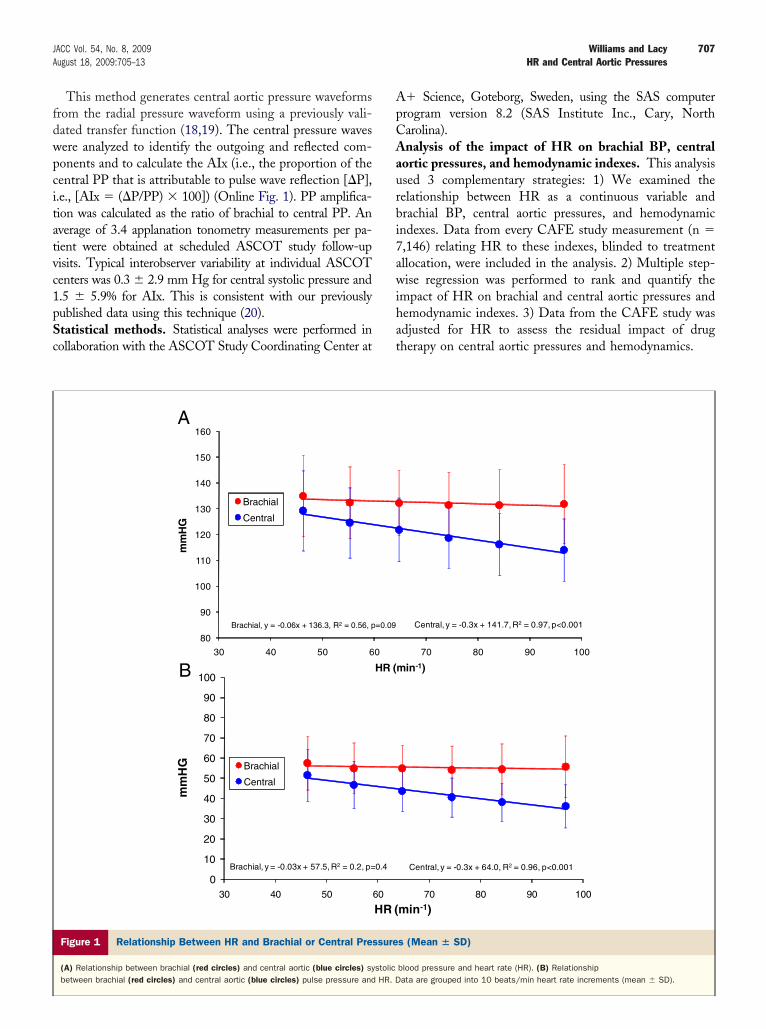
Figure 1 Relationship Between HR and Brachial or Central Pre
(A) Relationship between brachial (red circles) and central aortic (blue circles) sy
� Science, Goteborg, Sweden, using the SAS computerrogram version 8.2 (SAS Institute Inc., Cary, Northarolina).nalysis of the impact of HR on brachial BP, central
ortic pressures, and hemodynamic indexes. This analysissed 3 complementary strategies: 1) We examined theelationship between HR as a continuous variable andrachial BP, central aortic pressures, and hemodynamicndexes. Data from every CAFE study measurement (n �,146) relating HR to these indexes, blinded to treatmentllocation, were included in the analysis. 2) Multiple step-ise regression was performed to rank and quantify the
mpact of HR on brachial and central aortic pressures andemodynamic indexes. 3) Data from the CAFE study wasdjusted for HR to assess the residual impact of drugherapy on central aortic pressures and hemodynamics.
Central, y = -0.3x + 141.7, R2 = 0.97, p<0.001
70 80 90 100
min-1)
Central, y = -0.3x + 64.0, R2 = 0.96, p<0.001
70 80 90 100
min-1)
s (Mean � SD)
blood pressure and heart rate (HR). (B) Relationship
=0.09
0
HR (
0.4
60
HR (
ssure
stolicbetween brachial (red circles) and central aortic (blue circles) pulse pressure and HR. Data are grouped into 10 beats/min heart rate increments (mean � SD).

HvlaacBSumoc
R
Tacwc
TptmupiriiHcs1ba
capa
708 Williams and Lacy JACC Vol. 54, No. 8, 2009HR and Central Aortic Pressures August 18, 2009:705–13
For univariate analyses, data were grouped into deciles ofR, and the relationship between HR and hemodynamic
ariables was analyzed using linear regression. Regressionines were also fitted to plots of raw data. For multivariatenalysis, stepwise multiple linear regression was used. Vari-bles entered into the model were determined by linearorrelation analyses. Continuous data variables betweenP-lowering regimens were compared using nonpairedtudent t tests. Where stated, data were adjusted for HRsing general linear modeling before comparisons wereade. Data are presented as mean (95% confidence interval)
r mean � SD as stated and a value of p � 0.05 wasonsidered significant.
esults
he baseline characteristics of the CAFE study populationccording to their randomized BP-lowering treatment allo-ation are shown in Table 1. The 2 treatment groups wereell matched with respect to their demographics, clinical
haracteristics, and previous medication.
0
5
10
15
20
25
30
35
40
30 40 50 60
Bra
chia
l-C
entr
al S
BP
(m
mH
G)
0
5
10
15
20
25
30
35
40
30 40 50 60
Bra
chia
l-C
entr
al P
P (m
mH
G)
A
B
Figure 2 Relationship Between HR and the Difference Between
Relationship between heart rate (HR) and the difference between brachial and cen
he relationship between HR and brachial versus centralressures. The relationship of HR with brachial and cen-ral pressures is shown in Figure 1. The data encompass alleasurements performed during the CAFE study follow-
p. The data plots were very dense, thus for clarity ofresentation, the data were grouped into increments ofncreasing HR (10 beats/min increments). Importantly, theegressions of the relationships did not differ when compar-ng the grouped and raw data plots. There was no significantmpact of reducing HR on brachial systolic BP (�0.6 mm
g per 10 beats/min decrease in HR) (Fig. 1A). Byontrast, there was a 5-fold greater increase in centralystolic pressure per unit change in HR (�3.0 mm Hg per0 beats/min decrease in HR). A similar dissociationetween the impact of HR on brachial and central PP waslso observed (Fig. 1B).
Figure 2 shows the differences between brachial andentral pressures, plotted as a function of HR. Importantly,t lower HRs, the difference between brachial and centralressure progressively decreased, so that central pressurepproached brachial pressure at the lowest HRs.
y = 0.25x - 5.7, R2 = 0.3705, p<0.001
80 90 100 110 120
y = 0.28x - 6.7, R2 = 0.422, p<0.001
80 90 100 110 120in-1)
hial and Central Pressures
stolic blood pressure (SBP) (A) or brachial and central pulse pressure (PP) (B).
70
70HR (m
Brac
tral sy

Rpiniaeb(btwtr(Tht
pacoataimr(Caabdoa
709JACC Vol. 54, No. 8, 2009 Williams and LacyAugust 18, 2009:705–13 HR and Central Aortic Pressures
elationship between HR and components of the centralressure waveform. To investigate the mechanisms involved
n the changes in central pressure with variation in HR, weext analyzed the components of the central pressure waveform
n relation to HR. There was minimal impact of HR on themplitude of the outgoing pressure wave (P1 height). How-ver, there was a strong and significant inverse relationshipetween HR and the amplitude of pressure wave reflectionsaugmentation), which increased by �3 mm Hg per 10eats/min reduction in HR (Fig. 3A). This finding suggestshat the main impact of HR reduction was on the reflectedave, rather than the incident pressure wave. Consistent with
his observation, there was a marked increase in AIx witheducing HR: �4.9% per 10 beats/min reduction in HRFig. 3B).
he relative contribution of HR to central pressures andemodynamic variables. To further evaluate the contribu-
ion of HR to central pressures and hemodynamics, we
Outgoing, y = -0.03x + 32.9,R2 = 0.002, p<0.001
Reflected, y = -0.3x + 32.5,R2 = 0.3, p<0.001
0
10
20
30
40
50
60
70
80
0 20 40
mm
Hg
H
Outgoing
Reflected
y = -0.49x + 59.4, R2 = 0.34, p<0.0001
-10
0
10
20
30
40
50
60
30 40 50 60 7
AIx
(%)
H
A
B
Figure 3 Relationship Between HR and Outgoing or Reflected P
(A) Relationship between heart rate (HR) and the outgoing pressure wave (P1 heigthe reflected pressure wave (augmentation, blue circles). (B) Relationship betwee
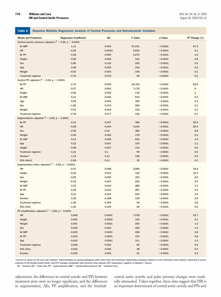
erformed stepwise multiple linear regression (Table 2). Afterccounting for brachial BP, HR was the major determinant ofentral systolic and pulse pressures, accounting for 5% and 9%f the variability in these parameters, respectively. HR was alsomajor determinant of pressure wave reflections (augmenta-
ion and AIx) and PP amplification, accounting for 26%, 34%,nd 54%, respectively of the variability in these parameters. Ofmportance, in this analysis, the BP treatment regimen was a
uch less powerful determinant of central pressures and waveeflections, accounting for no more than 0.5% of the variabilityi.e., at least 10-fold less important than the impact of HR).
omparison of central pressures and hemodynamic vari-bles between BP-lowering treatment arms before and afterdjustment for HR. To further evaluate the relative contri-ution of HR as a determinant of central pressures, theifferential impact of the 2 BP-lowering treatment regimensn central pressures and wave reflections was resolved afterdjusting the data for HR differences (Table 3). After HR
80 100 120in-1)
80 90 100 110 120
in-1)
ure Wave Components
circles) and HR andentation index (AIx) and HR.
60R (m
0
R (m
ress
ht, redn augm
ati
ce
S
*e
dard e
710 Williams and Lacy JACC Vol. 54, No. 8, 2009HR and Central Aortic Pressures August 18, 2009:705–13
djustment, the differences in central systolic and PPs betweenreatment arms were no longer significant, and the differences
tepwise Multiple Regression Analysis of Central Pressures and HeTable 2 Stepwise Multiple Regression Analysis of Central Pres
Model and Predictors Regression Coefficient S
Central systolic pressure; adjusted r2 � 0.95, p � 0.0001
Br SBP 1.11 0.0
HR 0.25 0.0
Br PP 0.28 0.0
Height 0.06 0.0
Sex 1.85 0.1
Age 0.08 0.0
Weight 0.03 0.0
Treatment regimen 0.76 0.0
Central PP; adjusted r2 � 0.94, p � 0.0001
Br PP 0.72 0.0
HR 0.27 0.0
Height 0.06 0.0
Br SBP 0.11 0.0
Age 0.09 0.0
Sex 1.85 0.1
Weight 0.03 0.0
Treatment regimen 0.78 0.0
Augmentation; adjusted r2 � 0.69, p � 0.0001
Br PP 0.13 0.0
HR 0.28 0.0
Sex 2.92 0.1
Weight 0.05 0.0
Br SBP 0.14 0.0
Age 0.12 0.0
Height 0.08 0.0
Treatment regimen 1.01 0.1
Smoker* 1.14 0.1
ECG other† 0.58 0.1
Augmentation index; adjusted r2 � 0.55, p � 0.0001
HR 0.47 0.0
Height 0.15 0.0
Sex 4.83 0.2
Weight 0.10 0.0
Br SBP 0.22 0.0
Br PP 0.20 0.0
Age 0.21 0.0
Smoker 2.30 0.1
Treatment regimen 1.46 0.1
ECG other 1.05 0.1
PP amplification; adjusted r2 � 0.66, p � 0.0001
HR 0.008 0.0
Height 0.002 0.0
Weight 0.001 0.0
Sex 0.046 0.0
Br SBP 0.003 0.0
Br PP 0.003 0.0
Age 0.003 0.0
Treatment regimen 0.018 0.0
ECG other 0.016 0.0
Smoker 0.019 0.0
Current or recent (in the last year) smoker; †abnormalities on electrocardiogram (ECG) other thvidence of left bundle branch block, and ST-T changes compatible with ischemic heart disease (Br � brachial; HR � heart rate; PP � pulse pressure; SBP � systolic blood pressure; SE � stan
n augmentation, AIx, PP amplification, and the brachial- a
entral aortic systolic and pulse pressure changes were mark-dly attenuated. Taken together, these data suggest that HR is
ynamic Variablesand Hemodynamic Variables
F Value p Value R2 Change (%)
67,241 �0.0001 87.3
5,630 �0.0001 5.1
3,076 �0.0001 1.6
123 �0.0001 0.8
255 �0.0001 0.2
232 �0.0001 0.2
145 �0.0001 0.1
93 �0.0001 0.1
20,790 �0.0001 82.5
7,176 �0.0001 9
116 �0.0001 1
634 �0.0001 0.4
305 �0.0001 0.3
266 �0.0001 0.1
153 �0.0001 0.1
100 �0.0001 0.1
380 �0.0001 29.3
4,240 �0.0001 26.4
382 �0.0001 6.8
178 �0.0001 2.1
634 �0.0001 1.9
319 �0.0001 1.1
133 �0.0001 0.5
99 �0.0001 0.5
108 �0.0001 0.5
30 �0.0001 0.1
3,806 �0.0001 34.4
140 �0.0001 10.3
322 �0.0001 2.5
226 �0.0001 2.0
489 �0.0001 1.3
287 �0.0001 0.9
334 �0.0001 1.6
135 �0.0001 0.9
64 �0.0001 0.5
29 �0.0001 0.2
7,978 �0.0001 53.7
124 �0.0001 6.1
205 �0.0001 1.3
200 �0.0001 1.0
495 �0.0001 0.8
315 �0.0001 0.8
311 �0.0001 1.3
65 �0.0001 0.4
48 �0.0001 0.3
61 �0.0001 0.3
ventricular hypertrophy including evidence of left ventricular strain pattern, abnormal Q waves,pression, negative or biphasic T waves).rror.
modsures
E
04
033
05
05
16
05
03
79
05
03
05
04
05
13
03
77
07
04
5
04
06
07
07
1
1
08
12
69
07
10
12
12
98
83
93
001
002
001
03
001
001
001
02
02
02
an leftST-T de
n important determinant of central aortic systolic and PPs and

wb
D
WdaWsdtrb
sPpIpib(cfpisH
isp5frHsIsipm
tdssvitMatrdiwBeoprA
awsrabisIicpAmfmttsa
CB
V
711JACC Vol. 54, No. 8, 2009 Williams and LacyAugust 18, 2009:705–13 HR and Central Aortic Pressures
as the main determinant of the difference between central andrachial pressures between treatment arms in the CAFE study.
iscussion
ithin a major clinical outcomes trial, this is the first study toefine the impact of drug-related changes in HR, on centralortic pressures and hemodynamics, in hypertensive patients.
ith over 2,000 patients and over 7,000 measurements, thistudy had abundant statistical power to test its hypotheses. Theata clearly demonstrate the powerful influence of HR, acrosshe physiological range, on central aortic pressures and waveeflections in hypertensive patients, despite minimal effects onrachial pressures.
We show that HR is inversely related to central aorticystolic and PPs. Lower HRs are also associated with reducedP amplification; thus, at lower HRs, the central aortic systolicressure becomes closer to the brachial systolic pressure.mportantly, there was minimal impact of HR on the outgoingressure wave height (P1 height), showing only a minorncrease with reduced HR. However, the inverse relationshipetween HR and indexes of central pressure wave reflectioni.e., augmentation) were much stronger, consistent with in-reased wave reflection at lower HRs. Remarkably, the slopesor the relationship between HR and central aortic systolicressure or magnitude of wave reflection (augmentation) weredentical (�3 mm Hg per 10 beats/min reduction in HR),uggesting the importance of wave reflection in mediating theR-related change in central aortic systolic pressure.This finding that central pressure wave reflection is strongly
nfluenced by HR is supported by data from cross-sectionaltudies with data stratified by HR (21) and studies of cardiacacing in humans, which suggested that AIx declines by 4% to% per 10 beats/min increase in HR (12,13); the data for AIxrom the present study are similar at 4.9% per 10 beats/mineduction in HR. Interestingly, in a recent population study,
R was the most powerful modifiable predictor of AIx, centralystolic pressure, and central PP (22).mpact of HR versus treatment regimen. Multiple regres-ion analysis confirmed the relative importance of HR after BPtself, as a key determinant for all central hemodynamicarameters. Moreover, adjusting the CAFE study data for HR
omparison of Central Pressures and Hemodynamic Variables BetwP-Lowering Treatment Arms Before and After Adjustment for HearTable 3 Comparison of Central Pressures and Hemodynamic VaBP-Lowering Treatment Arms Before and After Adjustm
Parameter
Unadjusted
Atenolol Amlodipine
Central systolic BP (mm Hg) 125.5 (124.7–126.3) 121.2 (120.5–12
Central pulse pressure (mm Hg) 46.4 (45.7–47.1) 43.4 (42.8–44.0
Augmentation (mm Hg) 15.4 (14.9–15.8) 11.5 (11.2–11.9
Augmentation index (%) 31.9 (31.3–32.4) 25.3 (24.8–25.9
Pulse pressure amplification 1.21 (1.2–1.21) 1.31 (1.3–1.32)
Brachial-central SBP (mm Hg) 8.3 (8.1–8.6) 12.0 (11.7–12.3
Brachial-central PP (mm Hg) 8.9 (8.6–9.1) 12.8 (12.5–13.1
alues are mean (95% confidence interval).BP � blood pressure; other abbreviations as in Table 2.
arkedly attenuated the difference in central pressures between s
he 2 BP-lowering treatment regimens. This suggests that thisifference was primarily driven by differences in HR, althoughome residual effects remained. It is conceivable that unmea-ured hemodynamic factors such as aortic stiffness and systemicascular resistance/remodeling, which could be differentiallynfluenced by drug treatments, may have accounted for some ofhis residual variability (10,23).
echanisms for the inverse relationship between HRnd central aortic pressures. We suggest 2 mechanismshat could account for the elevation in central pressures witheduced HR: first, reducing HR prolongs cardiac ejectionuration, but has no major effect on pulse velocity (7,24). This
ncreases the likelihood of a greater proportion of the reflectedave appearing in late systole for any given pulse wave velocity.eta-blockade also decreases the dP/dT during ventricularjection, and this could delay the time to the peak of theutgoing wave (10,25). This could also increase central systolicressure by increasing the probability of coincidence of theeflected wave with late systole. Our finding of an increasedIx with beta-blockade is consistent with this hypothesis.Second, the less effective lowering of central aortic systolic
nd pulse pressures in patients with lower HRs is consistentith basic physiology. According to the derivation of Poi-
euille’s law, BP is the product of cardiac output � peripheralesistance, where cardiac output is the product of stroke volumend HR. When HR is reduced by drug therapy (e.g., aeta-blocker) mean arterial pressure is maintained by anncrease in stroke volume (26)—a phenomenon readily ob-erved in patients with complete atrio-ventricular heart block.n younger patients with compliant conduit arteries, thisncrease in stroke volume can be accommodated. Indeed, inonditioned athletes, a combination of increased aortic com-liance and peripheral vasodilation prevents a marked rise inIx and central aortic pressure despite very low HRs andarkedly increased stroke volumes (27). This represents per-
ect physiological adaptation to a reduced HR. In contrast,ost hypertensive patients are not conditioned athletes, and in
he CAFE study were older with stiffened conduit arteries. Inhis setting, a reduction in HR will result in the increasedtroke volume being ejected into a less compliant proximalorta, resulting in a rise in central aortic systolic and PPs. We
ees Betweenor Heart Rate
Adjusted
p Value Atenolol Amlodipine p Value
�0.001 123.9 (123.1–124.6) 122.8 (122.1–123.6) 0.07
�0.001 44.6 (43.9–45.2) 45.2 (44.6–45.9) 0.2
�0.001 13.7 (13.4–14.1) 13.1 (12.8–13.5) 0.02
�0.001 29.3 (28.8–29.8) 27.8 (27.3–28.3) �0.001
�0.001 1.25 (1.24–1.26) 1.27 (1.26–1.28) �0.001
�0.001 9.6 (9.3–9.8) 10.8 (10.5–11.0) �0.001
�0.001 10.3 (10.0–10.5) 11.4 (11.2–11.7) �0.001
eent Ratriablent f
1.9)
)
)
)
)
)
uggest that these are the 2 principal mechanisms accounting

fstcebassIswatpsuiocsbltdtrfSHcfbropbcls
pIfsmtroeHdscpMb
pocdbaa
ttibccfirtCilttifvppcesPbntcr
C
IsaWbct
RDC6a
R
712 Williams and Lacy JACC Vol. 54, No. 8, 2009HR and Central Aortic Pressures August 18, 2009:705–13
or the inverse relationship between HR and central aorticystolic and PPs in the CAFE study. Moreover, we suggesthat this inverse relationship would be accentuated if HRhanges are restricted by drugs (i.e., beta-blockers) duringxercise, when the need to increase cardiac output could onlye met by an increase in stroke volume. These considerationsre of clinical importance given that central PP showed aignificant association with clinical outcomes in the CAFEtudy and other studies (28,29).s this data relevant to all beta-blockers? The presenttudy raises important questions as to whether similar effectsould have been observed with beta-blockers other than
tenolol, notably vasodilating beta-blockers. Our data suggesthat the impact of HR on central aortic pressure is veryowerful and consistent across the physiological range, irre-pective of treatment allocation in this study. Other studiessing invasive monitoring have shown that in patients receiv-ng beta-blockers, the use of powerful vasodilators cannotvercome the impact of HR reduction on wave reflection andentral pressures (30). By contrast, a small number of previoustudies comparing vasodilating and nonvasodilating beta-lockers have suggested a more beneficial influence of vasodi-ating beta-blockers on central pressures (31,32). However,hese studies were small scale and underpowered, and theifferences in central pressures and wave reflections betweenhe different beta-blockers could be accounted for by the lessereductions in HR with vasodilating beta-blockers and/or dif-erences in brachial BP.tudy limitations. We recruited predominantly white men.owever, our regression analysis suggests that the direction of
hange in central pressures and hemodynamics was the sameor women. It is unclear whether similar findings would haveeen observed in other ethnic groups. Nevertheless, we cannotationalize why a mechanism that appears to be so dependentn HR would be different in other ethnic groups. Our patientopulation was also older, with a mean age of 63 years ataseline. It is conceivable that in younger people with moreompliant conduit arteries there would be a lesser impact ofower HRs on central aortic pressure. These important con-iderations need further evaluation.
We used noninvasive methods to derive central aorticressure from the radial pulse wave, calibrated to brachial BP.t has to be considered whether the mathematical transferunction used to derive central hemodynamic indexes could beensitive to, or confounded by, changes in HR. The mathe-atics involved are beyond detailed discussion here but use a
ransfer function to calculate central pressures from individualadial pressure waveforms that is uninfluenced by the numberf waveforms as a function of time. Although, to our knowl-dge, there have been no specific studies to assess impact ofR on central pressures comparing the methods here with
irect invasive measurements, there have been invasive mea-urements of central aortic pressures in humans in response tohanges in HR. In these studies, increasing HR via cardiacacing has been shown to reduce central aortic pressure (12).oreover, previous invasive studies have shown that beta-
locker treatment increased (rather than reduced) central aortic
ressures (33). These directional changes are consistent withur findings. Furthermore, data from studies directly analyzingarotid or invasively acquired central pressure waves haveocumented reduced “pressure amplification” with beta-lockade (24,33), consistent with our data. Other studies havelso implicated HR as a major factor modulating pressuremplification (13,21,34,35).
Finally, our study examines the association between on-reatment HR and central pressures. It does not directly assesshe change in central pressure in response to a treatment-nduced change in HR in individual patients. This would haveeen difficult to do because of confounding due to associatedhanges in BP per se as a consequence of any treatmenthanges. Nevertheless, our multiple regression analysis identi-ed HR to be a powerful independent factor influencing theelationship between brachial and central aortic pressures, withhe latter being higher at lower HRs.
linical implications. These data have important clinicalmplications. There is a well-recognized association between aower HR and cardiovascular health reported from observa-ional studies (36–38). This is often used as a justification forherapeutic reductions in HR. In the setting of symptomaticschemic heart disease and in patients with chronic stable heartailure, HR lowering by beta-blockade has been shown to be aery effective treatment strategy. However, the data from theresent study question whether extending these assumptions toeople with hypertension, especially older people with stiffonduit arteries, is safe and appropriate. Moreover, because theffect of HR on central pressures seems so powerful, our datauggest that there will be less effective central aortic systolic andP reduction in older hypertensive patients with all beta-lockers, or other drugs that lower HR. In this regard, theewer generation of vasodilating beta-blockers must be showno be as effective as alternative treatments in preventingardiovascular events before they can be considered as a suitableoutine treatment for older people with hypertension.
onclusions
n summary, the CAFE-Heart Rate study has demon-trated that a lower HR is associated with higher centralortic systolic and PPs in patients with treated hypertension.
e suggest that this is the major reason why beta-blocker–ased therapy has been less effective at reducing cardiovas-ular events, especially stroke, when compared with otherreatments in patients with hypertension.
eprint requests and correspondence: Dr. Bryan Williams,epartment of Cardiovascular Sciences, University of Leicester,linical Sciences Building, Leicester Royal Infirmary, P.O. Box5, Leicester, LE2 7LX, United Kingdom. E-mail: [email protected].
EFERENCES
1. Lindholm LH, Carlberg B, Samuelsson O. Should beta blockers
remain first choice in the treatment of primary hypertension? Ameta-analysis. Lancet 2005;366:1545–53.
1
1
1
1
1
1
1
1
1
1
2
2
2
2
2
2
2
2
2
2
3
3
3
3
3
3
3
3
3
Kh
Fva
713JACC Vol. 54, No. 8, 2009 Williams and LacyAugust 18, 2009:705–13 HR and Central Aortic Pressures
2. Wiysonge C, Bradley H, Myose B, et al. Beta-blockers for hyperten-sion. Cochrane Data Base Syst Rev 2007;1:CD002003.
3. Bangalore S, Wild D, Parkar S, Kukin M, Messerli FH. Beta-blockersfor primary prevention of heart failure in patients with hypertension.J Am Coll Cardiol 2008;52:1062–72.
4. Bangalore S, Sawhney S, Messerli FH. Relation of �-blocker-inducedheart rate lowering and cardioprotection in hypertension. J Am CollCardiol 2008;52:1482–9.
5. Williams B. �-blockers and the treatment of hypertension. J Hyper-tens 2007;25:1351–3.
6. NICE/BHS. Clinical guideline 34: hypertension: management ofhypertension in adults in primary care: partial update. Available at:http://www.nice.org.uk/CG034guidance. Accessed March 10, 2007.
7. The CAFE Investigators for the ASCOT Investigators. Differentialimpact of blood pressure lowering drugs on central aortic pressure andclinical outcomes—principal results of the Conduit Artery FunctionEvaluation study: the CAFE study. Circulation 2006;113:1213–25.
8. Chen C-H, Ting C-T, Lin S-J, et al. Different effects of fosinopril andatenolol on wave reflections in hypertensive patients. Hypertension1995;25:1034–41.
9. Hirata K, Vlachopoulos C, Adji A, O’Rourke M. Benefits fromangiotensin-converting enzyme inhibitor ‘beyond blood pressure low-ering’: beyond blood pressure or beyond the brachial artery [erratum in:J Hypertens 2005;23:903–4]? J Hypertens 2005;23:551–6.
0. London GM, Asmar RG, O’Rourke M, Safar ME, on behalf of theREASON Investigators. Mechanism(s) of selective systolic bloodpressure reduction after a low-dose combination of perindopril/indapamide in hypertensive subjects: comparison with atenolol. J AmColl Cardiol 2004;43:92–9.
1. Deary AJ, Schumann AL, Murfet H, Haydock S, Foo RS, Brown M.Influence of drugs and gender on the arterial pulse wave and natriureticpeptide secretion in untreated patients with essential hypertension.Clin Sci 2002;103:493–9.
2. Wilkinson IB, MacCallum H, Flint L, Cockcroft JR, Newby DE,Webb DJ. The influence of heart rate on augmentation index andcentral arterial pressure in humans. J Physiol 2000;525:263–70.
3. Wilkinson IB, Mohammed NH, Tyrrell S, et al. Heart rate depen-dency of pulse pressure amplification and arterial stiffness. Am JHypertens 2002;15:24–30.
4. Dahlof B, Sever PS, Poulter NR, et al., for the ASCOT Investigators.Prevention of cardiovascular events with an antihypertensive regimenof amlodipine adding perindopril as required versus atenolol addingbendroflumethiazide as required, in the Anglo-Scandinavian CardiacOutcomes Trial-Blood Pressure Lowering Arm (ASCOT-BPLA): amulticentre randomised controlled trial. Lancet 2005;366:895–906.
5. Sever PS, Dahlof B, Poulter NR, et al., for the ASCOT Investigators.Rationale, design, methods and baseline demography of participants ofthe Anglo-Scandinavian Cardiac Outcomes Trial. J Hypertens 2001;19:1139–47.
6. Kelly R, Hayward CS, Ganis J. Non-invasive registration of thearterial pressure pulse waveform using high-fidelity applanationtonometry. J Vasc Biol 1989;1:142–9.
7. O’Rourke M, Gallagher DE. Pulse wave analysis. J Hypertens 1996;14Suppl 5:S147–57.
8. Chen C-H, Nevo E, Fetics B, et al. Estimation of central aorticpressure waveform by mathematical transformation of radial tonome-try pressure: validation of generalized transfer function. Circulation1997;95:1827–36.
9. Fetics B, Nevo E, Chen C-H, Kass DA. Parametric model derivationof transfer function for non-invasive estimation of aortic pressure byradial tonometry. IEEE Trans Biomed Eng 1999;46:698–706.
0. Siebenhofer A, Kemp CRW, Sutton AJ, Williams B. The reproduc-ibility of central aortic blood pressure measurements in healthy subjectsusing applanation tonometry and sphygmocardiography. J Hum Hy-
1. Laurent P, Albaladejo P, Blacher J, Rudnichi A, Smulyan H, SafarME. Heart rate and pulse pressure amplification in hypertensivesubjects. Am J Hypertens 2003;16:363–70.
2. McEniery CM, Yasmin, Hall IR, et al., on behalf of the ACCTInvestigators. Normal vascular aging; differential effects on wavereflections and aortic pulse wave velocity. The Anglo Cardiff Collab-orative Trial (ACCT). J Am Coll Cardiol 2005;46:1753–60.
3. Savoia C, Touyz RM, Endemann DH, et al. Angiotensin receptorblocker added to previous antihypertensive agents on arteries ofdiabetic hypertensive patients. Hypertension 2006;48:271–7.
4. Asmar RG, London GM, O’Rourke M, Safar ME, for the REASONProject Coordinators and Investigators. Improvement in blood pres-sure, arterial stiffness and wave reflections with a very-low-doseperindopril/indapamide combination in hypertensive patients: a com-parison with atenolol. Hypertension 2001;38:922–6.
5. O’Rourke M. Arterial stiffness, systolic blood pressure, and logicaltreatment of arterial hypertension. Hypertension 1990;15:339–47.
6. Nichols WW, Edwards DG. Arterial elastance and wave reflectionaugmentation of systolic blood pressure: deleterious effects and impli-cations for therapy. J Cardiovasc Pharmacol Therapeut 2001;6:5–21.
7. Edwards DG, Lang JT. Augmentation index and systolic load arelower in competitive endurance athletes. Am J Hypertens 2005;18:679–83.
8. Roman MJ, Devereux RB, Kizer JR, et al. Central pressure morestrongly relates to vascular disease and outcome than does brachialpressure: the Strong Heart study. Hypertension 2007;50:197–203.
9. Jankowski P, Kawecka-Jaszcz K, Czarnecka D, et al. Pulsatile but notsteady component of blood pressure predicts cardiovascular events incoronary patients. Hypertension 2008;51:848–55.
0. Ting C-T, Chen C-H, Chang M-S, Yin FCP. Short- and long-termeffects of antihypertensive drugs on arterial reflections, compliance andimpedance. Hypertension 1995;26:524–30.
1. Kelly R, Daley J, Avolio A, O’Rourke M. Arterial dilation and reducedwave reflection. Benefit of dilevanol in hypertension. Hypertension1989;14:14–21.
2. Dhakam Z, Yasmin, McEniery CM, Burton T, Brown MJ, WilkinsonIB. A comparison of atenolol and nebivolol in isolated systolichypertension. J Hypertens 2008;26:351–6.
3. Ting CT, Chou CY, Chang MS, Wang SP, Chiang BN, Yin FCP.Arterial hemodynamics in human hypertension: effects of adrenergicblockade. Circulation 1991;84:1049–57.
4. Protogerou AD, Blacher J, Mavrikakis M, Lekakis J, Safar ME.Increased pulse pressure amplification in treated hypertensive subjectswith metabolic syndrome. Am J Hypertens 2007;20:127–33.
5. Safar ME, Blacher J, Protogerou AD, Achimastos A. Arterial stiffnessand central hemodynamics in treated hypertensive subjects accordingto brachial blood pressure classification. J Hypertens 2008;26:130–7.
6. Dyer AR, Persky V, Stamler J, et al. Heart rate as a prognostic factorfor coronary heart disease and mortality: findings in three Chicagoepidemiologic studies. Am J Epidemiol 1980;112:736–49.
7. Gillum RF, Makuc DM, Feldman JJ. Pulse rate, coronary heart diseaseand death: the NHANES I epidemiologic follow-up study. AmHeart J 1991;121:172–7.
8. Palatini P, Thijs L, Staessen JA, et al., for the Systolic Hypertensionin Europe (Syst-Eur) Trial Investigators. Predictive value of clinic andambulatory heart rate for mortality in elderly subjects with systolichypertension. Arch Intern Med 2002;162:2313–21.
ey Words: beta-blocker y heart rate y central aortic pressure yypertension.
APPENDIX
or the text on the measurement of pressure waveforms and definition ofariables derived by pulse wave analysis, a list of the CAFE investigators,nd a figure on the central arterial pressure wave with derived parameters,
pertens 1999;13:625–9. please see the online version of this article.
Go to http://cme.jaccjournals.orgto take the CME quiz for this article.