Embed Size (px)
Citation preview

REVIEW Open Access
Immunotoxicity and intestinal effects ofnano- and microplastics: a review of theliteratureNell Hirt and Mathilde Body-Malapel*
Abstract
Background: Together with poor biodegradability and insufficient recycling, the massive production and use ofplastics have led to widespread environmental contamination by nano- and microplastics. These particlesaccumulate across ecosystems - even in the most remote habitats - and are transferred through food chains,leading to inevitable human ingestion, that adds to the highest one due to food processes and packaging.
Objective: The present review aimed at providing a comprehensive overview of current knowledge regarding theeffects of nano- and microplastics on intestinal homeostasis.
Methods: We conducted a literature search focused on the in vivo effects of nano- and microplastics on gutepithelium and microbiota, as well as on immune response.
Results: Numerous animal studies have shown that exposure to nano- and microplastics leads to impairments inoxidative and inflammatory intestinal balance, and disruption of the gut’s epithelial permeability. Other notableeffects of nano- and microplastic exposure include dysbiosis (changes in the gut microbiota) and immune celltoxicity. Moreover, microplastics contain additives, adsorb contaminants, and may promote the growth of bacterialpathogens on their surfaces: they are potential carriers of intestinal toxicants and pathogens that can potentiallylead to further adverse effects.
Conclusion: Despite the scarcity of reports directly relevant to human, this review brings together a growing bodyof evidence showing that nano- and microplastic exposure disturbs the gut microbiota and critical intestinalfunctions. Such effects may promote the development of chronic immune disorders. Further investigation of thisthreat to human health is warranted.
Keywords: Microplastics, Nanoplastics, Intestinal, Microbiota, Inflammation, Immunotoxicity
© The Author(s). 2020 Open Access This article is licensed under a Creative Commons Attribution 4.0 International License,which permits use, sharing, adaptation, distribution and reproduction in any medium or format, as long as you giveappropriate credit to the original author(s) and the source, provide a link to the Creative Commons licence, and indicate ifchanges were made. The images or other third party material in this article are included in the article's Creative Commonslicence, unless indicated otherwise in a credit line to the material. If material is not included in the article's Creative Commonslicence and your intended use is not permitted by statutory regulation or exceeds the permitted use, you will need to obtainpermission directly from the copyright holder. To view a copy of this licence, visit http://creativecommons.org/licenses/by/4.0/.The Creative Commons Public Domain Dedication waiver (http://creativecommons.org/publicdomain/zero/1.0/) applies to thedata made available in this article, unless otherwise stated in a credit line to the data.
* Correspondence: [email protected]. Lille, Inserm, CHU Lille, U1286- INFINITE - Institute for TranslationalResearch in Inflammation, F-59000 Lille, France
Hirt and Body-Malapel Particle and Fibre Toxicology (2020) 17:57 https://doi.org/10.1186/s12989-020-00387-7

BackgroundThe use of plastics has increased hugely over the pastfew decades. Indeed, the continuous production, use andconsumption of plastics since the 1950s has createdmajor environmental problems worldwide (Scheme 1).In 1960, half a million metric tons of plastics were re-leased each year in the world [1]. This tonnage has sincerisen exponentially and has reached 359 million metrictons in 2018 [2]. In view of their low price and attractivephysicochemical properties, plastics have become essen-tial in every industry (packaging, construction, transport,etc). At present, it is almost impossible to find plastic-free goods. Plastic is used extensively in our everydayobjects (packaging, cosmetics, household goods, elec-trical and electronic equipment, furniture, etc). Due tolimited recycling and the lack of regulations limitingplastic waste, plastics (and especially nano- and micro-plastics) have contaminated aquatic, terrestrial andatmospheric environments worldwide. Plastics arepresent in our oceans, seas, rivers, and lakes, and haveeven reached the Arctic sea ice [3–5].Microplastic pollution is ubiquitous in soil environ-
ments, including agricultural/farmland, greenhouse,home garden, coastal, industrial, and floodplain soils[6]. This pollution is due to the tremendous growthin plastic waste. In 2018, 25% of the 29.1 millionmetric tons of post-consumer plastic waste in Europeended up in landfills [2]. Soil microplastics comefrom the unsustainable use and inappropriate wastemanagement of plastics - especially those in pack-aging. Moreover, microplastics are released into thesoil by agricultural processes [7]; the use of plasticmulches and the application of sewage sludge to fieldsare major sources of soil microplastics. In order toprevent microplastics from entering the aquatic envir-onment, wastewater treatment plants remove micro-plastics from the wastewater but thus concentratethem in the sludge subsequently used as a fertilizeron agricultural soils [8].
In the marine environment, plastic debris can befound on the sea floor, surface and shoreline. Eriksenet al. estimated in 2014 that at least 5.25 trillionplastic particles including 35,500 metric tons ofmicroplastics were floating at sea [9]. It has been esti-mated that 80% of the plastic pollution in the oceansand seas comes from land [10] and the estimatedamount of land-based plastic debris entering theocean is between 4.8 and 12.7 million metric tons peryears [11]. Microplastics are detected in freshwater,including lakes, rivers, and groundwater. These parti-cles come mainly from urban pollution but also fromshipping, fisheries, tourism, oil and gas platforms,wastewater treatment plants, discharged personalhealth care products, textiles, and packaging [12].Rochman et al. (2015) calculated that in 2015, 8 tril-lion microbeads per day were emitted into aquatichabitats in the United States [13].Lastly, the atmosphere is a new recognized vehicle
through which microplastics enter the wider environ-ment [14, 15]. Microplastics have been measured inatmospheric fallout in both megacities [16, 17] andsparsely inhabited areas [18]. Suspended atmosphericmicroplastics have been also repeatedly detected inindoor air [19, 20].The omnipresence of microplastics in the environment
leads to human exposure largely by ingestion but also byinhalation and dermal contact [21]. This exposure is acause of concern for potential long-term health hazards.Recent research has highlighted the possible adverseeffects of nano- and microplastic exposure on intestinalhomeostasis, gut microbiota and immune response.Here, we review this emerging field. Firstly, nano- andmicroplastics were briefly defined, then the pathwaysthrough which they can interact with the intestine andthe immune system were described. Afterwards, studiesof in vivo exposure to nano -and microplastics on thegut epithelium, the intestinal microbiota, and theimmune system were detailed. The potential of
Scheme 1 The omnipresence of plastics
Hirt and Body-Malapel Particle and Fibre Toxicology (2020) 17:57 Page 2 of 22
microplastics as carriers of intestinal toxics and patho-gens was emphasized. Lastly, current research perspec-tives and future needs were discussed.
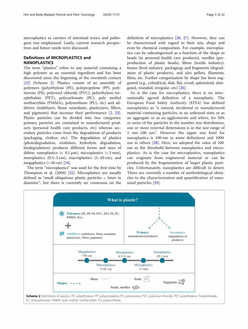
Definitions of MICROPLASTICS andNANOPLASTICSThe term “plastics” refers to any material containing ahigh polymer as an essential ingredient and has beendiscovered since the beginning of the twentieth century[22] (Scheme 2). Plastics consist of an assembly ofpolymers (polyethylene (PE), polypropylene (PP), poly-styrene (PS), polyvinyl chloride (PVC), polyethylene ter-ephthalate (PET), polycarbonate (PC), poly methylmethacrylate (PMMA), polyurethane (PU), etc) and ad-ditives (stabilizers, flame retardants, plasticizers, fillers,and pigments) that increase their performance [2, 23].Plastic particles can be divided into two categories:primary particles are contained in manufactured prod-ucts (personal health care products, etc) whereas sec-ondary particles come from the degradation of products(packaging, clothes, etc). The degradation of plastics(photodegradation, oxidation, hydrolytic degradation,biodegradation) produces different forms and sizes ofdebris; nanoplastics (≤ 0.1 μm), microplastics (< 5 mm),mesoplastics (0.5–5 cm), macroplastics (5–50 cm), andmegaplastics (> 50 cm) [24].The term “microplastics” was used for the first time by
Thompson et al. (2004) [25]. Microplastics are usuallydefined as “small ubiquitous plastic particles < 5mm indiameter”, but there is currently no consensus on the
definition of microplastics [26, 27]. However, they canbe characterized with regard to their size, shape andeven by chemical composition. For example, microplas-tics can be subcategorized as a function of the shape asbeads (in personal health care products), nurdles (pre-production of plastic beads), fibres (textile industry),foams (food industry, packaging) and fragments (degrad-ation of plastic products), and also pellets, filaments,films, etc. Further categorization by shape has been sug-gested (e.g., cylindrical, disk, flat, ovoid, spheruloid, elon-gated, rounded, irregular, etc) [26] .As is the case for microplastics, there is no inter-
nationally agreed definition of a nanoplastic. TheEuropean Food Safety Authority (EFSA) has definednanoplastics as “a natural, incidental or manufacturedmaterial containing particles, in an unbound state or asan aggregate or as an agglomerate and where, for 50%or more of the particles in the number size distribution,one or more external dimensions is in the size range of1 nm−100 nm”. However the upper size limit fornanoplastics is 100 nm in some definitions and 1000nm in others [28]. Here, we adopted the value of 100nm as the threshold between nanoplastics and micro-plastics. As is the case for microplastics, nanoplasticscan originate from engineered material or can beproduced by the fragmentation of larger plastic parti-cles. Unfortunately, nanoplastics are difficult to detect.There are currently a number of methodological obsta-cles to the characterization and quantification of nano-sized particles [29].
Scheme 2 Definitions of plastics. PE: polyethylene, PP: polypropylene, PS: polystyrene, PVC: polyvinyl-chloride, PET: polyethylene Terephthalate,PC: polycarbonate, PMMA: poly methyl methacrylate, PU: polyurethane
Hirt and Body-Malapel Particle and Fibre Toxicology (2020) 17:57 Page 3 of 22

Human exposure to NANO- and MICROPLASTICSHuman exposure to microplastics through ingestionThe human ingestion of microplastics was revealed bythe detection of microplastics in several dietary products(Scheme 3). Firstly, microplastics are ubiquitous in sur-face water, groundwater and wastewater [30]. Plasticsare also found in drinking water and this issue has beenreviewed recently [31, 32]. Koelmans et al. reported onthe types of plastics found in freshwater (in decreasingorder of frequency): fragments (35%), fibres (25%), films,foams, pellets, spheres, lines, beads, flakes, sheets, gran-ules, paints, foils and nurdles. Overall, the polymersmost frequently detected by researchers are PE ≈ PP >PS > PVC > PET, followed by polyamide (PA), acrylic oracrylic-related compounds, polyesters and PMMA. Des-pite the removal of microplastics by various water treat-ment processes, microplastics are also detected in tapwater [33]. In Kosuth et al.’s study of 159 samples of tapwater from all over the world, 81% contained microplas-tics; the mean concentration was 5.45 particles/L [34]. Inan analysis of tap water samples in China, the lowestmicroplastic particle count measured was 440/L. Mostof these particles were smaller than 50 μm fragments(followed by fibres and spheres) and composed mainlyof PE and PP [35]. Microplastic particles were also de-tected in mineral water contained in both plastic bottlesand glass bottles. The literature data on microplastics inmineral water were compiled recently [36]. The overallreported concentrations of microplastics were 0.6 μg/L[37] and 7.3 μg/L [38] in multi-use PET bottles, and0.1 μg/L [37] and 1.8 μg/L [38] in single-use PET bottles.The concentrations in water in glass bottles were evenhigher (2.6 μg/L [37] and 8.7 μg/L [38]). The overall par-ticle number ranged from 14 to 6290 particles/L [36].Particles smaller than 5 μm accounted for approximately
96% of the total in PET bottles and 78% in glass bottles[39]. In a recent analysis of microplastics in Thailand,there were 140 particles/L in water in PET bottles, 52particles/L in water in glass bottles, 81, 26 and 12 parti-cles/L for the 6.5–20 μm, 20–50 μm, and > 50 μm diam-eter sizes [40]. The estimated maximum annual uptakeby human adults is 458,000 microplastic particles for tapwater and 3,569,000 microplastic particles for bottledwater [32].Given the presence of microplastics in the oceans,
these particles are also detected in seafood products [41,42]. Indeed, some of the 220 species found to ingestmicroplastic debris in natura (such as mussels, oysters,clams, common shrimps, etc) are of commercial import-ance for fisheries and aquaculture [43]. In Hantoroet al.’s review of studies of microplastics in seafood, itwas estimated that the human intake can attain 66 × 103,28 × 103 and 36 × 103 particles/day through fish, crust-acean, and mollusk consumption, respectively [44].Furthermore, qualitative and quantitative measure-
ments of microplastics have been reported for otherfood products, such as honey and sugar. In samples ofthese basic products collected in Europe (Germany,France, Italy, and Spain) and Mexico, the fibre contentper kg was 166 for honey and 217 for sugar [45].Microplastic contamination has also been detected
in sea salt originating from various countries world-wide [34, 46–49], and these data have been reviewedby Toussaint et al. [26]. For example, 550–681 parti-cles/kg were detected in sea salt samples collectedacross China, [46]. The majority of the particles (55%)measured less than 200 μm in diameter. Fragmentsand fibres were more prevalent than pellets andsheets. The most common microplastics were PET,followed by PE and cellophane.
Scheme 3 Human ingestion of microplastics. PE: polyethylene, PP: polypropylene, PS: polystyrene, PVC: polyvinyl chloride, PET: polyethyleneterephthalate, PA: polyamide. Data on plastic polymers and shapes in freshwater are based on the number of studies reporting the presence of aparticular polymer or shape of microplastic particles in freshwater. Adapted from Koelmans, Water Research 155 (2019) 410–422
Hirt and Body-Malapel Particle and Fibre Toxicology (2020) 17:57 Page 4 of 22

Furthermore, Liebezeit et al. analyzed the content ofmicroplastics in German beers [50]. Microplasticcontamination was found in all cases, with counts ran-ging from 2 to 79 fibres/L, from 12 to 109 fragments/Land from 2 to 66 granules/L. The relative contributionsranged from 5 to 71% for granular material, from 14 to87% for fragments and from 3 to 57% for fibres.Microplastics have been also detected in cow milk
samples for adults and children. All samples containedmicroplastic particles, with differences in the amounts (1to 14 particles/L) [51]. Of the total detected microplas-tics, 97.5% were fibres and 2.5% were fragments: micro-plastics < 0.5 mm were dominant (40%) followed by thesizes 0.5–1 mm (28%) and 1–2mm (25%).Lastly, many researchers have attempted to estimate the
yearly human exposure to microplastics, However, inter-study differences in the types of plastic and the experi-mental methods mean that these estimations varymarkedly [26]. In such a context, another way to estimatehuman contamination is probably to measure the amountof plastic in human feces. This is what Schwabl et al. didin a recent study of stools samples from 8 healthy volun-teers: the mean number microplastic particles (from 50 to500 μm in size) was 20 per 10 g. Nine types of plastic weredetected, with PP and PET being the most abundant.Based on these results and an average production of 128 gof feces per day per person, the researchers estimate thatthe annual discharge of microplastic particles in the feces(reflecting at least in part the equivalent human body ex-posure) was over 90,000 [39].
Other routes of human exposure to nano- andmicroplasticsHuman exposure to microplastics also occurs throughinhalation, because microplastics are present in the in-door and outdoor air [52]. The sources of airbornemicroplastics have been reviewed recently [14, 15].Synthetic textiles, the erosion of synthetic rubbertires, and city dust are thought to be the most im-portant sources of airborne microplastics. Fibres werethe dominant shape, and PP, PE, PS and PET werethe dominant polymer components of microplastics inatmospheric fallout. Individual human exposure by in-halation has been estimated at 26 to 130 airbornemicroplastic particles a day [14].Lastly, microplastics reach humans by dermal contact.
Microplastic beads are included in the composition of fa-cial cleansers, facial scrubs, and toothpaste, where they areused as exfoliators for the skin and teeth [53]. A recentChinese study has identified personal care products con-taining microplastics. Overall, 7.1% of facial cleansers con-tained microplastics, with a mean ± standard deviationcontent of 25.04 ± 10.69mg microplastics/g and mean sizeof 313 ± 130 μm. The majority of these microplastics were
made of PE [54]. Microplastic beads are also used to regu-late the viscosity of films, condition the skin, and stabilizeemulsions: they are included into a wide range of prod-ucts, such as soaps, shampoos, deodorants, wrinklecreams, moisturizers, shaving creams, sunscreen lotions,facial masks, lipsticks, eye shadows, and children’s bubblebath [55]. The glitters which are used in significant vol-umes in make-up (and also craft activities and textileproducts) are usually PET-containing plastics [56]. More-over, the microbeads used in consumer products (such asscrubs and shampoos) are processed by mechanicalmeans, which may lead to their fragmentation into poten-tially more hazardous nanoplastics. The presence of nano-plastics has been confirmed in personal care productscontaining PE microbeads [57].
Relationships between NANO- and MICROPLASTICS,the intestinal mucosa, and the immune systemMany studies of various species have shown thatingested microplastics accumulate in the gut of variousspecies [58–61]. After a five-day oral course of 60 nm PSnanoparticles in rats, approximatively 10% of the dosewas found in the gastrointestinal tract [62]. Not much isknown on the distribution of nano- and microplasticsafter ingestion. Based on in vitro and in vivo data, know-ledge on the uptake of nano- and microplastics has beenreviewed by the European Food Safety Authority [63].Microplastics with a greatest dimension > 150 μm arenot absorbed, they remain bound to the intestinal mucuslayer and come into direct contact with the apical partof intestinal epithelial cells. This may lead to inflamma-tion of the gut and local effects on the immune system.The smaller particles (greatest dimension < 150 μm) cancross the mucus barrier. Indeed, several mechanismsresult in the size-dependent uptake of nano- and micro-particles: (i) endocytosis through enterocytes, (ii)transcytosis through microfold cells (also referred to asM-cells, a specific subset of intestinal epithelial cells ingut-associated lymphoid tissue), (iii) persorption (namelythe passage through “gaps” at the villous tip, followingthe loss of enterocytes), and (iv) paracellular uptake [64].Uptake of microparticles by endocytosis, transcytosisand paracellular diffusion between enterocytes has beenobserved in rodents without any disruption of the intes-tinal barrier [65, 66]. Peyer’s patches have a high propor-tion of M-cells and constitute the main site ofmicroplastic absorption [67]. The intestinal uptake ofmicroparticles is not very efficient: in one study, only0.3% orally administrated latex particles (greatest dimen-sion: 2 μm) crossed the epithelium [68]. Despite this lowlevel, intestinal absorption of particles may lead to sys-temic toxicologically relevant exposure. The small size ofnanoplastics allows them to penetrate deeply into or-gans. Data from animal studies have shown that once
Hirt and Body-Malapel Particle and Fibre Toxicology (2020) 17:57 Page 5 of 22
absorbed, nanoplastics can distribute to the liver, spleen,heart, lungs, thymus, reproductive organs, kidney, andeven the brain (i.e. they cross the blood–brain barrier[69]) [63].It must also be borne in mind that airborne microplas-
tics may also have an impact on the digestive tract andthe immune system. It is known that among airborneparticles, the smallest particles (i.e. the inhalable frac-tion) are absorbed via the pulmonary epithelium [70,71]. They reach the systemic circulation and exert animmune effect on the so called gut-lung axis [72]. A pro-portion of the larger particles (the extrathoracic fraction)is transported to the gastrointestinal tract by mucociliaryclearance, where it undergoes the fate of ingested parti-cles. Hence, depending of the particle size, both ingestedand inhaled plastics are able to interact with intestinaltissues, reach the bloodstream and (potentially) dysregu-late the immune response.
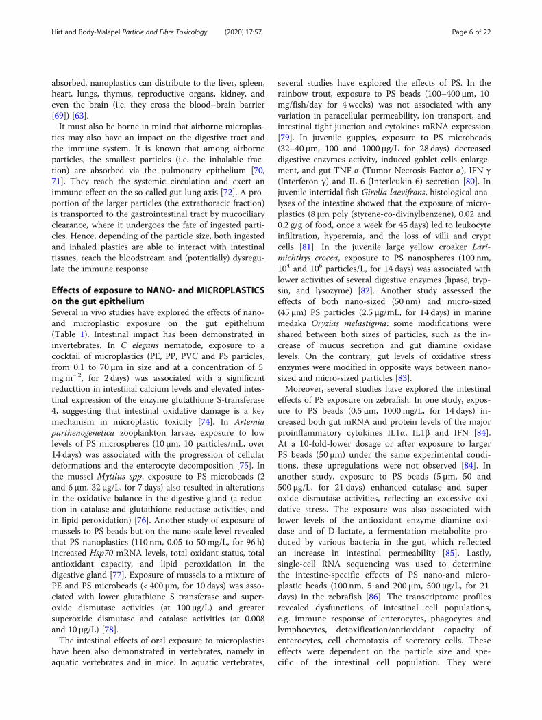
Effects of exposure to NANO- and MICROPLASTICSon the gut epitheliumSeveral in vivo studies have explored the effects of nano-and microplastic exposure on the gut epithelium(Table 1). Intestinal impact has been demonstrated ininvertebrates. In C elegans nematode, exposure to acocktail of microplastics (PE, PP, PVC and PS particles,from 0.1 to 70 μm in size and at a concentration of 5mgm− 2, for 2 days) was associated with a significantreducttion in intestinal calcium levels and elevated intes-tinal expression of the enzyme glutathione S-transferase4, suggesting that intestinal oxidative damage is a keymechanism in microplastic toxicity [74]. In Artemiaparthenogenetica zooplankton larvae, exposure to lowlevels of PS microspheres (10 μm, 10 particles/mL, over14 days) was associated with the progression of cellulardeformations and the enterocyte decomposition [75]. Inthe mussel Mytilus spp, exposure to PS microbeads (2and 6 μm, 32 μg/L, for 7 days) also resulted in alterationsin the oxidative balance in the digestive gland (a reduc-tion in catalase and glutathione reductase activities, andin lipid peroxidation) [76]. Another study of exposure ofmussels to PS beads but on the nano scale level revealedthat PS nanoplastics (110 nm, 0.05 to 50 mg/L, for 96 h)increased Hsp70 mRNA levels, total oxidant status, totalantioxidant capacity, and lipid peroxidation in thedigestive gland [77]. Exposure of mussels to a mixture ofPE and PS microbeads (< 400 μm, for 10 days) was asso-ciated with lower glutathione S transferase and super-oxide dismutase activities (at 100 μg/L) and greatersuperoxide dismutase and catalase activities (at 0.008and 10 μg/L) [78].The intestinal effects of oral exposure to microplastics
have been also demonstrated in vertebrates, namely inaquatic vertebrates and in mice. In aquatic vertebrates,
several studies have explored the effects of PS. In therainbow trout, exposure to PS beads (100–400 μm, 10mg/fish/day for 4 weeks) was not associated with anyvariation in paracellular permeability, ion transport, andintestinal tight junction and cytokines mRNA expression[79]. In juvenile guppies, exposure to PS microbeads(32–40 μm, 100 and 1000 μg/L for 28 days) decreaseddigestive enzymes activity, induced goblet cells enlarge-ment, and gut TNF α (Tumor Necrosis Factor α), IFN γ(Interferon γ) and IL-6 (Interleukin-6) secretion [80]. Injuvenile intertidal fish Girella laevifrons, histological ana-lyses of the intestine showed that the exposure of micro-plastics (8 μm poly (styrene-co-divinylbenzene), 0.02 and0.2 g/g of food, once a week for 45 days) led to leukocyteinfiltration, hyperemia, and the loss of villi and cryptcells [81]. In the juvenile large yellow croaker Lari-michthys crocea, exposure to PS nanospheres (100 nm,104 and 106 particles/L, for 14 days) was associated withlower activities of several digestive enzymes (lipase, tryp-sin, and lysozyme) [82]. Another study assessed theeffects of both nano-sized (50 nm) and micro-sized(45 μm) PS particles (2.5 μg/mL, for 14 days) in marinemedaka Oryzias melastigma: some modifications wereshared between both sizes of particles, such as the in-crease of mucus secretion and gut diamine oxidaselevels. On the contrary, gut levels of oxidative stressenzymes were modified in opposite ways between nano-sized and micro-sized particles [83].Moreover, several studies have explored the intestinal
effects of PS exposure on zebrafish. In one study, expos-ure to PS beads (0.5 μm, 1000mg/L, for 14 days) in-creased both gut mRNA and protein levels of the majorproinflammatory cytokines IL1α, IL1β and IFN [84].At a 10-fold-lower dosage or after exposure to largerPS beads (50 μm) under the same experimental condi-tions, these upregulations were not observed [84]. Inanother study, exposure to PS beads (5 μm, 50 and500 μg/L, for 21 days) enhanced catalase and super-oxide dismutase activities, reflecting an excessive oxi-dative stress. The exposure was also associated withlower levels of the antioxidant enzyme diamine oxi-dase and of D-lactate, a fermentation metabolite pro-duced by various bacteria in the gut, which reflectedan increase in intestinal permeability [85]. Lastly,single-cell RNA sequencing was used to determinethe intestine-specific effects of PS nano-and micro-plastic beads (100 nm, 5 and 200 μm, 500 μg/L, for 21days) in the zebrafish [86]. The transcriptome profilesrevealed dysfunctions of intestinal cell populations,e.g. immune response of enterocytes, phagocytes andlymphocytes, detoxification/antioxidant capacity ofenterocytes, cell chemotaxis of secretory cells. Theseeffects were dependent on the particle size and spe-cific of the intestinal cell population. They were
Hirt and Body-Malapel Particle and Fibre Toxicology (2020) 17:57 Page 6 of 22

Table 1 Overview of in vivo studies of the effects of nano- and microplastic exposure on the gut epithelium. *This has beencalculated by the authors based on Bachmanov AA et al. Behav Genet 2002 [73]Reference Nano-microplastics Dosage Duration
of exposureRoute ofexposure
Species Observed effects related to thegut epithelium
Invertebrates
Lei et al.,SciTotal Environ.2018 [74]
Polyamides,polyethylene,polypropylene,polyvinyl chlorideand polystyrene0.1 to 70 μm
5mgm− 2 2 days Added to thenematode’sgrowth medium
Nematode(Caenorhabditiselegans)
↓ intestinal calcium levels↑ glutathione S-transferase 4enzyme expression
Wang et al.,Chemosphere2019 [75]
Polystyrene10 μm
10 particles/mL 14 days Culture medium Zooplankton(Artemiaparthenogenetica)larvae
Histological deformation anddestructuring of the intestinalepithelium
Paul-Pont et al.,EnvironmentalPollution2016 [76]
Polystyrenemicrobeads(2 and 6 μm)
32 μg/L 7 days Supplied withChaetocerosmueller algae asa food source
Mussel(Mytiulus spp)
In digestive gland↓ catalase activity↓ glutathione reductase activity↓ lipid peroxidation
Brandts et al.,Sci Total Environ2018 [77]
Polystyrene110 nm
0.05 to 50 mg/L 96 h Tank water Mussel(Mytiulusgalloprovincialis)
In digestive gland↑ Hsp70 mRNA levels, total oxidantstatus, total antioxidant capacity,and lipid peroxidation
Revel et al.,Frontiers inEnvironmentalScience 2019 [78]
Commercialpolyethylene andpolystyrene mixture(< 400 μm)
0.008, 10,100 μg/l
10 days Tank water Mussel(Mytilus spp.)
↓glutathione S transferase andsuperoxide dismutase activities(at 100 μg/L)↑superoxide dismutase and catalaseactivities (at 0.008 and 10 μg/L)
Vertebrates
Asmoniate et al.,Environ.Sci.Technol. 2018 [79]
Polystyrene100–400 μm
10mg /fish/day 4 weeks Food Rainbow trout(Oncorhynchusmykiss)
No variations in paracellularpermeability,intestinal tight junction andcytokines mRNA expression,or ion transport
Huang et al.,Sc Total Environ.2020 [80]
Polystyrene;32–40μm
100 and1000 μg/L
28 days Tank water Juvenile guppy(Poeciliareticulata)
↓ digestive enzymes activity↑ goblet cells secretion↑ gut secretion of TNFα, IFNγand IL6
Ahrendt et al.,Mar Pollut Bull2020 [81]
Poly(styrene-co-divinylbenzene)8 μm
0.02 and0.2 g/g food
Once a dayfor45 days
Diet Juvenileintertidal fish(Girella laevifrons)
Dose-dependent whole intestinehistological damage:leukocyte infiltration, hyperemia,and crypt and villus cell loss
Gu et al.,J. Hazard. Mater.2020 [82]
Polystyrene100 nm
104 and 106
particles/L14 days Tank water Juvenile large
yellowcroaker
↓ digestive enzymes activity (lipase,trypsin, and lysozyme)
Kang et al.,J Hazard.Mater.2020 [83]
Polystyrene50 nm (NP)and 45 μm (MP)
2.5 μg/mL 14 days Artificial seawater
Medaka(Oryzias.melastigma)
↑ mucus secretion (NP and MP)No variation of villus length andwidth (NP and MP)↑ gut D-lactate levels (MP)↑ gut diamine oxidase levels(NP and MP)Gut oxidative stress:NP: ↓ ROS, ↑ SOD, ↑ CAT, ↑ GSTMP: ↑ ROS, ↓ SOD, ↓ CAT, novariation of GST
Jin et al.,Environ. Pollut.2018 [84]
Polystyrene0,5 and 50 μm
1000 mg/L 14 days Tank water Zebrafish(Danio rerio)
0.5 μm beads:↑ gut mRNA and protein levelsof IL1α,IL1β and IFN50 μm beads: no differences
Qiao, Sheng, et al.,Sci.Total Environ.2019 [85]
Polystyrene5 μm
50 & 500 μg/L 21 days Tank water Zebrafish(Danio rerio)
↑ catalase and superoxidedismutase activities↓gut D-lactate content
Gu et al.,Environ. Sci.Technol. 2020 [86]
Polystyrene100 nm,5 μm, 200 μm
500 μg/L 21 days Tank water Zebrafish(Danio rerio)
↑ intestinal level of TLR2 protein(100 nm, 200 μm)↑ mucus secretion (100 nm)Significant transcriptome variations:specific of the NP/MP type, andspecific of the intestinal cellpopulation (enterocytes, secretory
Hirt and Body-Malapel Particle and Fibre Toxicology (2020) 17:57 Page 7 of 22

associated with increased number of pathogenic intes-tinal bacteria.The influence of the microplastic’s shapes on their gut
toxicity has been analyzed in the zebrafish [59]. Expos-ure to PS beads, PS fragments or PP fibres (10 μg/L, for21 days) decreased intestinal D-lactate levels. Microplas-tic fibres also induced a steep decline in the volume ofmucus in the gut. Microplastic fibres and fragmentscaused intestinal inflammation, as characterized by thesignificant increase in the level of Il1α in the gut. Micro-plastic fragments, fibres, and beads also enhanced the
activity of superoxide dismutase [59]. Hence, a growingbody of evidence suggests that PS can induce oxidativestress and epithelial disruption in the intestine of aquaticspecies.The effects of other types or shapes of microplastic
have been also assessed in aquatic vertebrates. In theEuropean sea bass Dicentrarchus labrax L, exposureto PVC (< 0.3 mm, 1%w/w in food) for 90 days in-duced histological alterations in the intestine (mainlyin the distal part) [87], and contamination of the dietwith 500 mg/kg PVC for three weeks (40 to 150 μm)
Table 1 Overview of in vivo studies of the effects of nano- and microplastic exposure on the gut epithelium. *This has beencalculated by the authors based on Bachmanov AA et al. Behav Genet 2002 [73] (Continued)Reference Nano-microplastics Dosage Duration
of exposureRoute ofexposure
Species Observed effects related to thegut epithelium
cells, M1and M2 macrophages, Tand B cells)
Qiao, Deng, et al.,Chemosphere.2019 [59]
PolystyreneBeads 15 μmFragments 4-40 μmPolypropyleneFibres 20–200 μm
10 μg/L 21 days Tank water Zebrafish(Danio rerio)
↓ mucus secretion (fibres)↑ superoxide dismutase activity↓ D-lactate levels↑ Il1α levels (fragments and fibres)
Peda et al.,Environ Pollut2016 [87]
Polyvinylchloride< 0.3 mm
1%w/win food
90 days Food European seabass(Dicentrarchuslabrax L)
Histopathological alterations inthe distal intestine(edema, villus desquamation,detached epithelium, and lossof epithelial structure)
Espinosa et al., FishShellfish Immunol.2017 [88]
Polyvinyl chloridePolyethylene40–150 μm
100 and500 mg/kgof diet
3 weeks Food Giltheadseabream(Sparus aurata)
PVC 500 mg/kg:↑ goblet cells count, villus thickness,and expression of intestinalnuclear factorE2-related factor 2 Nrf2PE 100 and 500 mg/kg:↓ goblet cell count and villus height
Jabeen et al.,Chemosphere2018 [89]
Ethylene vinyl acetate0.7-5 mm fibres
55–76 fibres perfish/day
3 days aweek for 6weeks
Food Goldfish(Carassiusauratus)
Histologically documented inflammatoryinfiltration and breakage of epitheliumin the proximal and distal intestine
Limonta et al.,Sci rep2019 [90]
Irregularly shaped high densitypolyethylene and polystyreneparticles
100 and1000 μg/L
20 days Food Zebrafish(Danio rerio)
In the intestinal epithelium:epithelial detachment,↑ neutrophils count↓ goblet cell count
Lu et al.,Sci.Total Environ.2018 [91]
Polystyrene0.5 and 50 μm
100 and1000 μg/L~ 26 and266 μg/kg bw/day*
5 weeks Drinking water ICR mice(Mus musculus)
↓ mucus secretion↓ Muc1 transcript levels↓ Klf4 transcript levels (1000 μg/L only)
Jin et al.,Sci.Total Environ.2019 [60]
Polystyrene5 μm
100 and1000 μg/L~ 26 and266 μg/kg bw/day*
6 weeks Drinking water ICR mice(Mus musculus)
↓ Muc1 and Klf4 transcript levels↓ Cftr, Nkcc1 and Nhe3 transcription inthe colon↓ Ano1, Cftr, Slc26a6, Nkcc1and Nhe3transcription in the ileum
Stock et am. ArchToxicol2019 [92]
Polystyrene1,4, 10 μm
1.25,25 and 34 mg/kgbw
28 days Oral gavage C57BL/6NTacmice(Mus musculus)
Absence of histologicallydetectable lesionsor inflammatory responses.
Li et al.,Chemosphere.2020 [93]
Polyethylene10–150 μm
2–20-200 μg/gFood~ 0.0004, 0.004and 0.04 μg/kgbw/day*
5 weeks Food C57BL/6 mice(Mus musculus)
In both colon and duodenum(200 μg/g only)↑ histological score↑ TLR4, AP-1 and IRF5 protein expression
Deng et al.,EnvironmentInternational2020 [94]
Polyethylene45–53 μm
100mg/kg/day5.25 104 particles/day
30 days Gavage CD-1 mice(Mus musculus)
↑ serum D-Lactate levelsNo variation serum diamine oxidaseactivity↓ gut transcript levels of Cyp1a2,Cyp1a5, H2BMb2, H2Eb1, Aldh8a1, Scarb1↑ gut transcript levels of Rdh16, Gm8909
Hirt and Body-Malapel Particle and Fibre Toxicology (2020) 17:57 Page 8 of 22
increased the goblet cells number, the villus thickness,and expression of intestinal nuclear factor E2-relatedfactor 2 Nrf2. Contamination with PE at the samedosage and a lower one (100 mg/kg) decreased thegoblet cell number and the villus height [88]. In thegoldfish Carassius auratus, oral exposure to ethylenevinyl acetate fibres (0.7–5 mm fibres, 55–76 fibres perfish per day, three days a week for 6 weeks) inducedhistologically confirmed inflammatory infiltration andbreakage of epithelium in the proximal and distal in-testine [89]. Epithelial detachment, an increase in theneutrophil count, and a decrease in the goblet cellcount were observed in the intestine of zebrafish ex-posed for 20 days to irregularly shaped high-densityPE and PS particles (100 and 1000 μg/L) [90].Furthermore, evidence of the microplastics’ intestinal
toxicity is now emerging in mammals. Three studies ofthe effects of PS have been conducted in the mouse. Ex-posure to microspheres (0.5 and 50 μm in diameter, 100and 1000 μg/L, for 5 weeks) decreased the mucus secre-tion and the transcript levels of a major gene related tomucin expression, mucin 1 (Muc1) in colon [91]. Simi-larly, exposure of mice to PS microspheres microplastics(5 μm, 100 and 1000 μg/L, for 6 weeks) significantly de-creased the secretion of mucus and the transcript levelsof two genes related to mucus secretion, Mucin1 (Muc1)and Kruppel like factor 4 (Klf4) in the gut. After expos-ure to 1000 μg/L PS microspheres, there was significantdown-regulation of the genes related to ion transport,such as cystic fibrosis transmembrane conductance regu-lator (Cftr), Na-K-2Cl cotransporter 1 (Nkcc1) and Na+/H+ exchanger 3 (Nhe3) in the colon, and anoctamin 1(Ano1), Cftr, solute carrier family 26 member 6(Slc26a6), Nkcc1and Nhe3 in the ileum [60]. In contrast,no evidence of inflammation (from the duodenum to thecolon) was found in another study of mice exposed toPS microplastics (1, 4 and 10 μm, 1.25, 25 and 34 mg/kgbodyweight by oral gavage, three times per week for 28days); however, the mouse had a different genetic back-ground, the exposure schedule differed, and a muchhigher dosage was used [92].Two last studies in the mouse assessed the effects of
another type of microplastic: mice were exposed to dif-ferent amounts of polyethylene microplastics (10–150 μm, 2, 20, 200 μg/g of food, for 5 weeks). The miceshowed clear signs of histological inflammation in thecolon and duodenum, and expressed higher proteinlevels of the innate immune receptor toll-like receptor 4(TLR4), the proinflammatory transcription factor activa-tor protein 1 AP-1 (also known as c-Jun) and interferonregulatory factor 5 (IRF5) [93]. Another study of expos-ure of mice to polyethylene microplastiques (45–53 μm,100 mg/kg/day by gavage, for 30 days) revealed impair-ments of intestinal permeability (increase of serum D-
Lactate levels) and gene expression (decrease of guttranscript levels of Cyp1a2 (cytochrome P450, family 1,subfamily a, polypeptide 2), Cyp1a5 (cytochrome P450,family 1, subfamily a, polypeptide, H2-DMb2 (histocom-patibility 2, class II, locus Mb2), H2-Eb1 (histocompati-bility 2, class II antigen E beta), Aldh8a1 (aldehydedehydrogenase 8 family, member A1), Scarb1 (scavengerreceptor class B, member 1) and increase of gut tran-script levels of Rdh16 (retinol dehydrogenase 16),Gm8909) [94].
Effects of NANO- and MICROPLASTIC exposure onthe gut MICROBIOTAThe variations in the intestinal microbiota followingin vivo exposure to microplastics have been investigatedin several contexts. In the sea bass, an analysis of the gutmicroflora’s composition (using denaturing gradient gelelectrophoresis fingerprinting) failed to detect shifts inthe composition of the bacterial community after 90 daysof exposure to native and weathered PVC [95]. In theshrimp, the size, granularity, and viability of gut micro-bial cells were greater in a group exposed for 7 days toPS nanoparticles (44 nm, 50 μg/mL of tank seawater)than in a control group. The cell viability of the gutmicrobiota was still increased after 2 and 3 weeks ofexposure [96].Other studies have used metagenomic techniques to
explore changes in the gut microbiota (Table 2). Inthe common springtail Folsomia candida exposed toPVC microspheres (80 to 250 μm, 1 μg/kg dry soil for56 days), the gut microbial diversity was significantlyhigher and its composition differed significantly, withfewer Bacteroidetes and more Firmicutes [97]. Expos-ure of Folsomia candida to PE (< 500 μm, at concen-trations of 0.5% dry weight in the soil, for 28 days),significantly altered the microbial communities anddecreased bacterial diversity in the springtail gut [98].In the crab Eriocheir sinensis, 21 days of exposure toPS microspheres (5 μm, 40 mg/L) decreased the rela-tive abundance of the Firmicutes and Bacteroidetes,and increased the relative abundance of the Fusobac-teria and Proteobacteria [99].In the juvenile guppy, exposure to PS microspheres
(32–40 μm,100 and 1000 μg/L, for 28 days) induceddysbiosis, with a greater relative abundance of Proteo-bacteria and a lower relative abundance of Actinobac-teria [80]. In larval zebrafish, exposure to PSmicroplastics (5 and 50 μm, 1000 μg/L, for 7 days) in-duced a decrease in gut microbiota richness and pro-duced significant variations in the genus-levelabundance [100]. In the large yellow croaker fish, 14days of exposure to PS nanoplastics (100 nm, 5.5 ×10− 12 mg/L) enhanced the relative abundance of theFirmicutes and the Bacteroidetes, and diminished the
Hirt and Body-Malapel Particle and Fibre Toxicology (2020) 17:57 Page 9 of 22
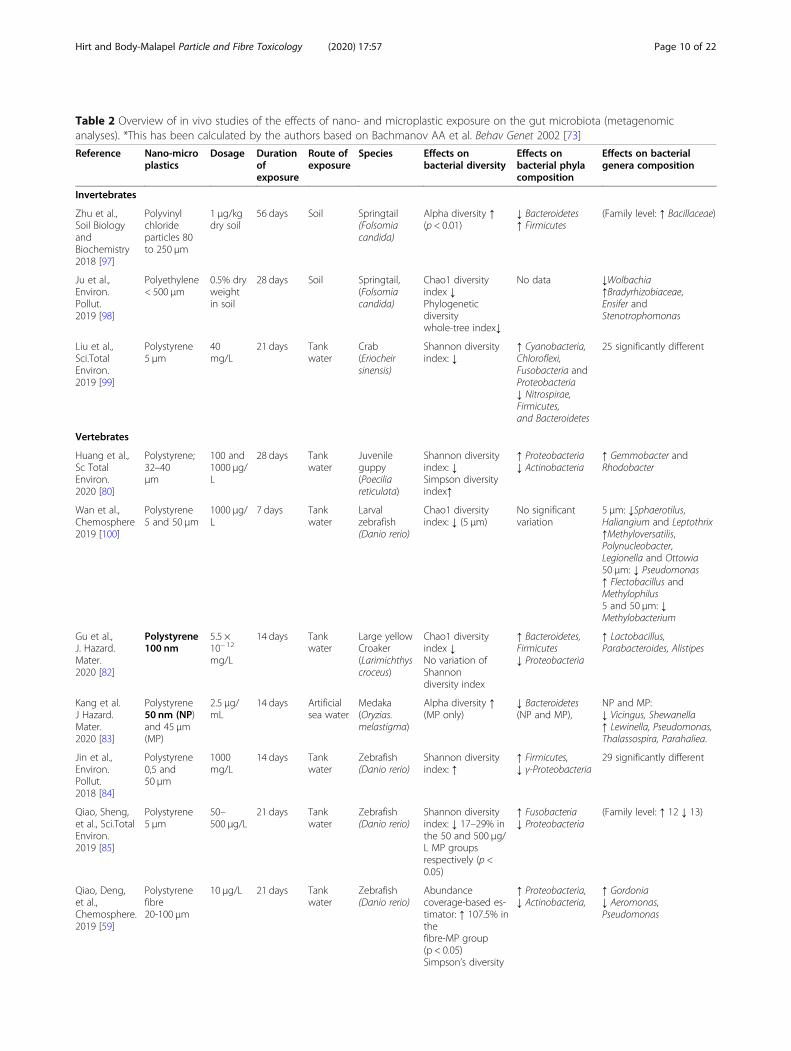
Table 2 Overview of in vivo studies of the effects of nano- and microplastic exposure on the gut microbiota (metagenomicanalyses). *This has been calculated by the authors based on Bachmanov AA et al. Behav Genet 2002 [73]
Reference Nano-microplastics
Dosage Durationofexposure
Route ofexposure
Species Effects onbacterial diversity
Effects onbacterial phylacomposition
Effects on bacterialgenera composition
Invertebrates
Zhu et al.,Soil BiologyandBiochemistry2018 [97]
Polyvinylchlorideparticles 80to 250 μm
1 μg/kgdry soil
56 days Soil Springtail(Folsomiacandida)
Alpha diversity ↑(p < 0.01)
↓ Bacteroidetes↑ Firmicutes
(Family level: ↑ Bacillaceae)
Ju et al.,Environ.Pollut.2019 [98]
Polyethylene< 500 μm
0.5% dryweightin soil
28 days Soil Springtail,(Folsomiacandida)
Chao1 diversityindex ↓Phylogeneticdiversitywhole-tree index↓
No data ↓Wolbachia↑Bradyrhizobiaceae,Ensifer andStenotrophomonas
Liu et al.,Sci.TotalEnviron.2019 [99]
Polystyrene5 μm
40mg/L
21 days Tankwater
Crab(Eriocheirsinensis)
Shannon diversityindex: ↓
↑ Cyanobacteria,Chloroflexi,Fusobacteria andProteobacteria↓ Nitrospirae,Firmicutes,and Bacteroidetes
25 significantly different
Vertebrates
Huang et al.,Sc TotalEnviron.2020 [80]
Polystyrene;32–40μm
100 and1000 μg/L
28 days Tankwater
Juvenileguppy(Poeciliareticulata)
Shannon diversityindex: ↓Simpson diversityindex↑
↑ Proteobacteria↓ Actinobacteria
↑ Gemmobacter andRhodobacter
Wan et al.,Chemosphere2019 [100]
Polystyrene5 and 50 μm
1000 μg/L
7 days Tankwater
Larvalzebrafish(Danio rerio)
Chao1 diversityindex: ↓ (5 μm)
No significantvariation
5 μm: ↓Sphaerotilus,Haliangium and Leptothrix↑Methyloversatilis,Polynucleobacter,Legionella and Ottowia50 μm: ↓ Pseudomonas↑ Flectobacillus andMethylophilus5 and 50 μm: ↓Methylobacterium
Gu et al.,J. Hazard.Mater.2020 [82]
Polystyrene100 nm
5.5 ×10− 12
mg/L
14 days Tankwater
Large yellowCroaker(Larimichthyscroceus)
Chao1 diversityindex ↓No variation ofShannondiversity index
↑ Bacteroidetes,Firmicutes↓ Proteobacteria
↑ Lactobacillus,Parabacteroides, Alistipes
Kang et al.J Hazard.Mater.2020 [83]
Polystyrene50 nm (NP)and 45 μm(MP)
2.5 μg/mL
14 days Artificialsea water
Medaka(Oryzias.melastigma)
Alpha diversity ↑(MP only)
↓ Bacteroidetes(NP and MP),
NP and MP:↓ Vicingus, Shewanella↑ Lewinella, Pseudomonas,Thalassospira, Parahaliea.
Jin et al.,Environ.Pollut.2018 [84]
Polystyrene0,5 and50 μm
1000mg/L
14 days Tankwater
Zebrafish(Danio rerio)
Shannon diversityindex: ↑
↑ Firmicutes,↓ γ-Proteobacteria
29 significantly different
Qiao, Sheng,et al., Sci.TotalEnviron.2019 [85]
Polystyrene5 μm
50–500 μg/L
21 days Tankwater
Zebrafish(Danio rerio)
Shannon diversityindex: ↓ 17–29% inthe 50 and 500 μg/L MP groupsrespectively (p <0.05)
↑ Fusobacteria↓ Proteobacteria
(Family level: ↑ 12 ↓ 13)
Qiao, Deng,et al.,Chemosphere.2019 [59]
Polystyrenefibre20-100 μm
10 μg/L 21 days Tankwater
Zebrafish(Danio rerio)
Abundancecoverage-based es-timator: ↑ 107.5% inthefibre-MP group(p < 0.05)Simpson’s diversity
↑ Proteobacteria,↓ Actinobacteria,
↑ Gordonia↓ Aeromonas,Pseudomonas
Hirt and Body-Malapel Particle and Fibre Toxicology (2020) 17:57 Page 10 of 22

abundance of the Proteobacteria [82]. In the medaka,exposure to PS particles (50 nm and 45 μm, 2.5 μg/mL,for 14 days) enhanced the abundance of Bacteroidetesphylum. At the genus level, PS nano- and microparti-cles decreased the abundance of Vicingus and Shewa-nella, and increased the abundance of Lewinella,Pseudomonas, Thalassospira, Parahaliea [83]. In thezebrafish, after 14-day exposure to high-dose of PS
(1000 mg/L), the abundance of γ-Proteobacteria de-creased significantly and the abundance of Firmicutesincreased for both microbead size (0.5 and 50 μm) [84].Another zebrafish study analyzed the effects of lower-dose exposure to PS microspheres (5 μm, 50 and500 μg/L, for 21 days); it showed a decrease of bacterialdiversity, an increase of the Fusobacteria abundanceand a decrease of the Proteobacteria abundance [85].
Table 2 Overview of in vivo studies of the effects of nano- and microplastic exposure on the gut microbiota (metagenomicanalyses). *This has been calculated by the authors based on Bachmanov AA et al. Behav Genet 2002 [73] (Continued)
Reference Nano-microplastics
Dosage Durationofexposure
Route ofexposure
Species Effects onbacterial diversity
Effects onbacterial phylacomposition
Effects on bacterialgenera composition
index: ↓ 45.7% inthe fibre-MPgroup (p < 0.05)
Jin et al.,Sci.TotalEnviron.2019 [60]
Polystyrene5 μm
1000 μg/L~266 μg/kg bw/day*
6 weeks Drinkingwater
ICRMice(Musmusculus)
Phylogeneticdiversitywhole-tree index↓
↓α−Proteobactriaγ-Proteobacteria
↓ Parabacteroides,Prevotella,Dehalobacterium,Turicibacter,Bifidobacterium,Phascolarctobacterium,Lachnospira, Haemophilus,Adlercreutzia,Megamonas, Blautia,Dialister and Veillonella↑ Coprococcus andAnaeroplasma
Lu et al.,Sci.TotalEnviron.2018 [91]
Polystyrene0.5 and50 μm
1000 μg/L~266 μg/kg bw/day*
5 weeks Drinkingwater
ICRMice(Musmusculus)
No data ↓ Firmicutes,α-Proteobacteria,Actinobacteria
↓ Oscillospira andAnaerostipes↑ Parabacteroides,Prevotella,Dehalobacterium,Ruminococcus, Bilophila,Bifidobacterium,Adlercreutzia, Plesiomona,Halomonas andAcinetobacter (after both0.5 and 50 μm polystyreneMP exposure)
Luo et al.,Environ. Sci.Technol2019 [101]
Pristinepolystyrenemicrospheres5 μm
1000 μg/L~266 μg/kg bw/day*
Gestationandlactation6 weeks(analysisof dams)
Drinkingwater
ICRMice(Musmusculus)
Shannon diversityindex: nosignificant variation
No significantvariation ofBacteroidetes,Proteobacteria,Firmicutes↑ Actinobacteria↑Epsilonbacteraeota
14 significantly different
Li et al.,Chemosphere.2020 [93]
Polyethylene10–150 μm
2–20-200 μg/gfeed~0.0004,0.004and0.04 μg/kg bw/day*
5 weeks Feed C57BL/6Mice(Musmusculus)
Shannon diversityindex: ↑in the 200 μg/g MP(p < 0.05)
↑ Firmicutes (20–200 μg/g),↑Melainabacteria(3 dosages)↓ Bacteroidetes(20–200)
↑ Staphylococcus↓ Parabacteroides(3 dosages)
Deng et al.,EnvironmentInternational2020 [94]
Polyethylene45–53 μm
100mg/kg/day5.25 104
particles/day
30 days Gavage CD-1 mice(Musmusculus)
Shannon diversityindex: nosignificant variation
↑ Actinobacteria ↑ Lactobacillus↑ Adlercreutzia↑ Butyricimonas↑ Parabacteroides
Hirt and Body-Malapel Particle and Fibre Toxicology (2020) 17:57 Page 11 of 22

The effects of PS fibres have also been assessed in thezebrafish: 21 days of exposure to 10 μg/L of PS fibres(20–100 μm) induced a diminution of bacterial diversityand variations in specific bacterial phyla (an enhance-ment in the Proteobacteria and a diminution in theActinobacteria) [59].Two studies in the mouse found a large number of sig-
nificant modifications in the bacterial phyla compositionafter chronic exposure to PS microspheres (5 μm,1000 μg/L, for 5 or 6 weeks) [60, 91]. The relative abun-dance of the α-Proteobacteria phylum was decreased bymicroplastic exposure in both studies, and the relativeabundance of Actinobacteria and Firmicutes phyla werealso reduced in the study by Lu et al. In contrast, themodifications in bacterial composition observed by Luoet al. were different: an increase in the abundance Acti-nobacteria but no significant variations in the Proteobac-teria and Firmicutes. However, it should be noted thatalthough Luo et al. used a very similar exposure protocol(5 μm PS beads, 1000 μg/L, for around 6 weeks), they ex-posed mice during gestation and lactation [101].Two studies focused on PE microplastics. Li et al.
(2020) observed that mice exposure to PE microplastics(10–150 μm, 2, 20 and 200 μg/g of food for 5 weeks) in-duced an increase in the abundance of the Firmicutesand Melainabacteria phyla and the Staphylococcusgenus, and a decrease in the abundance of the Bacteroi-detes phylum and the Parabacteroides genus [93]. Denget al. (2020) observed that mice exposure to PE micro-plastics (45–53 μm, 100 mg/kg/day by gavage, for 30days) increased the abundance of the Actinobacteriaphylum and the abundance of Lactobacillus, Adlercreut-zia, Butyricimonas and Parabacteroides genera [94].In conclusion, all the reports aiming to study the intes-
tinal microbiota in microplastic-exposed animals haveobserved dysbiosis. Even though the precise features ofthis dysbiosis vary from one context to another, the ob-served variations in microflora diversity and compositionare likely to cause functional impairments of the im-mune system.
IMMUNOTOXIC effects of ingested NANO- andMICROPLASTICSThe intestinal immune system interacts constantly withnon-pathogenic commensal organisms and innocuousfood antigens that must be tolerated immunologically.At the same time, the intestinal immune system mustretain the ability to respond rapidly to infectious threatsand toxins. This delicate task relies on several mecha-nisms involving myeloid cells, innate lymphoid cells, andT cells that reside in the intestinal lamina propria andthe draining mesenteric lymph node. These immunecells circuits are critical components of the immune sys-tem. Even though the immunotoxicity of plastics has not
been studied directly on the intestinal immune system,in vivo evidence of immunotoxicity of nano- and micro-plastics suggests that immune cells, including those ofintestinal immune system, could be target for plastic-induced damage. Indeed, studies conducted mainly in in-vertebrates (Table 3) but also in vertebrates (Table 4)have demonstrated that their immune system is compro-mised by exposure to nano- and microplastics.
In vivo immunotoxicity of nano- and microplastics ininvertebratesSeveral studies of invertebrates have linked PS exposureto disruption of the immune system. Exposure of cla-doceran Daphnia magna to carboxylate-modified PSnanoparticles (500 nm, 85mg/L, for 1 year) was associ-ated with higher hemocyte counts [102]. In musselhemolymph, exposure of PS particles (110 nm, 5 mg/L,for 96 h) decreased total antioxidant capacity and gaverise to DNA damage [77]. Exposure of mussels toamino-modified PS nanoparticles (50 nm, 10 μg/L) in-duced changes in hemocytes, depending on the durationof exposure. After a 24 h exposure, the hemocytes pre-sented mitochondrial and lysosomal disturbances [103].After two 24 h periods of exposure 72 h apart, levels ofbactericidal activity and immune-related gene transcrip-tion were found to be elevated. After 96 h of exposure,hemolymph phagocytosis, levels of oxidative stress, andthe microbiota were modified [104].Several research groups have explored the effects of PS
on the microscale. In mussels, hemocytes mortality andreactive oxygen species production were increased by ex-posure to PS microbeads (2 and 6 μm, 32 μg/L, for 7days) [76]. In crab hemolymph, the hemocyanin contentand the levels of activity of several enzymes related tothe immune system (acid phosphatase, alkaline phos-phatase, lysozyme and phenoloxidase) were significantlymodified by PS exposure (5 μm, 0.04 to 40mg/mL, for 7,14 or 21 days), although the direction of change (i.e. anincrease or a decrease) varied with the duration or doselevel of PS exposure [99]. In Mediterranean sea urchin,exposure to PS microbeads (10 μm and 4 5 μm, 10 parti-cles/mL, for 24 h) increased the total coelomocyte countand the intracellular levels of reactive oxygen and nitro-gen species, indicating a stress-related impact on thesecirculating immune cells [105]. In the bivalve molluskTegillarca granosa, two studies have shown that PS ex-posure (500 nm and 30 μm, 0.29 and 1mg/L, during 14and 4 days, respectively) leads to several disturbances inhemocytes, with notably a decrease in the cell count andin phagocytosis activity, and numerous variations in im-mune parameters related to oxidative stress, apoptosis,and the inflammatory response [109, 110]. In both stud-ies, PS nanoparticles caused more damage than PS mi-croparticles did.
Hirt and Body-Malapel Particle and Fibre Toxicology (2020) 17:57 Page 12 of 22

Table 3 Overview of in vivo studies of the immunotoxic effects of nano- and microplastics in invertebrates
Reference Nano-microplastics
Dosage Durationofexposure
Route of exposure Species Observed immunotoxic effects
Nanoplastics
Sadler et al., Environ.Pollut. 2019 [102]
Carboxylate-modifiedpolystyrene beads500 nm
1.25 ± 0.205particles/L,or 85.6 ±14.0 mg/L
1 year Tank water Cladoceran(Daphniamagna)
↑ Hemocyte counts
Brandts et al.,Sci.Total Environ.2018 [77]
Polystyrene~ 110 nm
0.005–0.05-0.5-5-50mg/L
96 h Tank water Mussel(Mytiulusgalloprovincialis)
Hemolymph↓total antioxidant capacity (5 mg/L)↑ DNA damage (all dosages)
Auguste et al.,Front.Immunol2020 [103]
Amino-modifiednanopolystyrene50 nm
10 μg/L 24 h Tank water Mussel(Mytilus.galloprovincialis)
HemocytesOne exposure:↓ mitochondrial membrane potential(MMP),↑ lysosomal acidification↓ lysosomal membrane stability↑ lysozyme releaseNo changes in total hemocyte count,subpopulations, phagocytic activityand ROS production↓ transcription of PCNA and p53No change in hemolymphbactericidal activityTwo exposures with 72 h resting periodbetween:normal hemocyte lysosomal stability,MMP, and lysozyme activity↓ lysosomal membranedestabilization↓ fully mature phagocytes↑ bactericidal activity↑ transcription of immune-relatedgenes
Auguste et al.MarineEnvironmentalResearch2020 [104]
Amino-modifiednanopolystyrene50 nm
10 μg/L 96 h Tank water Mussel(Mytilusgalloprovincialis)
Hemolymph↓ phagocytosis,↑ ROS and lysozyme activity↓ NO production.Hemolymph microbiota compositionshift
Microplastics
Paul-Pont et al.,EnvironmentalPollution2016 [76]
Polystyrenemicrobeads(2 and 6 μm)
32 μg/L 7 days Supplied withChaetoceros muelleralgae as a foodsource
Mussel(Mytilus spp)
↑ hemocytes mortality and ROSproduction
Liu et al.,Sci. Total Environ.,2019 [99]
Polystyrene5 μm
0.04–0.4-4-40 mg/L
7, 14, and21days
Tank water Crab(EriocheirSinensis)
Immune parameters in thehemolymphHemocyanin contentAfter 7 days: ↑ at 0.04 mg/LAfter 14 days: ↑ at 0.04 and 0.4 mg/LAfter 21 days: ↓at all dosagesAcid phosphatase activityAfter 7 days: ↑ at 4 and 40mg/LAfter 14 days: ↑ at 0.04 and 0.4 mg/L,↓ 4 and 40 mg/LAfter 21 days: ↑ at 0.04 mg/L, ↓ 4 and40 mg/LAlkaline phosphatase activityAfter 7 days: ↑ at 0.04 mg/L, ↓ 4 and40 mg/LAfter 14 days: ↑ at 0.04 mg/LAfter 21 days: ↓ at all dosagesLysozyme activityAfter 7 days: ↓ at 40 mg/LAfter 14 days: ↓ at 0.4 and 4mg/L
Hirt and Body-Malapel Particle and Fibre Toxicology (2020) 17:57 Page 13 of 22

Table 3 Overview of in vivo studies of the immunotoxic effects of nano- and microplastics in invertebrates (Continued)
Reference Nano-microplastics
Dosage Durationofexposure
Route of exposure Species Observed immunotoxic effects
After 21 days: ↓ at 4 et 40 mg/LPhenoloxidase activityAfter 7 days: ↑ at 0.04, 0.4, 4 mg/L, ↓40 mg/LAfter 14 days: ↓ at 0.4, 4, and 40mg/LAfter 21 days: ↓ at all dosagesExpression of immune-relatedgenes in the hemocytesHemocyanin and lysozyme: dosedependent ↓Caspase: ↑ 0.04 and 4 mg/L, ↓ at 40mg/LMyD88: ↑ at all dosages
Murano et al.EnvironmentalPollution 2020 [105]
Polystyrenemicrobeads(10 and 45 μm)
10 particles/mL
24 h48 h72 h
Tank water Mediterraneansea urchin(Paracentrotuslividus)
↑ total number of immune cells↑ ratio between red and whiteamoebocyte(at 3 times for 10 μm beads and onlyat 48 and 72 h for 45 μm beads)↑ intracellular levels of reactiveoxygen and nitrogen species(at 24 h only for both 10 and 45 μmbeads)↑ total antioxidant capacity (at 72 hfor 10 μm beads)
Revel et al., EnvironSci Pollut Res Int.2020 [106]
Polyethylene andPolypropylene0.4–400 μm
10–100 μg/L
10 days Soil Ragworm(Hedistediversicolor)
CoelomocytesNo variation of phagocytosis activity,phenoloxydase,and acid phosphatase
Revel et al., Mar.Pollut. Bull. 2020[107]
Polyethylene andPolypropylenefragments <400 μm
0.008–10-100μg ofparticles/L
10 days Tank water Pacific oyster(Crassostreagigas)
HemolymphNo variation of ROS production, acidphosphatase activity,and DNA damage
Green et al., Environ.Pollut. 2019 [108]
High densityPolyethylene(HDPE)0.48–316 μmPolylactic acid(PLA)0.6–363 μm
HDPE 845particles/LPLA 1296particles/L
52 days2 h/day
MP-dosed microalgaeIsochrysis galbana
Blue mussel(Mytilus edulis)
Hemolymph proteomeHDPE groupDysregulation of 6 protein involved inimmune response↑ three complement C1q domain-containing (C1qDC) proteins(FR715598.1; FR715581; HE609753.1),and fibrinogen-related protein(OPL33687.1)↓ macrophage migration inhibitoryfactor (HE609105.1),Microfibril-Associated Glyco 4(OPL32613.1)PLA groupdysregulation of 3 protein involved inimmune response↑ C1Q Domain Containing 1Q19(FR715598.1)and Fibrinogen-Related (OPL33687.1)↓ Microfibril-Associated Glyco 4(OPL32613.1)
Revel et al. Frontiersin EnvironmentalScience 2019 [78]
Commercialpolyethylene andpolystyrenemixture(< 400 μm)
0.008, 10,100 μg/L
10 days Tank water Mussel(Mytilus spp.)
HemolymphNo variation hemocyte count↑ acide phosphatase activity (0.008and 10 μg/L)↑ DNA damage (10 and 100 μg/L)
Both nanoplastics and microplastics
Shi et al., J. Hazard.Mater. 2020 [109]
Polystyrene beads500 nm (NP) and30 μm (MP)
0.29 mg/L 14 days Tank water Bivalve mollusk(Tegillarcagranosa)
Hemocytes↓ total hemocytes count↓ phagocytosis
Hirt and Body-Malapel Particle and Fibre Toxicology (2020) 17:57 Page 14 of 22

Other microplastics have been studied individually.Polyethylene and PP fragments (< 400 μm, 0.008 to100 μg/L, for 10 days) did not show significant immunedamage in ragworm and oyster [106, 107]. Specific dys-regulation of proteins involved in immune responsewere observed in hemolymph of mussels exposed to PEand polylactic acid microplastics (845 and 1296 parti-cles/L respectively, 2 h/day, for 52 days) [108].Lastly, in Mytilus spp, exposure to a mixture of PE
and PS microbeads (< 400 μm, 10 μg/L, for 10 days)enhanced hemolymph acid phosphatase activity andDNA damage [78].
In vivo immunotoxicity of nano- and microplastics invertebratesTwo studies have assessed the in vivo immunotoxicity ofnanoplastics in fishes. In vitro neutrophil function assaysshowed dose-dependent increases in myeloperoxidaseactivity and neutrophil extracellular trap release in fat-head minnows Pimephales promelas exposed to PSnanoparticles (41 nm, 0.025 to 0.2 μg/μL) [111]. Acuteexposure to PMMA nanoparticles (45 nm, 0.02 and 0.2mg/L, for 96 h) diminished the level of oxidative stressin the plasma [112].Several studies have explored the in vivo toxicity of
micro scale plastics. Polycarbonate microplastics (159nm, 0.025 to 0.2 μg/ml, 2 h) dose-dependently disturbedneutrophil function in fathead minnows [111]. Incontrast, no significant immunotoxicity was observed introut exposed for 4 weeks to PS microbeads (100–400 μm, 10mg/fish/day) [79]. Exposure to PE micropar-ticles (250 and 500 μg/L, for 30 days) impaired thecomplement system and the levels of activity ofimmunity-related enzymes in the plasma of carp [113].Espinosa et al. showed in fishes that exposure to PVCmicroplastics (40–150 μm, 1 to 500 mg/kg, for between1 h and 3 weeks) disturbed phagocytic capacity and
increased the head-kidney leukocytes’ respiratory burst[88, 114, 115]. Polyethylene also enhanced the head-kidney leukocytes’ respiratory burst in fishes, anddysregulated major immune response proteins in thehemolymph of mussels [88, 115]. In zebrafish, exposureto high-density PE and PS particles (100 and 1000 μg/L,for 20 days) decreased the liver transcript levels of 2immune genes leukotriene B4 receptor (ltb4r) and inter-feron induced transmembrane protein (ifitm1) [90].In mice, exposure to PE microplastics (10–150 μm, 20
and 200 μg/g, for 5 weeks) modified the serum levels ofIL1α and granulocyte colony-stimulating factor G-CSF,decreased the regulatory T cell count, and increased theproportion of Th17 cells in splenocytes [93]. The cross-generational effects of PE exposure (7 μm, 0.125 to 2mg/day/mouse, for 90 days) have been also studied inthe mouse: blood neutrophil counts and IgA levels wereelevated in dams, and spleen lymphocytes were alteredin both dams and offspring [116].Lastly, Mancia et al. studied the small-spotted cat-
shark (Scyliorhinus canicula) in the MediterraneanSea: the presence of macroplastics in the gastrointes-tinal tract was associated with significant upregulationof the expression of T cell receptors beta and delta(TCRβ and TCRδ) and immunoglobulin M (IgM) inthe spleen [117].The many alterations observed in these studies not
only demonstrate that the immune system is alteredby plastics but also highlight the need for moreimmunotoxicity studies of species more closely relatedto the human.
MICROPLASTICS as carriers of intestinal toxics andpathogensMicroplastics may contain additives on average (4% w/w, on average) and can adsorb contaminants [63, 118].Both additives and contaminants can be of organic as
Table 3 Overview of in vivo studies of the immunotoxic effects of nano- and microplastics in invertebrates (Continued)
Reference Nano-microplastics
Dosage Durationofexposure
Route of exposure Species Observed immunotoxic effects
↓ viability (NP only)↑ ROS content↑ Caspase 3 activity↑ malondialdehyde content↓ ATP content (NP only)↓ pyruvate kinase activity↑ GABA content
Tang et al., Environ.Pollut. 2020 [110]
Polystyrene500 nm (NP) and30 μm (MP) and
1mg/L 4 days Tank water Bivalve mollusk(Tegillarcagranosa)
Hemocytes↓ hemocytes count, basophils count,phagocytosis↓ lysozyme (NP only)↓ TLR4 (NP only), TRAF6, IKKα, NFκBgene expression↑ Bcl2 (NP only), Caspase 3,Calmodulin gene expression
Hirt and Body-Malapel Particle and Fibre Toxicology (2020) 17:57 Page 15 of 22
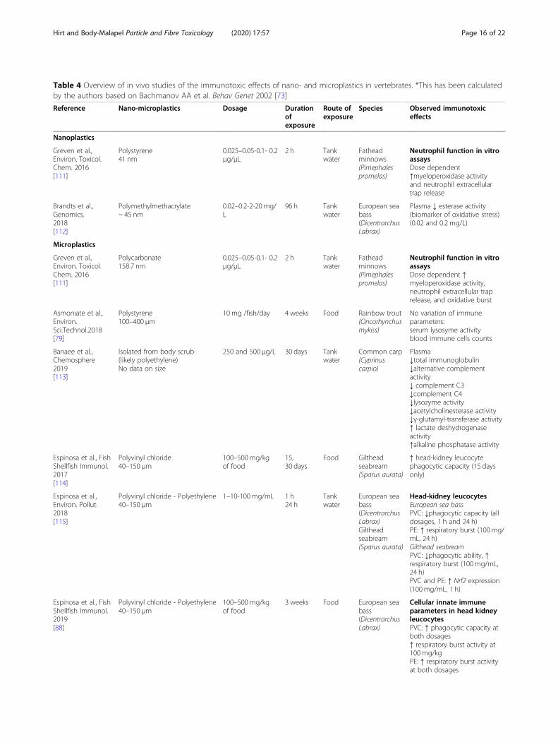
Table 4 Overview of in vivo studies of the immunotoxic effects of nano- and microplastics in vertebrates. *This has been calculatedby the authors based on Bachmanov AA et al. Behav Genet 2002 [73]
Reference Nano-microplastics Dosage Durationofexposure
Route ofexposure
Species Observed immunotoxiceffects
Nanoplastics
Greven et al.,Environ. Toxicol.Chem. 2016[111]
Polystyrene41 nm
0.025–0.05-0.1- 0.2μg/μL
2 h Tankwater
Fatheadminnows(Pimephalespromelas)
Neutrophil function in vitroassaysDose dependent↑myeloperoxidase activityand neutrophil extracellulartrap release
Brandts et al.,Genomics.2018[112]
Polymethylmethacrylate~ 45 nm
0.02–0.2-2-20 mg/L
96 h Tankwater
European seabass(DicentrarchusLabrax)
Plasma ↓ esterase activity(biomarker of oxidative stress)(0.02 and 0.2 mg/L)
Microplastics
Greven et al.,Environ. Toxicol.Chem. 2016[111]
Polycarbonate158.7 nm
0.025–0.05-0.1- 0.2μg/μL
2 h Tankwater
Fatheadminnows(Pimephalespromelas)
Neutrophil function in vitroassaysDose dependent ↑myeloperoxidase activity,neutrophil extracellular traprelease, and oxidative burst
Asmoniate et al.,Environ.Sci.Technol.2018[79]
Polystyrene100–400 μm
10mg /fish/day 4 weeks Food Rainbow trout(Oncorhynchusmykiss)
No variation of immuneparameters:serum lysosyme activityblood immune cells counts
Banaee et al.,Chemosphere2019[113]
Isolated from body scrub(likely polyethylene)No data on size
250 and 500 μg/L 30 days Tankwater
Common carp(Cyprinuscarpio)
Plasma↓total immunoglobulin↓alternative complementactivity↓ complement C3↓complement C4↓lysozyme activity↓acetylcholinesterase activity↓γ-glutamyl-transferase activity↑ lactate deshydrogenaseactivity↑alkaline phosphatase activity
Espinosa et al., FishShellfish Immunol.2017[114]
Polyvinyl chloride40–150 μm
100–500mg/kgof food
15,30 days
Food Giltheadseabream(Sparus aurata)
↑ head-kidney leucocytephagocytic capacity (15 daysonly)
Espinosa et al.,Environ. Pollut.2018[115]
Polyvinyl chloride - Polyethylene40–150 μm
1–10-100mg/mL 1 h24 h
Tankwater
European seabass(DicentrarchusLabrax)Giltheadseabream(Sparus aurata)
Head-kidney leucocytesEuropean sea bassPVC: ↓phagocytic capacity (alldosages, 1 h and 24 h)PE: ↑ respiratory burst (100mg/mL, 24 h)Gilthead seabreamPVC: ↓phagocytic ability, ↑respiratory burst (100mg/mL,24 h)PVC and PE: ↑ Nrf2 expression(100mg/mL, 1 h)
Espinosa et al., FishShellfish Immunol.2019[88]
Polyvinyl chloride - Polyethylene40–150 μm
100–500mg/kgof food
3 weeks Food European seabass(DicentrarchusLabrax)
Cellular innate immuneparameters in head kidneyleucocytesPVC: ↑ phagocytic capacity atboth dosages↑ respiratory burst activity at100 mg/kgPE: ↑ respiratory burst activityat both dosages
Hirt and Body-Malapel Particle and Fibre Toxicology (2020) 17:57 Page 16 of 22
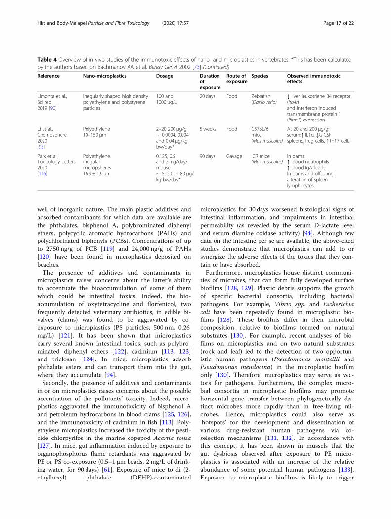
well of inorganic nature. The main plastic additives andadsorbed contaminants for which data are available arethe phthalates, bisphenol A, polybrominated diphenylethers, polycyclic aromatic hydrocarbons (PAHs) andpolychlorinated biphenyls (PCBs). Concentrations of upto 2750 ng/g of PCB [119] and 24,000 ng/g of PAHs[120] have been found in microplastics deposited onbeaches.The presence of additives and contaminants in
microplastics raises concerns about the latter’s abilityto accentuate the bioaccumulation of some of themwhich could be intestinal toxics. Indeed, the bio-accumulation of oxytetracycline and florfenicol, twofrequently detected veterinary antibiotics, in edible bi-valves (clams) was found to be aggravated by co-exposure to microplastics (PS particles, 500 nm, 0.26mg/L) [121]. It has been shown that microplasticscarry several known intestinal toxics, such as polybro-minated diphenyl ethers [122], cadmium [113, 123]and triclosan [124]. In mice, microplastics adsorbphthalate esters and can transport them into the gut,where they accumulate [94].Secondly, the presence of additives and contaminants
in or on microplastics raises concerns about the possibleaccentuation of the pollutants’ toxicity. Indeed, micro-plastics aggravated the immunotoxicity of bisphenol Aand petroleum hydrocarbons in blood clams [125, 126],and the immunotoxicity of cadmium in fish [113]. Poly-ethylene microplastics increased the toxicity of the pesti-cide chlorpyrifos in the marine copepod Acartia tonsa[127]. In mice, gut inflammation induced by exposure toorganophosphorus flame retardants was aggravated byPE or PS co-exposure (0.5–1 μm beads, 2 mg/L of drink-ing water, for 90 days) [61]. Exposure of mice to di (2-ethylhexyl) phthalate (DEHP)-contaminated
microplastics for 30 days worsened histological signs ofintestinal inflammation, and impairments in intestinalpermeability (as revealed by the serum D-lactate leveland serum diamine oxidase activity) [94]. Although fewdata on the intestine per se are available, the above-citedstudies demonstrate that microplastics can add to orsynergize the adverse effects of the toxics that they con-tain or have absorbed.Furthermore, microplastics house distinct communi-
ties of microbes, that can form fully developed surfacebiofilms [128, 129]. Plastic debris supports the growthof specific bacterial consortia, including bacterialpathogens. For example, Vibrio spp. and Escherichiacoli have been repeatedly found in microplastic bio-films [128]. These biofilms differ in their microbialcomposition, relative to biofilms formed on naturalsubstrates [130]. For example, recent analyses of bio-films on microplastics and on two natural substrates(rock and leaf) led to the detection of two opportun-istic human pathogens (Pseudomonas monteilii andPseudomonas mendocina) in the microplastic biofilmonly [130]. Therefore, microplastics may serve as vec-tors for pathogens. Furthermore, the complex micro-bial consortia in microplastic biofilms may promotehorizontal gene transfer between phylogenetically dis-tinct microbes more rapidly than in free-living mi-crobes. Hence, microplastics could also serve as‘hotspots’ for the development and dissemination ofvarious drug-resistant human pathogens via co-selection mechanisms [131, 132]. In accordance withthis concept, it has been shown in mussels that thegut dysbiosis observed after exposure to PE micro-plastics is associated with an increase of the relativeabundance of some potential human pathogens [133].Exposure to microplastic biofilms is likely to trigger
Table 4 Overview of in vivo studies of the immunotoxic effects of nano- and microplastics in vertebrates. *This has been calculatedby the authors based on Bachmanov AA et al. Behav Genet 2002 [73] (Continued)
Reference Nano-microplastics Dosage Durationofexposure
Route ofexposure
Species Observed immunotoxiceffects
Limonta et al.,Sci rep2019 [90]
Irregularly shaped high densitypolyethylene and polystyreneparticles
100 and1000 μg/L
20 days Food Zebrafish(Danio rerio)
↓ liver leukotriene B4 receptor(ltb4r)and interferon inducedtransmembrane protein 1(ifitm1) expression
Li et al.,Chemosphere.2020[93]
Polyethylene10–150 μm
2–20-200 μg/g~ 0.0004, 0.004and 0.04 μg/kgbw/day*
5 weeks Food C57BL/6mice(Mus musculus)
At 20 and 200 μg/g:serum:↑ IL1α, ↓G-CSFspleen:↓Treg cells, ↑Th17 cells
Park et al.,Toxicology Letters2020[116]
Polyethyleneirregularmicropsheres16.9 ± 1.9 μm
0.125, 0.5and 2mg/day/mouse~ 5, 20 an 80 μg/kg bw/day*
90 days Gavage ICR mice(Mus musculus)
In dams:↑ blood neutrophils↑ blood IgA levelsIn dams and offspring:alteration of spleenlymphocytes
Hirt and Body-Malapel Particle and Fibre Toxicology (2020) 17:57 Page 17 of 22

changes in the gut microbiota and activation of theimmune system, although this field has not yet beenexplored.
ConclusionsThe gut epithelium encounters a broad range of plastics.Faced with this complexity, we still have too few data onthe amounts and features of ingested plastics. Analyticallimitations on the detection of particles sizing few μmmean that the environmental exposure data relates tomicro-size plastics only. Moreover, these exposure dataare subject to debate because of the limited number ofstudies of food products, poor data quality (due to con-tamination, for example), and the absence of data onsmall particles [36, 134]. Furthermore, interstudy com-parisons are not valid because of the lack of standardizedtechnical methods for collection and analysis. Today’savailable data give us a few clues about the plastic pol-lutants in drinking water and in a small number of foodproducts. Larger studies of plastics in the general diets,(i.e. providing a realistic estimate of overall oral contam-ination by plastics) are still lacking. With regards to thischallenge, we suggest that research should initially focuson the microplastics present in human stools. This strat-egy is particularly useful for identifying the plastics thatexert their harmful effects via direct contact with the in-testinal mucosa. Furthermore, it is essential to study thenano- and microplastics that cross the intestinal barrier;even small quantities of translocated plastic may be asdangerous as or even more dangerous than plastics ex-creted in the stools. Lastly, in view of the recent litera-ture on the effects of particulate matter in theatmosphere, the impact of airborne microplastics on theintestine should also be assessed [135–137].
To the best of our knowledge, the intestinal andimmunotoxic effects of nanoplastic ingestion by mam-mals have never been studied. However, ingestion ofnon-plastic nanoparticles is known to have many harm-ful effects. For example, ingestion of TiO2 nanoparticlesby mammals impairs intestinal and systemic immunehomeostasis and induces variations in the gut microbiotaand gut-associated metabolism [138, 139]. One cantherefore reasonably hypothesize that contamination ofthe diet by some nanoplastics is likely to harm intestinaland immune systems; this topic requires more attention.Furthermore, the microplastics most frequently studiedfor their in vivo effects are PS and (to a lesser extent)PP: more efforts are required for PP and PET, the twomost abundant microplastics in human feces.Despite the lack of data on plastic levels in the human
diet, it is clear that plastics contaminate water and thefood chain; the toxicological effects of certain types andshapes of microplastics are now starting to be assessed.The current data are insufficient and do not allow robustscientific conclusions to be drawn for humans in generaland intestinal health in particular. In this respect, thelack of reliable data on human dietary exposure meansthat relevant doses have yet to be defined. Furthermore,the effects of chronic exposure to microplastics appearto be very variable and dependent on the latter’s typeand shape. It is also very likely that the effects of a cock-tail of microplastics (as encountered in real life) are dif-ferent from those of individual components - furthercomplicating the problem. Likewise, the biological rele-vance of the research results – notably concerning theimpact on the immune system - would be increased bystudies of nano- and microplastics bearing biofilms.However, most of the literature studies suggest that
Scheme 4 Overview of the potential effects of nano- and microplastic contamination on intestinal health and the immune response
Hirt and Body-Malapel Particle and Fibre Toxicology (2020) 17:57 Page 18 of 22

nano- and microplastics have several effects on intestine:the disturbance of intestinal homeostasis, alterations ingut permeability, and changes in the recruitment of im-mune cells or in levels of cytokine secretion. The intes-tinal dysbiosis which has been observed followingmicroplastic ingestion, sometimes differs from one studyto another but reflects the deregulation of a crucial par-ameter for host defense, intestinal metabolism and in-flammation. The immune system’s susceptibility toplastics constitutes an additional threat to health.In conclusion, a growing body of evidence shows that
the omnipresence of plastics in our daily life is associ-ated with chronic, evolving exposure to microplastics.Furthermore, many animal experiments suggest that theingestion of microplastics disrupts essential intestinalfunctions, such as the gut barrier function and regula-tion of the gut microbiota (Scheme 4). Due to the multi-functional nature of the intestinal system, these plastic-associated disruptions may promote immune, inflamma-tory and metabolic disorders and therefore warrant fur-ther investigation.
AcknowledgementsNot applicable.
Authors’ contributionsNH was involved in the initial write-up of the background and gut micro-biota sections of the manuscript. NH created part of the schemes and laidout part of the Tables. MB-M double-checked the literature search resultsand revised the draft provided by NH. MB-M performed the literaturesearches and wrote the others sections of the review. NH and MB-M readand approved the final manuscript.
Authors’ informationNot applicable.
FundingThe authors thank the Hauts de France Regional Council and the frenchassociation François Aupetit for their financial support.
Availability of data and materialsNot applicable.
Ethics approval and consent to participateNot applicable.
Consent for publicationNot applicable.
Competing interestsThe authors declare that they have no competing interests.
Received: 25 June 2020 Accepted: 26 October 2020
References1. Wu P, Huang J, Zheng Y, Yang Y, Zhang Y, He F, et al. Environmental
occurrences, fate, and impacts of microplastics. Ecotoxicol Environ Saf. 2019;184:109612.
2. Publications :: PlasticsEurope [Internet]. [cited 2020 Sep 7]. Available from:https://www.plasticseurope.org/fr/resources/publications/1804-plastics-facts-2019.
3. Alimba CG, Faggio C. Microplastics in the marine environment: currenttrends in environmental pollution and mechanisms of toxicological profile.Environ Toxicol Pharmacol. 2019;68:61–74.
4. Lambert S, Wagner M. Microplastics are contaminants of emerging concernin freshwater environments: an overview. Freshw Microplastics. Cham:Springer; 2018. p. 1–23.
5. Peeken I, Primpke S, Beyer B, Gütermann J, Katlein C, Krumpen T, et al.Arctic Sea ice is an important temporal sink and means of transport formicroplastic. Nat Commun. 2018;9:1505.
6. Xu B, Liu F, Cryder Z, Huang D, Lu Z, He Y, et al. Microplastics in the soilenvironment: occurrence, risks, interactions and fate – a review. Crit RevEnviron Sci Technol Taylor & Francis. 2019;0:1–48.
7. Wang J, Liu X, Li Y, Powell T, Wang X, Wang G, et al. Microplastics ascontaminants in the soil environment: a mini-review. Sci Total Environ. 2019;691:848–57.
8. Corradini F, Meza P, Eguiluz R, Casado F, Huerta-Lwanga E, Geissen V.Evidence of microplastic accumulation in agricultural soils from sewagesludge disposal. Sci Total Environ. 2019;671:411–20.
9. Eriksen M, Lebreton LCM, Carson HS, Thiel M, Moore CJ, Borerro JC, et al.lastic Pollution in the World’s Oceans: More than 5 Trillion Plastic PiecesWeighing over 250,000 Tons Afloat at Sea. PLOS ONE. Public Libr Sci. 2014;9:e111913.
10. Schwarz AE, Ligthart TN, Boukris E, van Harmelen T. Sources, transport, andaccumulation of different types of plastic litter in aquatic environments: areview study. Mar Pollut Bull. 2019;143:92–100.
11. Jambeck JR, Geyer R, Wilcox C, Siegler TR, Perryman M, Andrady A, et al.Plastic waste inputs from land into the ocean. Sci Am Assoc AdvancementSci. 2015;347:768–71.
12. Bouwmeester H, Hollman PCH, Peters RJB. Potential health impact ofenvironmentally released micro- and Nanoplastics in the human foodproduction chain: experiences from Nanotoxicology. Environ Sci TechnolAm Chem Soc. 2015;49:8932–47.
13. Rochman CM, Kross SM, Armstrong JB, Bogan MT, Darling ES, Green SJ,et al. Scientific evidence supports a ban on microbeads. Environ SciTechnol. 2015;49:10759–61.
14. Prata JC. Airborne microplastics: consequences to human health? EnvironPollut. 2018;234:115–26.
15. Chen G, Feng Q, Wang J. Mini-review of microplastics in the atmosphereand their risks to humans. Sci Total Environ. Elsevier. 2020;703:135504.
16. Wright SL, Ulke J, Font A, Chan KLA, Kelly FJ. Atmospheric microplasticdeposition in an urban environment and an evaluation of transport. EnvironInt. 2020;136:105411.
17. Liu K, Wang X, Fang T, Xu P, Zhu L, Li D. Source and potential riskassessment of suspended atmospheric microplastics in Shanghai. Sci TotalEnviron. 2019;675:462–71.
18. Allen S, Allen D, Phoenix VR, Le Roux G, Durántez Jiménez P,Simonneau A, et al. Atmospheric transport and deposition ofmicroplastics in a remote mountain catchment. Nat Geosci NaturePublishing Group. 2019;12:339–44.
19. Vianello A, Jensen RL, Liu L, Vollertsen J. Simulating human exposure toindoor airborne microplastics using a Breathing Thermal Manikin. Sci RepNat Publ Group. 2019;9:8670.
20. Zhang Q, Zhao Y, Du F, Cai H, Wang G, Shi H. Microplastic fallout indifferent indoor environments. Environ Sci Technol Am Chem Soc. 2020;54:6530–9.
21. Prata JC, da Costa JP, Lopes I, Duarte AC, Rocha-Santos T. Environmentalexposure to microplastics: An overview on possible human health effects.Sci Total Environ. 2020;702:134455.
22. Andrady AL, Neal MA. Applications and societal benefits of plastics. PhilosTrans R Soc Lond Ser B Biol Sci. 2009;364:1977–84.
23. Rochman CM, Brookson C, Bikker J, Djuric N, Earn A, Bucci K, et al.Rethinking microplastics as a diverse contaminant suite. Environ ToxicolChem. 2019;38:703–11.
24. Lebreton L, Slat B, Ferrari F, Sainte-Rose B, Aitken J, Marthouse R, et al.Evidence that the Great Pacific Garbage Patch is rapidly accumulatingplastic. Sci Rep Nat Publ Group. 2018;8:4666.
25. Thompson RC, Olsen Y, Mitchell RP, Davis A, Rowland SJ, John AWG, et al.Lost at sea: where is all the plastic? Sci Am Assoc Advanc Sci. 2004;304:838.
26. Toussaint B, Raffael B, Angers-Loustau A, Gilliland D, Kestens V, PetrilloM, et al. Review of micro- and nanoplastic contamination in the foodchain. Food Addit Contam Part Chem Anal Control Expo Risk Assess.2019;36:639–73.
27. Gigault J, ter Halle A, Baudrimont M, Pascal P-Y, Gauffre F, Phi T-L, et al.Current opinion: what is a nanoplastic? Environ Pollut. 2018;235:1030–4.
Hirt and Body-Malapel Particle and Fibre Toxicology (2020) 17:57 Page 19 of 22
28. Lehner R, Weder C, Petri-Fink A, Rothen-Rutishauser B. Emergence ofNanoplastic in the environment and possible impact on human health.Environ Sci Technol. 2019;53:1748–65.
29. Schwaferts C, Niessner R, Elsner M, Ivleva NP. Methods for the analysis ofsubmicrometer- and nanoplastic particles in the environment. TrAC TrendsAnal Chem. 2019;112:52–65.
30. Koelmans B, Pahl S, Backhaus T, Bessa F, van Calster G, Contzen N, et al. Ascientific perspective on microplastics in nature and society. SAPEA; 2019.
31. Koelmans AA, Mohamed Nor NH, Hermsen E, Kooi M, Mintenig SM, DeFrance J. Microplastics in freshwaters and drinking water: critical review andassessment of data quality. Water Res. 2019;155:410–22.
32. Danopoulos E, Twiddy M, Rotchell JM. Microplastic contamination ofdrinking water: A systematic review. PloS One. Public Library of Science SanFrancisco, CA USA. 2020;15:e0236838.
33. Novotna K, Cermakova L, Pivokonska L, Cajthaml T, Pivokonsky M.Microplastics in drinking water treatment – current knowledge and researchneeds. Sci Total Environ. 2019;667:730–40.
34. Kosuth M, Mason SA, Wattenberg EV. Anthropogenic contamination of tapwater, beer, and sea salt. PLOS ONE Public Libr Sci. 2018;13:e0194970.
35. Tong H, Jiang Q, Hu X, Zhong X. Occurrence and identification ofmicroplastics in tap water from China. Chemosphere. 2020;252:126493.
36. Welle F, Franz R. Microplastic in bottled natural mineral water -literature review and considerations on exposure and risk assessment.Food Addit Contam Part Chem Anal Control Expo Risk Assess. 2018;35:2482–92.
37. Oßmann BE, Sarau G, Holtmannspötter H, Pischetsrieder M, Christiansen SH,Dicke W. Small-sized microplastics and pigmented particles in bottledmineral water. Water Res. 2018;141:307–16.
38. Schymanski D, Goldbeck C, Humpf H-U, Fürst P. Analysis of microplastics inwater by micro-Raman spectroscopy: release of plastic particles fromdifferent packaging into mineral water. Water Res. 2018;129:154–62.
39. van Raamsdonk LWD, van der Zande M, Koelmans AA, Hoogenboom RLAP,Peters RJB, Groot MJ, et al. Current insights into monitoring,bioaccumulation, and potential health effects of microplastics present in thefood chain. Foods Basel Switz. 2020;9:72.
40. Kankanige D, Babel S. Smaller-sized micro-plastics (MPs) contamination insingle-use PET-bottled water in Thailand. Sci Total Environ. Elsevier. 2020;717:137232.
41. Van Cauwenberghe L, Janssen CR. Microplastics in bivalves cultured forhuman consumption. Environ Pollut Barking Essex 1987. 2014;193:65–70.
42. Smith M, Love DC, Rochman CM, Neff RA. Microplastics in seafood and theimplications for human health. Curr Environ Health Rep. 2018;5:375–86.
43. Lusher A, Hollman PCH, Mendoza-Hill J. Microplastics in fisheries andaquaculture: status of knowledge on their occurrence and implications foraquatic organisms and food safety. Rome: Food and AgricultureOrganization of the United Nations; 2017.
44. Hantoro I, Löhr AJ, Van Belleghem FGAJ, Widianarko B, Ragas AMJ.Microplastics in coastal areas and seafood: implications for food safety.Food Addit Contam Part Chem Anal Control Expo Risk Assess. 2019;36:674–711.
45. Liebezeit G, Liebezeit E. Non-pollen particulates in honey and sugar. FoodAddit Contam Part A Taylor & Francis. 2013;30:2136–40.
46. Yang D, Shi H, Li L, Li J, Jabeen K, Kolandhasamy P. Microplastic pollution intable salts from China. Environ Sci Technol. 2015;49:13622–7.
47. Iñiguez ME, Conesa JA, Fullana A. Microplastics in Spanish table salt. Sci Rep.2017;7:8620.
48. Karami A, Golieskardi A, Ho YB, Larat V, Salamatinia B. Microplastics ineviscerated flesh and excised organs of dried fish. Sci Rep. 2017;7:5473.
49. Gündoğdu S. Contamination of table salts from Turkey with microplastics.Food Addit Contam Part Chem Anal Control Expo Risk Assess. 2018;35:1006–14.
50. Liebezeit G, Liebezeit E. Synthetic particles as contaminants in Germanbeers. Food Addit Contam Part A. Taylor & Francis. 2014;31:1574–8.
51. Kutralam-Muniasamy G, Pérez-Guevara F, Elizalde-Martínez I, Shruti VC.Branded milks – are they immune from microplastics contamination? SciTotal Environ. 2020;714:136823.
52. Gasperi J, Wright SL, Dris R, Collard F, Mandin C, Guerrouache M, et al.Microplastics in air: are we breathing it in? Curr Opin Environ Sci Health.2018;1:1–5.
53. Strand J. Contents of polyethylene microplastic in some selected personalcare products in Denmark; 2014.
54. Lei K, Qiao F, Liu Q, Wei Z, Qi H, Cui S, et al. Microplastics releasing frompersonal care and cosmetic products in China. Mar Pollut Bull. 2017;123:122–6.
55. Leslie HA. Review of microplastics in cosmetics. IVM Inst Environ Stud. 2014;476:1–33.
56. Yurtsever M. Tiny, shiny, and colorful microplastics: are regular glitters asignificant source of microplastics? Mar Pollut Bull. 2019;146:678–82.
57. Hernandez LM, Yousefi N, Tufenkji N. Are there Nanoplastics in yourpersonal care products? Environ Sci Technol Lett. 2017;4:280–5.
58. Deng Y, Zhang Y, Lemos B, Ren H. Tissue accumulation of microplastics inmice and biomarker responses suggest widespread health risks of exposure.Sci Rep. 2017;7:46687.
59. Qiao R, Deng Y, Zhang S, Wolosker MB, Zhu Q, Ren H, et al. Accumulationof different shapes of microplastics initiates intestinal injury and gutmicrobiota dysbiosis in the gut of zebrafish. Chemosphere. 2019;236:124334.
60. Jin Y, Lu L, Tu W, Luo T, Fu Z. Impacts of polystyrene microplastic on thegut barrier, microbiota and metabolism of mice. Sci Total Environ. 2019;649:308–17.
61. Deng Y, Zhang Y, Qiao R, Bonilla MM, Yang X, Ren H, et al. Evidence thatmicroplastics aggravate the toxicity of organophosphorus flame retardantsin mice (Mus musculus). J Hazard Mater. 2018;357:348–54.
62. Hillery A, Jani P, Florence A. Comparative, quantitative study of lymphoidand non-lymphoid uptake of 60 nm polystyrene particles. J Drug TargetTaylor & Francis. 1994;2:151–6.
63. EFSA. Presence of microplastics and nanoplastics in food, with particularfocus on seafood. EFSA J. 2016;14:e04501.
64. Powell JJ, Faria N, Thomas-McKay E, Pele LC. Origin and fate of dietarynanoparticles and microparticles in the gastrointestinal tract. J Autoimmun.2010;34:J226–33.
65. Coméra C, Cartier C, Gaultier E, Catrice O, Panouille Q, Hamdi SE, et al. Jejunalvillus absorption and paracellular tight junction permeability are major routesfor early intestinal uptake of food-grade TiO 2 particles: an in vivo and ex vivostudy in mice. Part Fibre Toxicol BioMed Central. 2020;17:1–15.
66. Smith MW, Thomas NW, Jenkins PG, Miller NG, Cremaschi D, Porta C.Selective transport of microparticles across Peyer’s patch follicle-associatedM cells from mice and rats. Exp Physiol. 1995;80:735–43.
67. Galloway TS. Micro-and nano-plastics and human health. Mar AnthropogLitter. Cham: Springer; 2015. p. 343–66.
68. Carr KE, Smyth SH, McCullough MT, Morris JF, Moyes SM. Morphologicalaspects of interactions between microparticles and mammalian cells:intestinal uptake and onward movement. Prog Histochem CytochemElsevier. 2012;46:185–252.
69. Prüst M, Meijer J, Westerink RHS. The plastic brain: neurotoxicity of micro-and nanoplastics. Part Fibre Toxicol BioMed Central. 2020;17:1–16.
70. Smith JRH, Etherington G, Shutt AL, Youngman MJ. A study of aerosoldeposition and clearance from the human nasal passage. Ann Occup HygOxford University Press. 2002;46:309–13.
71. Asgharian B, Hofmann W, Miller FJ. Mucociliary clearance of insolubleparticles from the tracheobronchial airways of the human lung. J AerosolSci Elsevier. 2001;32:817–32.
72. Enaud R, Prevel R, Ciarlo E, Beaufils F, Wieërs G, Guery B, et al. The gut-lungaxis in health and respiratory diseases: a place for inter-organ and inter-kingdom crosstalks. Front cell infect Microbiol. Frontiers. 2020;10:9.
73. Bachmanov AA, Reed DR, Beauchamp GK, Tordoff MG. Food intake, waterintake, and drinking spout side preference of 28 mouse strains. BehavGenet. 2002;32:435–43.
74. Lei L, Wu S, Lu S, Liu M, Song Y, Fu Z, et al. Microplastic particles causeintestinal damage and other adverse effects in zebrafish Danio rerio andnematode Caenorhabditis elegans. Sci Total Environ. 2018;619–620:1–8.
75. Wang Y, Mao Z, Zhang M, Ding G, Sun J, Du M, et al. The uptake andelimination of polystyrene microplastics by the brine shrimp, Artemiaparthenogenetica, and its impact on its feeding behavior and intestinalhistology. Chemosphere. 2019;234:123–31.
76. Paul-Pont I, Lacroix C, González Fernández C, Hégaret H, Lambert C, Le GoïcN, et al. Exposure of marine mussels Mytilus spp. to polystyrenemicroplastics: toxicity and influence on fluoranthene bioaccumulation.Environ Pollut. 2016;216:724–37.
77. Brandts I, Teles M, Gonçalves AP, Barreto A, Franco-Martinez L,Tvarijonaviciute A, et al. Effects of nanoplastics on Mytilus galloprovincialisafter individual and combined exposure with carbamazepine. Sci TotalEnviron. 2018;643:775–84.
Hirt and Body-Malapel Particle and Fibre Toxicology (2020) 17:57 Page 20 of 22

78. Revel M, Lagarde F, Perrein-Ettajani H, Bruneau M, Akcha F, Sussarellu R,et al. Tissue-Specific Biomarker Responses in the Blue Mussel Mytilus spp.Exposed to a Mixture of Microplastics at Environmentally RelevantConcentrations. Front Environ Sci. 2019;7. [cited 2020 Aug 13]. https://doi.org/10.3389/fenvs.2019.00033/full.
79. Ašmonaitė G, Sundh H, Asker N, Carney AB. Rainbow trout maintainintestinal transport and barrier functions following exposure topolystyrene microplastics. Environ Sci Technol ACS Publications. 2018;52:14392–401.
80. Huang J-N, Wen B, Zhu J-G, Zhang Y-S, Gao J-Z, Chen Z-Z. Exposure tomicroplastics impairs digestive performance, stimulates immune responseand induces microbiota dysbiosis in the gut of juvenile guppy (Poeciliareticulata). Sci Total Environ. 2020;733:138929.
81. Ahrendt C, Perez-Venegas DJ, Urbina M, Gonzalez C, Echeveste P, Aldana M,et al. Microplastic ingestion cause intestinal lesions in the intertidal fishGirella laevifrons. Mar Pollut Bull. 2020;151:110795.
82. Gu H, Wang S, Wang X, Yu X, Hu M, Huang W, et al. Nanoplastics impair theintestinal health of the juvenile large yellow croaker Larimichthys crocea. JHazard Mater. 2020;397:122773.
83. Kang H-M, Byeon E, Jeong H, Kim M-S, Chen Q, Lee J-S. Different effects ofnano- and microplastics on oxidative status and gut microbiota in themarine medaka Oryzias melastigma. J Hazard Mater. 2020;124207.
84. Jin Y, Xia J, Pan Z, Yang J, Wang W, Fu Z. Polystyrene microplastics inducemicrobiota dysbiosis and inflammation in the gut of adult zebrafish. EnvironPollut Barking Essex 1987. 2018;235:322–9.
85. Qiao R, Sheng C, Lu Y, Zhang Y, Ren H, Lemos B. Microplastics induceintestinal inflammation, oxidative stress, and disorders of metabolome andmicrobiome in zebrafish. Sci Total Environ. 2019;662:246–53.
86. Gu W, Liu S, Chen L, Liu Y, Gu C, Ren H-Q, et al. Single-cell RNA sequencingreveals size-dependent effects of polystyrene microplastics on immune andsecretory cell populations from Zebrafish intestines. Environ Sci Technol.2020;54:3417–27.
87. Pedà C, Caccamo L, Fossi MC, Gai F, Andaloro F, Genovese L, et al.Intestinal alterations in European sea bass Dicentrarchus labrax(Linnaeus, 1758) exposed to microplastics: preliminary results. EnvironPollut. 2016;212:251–6.
88. Espinosa C, Esteban MÁ, Cuesta A. Dietary administration of PVC and PEmicroplastics produces histological damage, oxidative stress andimmunoregulation in European sea bass (Dicentrarchus labrax L.). FishShellfish Immunol. 2019;95:574–83.
89. Jabeen K, Li B, Chen Q, Su L, Wu C, Hollert H, et al. Effects of virginmicroplastics on goldfish (Carassius auratus). Chemosphere. 2018;213:323–32.
90. Limonta G, Mancia A, Benkhalqui A, Bertolucci C, Abelli L, Fossi MC, et al.Microplastics induce transcriptional changes, immune response andbehavioral alterations in adult zebrafish. Sci Rep. 2019;9:15775.
91. Lu L, Wan Z, Luo T, Fu Z, Jin Y. Polystyrene microplastics induce gutmicrobiota dysbiosis and hepatic lipid metabolism disorder in mice. SciTotal Environ. 2018;631–632:449–58.
92. Stock V, Böhmert L, Lisicki E, Block R, Cara-Carmona J, Pack LK, et al. Uptakeand effects of orally ingested polystyrene microplastic particles in vitro andin vivo. Arch Toxicol. 2019;93:1817–33.
93. Li B, Ding Y, Cheng X, Sheng D, Xu Z, Rong Q, et al. Polyethylenemicroplastics affect the distribution of gut microbiota and inflammationdevelopment in mice. Chemosphere. 2020;244:125492.
94. Deng Y, Yan Z, Shen R, Wang M, Huang Y, Ren H, et al. Microplastics releasephthalate esters and cause aggravated adverse effects in the mouse gut.Environ Int. 2020;143:105916.
95. Caruso G, Pedà C, Cappello S, Leonardi M, La Ferla R, Lo Giudice A, et al.Effects of microplastics on trophic parameters, abundance and metabolicactivities of seawater and fish gut bacteria in mesocosm conditions. EnvironSci Pollut Res. 2018;25:30067–83.
96. Chae Y, Kim D, Choi M-J, Cho Y, An Y-J. Impact of nano-sized plastic on thenutritional value and gut microbiota of whiteleg shrimp Litopenaeusvannamei via dietary exposure. Environ Int. 2019;130:104848.
97. Zhu D, Chen Q-L, An X-L, Yang X-R, Christie P, Ke X, et al. Exposure of soilcollembolans to microplastics perturbs their gut microbiota and alters theirisotopic composition. Soil Biol Biochem. 2018;116:302–10.
98. Ju H, Zhu D, Qiao M. Effects of polyethylene microplastics on the gutmicrobial community, reproduction and avoidance behaviors of the soilspringtail, Folsomia candida. Environ Pollut. 2019;247:890–7.
99. Liu Z, Yu P, Cai M, Wu D, Zhang M, Chen M, et al. Effects of microplastics onthe innate immunity and intestinal microflora of juvenile Eriocheir sinensis.Sci Total Environ. 2019;685:836–46.
100. Wan Z, Wang C, Zhou J, Shen M, Wang X, Fu Z, et al. Effects of polystyrenemicroplastics on the composition of the microbiome and metabolism inlarval zebrafish. Chemosphere. 2019;217:646–58.
101. Luo T, Wang C, Pan Z, Jin C, Fu Z, Jin Y. Maternal polystyrene microplasticexposure during gestation and lactation altered metabolic homeostasis inthe dams and their F1 and F2 offspring. Environ Sci Technol. 2019;53:10978–92.
102. Sadler DE, Brunner FS, Plaistow SJ. Temperature and clone-dependenteffects of microplastics on immunity and life history in Daphnia magna.Environ Pollut Barking Essex 1987. 2019;255:113178.
103. Auguste M, Balbi T, Ciacci C, Canonico B, Papa S, Borello A, et al. Shift inImmune Parameters After Repeated Exposure to Nanoplastics in the MarineBivalve Mytilus. Front Immunol. 2020;11. [cited 2020 Aug 10]. https://doi.org/10.3389/fimmu.2020.00426/full?report=reader.
104. Auguste M, Lasa A, Balbi T, Pallavicini A, Vezzulli L, Canesi L. Impactof nanoplastics on hemolymph immune parameters and microbiotacomposition in Mytilus galloprovincialis. Mar Environ Res. 2020;159:105017.
105. Murano C, Agnisola C, Caramiello D, Castellano I, Casotti R, Corsi I, et al.How sea urchins face microplastics: uptake, tissue distribution and immunesystem response. Environ Pollut. 2020;264:114685.
106. Revel M, Yakovenko N, Caley T, Guillet C, Châtel A, Mouneyrac C.Accumulation and immunotoxicity of microplastics in the estuarine wormHediste diversicolor in environmentally relevant conditions of exposure.Environ Sci Pollut Res Int. 2020;27:3574–83.
107. Revel M, Châtel A, Perrein-Ettajani H, Bruneau M, Akcha F, Sussarellu R, et al.Realistic environmental exposure to microplastics does not inducebiological effects in the Pacific oyster Crassostrea gigas. Mar Pollut Bull.2020;150:110627.
108. Green DS, Colgan TJ, Thompson RC, Carolan JC. Exposure to microplasticsreduces attachment strength and alters the haemolymph proteome of bluemussels (Mytilus edulis). Environ Pollut Barking Essex 1987. 2019;246:423–34.
109. Shi W, Han Y, Sun S, Tang Y, Zhou W, Du X, et al. Immunotoxicities ofmicroplastics and sertraline, alone and in combination, to a bivalve species:size-dependent interaction and potential toxication mechanism. J HazardMater. 2020;396:122603.
110. Tang Y, Rong J, Guan X, Zha S, Shi W, Han Y, et al. Immunotoxicity ofmicroplastics and two persistent organic pollutants alone or incombination to a bivalve species. Environ Pollut Barking Essex 1987.2020;258:113845.
111. Greven A-C, Merk T, Karagöz F, Mohr K, Klapper M, Jovanović B, et al.Polycarbonate and polystyrene nanoplastic particles act as stressors to theinnate immune system of fathead minnow (Pimephales promelas). EnvironToxicol Chem. 2016;35:3093–100.
112. Brandts I, Teles M, Tvarijonaviciute A, Pereira ML, Martins MA, Tort L, et al.Effects of polymethylmethacrylate nanoplastics on Dicentrarchus labrax.Genomics. 2018;110:435–41.
113. Banaee M, Soltanian S, Sureda A, Gholamhosseini A, Haghi BN, Akhlaghi M,et al. Evaluation of single and combined effects of cadmium and micro-plastic particles on biochemical and immunological parameters of commoncarp (Cyprinus carpio). Chemosphere. 2019;236:124335.
114. Espinosa C, Cuesta A, Esteban MÁ. Effects of dietary polyvinylchloridemicroparticles on general health, immune status and expression of severalgenes related to stress in gilthead seabream (Sparus aurata L.). Fish ShellfishImmunol. 2017;68:251–9.
115. Espinosa C, García Beltrán JM, Esteban MA, Cuesta A. In vitro effects ofvirgin microplastics on fish head-kidney leucocyte activities. Environ PollutBarking Essex 1987. 2018;235:30–8.
116. Park E-J, Han J-S, Park E-J, Seong E, Lee G-H, Kim D-W, et al. Repeated-oraldose toxicity of polyethylene microplastics and the possible implications onreproduction and development of the next generation. Toxicol Lett. 2020;324:75–85.
117. Mancia A, Chenet T, Bono G, Geraci ML, Vaccaro C, Munari C, et al. Adverseeffects of plastic ingestion on the Mediterranean small-spotted catshark(Scyliorhinus canicula). Mar Environ Res. 2020;155:104876.
118. Teuten EL, Saquing JM, Knappe DRU, Barlaz MA, Jonsson S, Björn A, et al.Transport and release of chemicals from plastics to the environment and towildlife. Philos Trans R Soc Lond Ser B Biol Sci. 2009;364:2027–45.
Hirt and Body-Malapel Particle and Fibre Toxicology (2020) 17:57 Page 21 of 22
119. Global Pollution Map [] :: International Pellet Watch [Internet]. [cited 2020Oct 22]. Available from: http://www.pelletwatch.org/maps/map-1.html.
120. Global Pollution Map [] :: International Pellet Watch [Internet]. [cited 2020Oct 22]. Available from: http://www.pelletwatch.org/maps/map-5.html.
121. Zhou W, Han Y, Tang Y, Shi W, Du X, Sun S, et al. Microplastics aggravatethe bioaccumulation of two waterborne veterinary antibiotics in an ediblebivalve species: potential mechanisms and implications for human health.Environ Sci Technol American Chemical Society. 2020;54:8115–22.
122. Scopetani C, Cincinelli A, Martellini T, Lombardini E, Ciofini A, Fortunati A,et al. Ingested microplastic as a two-way transporter for PBDEs in Talitrussaltator. Environ Res. 2018;167:411–7.
123. Wen B, Jin S-R, Chen Z-Z, Gao J-Z, Liu Y-N, Liu J-H, et al. Single andcombined effects of microplastics and cadmium on the cadmiumaccumulation, antioxidant defence and innate immunity of the discus fish(Symphysodon aequifasciatus). Environ Pollut. 2018;243:462–71.
124. Syberg K, Nielsen A, Khan FR, Banta GT, Palmqvist A, Jepsen PM.Microplastic potentiates triclosan toxicity to the marine copepod Acartiatonsa (Dana). J Toxicol Environ Health A. 2017;80:1369–71.
125. Tang Y, Zhou W, Sun S, Du X, Han Y, Shi W, et al. Immunotoxicity andneurotoxicity of bisphenol a and microplastics alone or in combination to abivalve species. Tegillarca granosa Environ Pollut. 2020;265:115115.
126. Sun S, Shi W, Tang Y, Han Y, Du X, Zhou W, et al. Immunotoxicity ofpetroleum hydrocarbons and microplastics alone or in combination to abivalve species: synergic impacts and potential toxication mechanisms. SciTotal Environ. 2020;138852. in press.
127. Bellas J, Gil I. Polyethylene microplastics increase the toxicity of chlorpyrifosto the marine copepod Acartia tonsa. Environ Pollut. 2020;260:114059.
128. Bowley J, Baker-Austin C, Porter A, Hartnell R, Lewis C. Oceanic hitchhikers–assessing pathogen risks from marine microplastic. Trends Microbiol.Elsevier. 2020. https://doi.org/10.1016/j.tim.2020.06.011.
129. Yang Y, Liu W, Zhang Z, Grossart H-P, Gadd GM. Microplastics provide newmicrobial niches in aquatic environments. Appl Microbiol Biotechnol.Springer. 2020;104:6501–11.
130. Wu X, Pan J, Li M, Li Y, Bartlam M, Wang Y. Selective enrichment of bacterialpathogens by microplastic biofilm. Water Res. Elsevier. 2019;165:114979.
131. Zhang Y, Lu J, Wu J, Wang J, Luo Y. Potential risks of microplasticscombined with superbugs: Enrichment of antibiotic resistant bacteria onthe surface of microplastics in mariculture system. Ecotoxicol Environ Saf.Elsevier. 2020;187:109852.
132. Md I, Das KR, Naik MM. Co-selection of multi-antibiotic resistance inbacterial pathogens in metal and microplastic contaminated environments:An emerging health threat. Chemosphere. 2019;215:846–57.
133. Li L-L, Amara R, Souissi S, Dehaut A, Duflos G, Monchy S. Impacts ofmicroplastics exposure on mussel (Mytilus edulis) gut microbiota. Sci TotalEnviron. 2020;745:141018.
134. Weis JS. Aquatic Microplastic Research—A Critique and Suggestions for theFuture. Water. Multidisciplinary Digital Publishing Institute. 2020;12:1475.
135. Vignal C, Pichavant M, Alleman LY, Djouina M, Dingreville F, Perdrix E, et al.Effects of urban coarse particles inhalation on oxidative and inflammatoryparameters in the mouse lung and colon. Part Fibre Toxicol. 2017;14:46.
136. Li X, Cui J, Yang H, Sun H, Lu R, Gao N, et al. Colonic injuries induced byinhalational exposure to particulate-matter air pollution. Adv Sci WeinhBaden-Wurtt Ger. 2019;6:1900180.
137. Li X, Sun H, Li B, Zhang X, Cui J, Yun J, et al. Probiotics ameliorate Colonepithelial injury induced by ambient ultrafine particles exposure. Adv SciWeinh Baden-Wurtt Ger. 2019;6:1900972.
138. Bettini S, Boutet-Robinet E, Cartier C, Coméra C, Gaultier E, Dupuy J, et al.Food-grade TiO2 impairs intestinal and systemic immune homeostasis,initiates preneoplastic lesions and promotes aberrant crypt development inthe rat colon. Sci Rep. 2017;7:40373.
139. Chen Z, Han S, Zhou D, Zhou S, Jia G. Effects of oral exposure to titaniumdioxide nanoparticles on gut microbiota and gut-associated metabolismin vivo. Nanoscale. 2019;11:22398–412.
Publisher’s NoteSpringer Nature remains neutral with regard to jurisdictional claims inpublished maps and institutional affiliations.
Hirt and Body-Malapel Particle and Fibre Toxicology (2020) 17:57 Page 22 of 22


![[PPT]OBSTRUCCION INTESTINAL - semio2013 | This … · Web viewOBSTRUCCION INTESTINAL OBSTRUCCION INTESTINAL OBSTACULO AL TRANSITO DEL CONTENIDO INTESTINAL Adinámico o paralítico](https://img.dokumen.tips/doc/110x75/5b36ceb57f8b9a4a728b5103/pptobstruccion-intestinal-semio2013-this-web-viewobstruccion-intestinal.jpg)