Embed Size (px)
Citation preview

ZOOLOGICAL SCIENCE 4: 387-390 (1987) © 1987 Zoological Society of Japan
[COMMUNICATION]
Immunoreactive Angiotensin II in the Corpuscles of Stannius of the Rainbow Trout, Salmo gairdneri
CHIFUMI YAMADA and H I D E S H I KOBAYASHI1
Department of Biology, Faculty of Science, Toho University, Funabashi, Chiba 274, Japan
ABSTRACT—The localization of immunoreactive angiotensin II (All) in the corpuscles of Stannius (CS) was examined in five species of freshwater teleosts, the rainbow trout, Salmo gairdneri, the biwa trout, Oncorhynchus masou, the Japanese char, Salvelinus leucomaenis, the carp, Cryprinus carpio and the goldfish, Carassius auratus. An All-like substance was found in the cells of CS of the rainbow trout. In four other species, All-immunoreactivity could not be detected.
INTRODUCTION
The corpuscles of Stannius (CS) situated near or in the kidney of ganoids and teleosts are considered to constitute an endocrine gland on the basis of various observations. Histologically, granules in the cells of CS are stained with Bowie's staining, which also stains granules containing renin in the juxtaglomerular cells of the teleostean kidney [see 1]. Electron microscopically, CS cells contain many proteinous granules [2-5]. Pharmacologically, Chester Jones et al. [6] reported extracts of CS of the European eel, Anguilla anguilla, to possess pressor activity such as that of renin. Sokabe et al. [7] and Ogawa and Sokabe [8] have also described that homogenate of CS contains a substance showing renin-like activity in several species of teleosts. Further, chemically, [Asp1, Val5, Asn9] angiotensin I (Al) and [Asn1, Val5, Asn9] Al were found to be produced by incubating tissue extracts of CS and kidney with homologous plasma in the
chum salmon, Oncorhynchus keta [9] and [Asn1, Val5, His9] Al in the Japanese goosefish, Lophius litulon [10]. Thus, it is clear that CS contain a renin-like enzyme. This raises a question as to whether this enzyme produces angiotensin II (AII) in the same cells of CS, since it has been demonstrated that renin and AII coexist in gonadotrophs in the rat [11]. In the present study, the presence of AII, a member of the renin-angiotensin system, was examined immunohis-tochemically in the CS of five species of fresh water teleosts.
MATERIALS AND METHODS
Tissue preparations
The rainbow trout, Salmo gairdneri (about 20 cm in total length), the biwa trout, Oncorhynchus masou (about 20 cm), the Japanese char, Salvelinus leucomaenis (about 20 cm), the carp, Cyprinus carpio (about 40 cm) and the goldfish, Carassius auratus (about 10 cm) were obtained commercially in the spring and summer. Following decapitation, CS were quickly removed from the kidney and fixed in Bouin's solution overnight. Tissue preparations were dehydrated through a series of etha-nols, cleared in xylol and embedded in paraffin. Six or eight µm thick sections were made.
Immunohistochemistry
An antiserum to AII raised by Yamaguchi [12] against synthetic Asp1-Ileu5-AII in rabbits was
Accepted December 12, 1986 Received November 20, 1986
1 To whom request of reprints should be addressed.

388 C. YAMADA AND H. KOBAYASHI
used for the immunohistochemistry. This antiserum was fully cross-reactive with Asp1-Val5-AII [12] and Asn1-Val5-AII (Okawara, unpublished data), both of which are teleostean type All [9, 13, 14].
Deparaffinized tissue preparations were im-munostained according to the peroxidase-anti-peroxidase method of Sternberger et al. [15]. After incubation in 0.3% H2O2 for 30min, the tissue preparations were rinsed in 0.1 M phosphate-buffered saline (PBS; pH7.2) containing 0.3% Triton X-100 for 15 min and incubated in All antiserum diluted at 1:1000 overnight at 4°C. They were then washed in 0.1 M PBS containing 0.3% Triton X-100 and incubated for 90 min at room temperature in goat anti-rabbit IgG (Polysci-ences Inc.) diluted at 1:200. After being washed in 0.1 M PBS containing 0.3% Triton X-100, they were incubated in peroxidase-anti-peroxidase complex (Dako Corp. or Cappel Laboratories) diluted at 1:200 for 90 min at room temperature. Staining was carried out by incubating the sections in a solution of 0.02% 3,3'-diaminobenzidine (DAB) in 0.05 M Tris buffer (pH7.6) containing 0.006% H2O2 for 10-15 min at room temperature. To examine the specificity of the immunoreaction by AII antiserum, immunostaining with AII antiserum preabsorbed by Asn1-Val5-AII (20µg/ml antiserum; Hypertensin Ciba) was performed.
RESULTS
There are 4 to 6 CS in the rainbow trout. CS belong to Type IV as defined by Krishnamurthy and Bern [16] and are comprised of many small lobes. Each lobe consists of a number of incomplete lobules (Fig. 1). Cells showing AII-immunoreactivity were observed singly (Fig. 1) or in clusters (Fig. 2) among the cells of the lobules (Fig. 2) . Some All-immunoreactive cells were observed to have cytoplasmic projections (Fig. 3). The intensity of All-immunoreactivity and number of All-immunoreactive cells varied with the individual. The All-immunoreactivity observed in the rainbow trout was abolished when All antiserum was preincubated with AII Thus, the immunoreaction is thought to be specific to AII In some trout specimens, no All-immunoreactive
cells were detected in CS. No AII-immunoreactivity was detected in the CS of the
FIG. 1. Corpuscles of Stannius of the rainbow trout, Salmo gairdneri, containing an All-immunoreactive cell (IRAII). L, lobule. CT, connective tissue between lobes. Scale, 20 µm.
FIG. 2. All-immunoreactive cells (IRAII) in loose clusters in the CS of the rainbow trout. Scale, 20µm.
FIG. 3. Some All-immunoreactive cells (IRAII) with cytoplasmic projections (arrow heads). Scale, 20 µm.
389 AII in the Corpuscles of Stannius
four other species.
DISCUSSION
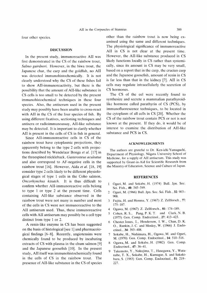
In the present study, immunoreactive AII was first demonstrated in the CS of the rainbow trout, Salmo gairdneri. However, in the biwa trout, the Japanese char, the carp and the goldfish, no AII was detected immunohistochemically. It is not clearly understood why the CS of these fishes fail to show All-immunoreactivity, but there is the possibility that the amount of All-like substance in CS cells is too small to be detected by the present immunohistochemical techniques in these four species. Also, the antiserum used in the present study may possibly have been unable to cross-react with AII in the CS of the four species of fish. By using different fixatives, sectioning techniques and antisera or radioimmunoassay, All-like substance may be detected. It is important to clarify whether AII is present in the cells of CS in fish in general.
Since All-immunoreactive cells in CS of the rainbow trout have cytoplasmic projections, they apparently belong to the type 2 cells with projections described by Wendelaar Bonga et al. [17] in the threespined stickleback, Gasterosteus aculeatus and also correspond to AF-negative cells in the rainbow trout [16]. However, Aida et al. [18, 19] consider type 2 cells likely to be different physiological stages of type 1 cells in the Coho salmon, Oncorhynchus kisutch. It is thus difficult to confirm whether All-immunoreactive cells belong to type 1 or type 2 at the present time. Cells containing All-like substance observed in the rainbow trout were not many in number and most of the cells in CS were not immunoreactive to the AII antiserum used. Thus, these immunoreactive cells with AII antiserum may possibly be a cell type distinct from type 1 or 2.
A renin-like enzyme in CS has been suggested on the basis of histological [see 1] and pharmacological findings [6-8]. Recently, angiotensins were chemically found to be produced by incubating extracts of CS with plasma in the chum salmon [9] and the Japanese goosefish [10]. In the present study, AII itself was immunohistochemically found in the cells of CS in the rainbow trout. The presence of All-like substance in the CS of species
other than the rainbow trout is now being examined using the same and different techniques. The physiological significance of immunoreactive AII in CS is not clear at the present time. However, the All-like substance produced in CS likely functions locally in CS rather than systemi-cally, since its amount in CS may be very small, based on a report that in the carp, the crucian carp and the Japanese goosefish, amount of renin in CS is far less than that in the kidney [7]. AII in CS cells may regulate intracellularly the secretion of CS hormones.
The CS of the eel were recently found to synthesize and secrete a mammalian parathyroidlike hormone called parathyrin of CS (PCS), by immunofluorescence techniques, to be located in the cytoplasm of all cells in CS [20]. Whether the CS of the rainbow trout contain PCS or not is not known at the present. If they do, it would be of interest to examine the distribution of All-like substance and PCS in CS.
ACKNOWLEDGMENTS
The authors are grateful to Dr. Ken-ichi Yamaguchi, Department of Physiology, Niigata University School of Medicine, for a supply of AII antiserum. This study was supported by Grant-in-Aid for Scientific Research from the Ministry of Education, Science and Culture of Japan.
REFERENCES
1 Oguri, M. and Sokabe, H. (1974) Bull. Jpn. Soc. Sci. Fish., 40: 545-549.
2 Oguri, M. (1966) Bull. Jpn. Soc. Sci. Fish., 32: 903-908.
3 Fujita, H. and Honma, Y. (1967) Z. Zellforsch., 77: 175-187.
4 Ogawa, M. (1967) Z. Zellforsch., 81: 174-189. 5 Cohen, R. S., Pang, P. K. T. and Clark, N. B.
(1975) Gen. Comp. Endocrinol., 27: 413-423. 6 Chester Jones, I., Henderson, I. W., Chan, D. K.
O., Rankin, J. C. and Mosley, W. (1966) J. Endocrinol., 34: 393-408.
7 Sokabe, H., Nishimura, H., Ogawa, M. and Oguri, M. (1970) Gen. Comp. Endocrinol., 14: 510-516.
8 Ogawa, M. and Sokabe, H. (1982) Gen. Comp. Endocrinol., 47: 36-41.
9 Takemoto, Y., Nakajima, T., Hasegawa, Y., Wata-nabe, T. X., Sokabe, H., Kumagae, S. and Sakaki-bara, S. (1983) Gen. Comp. Endocrinol., 51: 219-227.

390 C. YAMADA AND H. KOBAYASHI
10 Hasegawa, Y., Watanabe, T. X., Nakajima, T. and Sokabe,H. (1984) Gen. Comp. Endocrinol., 54: 264-269.
11 McKenzie, J. C , Naruse, K. and Inagami, T. (1985) Anat. R e c , 212: 161-166.
12 Yamaguchi, K. (1981) Acta Endocrinol., 97: 137-144.
13 Hayashi,T., Nakayama, T., Nakajima, T. and Sokabe,H. (1978) Chem. Pharm. Bull., 26: 215-219.
14 Hasegawa, Y., Nakajima, T. and Sokabe, H. (1983) Biomed. Res., 4: 417-420.
15 Sternberger, L. A., Hardy, P. H., Cuculis, J. J., Jr. and Meyer, H. G. (1970) J. Histochem. Cytochem.,
18: 315-333. 16 Krishnamurthy, V. G. and Bern, H. A. (1969) Gen.
Comp. Endocrinol., 13: 313-335. 17 Wendelaar Bonga, S. E., Greven, J. A. A. and
Veenhuis,M. (1976) Cell Tissue. Res., 175: 297-312.
18 Aida, K., Nishioka, R. S. and Bern, H. A. (1980) Gen. Comp. Endocrinol., 41: 296-304.
19 Aida, K., Nishioka, R. S. and Bern, H. A. (1980) Gen. Comp. Endocrinol, 41: 305-313.
20 Lopez, E., Tisserand-Jochem, E-M., Eyquem,A., Milet, C , Hillyard, C , Laliier, F. , Vidal, B. and Maclntyre, I. (1984) Gen. Comp. Endocrinol., 53: 28-36.