Embed Size (px)
Citation preview

Immunopathology and Infectious Diseases
Tropism and Innate Host Responses of the 2009Pandemic H1N1 Influenza Virus in ex Vivo andin Vitro Cultures of Human Conjunctiva andRespiratory Tract
Michael C.W. Chan,* Renee W.Y. Chan,*†
Wendy C.L. Yu,* Carol C.C. Ho,* Kit M. Yuen,*†
Joanne H.M. Fong,* Lynsia L.S. Tang,*Wico W. Lai,‡ Amy C.Y. Lo,‡ W.H. Chui,§
Alan D.L. Sihoe,§ Dora L.W. Kwong,¶
David S.H. Wong,‡ George S.W. Tsao,!
Leo L.M. Poon,* Yi Guan,* John M. Nicholls,†
and Joseph S.M. Peiris**From the Departments of Microbiology* and Pathology,† the EyeInstitute,‡ and the Departments of Cardiothoracic Surgery,§
Clinical Oncology,¶ and Anatomy,! the University of Hong Kong,Li Ka Shing Faculty of Medicine, Queen Mary Hospital,Pokfulam, Hong Kong SAR, China; and HKU-Pasteur ResearchCentre,** Hong Kong SAR, China
The novel pandemic influenza H1N1 (H1N1pdm) vi-rus of swine origin causes mild disease but occasion-ally leads to acute respiratory distress syndrome anddeath. It is important to understand the pathogenesisof this new disease in humans. We compared thevirus tropism and host-responses elicited by pan-demic H1N1pdm and seasonal H1N1 influenza virusesin ex vivo cultures of human conjunctiva, nasophar-ynx, bronchus, and lung, as well as in vitro culturesof human nasopharyngeal, bronchial, and alveolarepithelial cells. We found comparable replication andhost-responses in seasonal and pandemic H1N1 vi-ruses. However, pandemic H1N1pdm virus differsfrom seasonal H1N1 influenza virus in its ability toreplicate in human conjunctiva, suggesting subtle dif-ferences in its receptor-binding profile and highlight-ing the potential role of the conjunctiva as an addi-tional route of infection with H1N1pdm. A greaterviral replication competence in bronchial epitheliumat 33°C may also contribute to the slight increase invirulence of the pandemic influenza virus. In contrastwith highly pathogenic influenza H5N1 virus, pan-demic H1N1pdm does not differ from seasonal influ-
enza virus in its intrinsic capacity for cytokine dys-regulation. Collectively, these results suggest thatpandemic H1N1pdm virus differs in modest but subtleways from seasonal H1N1 virus in its intrinsic viru-lence for humans, which is in accord with the epide-miology of the pandemic to date. These findings aretherefore relevant for understanding transmissionand therapy. (Am J Pathol 2010, 176:000–000; DOI:10.2353/ajpath.2010.091087)
The recent pandemic caused by a novel H1N1 virus(H1N1pdm) arose from the reassortment of three or moreviruses of swine origin, including the North Americantriple reassortant H3N2 and H1N2 viruses, classicalswine H1N1, and European swine H1N1/H3N2 viruses.1,2
Most patients with pandemic H1N1pdm have mild influ-enza-like illness, but a minority of patients develop aprimary viral pneumonia, sometimes leading to acuterespiratory distress syndrome and death.3,4 Many, butnot all, patients with severe disease have pregnancy,obesity, or underlying disease states such as asthma,
Supported by Research Fund for Control of Infectious Disease grant (Ref:LAB-15, RFCID commissioned study on human swine influenza virus andRFCID grant, reference no: 06060552, 08070842) from the ResearchFund for Control of Infectious Disease, Health, Welfare, and Food Bureau,Hong Kong SAR Government, and the General Research Fund (HKU7612/08M and 7610/09M to M.C.W.C., HKU 7530/06M to L.L.M.P andHKU 7735/07M to J.M.N), Research Grants Council, Hong Kong SARGovernment; Small project funding (reference no: 200907176007 toR.W.Y.C), The University of Hong Kong; National Institutes of Health(NIAID contract HHSN266200700005C) and AoE Funding (AoE/M-12/06)from the Area of Excellence Scheme of the University Grants Committee,Hong Kong SAR Government.
M.C.W.C. and R.W.Y.C. contributed equally to this study.
Accepted for publication December 15, 2009.
Address reprint requests to Dr. Joseph Malik Sriyal Peiris, M.D. D.Phil.or Dr. Michael Chi Wai Chan, Ph.D., Department of Microbiology, Univer-sity Pathology Building, Queen Mary Hospital, Pokfulam, Hong Kong SAR,China. E-mail: [email protected] or [email protected].
The American Journal of Pathology, Vol. 176, No. 4, April 2010
Copyright © American Society for Investigative Pathology
DOI: 10.2353/ajpath.2010.091087
1
Uncorrected Version. Published on January 28, 2010 as DOI:10.2353/ajpath.2010.091087
Copyright 2010 by the American Society for Investigative Pathology.

obstructive airways disease, diabetes, and chronic car-diovascular or renal disease. The disease associatedwith H1N1pdm so far appears to be comparable with thatof seasonal influenza and less severe than that seen inthe 1918 pandemic or in zoonotic disease caused byhighly pathogenic avian influenza (HPAI) H5N1. How-ever, unlike seasonal influenza where morbidity and mor-tality are mainly seen in the elderly, pandemic H1N1pdmappears to spare this age-group, possibly because of thepresence of cross-neutralizing antibody generated byprior repeated seasonal H1N1 infection.5 In California,the median age of all cases was 17 years, of hospitalizedcases 26 years, and for fatal cases was 45 years.
It is therefore important to understand how the pathogen-esis and tissue tropism of H1N1pdm virus in humans differsfrom seasonal influenza viruses. However, there is so farlimited information in this regard. The H1N1pdm virus doesnot possess the genetic motifs of virulence associated witheither the HPAI H5N1 or 1918 H1N1 viruses.2 In experimen-tally infected ferrets, macaques, and mice, H1N1pdmcauses moderately more severe illness compared with sea-sonal influenza although being much less virulent than HPAIH5N1 or the 1918 pandemic Spanish flu virus.6–8 In theseanimal models, H1N1pdm virus was able to infect the alve-olar epithelium more readily than seasonal H1N1 virus, butwhether this holds true for humans is not known.7 ThoughH1N1pdm was initially reported to have a predominantly!2-6 sialic acid (Sia) receptor binding preference8 similar tohuman seasonal influenza viruses, recent glycan array dataindicates that there is binding to both “human” Sia !2-6 and“avian” Sia !2-3.9 H1N1pdm virus differs from seasonalinfluenza viruses in their ability to infect and cause illness inmice without prior adaptation. As the mouse respiratory tracthas a predominance of Sia !2-3, rather than Sia !2-6 recep-tors, these findings support the contention that H1N1pdm vi-ruses have a broader Sia receptor binding profile.8 Takentogether, these observations suggest that H1N1pdm virus dif-fers in subtle but important ways from seasonal influenza vi-ruses in receptor usage and tissue tropism, and this may beimportant in its pathogenesis and transmission.
Cytokine dysregulation is believed to contribute to thepathogenesis of human disease caused by HPAI H5N1as well as the 1918 pandemic H1N1 viruses.10–14 It is notknown whether the H1N1pdm virus differs from seasonalinfluenza in the induction of proinflammatory host re-sponses in human tissues. The lungs of H1N1pdm-in-fected mice had a markedly different cytokine profilewhen compared with seasonal influenza infected animalswith elevated levels of interleukin (IL)-4, IL-10, and inter-feron (IFN)-". The lungs of H1N1pdm-infected macaquesalso had higher levels of chemokines MCP-1, MIP-1!,IL-6, and IL-18.6 However, it is not known whether thesehost responses simply reflect the greater or more exten-sive replication of the H1N1pdm virus in the lung whencompared with seasonal influenza viruses or are attribut-able to intrinsic differences in the virus itself being able toinduce a more potent innate host response as occurswith the highly pathogenic avian influenza H5N1 virus.When primary human cells (macrophages and type I-likepneumocytes) are infected with seasonal and HPAI H5N1influenza viruses of comparable infectious titers, the HPAI
H5N1 viruses differentially hyperinduce a range of proin-flammatory responses over a single virus replication cy-cle.10,11,14 Thus it is clear that the H5N1 virus has inher-ent properties that lead to an exaggerated innate immuneresponse. It is relevant to use a similar approach to inves-tigate the host innate immune responses induced by pan-demic H1N1pdm compared with that of seasonal influenzaH1N1 virus in primary human respiratory epithelium.
We have previously used ex vivo cultures of nasopharynx,tonsillar tissue, and lung for investigating virus tropism.15
We have also established in vitro cultures of polarizedprimary human respiratory epithelial cells, including typeI–like alveolar epithelial cells, nasopharyngeal epithelialcells, and differentiated bronchial epithelial cells for in-vestigating tissue tropism and innate immune host re-sponses elicited by influenza viruses.10,14,15 These invitro cultures of bronchial epithelium differentiated at anair–liquid interface (ALI) provide a good representation ofthe human bronchial epithelium and have a ciliated epi-thelium as well as mucus producing goblet cells. Wehave also recently established ex vivo tissue culture mod-els of human conjunctival epithelium. We now use theseex vivo human tissue cultures as well as the primaryhuman respiratory epithelial cell cultures to compare thevirus replication competence, cell tropism, and host innateimmune responses of the pandemic H1N1pdm virus withthat of seasonal influenza H1N1 viruses and, where rele-vant, avian HPAI H5N1 and H7N7 viruses.
We demonstrate that whereas seasonal H1N1 andpandemic H1N1pdm viruses replicate comparably in exvivo cultures of human nasopharynx and lung tissues, thehuman conjunctiva is preferentially infected by H1N1pdmrather than seasonal influenza H1N1 or H3N2 viruses.Pandemic H1N1pdm replicates more efficiently than sea-sonal H1N1 virus in differentiated bronchial epithelialcells in vitro at 33°C, but the two viruses replicate com-parably at 37°C. We also demonstrate that the pandemicH1N1pdm virus does not differ from the human seasonalinfluenza viruses in their ability to induce proinflammatorycytokines and therefore does not appear to have thesame potential to induce cytokine dysregulation as thatmanifested by HPAI H5N1 or the 1918 H1N1 virus.
Materials and Methods
Viruses
The viruses used in these studies were the pandemicvirus A/HongKong/415742/09 (H1N1pdm), seasonal vi-ruses A/HongKong/54/98 (H1N1), A/HongKong/403721/2009 (H1N1) and A/Oklahoma/1992/05 (H3N2), and twoavian influenza viruses isolated from humans, A/Vietnam/3046/04 (H5N1) and A/Netherland/33/03 (H7N7). A/Okla-homa/1992/05 (H3N2) was chosen because of the avail-ability of glycan array data demonstrating a restricted Sia!2-6Gal–binding profile.16 The viruses were initially iso-lated and passaged in Madin-Darby canine kidney(MDCK) cells. The virus stock was aliquoted and thentitrated to determine tissue culture infection dose 50%(TCID50) in MDCK cells. The experiments were per-
2 Chan et alAJP April 2010, Vol. 176, No. 4

formed in a Bio-safety level 3 (BSL-3) facility at the De-partment of Microbiology, The University of Hong Kong.Table 1 summarizes all of the influenza A viruses used inthis study and their abbreviations.
In Vitro and ex Vivo Cell Cultures
Primary human alveolar type I–like pneumocytes, well-differentiated bronchial epithelial cells and nasopharyn-geal epithelial cells were derived and cultured as previ-ously described10,14,15,17–20 with modifications.
Alveolar Type I–Like Pneumocytes Isolation
Primary type I–like pneumocytes were isolated using hu-man nonmalignant lung tissue obtained from patientsundergoing lung resection in the Department of Cardio-thoracic Surgery, Queen Mary Hospital, Hong Kong SAR,under a study approved by the Institutional Review Boardof the University of Hong Kong and Hospital AuthorityHong Kong West Cluster, and written informed consentwas provided by each patient. Briefly, after removingvisible bronchi, the lung tissue was minced into pieces of!0.5 mm thickness using a tissue chopper and washedwith BSS containing Hanks balanced salt solution (Gibco,Grand Island, NY) with 0.7 mmol/L sodium bicarbonate(Gibco) at pH 7.4 for 3 times to partially remove macro-phages and blood cells. The tissue was digested using acombination of 0.5% trypsin (GIBCO BRL, Gaithersburg,MD) and 4 U/ml elastase (Worthington Biochemical Cor-poration, Lakewood, NJ) for 40 minutes at 37°C in ashaking water-bath. The digestion was stopped by add-ing DMEM/F12 medium (Gibco) with 40% FBS in andDNase I (350 U/ml) (Sigma-Aldrich, St Louis, MO). Cellclumps were dispersed by repeatedly pipetting the cellsuspension for 10 minutes. A disposable cell strainer(gauze size of 50 #m; BD Science, Palto-Alto, CA) wasused to separate large undigested tissue fragments. Thesingle cell suspension in the flow-through was centrifugedand resuspended in a 1:1 mixture of DMEM/F12 mediumand small airway basal medium (Lonza, Walkersville, MD)supplemented with 0.5 ng/ml epidermal growth factor(hEGF), 500 ng/ml epinephrine, 10 #g/ml transferrin, 5#g/ml insulin, 0.1 ng/ml retinoic acid, 6.5 ng/ml triiodothy-ronine, 0.5 #g/ml hydrocortisone, 30 #g/ml bovine pituitaryextract, and 0.5 mg/ml BSA together with 5% FBS and 350U/ml DNase I. The cell suspension was plated on tissueculture–grade plastic flask (Corning Inc., Corning, NY) andincubated in a 37°C water-jacketed incubator with 5% CO2
supply for 90 minutes. The nonadherent cells were layered
on a discontinuous cold Percoll density gradient (densities1.089 and 1.040 g/ml) and centrifuged at 25g for 20 minuteswithout brake. The cell layer at the interface of the twogradients was collected and washed four times with BSS toremove the Percoll. The cell suspension was incubated withmagnetic beads coated with anti-CD14 antibodies at roomtemperature for 20 minutes under constant mixing. Afterthe removal of the beads using a magnet (MACS CD14MicroBeads, Miltenyi Biotech GmbH, Gladbach, Ger-many), cell viability was assessed by trypan-blue exclu-sion. The purified pneumocyte suspension was resus-pended in small airway growth medium (Lonza)supplemented with 1% FBS, 100 U/ml penicillin, and 100#g/ml streptomycin, and plated at a cell density of 3 "105 cells/cm2. The cells were maintained in a humidifiedatmosphere (5% CO2, 37°C) under liquid-covered con-ditions, and growth medium was changed daily startingfrom 60 hours after plating the cells. When the cell layerapproached 75% confluence, the pneumocytes were de-tached Hanks buffered saline solution trypsin/EDTA andsubcultured for the experiments.
Culture of Well-Differentiated Normal HumanBronchial Epithelial Cells
Human bronchial epithelial (NHBE) cells were purchasedas cryopreserved vials (Lonza). NHBE cells were platedinto a T175 tissue culture grade culture flask in a densityof 500 cells/cm2 for cell proliferation. Bronchial epithelialbasal medium (Lonza Walkersville, Inc.) was supple-mented with growth factor and hormones as stated in thesuppliers instructions. After subculture, they were platedto a cell density of 2.5 " 105 cells/cm2 on human colla-gen IV (BD Science)–coated transwell inserts (Corning).Bronchial epithelial growth medium (Lonza Walkersville,Inc.) medium was supplemented with the retinoic acidconcentration adjusted to 10#7 mol/L. Medium waschanged every 48 hours until the cell layer reached con-fluence. An ALI was then established by removing theculture medium from the apical compartment. Thereafter,medium was changed in the basolateral compartmentevery 48 hours until day 21 of ALI culture. The apicalcompartment was gently washed with phosphate-buff-ered saline (PBS) once a week to remove accumulatedmucus and debris. The transepithelial resistance wasmeasured by epithelial voltohmeter (World Precision In-struments, Sarasota, Fla.). At day 21 of ALI culture, theNHBE cells became well differentiated and ready for use.The transwell cultures were collected and fixed with 10%formalin, paraffin embedded, and then cross-sectioned forhistological examination. Slides were stained using hema-toxylin-eosin. Ciliated cells were further identified by FITC-conjugated $-tubulin antibody (Sigma, Saint Louis, MO)and goblet cells were identified by biotinylated MUC5ACantibody (Invitrogen, San Francisco, CA).
Culture of Nasopharyngeal Epithelial (NPE) Cells
The nasopharyngeal biopsy was cut into 2- to 3-mm frag-ments and placed onto a 6-cm-sized Falcon culture dish
Table 1. List of Influenza A Viruses Used in this Study
Subtype Name Abbreviation
H1N1 A/Hong Kong/54/1998 HK98/H1N1H1N1 A/Hong Kong/463721/2009 HK09/H1N1H1N1 A/Hong Kong/415742/2009 HK09/H1N1pdmH3N2 A/Oklahoma/1992/2005 OK05/H3N2H7N7 A/Netherland/33/2003 NL03/H7N7H5N1 A/Vietnam/3046/2004 VN04/H5N1
Tropism of Pandemic Influenza H1N1 Virus 3AJP April 2010, Vol. 176, No. 4

(BD Biosciences, Palo Alto, CA) containing 2 ml of RPMI1640 culture medium (Gibco) supplemented with 1% dia-lyzed fetal bovine serum (FBS), 0.25 #mol/L transferrin, 3.32nmol/L hEGF, 0.7 #mol/L insulin, 0.5 #mol/L phosphoeth-anolamine, 0.5 #mol/L ethanolamine, 10 nmol/L tiodo-L-thyronine, 2 #mol/L hydrocortisone, 2.7 #mol/L L-epineph-rine, 2 mmol/L L-glutamine, 0.03 #mol/L sodium selenium, 1nmol/L molybdic acid, 0.5 nmol/L stannous chloride, and0.75 nmol/L nickel sulfate. The medium had 0.4 mmol/Lcalcium, which facilitated the attachment of explants to theculture surfaces. After 7 to 10 days of culture, the prolifer-ative epithelial outgrowth from the explants were trypsinizedand propagated at a splitting-ratio of 3 in a 1:1 mixture ofDefined Keratinocyte-SFM (Gibco) and EpiLife medium withfull supplements (Sigma-Aldrich). The cells were main-tained at 37°C with 5% CO2 in a water-jacketed incubator.The calcium concentration in this mixed medium formula-tion was below 0.08 mmol/L to stimulate the proliferation ofthe primary nasopharyngeal epithelial cells and suppressthe growth of contaminating fibroblasts.
Ex Vivo Culture of Conjunctiva, Nasopharynx,Bronchi, and Lung
Fresh conjunctiva tissues were obtained from 20 individ-uals who were undergoing excision for pterygium duringsurgical management. Biopsies of nasopharyngeal tissue(n $ 6) were obtained from patients undergoing electivenasopharyngoscopy as detailed earlier.15 Bronchi (n $18) and lung tissues (n $ 18) were obtained from lungcarcinoma patients having surgical resection of lung tis-sue. The biopsies or tissue fragments of normal nonma-lignant tissue that was excess to the requirements ofclinical diagnosis were used. All of the studies were ap-proved by the Institutional Review Board of the University ofHong Kong and Hospital Authority Hong Kong West Clus-ter, and a written informed consent was provided by eachpatients. The lung tissue fragments were placed into culturemedium (F-12K nutrient mixture with L-glutamine, and anti-biotics) in 24-well tissue culture plates incubated at 37°C forviral infection experiments. The conjunctival, nasopharyn-geal, and bronchial biopsies or tissue fragments wereplaced into culture medium (F-12K nutrient mixture withL-glutamine, and antibiotics) incubated at 33°C (except thebronchial biopsy, which was incubated at 37°C) with a
sterile surgical pathology sponge to establish an ALI con-dition in 24-well culture plates for both conjunctiva, naso-pharynx and bronchi ex vivo culture (Corning, New York,NY) for viral infection experiments.
Experimental Infection with Influenza Viruses
The experimental conditions and multiplicity of infection(MOI) used in the ex vivo and in vitro experiments issummarized in Table 2.
Infection of Respiratory Epithelial Cell in Vitro
Respiratory epithelial cell cultures were infected with in-fluenza A viruses at a MOI of 0.01 for the analysis of virusreplication and MOI of 2 for the analysis of cytokine andchemokine expression. MEM medium (Gibco) with 100U/ml penicillin and 100 #g/ml streptomycin was used asinoculum in the mock infected cells. The cell culture wasincubated with the virus inoculum for 1 hour in a water-jacketed 37°C or 33°C incubator with 5% CO2 (Table 2).After the incubation period the cells were rinsed 3 timeswith warm PBS and replenished with the appropriategrowth medium. The infected cells were harvested formRNA collection at 6 hours and 24 hours postinfection andviral M gene was quantified using qPCR as described.10,14
Productive viral replication was assayed by titrating thecell-free supernatant collected at 1, 24, 48, 72 hours bytitration in quadruplicate in MDCK cells and expressed as50% tissue culture infectious dose (TCID50).
Influenza Virus Infection of Ex Vivo Culture
Ex vivo conjunctival cultures were infected with influenzaA viruses of subtypes H7N7, H5N1, H3N2, H1N1, andH1N1pdm within 2 hours of collection. Five biopsiesserved as uninfected controls, and two were infected withUV light inactivated VN04/H5N1 virus as controls. Theviruses were used at a titer of 1 " 106 TCID50/ml (a similartiter to that used by Shinya et al21) for infecting the ex vivocultures. The biopsy or tissue fragments were incubatedat 33°C on the culture sponge to establish an ALI for timeperiods ranging from 0 to 24 hours at which time thetissues were fixed in 10% neutral buffered formalin and
Table 2. Summary Table Showing the Experimental Conditions, MOIs, and Virus Titer Used in the ex Vivo and in VitroExperiments
Experiment model Region Incubation temperature MOI
Ex vivo Conjunctiva* 33°C UDNasopharynx 33°C UDBronchi 37°C UDLung 37°C UD
In vitro Nasopharyngeal epithelial cells 33°C and 37°C 0.01† and 2‡
Bronchial epithelial cells 33°C and 37°C 0.01† and 2‡
Type I-like pneumocytes 37°C 0.01† and 2‡
*Conjunctiva tissues were infected with all the viruses listed in Table 1, while the other respiratory tissue and epithelial cells were infected withHK98/H1N1 and HK09/H1N1pdm.
MOI† used for replication kinetics study and MOI‡ used for cytokine study. The MOI used in the ex vivo model cannot be determined (UD).Influenza viruses were used at a titer of 1 " 106 50% tissue culture infectious doses (TCID50)/ml (a similar titer as used previously15,21) for infecting theex vivo cultures.
4 Chan et alAJP April 2010, Vol. 176, No. 4

processed for paraffin embedding and immunohisto-chemistry using a mouse anti-influenza nucleoprotein an-tibody (HB65, EVL Laboratories, The Netherlands). Todetermine productive viral replication, conjunctival andbiopsies were cultured in 1 ml of F12K with 100 units/mlpenicillin and 100 #g/ml streptomycin at 33°C, and in-fected with influenza A viruses at a titer of 1 " 106
TCID50/ml. After 2 hours infection the unattached viruswas removed by washing. At 0 and 24 hours after wash-ing, 130 #l of medium was removed and stored at #80°Cfor virus titration in MDCK cells. Increasing virus titers inthe cell supernatants provided evidence of productivevirus replication. At the end of 24 hours the tissues werethen fixed in formalin, and immunohistochemistry for in-fluenza antigen was performed.
Fragments of normal nasopharynx, bronchi, and lungtissues were cut into multiple 2- to 3-mm fragments andinfected in parallel with influenza A viruses at a titer of 1 "106 TCID50/ml. Human influenza viruses HK98/H1N1 andHK09/H1N1pdm were tested in parallel. These tissuesfragments were infected as above and incubated for 1,24, and 48 hours at 37°C. They were then fixed in 10%neutral buffered formalin and processed for paraffin em-bedding and immunohistochemistry (see below). Addi-tional tissue fragments were infected with influenza Avirus at 37°C and after 2 hours, unbound virus was re-moved, and viral yield in the cell-free supernatant wasassessed at 1 and 24 hours (for nasopharynx) and 1, 24,and 48 hours (for lung and bronchi) by titration in qua-druplicate in MDCK cells.
Viral Titration by TCID50 Assay
A confluent 96-well tissue culture plate of MDCK cellswas prepared one day before the virus titration (TCID50)assay. Cells were washed once with PBS and replen-ished with serum-free MEM medium supplemented with100 units/ml penicillin and 100 #g/ml streptomycin and2 #g/ml of tosylsulfonyl phenylalanylchloromethyl ke-tone (TPCK)–treated trypsin. Serial dilutions of virussupernatant, from 0.5 log to 7 log, were performedbefore adding the virus dilutions onto the plates inquadruplicate. The plates were observed for cyto-pathic effect daily. The end point of viral dilution lead-ing to CPE in 50% of inoculated wells was estimatedusing the Karber method.22
Immunohistochemical Staining for Influenza AVirus Antigen
Immunohistochemical staining of the conjunctiva and re-spiratory tract tissue was performed for the influenzanucleoprotein as follows. The tissue sections with incu-bated with 0.05% Pronase (Roche, Mannheim, Germany)in 0.1% CaCl2 pH 7.8 at 37°C for 2 minutes, blocked with3% H2O2 in TBS for 10 minutes, followed by treatmentwith an avidin/biotin blocking kit (Vector Lab, Burlingame,CA). After blocking with 10% normal rabbit serum for 10minutes at room temperature, the sections were incu-bated with 1/100 (15 #g/ml) HB65 (EVL anti-influenza NP,
subtype A) antibody for 1 hour at room temperature fol-lowed by biotinylated rabbit anti-mouse (Dako Cytoma-tion, Glostrup, Denmark) diluted 1/100 for 30 minutesat room temperature. After incubation with strep-ABCcomplex (Dako Cytomation) diluted 1/100 for 30 min-utes at room temperature, the sections were developedwith 0.5 mg/ml DAB (Sigma-Aldrich) in 0.02% H2O2 for20 minutes and then microwaved in 10 mmol/L citratebuffer pH 6.0 for 20 minutes to expose the secondantigen.
Lectin Histochemistry
Sections of ex vivo conjunctival biopsies used for the virusinfections experiments were processed similar to the pro-cedure for immunohistochemistry. The detection of Sia onex vivo tissue sections was performed using lectin cyto-chemistry. Tissue fragments were washed and fixation in10% neutral buffered formalin followed by staining withthe Roche Dig Glycan Detection Kit as per manufactur-er’s instructions. Lectins recognizing different penulti-mate structures as well as the Sia linkages (!2-6 or !2-3to either Gal$1-4GlcNAc or Gal$1-3GalNAc) were used:Peanut agglutinin (PNA; binding to Gal$1-3GalNAc),Maackia amurensis agglutinin (MAA; which binds to anumber of glycans including !2-3-linked Sia), and HRP-conjugated Sambucus nigra agglutinin (SNA; which binds!2-6-linked Sia).
Desialylation of Conjunctival ex Vivo Tissues
The detection of surface Sia on ex vivo tissue sectionswas performed using lectin cytochemistry. Tissue frag-ments were exposed to 100 U/ml of DAS181, a sialidasefusion protein provided by NexBio Inc (CA) for 2 hoursfollowed by washing and fixation in 10% neutral bufferedformalin followed by staining with the lectins SNA, MAA,and PNA as indicated above. Infectious viral yield in thecell-free supernatant was assessed at 1 and 24 hours inthe cell culture experiments with and without the treat-ment of DAS181 before influenza virus infection by viraltitration described above. Lectin binding was also per-formed to detect total Sia in the ex vivo tissues before andafter DAS181 treatment.
Quantification of Cytokine and ChemokineProfile by RT-qPCR and ELISA
mRNA from infected cells was extracted at 6, 8, and 24hours postinfection using RNeasy Mini kit (Qiagen,Hilden, Germany) and treated with DNase. cDNA wassynthesized with Oligo-dT primers and Superscript IIIreverse transcriptase (Invitrogen) and quantified by real-time quantitative PCR analysis with a LightCycler(Roche). The gene expression profile for cytokines, tumornecrosis factor-! and IFN-$ and chemokines (IP-10,RANTES) and viral matrix gene were quantified and nor-malized using the housekeeping gene product $-actinmRNA. The primers and methods used for these assays
Tropism of Pandemic Influenza H1N1 Virus 5AJP April 2010, Vol. 176, No. 4
have been reported previously.10,11 The concentrationsof IFN-$, RANTES, and IP-10 proteins in culture super-natants collected at 8 hours or 24 hours postinfection ofthe influenza viruses, and infected cells were measuredby ELISA assay as recommended by the manufacturer(R&D Systems, Minneapolis, MN).
Statistical Analysis
The differences of log10-transformed viral titers amongdifferent viruses at different time points of postinfectionand the quantitative cytokine and chemokine mRNAand protein expression profile of influenza virus–in-fected cells was compared using one-way analysis ofvariance followed by Bonferroni multiple-comparisontest. Differences were considered significant at P %0.05. The statistical analysis was performed usingGraph-pad Prism 5.
Results
HK09/H1N1pdm Infection in Conjunctiva BiopsyEx Vivo
We infected cultures of ex vivo tissues within 2 hours ofcollection with human seasonal influenza viruses HK98/H1N1 and HK09/H1N1, OK05/H3N2, pandemic HK09/H1N1pdm, and avian influenza viruses VN04/H5N1 andNL03/H7N7. Immunohistochemistry (Figure 1, A–C) re-vealed that pandemic influenza HK09/H1N1pdm as wellas avian VN04/H5N1 and NL03/H7N7 viruses readily in-fected conjunctival epithelial cells resulting in !10-foldincrease in viral titers at 24 hours after infection (Figure1G) indicating productive viral replication. There wasno evidence of infection when UV-inactivated HK09/H1N1pdm, NL03/H7N7, or VN04/H5N1 viruses wereused to infect the conjunctival cultures (data not shown).H7 and H5 subtype avian influenza viruses are known toreplicate in human ocular epithelium,23,24 but there are
Figure 1. Ex vivo organ cultures of conjunctiva infected with (A) VN04/H5N1, (B) NL03/H7N7, and(C) HK09/H1N1pdm at 24 hours postinfection showing positive staining for influenza A nucleopro-tein (reddish brown with arrow). Negative staining for influenza A nucleoprotein in (D) HK98/H1N1,(E) OK05/H3N2, and (F) HK09/H1N1 infected conjunctival tissues (G). Conjunctival biopsies infectedwith 106 TCID50/ml of influenza VN04/H5N1, HK09/H1N1pdm, and NL03/H7N7 viruses demonstratean increase in viral yield, whereas HK98/98 and OK05/H3N2 demonstrate no productive replication.The chart shows the mean and the SE of the virus titer pooled from three independent experiments.Single asterisk indicates statistically significant difference of means with P % 0.05, double asterisksindicate statistically significant differences of means with P % 0.01. Horizontal dotted line denotesdetection limit of TCID50 assay.
6 Chan et alAJP April 2010, Vol. 176, No. 4

no previous data on the replication of pandemicH1N1pdm at this site. On the other hand, there was noexpression of viral antigen (Figure 1D to F) or increase invirus titers (Figure 1G) in conjunctival ex vivo culturesinfected with seasonal HK98/H1N1 (Figure 1D), HK09/H1N1 (Figure 1E), or OK05/H3N2 (Figure 1F) virus–in-fected tissues or in mock-infected tissues.
Sia Receptor Distribution on HumanConjunctival Epithelium
Lectin histochemistry on the ex vivo conjunctival biopsiesusing SNA (which recognizes the human influenza recep-tor Sia !2-6) and MAA (which recognizes the avian influ-enza receptor Sia !2-3 but also binds non-sialyated gly-cans25) showed that SNA binding was similar to thatreported previously in normal human conjunctival epithe-lium26,27 with weak binding to the basal epithelium andthe mucins present in the secretory goblet cells of theconjunctiva (Figure 2A). The MAA bound strongly to thebasal and surface epithelium, and there was strong bind-ing to the mucins present within the goblet cells (Figure2B). Minimal PNA binding was seen in the epithelium, butit bound strongly to the mucins in the goblets cells(Figure 2C). After the removal of Sia from the adjacentglycan structures present on the surface of cells28 bysialidase DAS181 treatment, we showed that SNAbinding was totally abolished, MAA binding was signif-icantly reduced, but still with some binding to the mu-cins present within the cytoplasm of the goblet cells
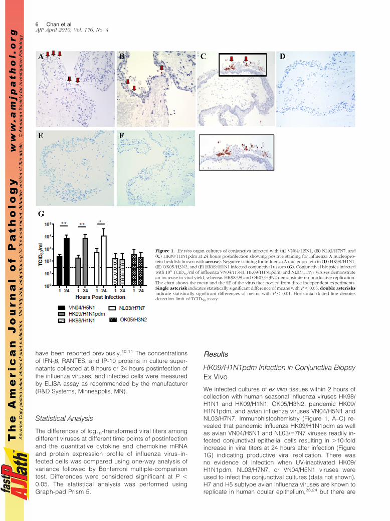
(Figure 3, A and B). DAS181 treatment led to increasedexposure of the subterminal polysaccharide structures(such as Gal$1-3GalNAc) and thus an increased bind-ing with PNA (Figure 3C). There was inhibition of rep-lication of HK09/H1N1pdm in human conjunctiva exvivo after sialidase treatment (Figure 3D) as deter-mined by viral yield in culture supernatants, which is inkeeping with the glycan array data that HK09/H1N1pdmmay use Sia !2-3Gal in addition to Sia !2-6Gal receptors forinfection.9
HK09/H1N1pdm Infection in Nasopharynx,Bronchi, and Lung Biopsies Ex Vivo
By immunohistochemistry, we found evidence that pan-demic HK09/H1N1pdm (Figure 4, A, D, and G) and sea-sonal HK98/H1N1 (Figure 4, B, E, and H) readily infectedthe epithelium of the nasopharynx, bronchi, and lung. Inparallel, there was evidence of productive virus replica-tion with increasing virus titers in pandemic H1N1pdmand seasonal H1N1-infected nasopharygeal, bronchial,and lung ex vivo cultures (Figure 4, C, F, and I, respec-tively). Pandemic HK09/H1N1pdm titers in the bronchushad peaked by 24 hours, whereas seasonal H1N1 virustiters continued to further increase until 48 hours postin-fection (Figure 4F). Mock infected control tissues re-vealed no evidence of infection by immunohistochemistryor virus yield (data not shown).
Figure 2. Distribution of Sia on conjunctival tissue ex vivo. Sia in !2-6 linkages were stained using SNA, Sia in !2-3 linkages were stained using MAA, and theGal$1-3GalNAc were stained using PNA. The conjunctival tissues show negative/weak positive binding of SNA on the conjunctival epithelium (A), whereas thereis a strong positive binding of the conjunctival epithelium for MAA (B) and a weak PNA staining (C). The goblet cells of the conjunctival epithelium (B and C)show a strongly positive intracytoplasmic binding for both MAA and PNA (arrow). AEC stain with hematoxylin counterstain.
Tropism of Pandemic Influenza H1N1 Virus 7AJP April 2010, Vol. 176, No. 4
Replication Kinetics of HK09/H1N1pdm andSeasonal Influenza HK98/H1N1 Virus in NPECells, NHBE cells, and Alveolar Type I-LikePneumocytes in Vitro
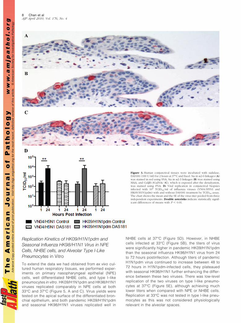
To extend the data we had obtained from ex vivo cul-tured human respiratory tissues, we performed exper-iments on primary nasopharyngeal epithelial (NPE)cells, well differentiated NHBE cells, and type I–likepneumocytes in vitro. HK09/H1N1pdm and HK98/H1N1viruses replicated comparably in NPE cells at both33°C and 37°C (Figure 5, A and C). Virus yields weretested on the apical surface of the differentiated bron-chial epithelium, and both pandemic HK09/H1N1pdmand seasonal HK98/H1N1 viruses replicated well in
NHBE cells at 37°C (Figure 5D). However, in NHBEcells infected at 33°C (Figure 5B), the titers of viruswere significantly higher in pandemic HK09/H1N1pdmthan the seasonal influenza HK98/H1N1 virus from 24to 72 hours postinfection. Although titers of pandemicH1N1pdm virus continued to increase between 48 to72 hours in H1N1pdm-infected cells, they plateauedwith seasonal HK98/H1N1 further enhancing the differ-ence between these two viruses. There was low-levelreplication of the two viruses on type I-like pneumo-cytes at 37°C (Figure 5E), although achieving muchlower titers when compared with NPE or NHBE cells.Replication at 33°C was not tested in type I-like pneu-mocytes as this was not considered physiologicallyrelevant in the alveolar spaces.
Figure 3. Human conjunctival tissues were incubated with sialidase,DAS181 (100 U/ml) for 2 hours at 37°C and fixed. Sia in !2-6 linkages (A)was stained in red using SNA, Sia in !2-3 linkages (B) was stained usingMAA, and Gal$1-3GalNAc (C), which is exposed after the desialyation,was stained using PNA. D: Viral replication in conjunctival biopsiesinfected with 106 TCID50/ml of influenza viruses (VN04/H5N1 andHK09/H1N1pdm) with and without DAS181 treatment by TCID50 assay.The chart shows the mean and the SE of the virus titer pooled from threeindependent experiments. Double asterisks indicate statistically signif-icant differences of means with P % 0.01.
8 Chan et alAJP April 2010, Vol. 176, No. 4

Cytokine and Chemokine Expression Profile inNHBE and Alveolar Type I–Like PneumocytesInfected with Pandemic HK09/H1N1pdm andSeasonal Influenza HK98/H1N1 VirusWe next investigated the expression of cytokine and che-mokine induction by pandemic HK09/H1N1pdm and sea-sonal influenza HK98/H1N1 virus–infected NHBE cellsand alveolar type I–like pneumocytes (Figures 6 and 7,respectively). The efficiency of influenza replication in
these cells was assayed using quantitative real-time PCRfor the influenza matrix gene (Figures 6E and 7E).
The cytokine (IFN-$ and IL-6) chemokine (RANTESand IP-10) mRNA expression profiles at 6 and 24 hourspostinfection were similar in pandemic HK09/H1N1pdmand seasonal HK98/H1N1 virus–infected NHBE cells(Figure 6, A–D). IL-6, RANTES, and IP-10 protein levels inthe culture supernatant measured by ELISA on the apicalaspect of the polarized NHBE cells at 24 hours postin-fection were also comparable (Figure 6, F–H). We failed
Figure 4. Cellular localization of ex vivo infection of the upper and lower respiratory tract by influenza A viruses(A and B) nasopharynx, (D and E) bronchi, and (G and H) lung infected with influenza HK09/H1N1pdm andHK98/H1N1 viruses, showing positive staining for influenza A nucleoprotein (reddish brown). Viral replicationkinetics in ex vivo cultures of (C) nasopharynx, (F) bronchi, and (I) lung biopsies infected with 106 TCID50/ml ofinfluenza viruses by virus titration. The chart showed the mean and the SE of mean of the virus titer pooled fromthree independent experiments.
Tropism of Pandemic Influenza H1N1 Virus 9AJP April 2010, Vol. 176, No. 4
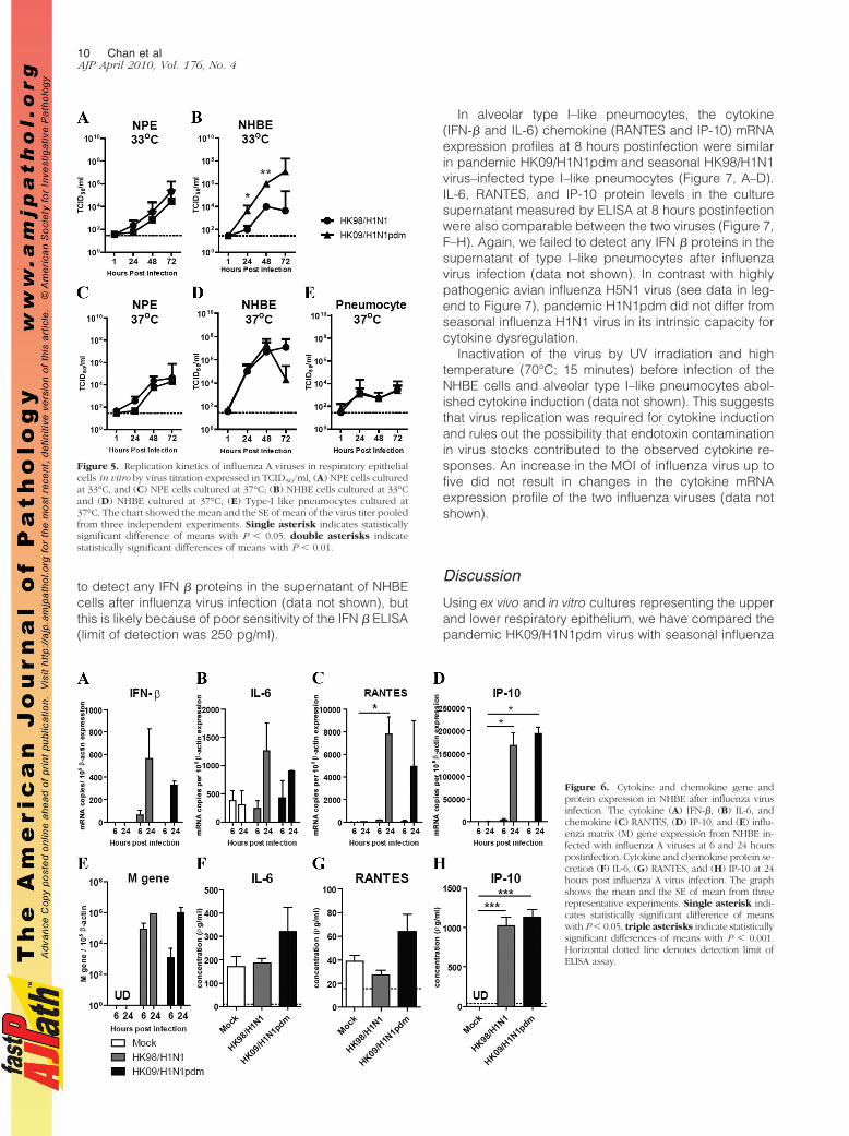
to detect any IFN $ proteins in the supernatant of NHBEcells after influenza virus infection (data not shown), butthis is likely because of poor sensitivity of the IFN $ ELISA(limit of detection was 250 pg/ml).
In alveolar type I–like pneumocytes, the cytokine(IFN-$ and IL-6) chemokine (RANTES and IP-10) mRNAexpression profiles at 8 hours postinfection were similarin pandemic HK09/H1N1pdm and seasonal HK98/H1N1virus–infected type I–like pneumocytes (Figure 7, A–D).IL-6, RANTES, and IP-10 protein levels in the culturesupernatant measured by ELISA at 8 hours postinfectionwere also comparable between the two viruses (Figure 7,F–H). Again, we failed to detect any IFN $ proteins in thesupernatant of type I–like pneumocytes after influenzavirus infection (data not shown). In contrast with highlypathogenic avian influenza H5N1 virus (see data in leg-end to Figure 7), pandemic H1N1pdm did not differ fromseasonal influenza H1N1 virus in its intrinsic capacity forcytokine dysregulation.
Inactivation of the virus by UV irradiation and hightemperature (70°C; 15 minutes) before infection of theNHBE cells and alveolar type I–like pneumocytes abol-ished cytokine induction (data not shown). This suggeststhat virus replication was required for cytokine inductionand rules out the possibility that endotoxin contaminationin virus stocks contributed to the observed cytokine re-sponses. An increase in the MOI of influenza virus up tofive did not result in changes in the cytokine mRNAexpression profile of the two influenza viruses (data notshown).
Discussion
Using ex vivo and in vitro cultures representing the upperand lower respiratory epithelium, we have compared thepandemic HK09/H1N1pdm virus with seasonal influenza
Figure 5. Replication kinetics of influenza A viruses in respiratory epithelialcells in vitro by virus titration expressed in TCID50/ml, (A) NPE cells culturedat 33°C, and (C) NPE cells cultured at 37°C; (B) NHBE cells cultured at 33°Cand (D) NHBE cultured at 37°C; (E) Type-I like pneumocytes cultured at37°C. The chart showed the mean and the SE of mean of the virus titer pooledfrom three independent experiments. Single asterisk indicates statisticallysignificant difference of means with P % 0.05, double asterisks indicatestatistically significant differences of means with P % 0.01.
Figure 6. Cytokine and chemokine gene andprotein expression in NHBE after influenza virusinfection. The cytokine (A) IFN-$, (B) IL-6, andchemokine (C) RANTES, (D) IP-10, and (E) influ-enza matrix (M) gene expression from NHBE in-fected with influenza A viruses at 6 and 24 hourspostinfection. Cytokine and chemokine protein se-cretion (F) IL-6, (G) RANTES, and (H) IP-10 at 24hours post influenza A virus infection. The graphshows the mean and the SE of mean from threerepresentative experiments. Single asterisk indi-cates statistically significant difference of meanswith P % 0.05, triple asterisks indicate statisticallysignificant differences of means with P % 0.001.Horizontal dotted line denotes detection limit ofELISA assay.
10 Chan et alAJP April 2010, Vol. 176, No. 4

virus HK98/H1N1 in regard to their tropism, replicationcompetence, and innate host responses. The most strik-ing difference between these two viruses was the abilityof the pandemic H1N1pdm virus to efficiently infect andreplicate in ex vivo cultures of the human conjunctivalepithelium. Two seasonal H1N1 influenza viruses (HK98/H1N1 and HK09/H1N1) and H3N2 virus (OK05/H3N2)failed to infect the conjunctiva, whereas the avian VN04/H5N1 and NL03/H7N7 viruses did. The human conjunc-tiva has been reported to lack Sia !2-6 that human influ-enza viruses bind to but expresses the Sia !2-3 thatallows binding of avian influenza viruses. Our own studiesusing the lectins SNA and MAA (Figure 2) essentiallyconfirm that these receptor distributions remain true inour ex vivo cultures when compared with human conjunc-tival epithelium.26,27,29 The lack of infection with the sea-sonal influenza and the ability of avian influenza virusesVN05/H5N1 and NL03/H7N7 that preferentially bind Sia!2-3 are in line with these expectations. The markeddifferences in conjunctival tissue tropism of pandemicH1N1pdm and seasonal H1N1 viruses suggest differ-ences in receptor preference or virus replication compe-tence in these cells. Significant differences in receptorbinding profiles between seasonal and pandemic H1N1viruses has been recently confirmed by glycan arraydata.9 Two different pandemic H1N1pdm virus strainshad an affinity for both Sia !2-3 as well as Sia !2-6glycans, whereas seasonal H1N1 had a more restrictedbinding profile to a narrower range of Sia !2-6 glycans.These findings may also explain the ability of pandemicH1N1pdm virus to cause disease in mice without prior
adaptation.6 The receptors that mediate pandemicH1N1pdm infection of the conjunctiva remain to be de-fined, but the finding that the sialidase DAS181 efficientlyblocks virus infection in the conjunctiva (Figure 3D) sug-gests that it is likely to be Sia in nature. Because SNAlectin staining does not recognize all of the Sia !2-6Galglycans that may present in human conjunctiva, it is stillpossible that other conformations of Sia !2-6Gal may yetbe used by the virus to gain entry to the conjunctivalepithelium. In any event, our observation may have thepractical implication that the pandemic HK09/H1N1pdmvirus may have a greater potential than seasonal influ-enza viruses to transmit via the conjunctival route. Therole of the viral hemagglutinin, neuraminidase, or othergenes in the ability of the novel pandemic influenza virusto infect human conjunctival tissue can be systematicallyinvestigated using reverse genetics approaches andthese studies are currently ongoing.
We furthermore demonstrate that both pandemicHK09/H1N1pdm and seasonal HK98/H1N1 viruses canefficiently and comparably infect ex vivo cultures of hu-man nasopharynx, bronchi, and lung tissues (Figure 4).They also have comparable growth curves when infect-ing in vitro cultures of primary human nasopharyngealepithelium at both 33°C and 37°C (Figure 5, A and C).Immunohistochemistry and virus growth curves show thatthe alveolar epithelium of ex vivo lung cultures is compa-rably infected by both pandemic HK09/H1N1pdm andseasonal HK98/H1N1 viruses (Figure 4, D, E, and I). Thisis also reflected in the in vitro cultures of type I–likepneumocytes (Figure 5E). Interestingly, HK09/H1N1pdm
Figure 7. Cytokine and chemokine gene andprotein expression in alveolar type I–like pneu-mocytes after influenza A virus infection. Thecytokine (A) IFN-$, (B) IL-6, and chemokine (C)RANTES, (D) IP-10, and (E) influenza matrix (M)gene expression from alveolar type I–like pneu-mocytes infected with influenza A viruses at 8hours postinfection. Cytokine and chemokineprotein secretion, (F) IL-6, (G) RANTES, and (H)IP-10 at 8 hours postinfection of the pandemicinfluenza H1N1pdm and seasonal influenzaH1N1 viruses. For comparison, the cytokine pro-teins secretion inducted by the infection ofhighly pathogenic influenza H5N1 virus in alve-olar type I–like pneumocytes are: IL-6 (602 & 21pg/ml), RANTES (526 & 75 pg/ml), and IP-10(1313 & 161 pg/ml). The graph shows the meanand the SE of mean from three representativeexperiments. Asterisk indicates significant dif-ference with P % 0.05.
Tropism of Pandemic Influenza H1N1 Virus 11AJP April 2010, Vol. 176, No. 4
virus appears to replicate significantly better than sea-sonal H1N1 in differentiated bronchial epithelium at 33°C(Figure 5B), whereas there is no difference in growthcurves of these two viruses in these cells at 37°C in thefirst 48 hours postinfection (Figure 5D). As the physio-logical temperature in the upper respiratory tract andconducting airways may be lower than the core-bodytemperature of 37°C, the differences in replicationcompetence at 33°C in differentiated human bronchialepithelial cells may be relevant in contributing to themodestly increased virulence of HK09/H1N1pdm virusin humans and in animal models.
In contrast to HPAI H5N1 viruses,10,11,14 the pandemicHK09/H1N1pdm virus did not induce higher levels ofproinflammatory cytokines and chemokines from NHBEcells or type I–like pneumocytes when compared withseasonal influenza viruses (Figures 6 and 7). Therefore,the pandemic HK09/H1N1pdm virus is unlikely to have anintrinsic capacity for cytokine hyperinduction and en-hanced pathogenesis by the route of immunopathogen-esis. In any event, these findings would suggest thatthere is little biological rationale for immunomodulatorytreatment in pandemic H1N1pdm disease on the basis ofintrinsic properties of this virus. It is to be noted, however,that extensive replication of a virus in the alveolar epithe-lium is by itself likely to induce alveolar damage andsubsequent host responses that may lead to acute respi-ratory distress syndrome, and these complications mayhave to be managed on their own merits.
In conclusion, our finding that the pandemic HK09/H1N1pdm virus (but not seasonal HK98/H1N1 virus) in-fects conjunctival epithelium suggests that the eye maybe an important route for acquiring infection with HK09/H1N1pdm as compared with seasonal influenza viruses.Furthermore, this observation implies important differ-ences in receptor preference and tissue tropism betweenthe pandemic H1N1 and seasonal influenza viruses,which may have relevance in pathogenesis. The in-creased replication competence of the HK09/H1N1pdmvirus in the bronchial epithelium at 33°C together withdifferences in tissue tropism in human may be relevant tothe modest increase in virulence of this virus comparedwith seasonal influenza viruses in the current influenzapandemic. On the other hand, the 2009 pandemic H1N1influenza virus is comparable with seasonal influenza ininducing host innate responses and does not have theintrinsic properties of cytokine dysregulation possessedby HPAI H5N1 virus or the 1918 pandemic H1N1 influ-enza virus.11–13 The virus receptor expression in the exvivo lung cultures mimics what is seen in human tis-sue,17,25 highlighting the value of ex vivo cultures in pro-viding physiologically relevant insights into pathogenesisin humans. However, no ex vivo culture or animal modelcan completely reproduce the state of the human respi-ratory tract, and our results therefore need to be inter-preted in the context of these limitations. Overall theexperimental findings are compatible with the clinical andepidemiological observations on pandemic influenzaH1N1pdm virus, which suggests that its severity is notmarkedly different from that of seasonal influenza H1N1viruses. The increased burden in hospitalization and
need for intensive care management may well be morelikely attributable to the much larger number of peopleinfected with an antigenically novel virus toward which asubstantial segment of the population remains immuno-logically naïve together with subtle differences in virustropism and replication competence.
Acknowledgments
We are grateful for the help of Kevin Fung and Yi Jun Chufor the immunohistochemistry and lectin staining. Wethank Elaine YL Yip and Deng Wen for the technicalassistance in the primary culture of nasopharyngeal ep-ithelial cells. We thank Gillian M. Air (Department of Bio-chemistry and Molecular Biology, University of OklahomaHealth Science Center) for providing the influenza H3N2virus strain and Mang Yu (NexBio Inc.) for providing thesialidase fusion protein, DAS181, used in this study.
References
1. Smith GJ, Vijaykrishna D, Bahl J, Lycett SJ, Worobey M, Pybus OG,Ma SK, Cheung CL, Raghwani J, Bhatt S, Peiris JS, Guan Y, RambautA: Origins and evolutionary genomics of the 2009 swine-origin H1N1influenza A epidemic. Nature 2009, 459:1122–1125
2. Neumann G, Noda T, Kawaoka Y: Emergence and pandemic poten-tial of swine-origin H1N1 influenza virus. Nature 2009, 459:931–939
3. Dawood FS, Jain S, Finelli L, Shaw MW, Lindstrom S, Garten RJ,Gubareva LV, Xu X, Bridges CB, Uyeki TM: Emergence of a novelswine-origin influenza A (H1N1) virus in humans. N Engl J Med 2009,360:2605–2615
4. Kumar A, Zarychanski R, Pinto R, Cook DJ, Marshall J, Lacroix J,Stelfox T, Bagshaw S, Choong K, Lamontagne F, Turgeon AF,Lapinsky S, Ahern SP, Smith O, Siddiqui F, Jouvet P, Khwaja K,McIntyre L, Menon K, Hutchison J, Hornstein D, Joffe A, Lauzier F,Singh J, Karachi T, Wiebe K, Olafson K, Ramsey C, Sharma S, DodekP, Meade M, Hall R, Fowler R: Critically ill patients with 2009 influenzaA(H1N1) infection in Canada. JAMA 2009, 302:1872–1879
5. Hancock K, Veguilla V, Lu X, Zhong W, Butler EN, Sun H, Liu F, DongL, Devos JR, Gargiullo PM, Brammer TL, Cox NJ, Tumpey TM, KatzJM: Cross-reactive antibody responses to the 2009 pandemic H1N1influenza virus. N Engl J Med 2009, 361:1945–1952
6. Itoh Y, Shinya K, Kiso M, Watanabe T, Sakoda Y, Hatta M, MuramotoY, Tamura D, Sakai-Tagawa Y, Noda T, Sakabe S, Imai M, Hatta Y,Watanabe S, Li C, Yamada S, Fujii K, Murakami S, Imai H, KakugawaS, Ito M, Takano R, Iwatsuki-Horimoto K, Shimojima M, Horimoto T,Goto H, Takahashi K, Makino A, Ishigaki H, Nakayama M, OkamatsuM, Warshauer D, Shult PA, Saito R, Suzuki H, Furuta Y, Yamashita M,Mitamura K, Nakano K, Nakamura M, Brockman-Schneider R,Mitamura H, Yamazaki M, Sugaya N, Suresh M, Ozawa M, NeumannG, Gern J, Kida H, Ogasawara K, Kawaoka Y: In vitro and in vivocharacterization of new swine-origin H1N1 influenza viruses. Nature2009, 460:1021–1025
7. Munster VJ, de Wit E, van den Brand JM, Herfst S, Schrauwen EJ,Bestebroer TM, van de Vijver D, Boucher CA, Koopmans M,Rimmelzwaan GF, Kuiken T, Osterhaus AD, Fouchier RA: Pathogen-esis and transmission of swine-origin 2009 A(H1N1) influenza virus inferrets. Science 2009, 325:481–483
8. Maines TR, Jayaraman A, Belser JA, Wadford DA, Pappas C, Zeng H,Gustin KM, Pearce MB, Viswanathan K, Shriver ZH, Raman R, CoxNJ, Sasisekharan R, Katz JM, Tumpey TM: Transmission and Patho-genesis of Swine-Origin 2009 A(H1N1) Influenza Viruses in Ferretsand Mice. Science 2009, 325:484–487
9. Childs RA, Palma AS, Wharton S, Matrosovich T, Liu Y, Chai W,Campanero-Rhodes MA, Zhang Y, Eickmann M, Kiso M, Hay A,Matrosovich M, Feizi T: Receptor-binding specificity of pandemicinfluenza A (H1N1) 2009 virus determined by carbohydrate microar-ray. Nature Biotechnol 2009, 27:797–799
12 Chan et alAJP April 2010, Vol. 176, No. 4

10. Chan MC, Cheung CY, Chui WH, Tsao SW, Nicholls JM, Chan YO,Chan RW, Long HT, Poon LL, Guan Y, Peiris JS: Proinflammatorycytokine responses induced by influenza A (H5N1) viruses in primaryhuman alveolar and bronchial epithelial cells. Respir Res 2005, 6:135
11. Cheung CY, Poon LL, Lau AS, Luk W, Lau YL, Shortridge KF, GordonS, Guan Y, Peiris JS: Induction of proinflammatory cytokines in humanmacrophages by influenza A (H5N1) viruses: a mechanism for theunusual severity of human disease? Lancet 2002, 360:1831–1837
12. Kash JC, Tumpey TM, Proll SC, Carter V, Perwitasari O, Thomas MJ,Basler CF, Palese P, Taubenberger JK, Garcia-Sastre A, Swayne DE,Katze MG: Genomic analysis of increased host immune and celldeath responses induced by 1918 influenza virus. Nature 2006,443:578–581
13. Perrone LA, Plowden JK, Garcia-Sastre A, Katz JM, Tumpey TM:H5N1 and 1918 pandemic influenza virus infection results in earlyand excessive infiltration of macrophages and neutrophils in thelungs of mice. PLoS Pathog 2008, 4:e1000115
14. Chan MC, Chan RW, Yu WC, Ho CC, Chui WH, Lo CK, Yuen KM,Guan Y, Nicholls JM, Peiris JM: Influenza H5N1 virus infection ofpolarized human alveolar epithelial cells and lung microvascular en-dothelial cells. Respir Res 2009, 10:102
15. Nicholls JM, Chan MC, Chan WY, Wong HK, Cheung CY, Kwong DL,Wong MP, Chui WH, Poon LL, Tsao SW, Guan Y, Peiris JS: Tropism ofavian influenza A (H5N1) in the upper and lower respiratory tract. NatMed 2007, 13:147–149
16. Kumari K, Gulati S, Smith DF, Gulati U, Cummings RD, Air GM:Receptor binding specificity of recent human H3N2 influenza viruses,Virol J 2007, 4:42
17. Chan RW, Chan MC, Wong AC, Karamanska R, Dell A, Haslam SM,Sihoe AD, Chui WH, Triana-Baltzer G, Li Q, Peiris JS, Fang F, NichollsJM: DAS181 inhibits H5N1 influenza virus infection of human lungtissues. Antimicrob Agents Chemother 2009, 53:3935–3941
18. Gray TE, Guzman K, Davis CW, Abdullah LH, Nettesheim P: Muco-ciliary differentiation of serially passaged normal human tracheobron-chial epithelial cells. Am J Respir Cell Mol Biol 1996, 14:104–112
19. Zhen G, Park SW, Nguyenvu LT, Rodriguez MW, Barbeau R, Paquet
AC, Erle DJ: IL-13 and epidermal growth factor receptor have criticalbut distinct roles in epithelial cell mucin production. Am J Respir CellMol Biol 2007, 36:244–253
20. Tsao SW, Wang X, Liu Y, Cheung YC, Feng H, Zheng Z, Wong N,Yuen PW, Lo AK, Wong YC, Huang DP: Establishment of two immor-talized nasopharyngeal epithelial cell lines using SV40 large T andHPV16E6/E7 viral oncogenes. Biochim Biophys Acta 2002, 1590:150–158
21. Shinya K, Ebina M, Yamada S, Ono M, Kasai N, Kawaoka Y: Avian flu:influenza virus receptors in the human airway. Nature 2006,440:435–436
22. Karber G: 50% end-point calculation. Arch Exp Pathol Pharmak 1931,162:480–483
23. Belser JA, Wadford DA, Xu J, Katz JM, Tumpey TM: Ocular infectionof mice with influenza A (H7) viruses: a site of primary replication andspread to the respiratory tract. J Virol 2009, 83:7075–7084
24. Michaelis M, Geiler J, Klassert D, Doerr HW, Cinatl J: Infection ofhuman retinal pigment epithelial cells with influenza A viruses. InvestOphthalmol Vis Sci 2009, 50:5419–5425
25. Nicholls JM, Bourne AJ, Chen H, Guan Y, Peiris JS: Sialic acidreceptor detection in the human respiratory tract: evidence for wide-spread distribution of potential binding sites for human and avianinfluenza viruses. Respir Res 2007, 8:73
26. Terraciano AJ, Wang N, Schuman JS, Haffner G, Panjwani N, Zhao Z,Yang Z: Sialyl Lewis X. Lewis X, and N-acetyllactosamine expressionon normal and glaucomatous eyes. Curr Eye Res 1999, 18:73–78
27. Berry M, Ellingham RB, Corfield AP: Polydispersity of normal humanconjunctival mucins. Invest Ophthalmol Vis Sci 1996, 37:2559–2571
28. Malakhov MP, Aschenbrenner LM, Smee DF, Wandersee MK, SidwellRW, Gubareva LV, Mishin VP, Hayden FG, Kim DH, Ing A, CampbellER, Yu M, Fang F: Sialidase fusion protein as a novel broad-spectruminhibitor of influenza virus infection. Antimicrob Agents Chemother2006, 50:1470–1479
29. Paulsen F, Thale A, Kohla G, Schauer R, Rochels R, Parwaresch R,Tillmann B: Functional anatomy of human lacrimal duct epithelium.Anat Embryol (Berl) 1998, 198:1–12
Tropism of Pandemic Influenza H1N1 Virus 13AJP April 2010, Vol. 176, No. 4