Embed Size (px)
Citation preview

INFECTION AND IMMUNITY, Jan. 2009, p. 223–231 Vol. 77, No. 10019-9567/09/$08.00�0 doi:10.1128/IAI.00526-08Copyright © 2009, American Society for Microbiology. All Rights Reserved.
Immunogenicity and Protective Efficacy of “Mycobacterium w” againstMycobacterium tuberculosis in Mice Immunized with
Live versus Heat-Killed M. w by the Aerosolor Parenteral Route�
Ankan Gupta,1† Nishamol Geetha,1†‡ Jiju Mani,1 Pramod Upadhyay,1 V. M. Katoch,2M. Natrajan,2 U. D. Gupta,2 and Sangeeta Bhaskar1*
National Institute of Immunology, Aruna Asaf Ali Marg, New Delhi 110067, India,1 and National JALMA Institute forLeprosy and Other Mycobacterial Diseases, Tajganj, Agra, UP 282001, India2
Received 29 April 2008/Returned for modification 23 June 2008/Accepted 26 October 2008
As the disease caused by Mycobacterium tuberculosis continues to be a burden, there is a concerted effortto find new vaccines to combat this problem. One of the important vaccine strategies is whole bacterialvaccines. This approach relies on multiple antigens and built-in adjuvanticity. Other mycobacterialstrains which share cross-reactive antigens with M. tuberculosis have been considered as alternatives to M.bovis for vaccine use. One such strain, “Mycobacterium w”, had been evaluated for its immunomodulatoryproperties in leprosy. A vaccine against leprosy based on killed M. w is approved for human use, where ithas resulted in clinical improvement, accelerated bacterial clearance, and increased immune responses toMycobacterium leprae antigens. M. w shares antigens not only with M. leprae but also with M. tuberculosis,and initial studies have shown that vaccination with killed M. w induces protection against tuberculosisin Mycobacterium bovis BCG responder, as well as BCG nonresponder, strains of mice. Hence, we furtherstudied the protective potential of M. w and the underlying immune responses in the mouse model oftuberculosis. We analyzed the protective efficacy of M. w immunization in both live and killed formsthrough the parenteral route and by aerosol immunization, compared with that of BCG. Our findingsprovide evidence that M. w has potential protective efficacy against M. tuberculosis. M. w activatesmacrophage activity, as well as lymphocytes. M. w immunization by both the parenteral route and aerosoladminstration gives higher protection than BCG given by the parenteral route in the mouse model oftuberculosis.
The current vaccine against tuberculosis (TB), Mycobacte-rium bovis BCG, fails to protect against the most prevalentdisease form, pulmonary TB in adults. It is generally assumedthat active TB occurs because of a weakening of the immunesystem, which keeps Mycobacterium tuberculosis in check aslong as it is fully competent. M. tuberculosis does not induce theoptimum protection because the pathogen is not eradicated,and it has now been shown that exogenous reinfection doesoccur, suggesting that natural immunity is insufficient (26) andfails to control the pathogen in the long run. Hence, othermycobacterial strains which share cross-reactive antigens (Ags)with M. tuberculosis have also been considered as alternativesto M. bovis for vaccine use. One strain, “Mycobacterium w,”had been evaluated for its immunomodulatory properties inleprosy. M. w is a nonpathogenic, cultivable mycobacterium(18) which has been found to improve immunity to leprosy(30). A vaccine against leprosy based on M. w is approved forhuman use, where it has resulted in clinical improvement,
accelerated bacterial clearance, and increased immune re-sponses to Mycobacterium leprae Ags (13, 21, 25). M. w sharesAgs not only with M. leprae but also with M. tuberculosis (29),and initial studies have shown that vaccination with killed M. winduces protection against TB in animal models (22, 23) andalso resulted in early sputum conversion in TB patients (17).Recently it has been suggested that M. w be referred to asMycobacterium indicus pranii to avoid confusion with M. tuber-culosis-W (Beijing strain) (24). It is generally known that livebacteria impart greater protection than killed bacteria. It maybe that persistence of live bacteria in the host for some timeresults in a robust memory response (12). Another importantfactor is that secretory proteins which are absent in the killedbacterial vaccines have been shown to play an important role inprotection. In this study, we analyzed the M. tuberculosis-spe-cific immune response induced in mice immunized with live orkilled M. w and compared it with the BCG-induced immuneresponse and also compared the protective efficacies of the twomycobacteria.
As the lung is the primary target organ of this disease,immunization potential by the aerogenic route was also stud-ied. Inhalation of aerosols provides a noninvasive delivery sys-tem that physically targets the lung as the desired site of thepharmacological effect. This route of immunization hasemerged a very attractive route of vaccine delivery, inducingboth local and systemic immunity (7, 10). The immune re-
* Corresponding author. Mailing address: Product DevelopmentCell, National Institute of Immunology, Aruna Asaf Ali Marg, NewDelhi 110067, India. Phone: 91-11-26703670. Fax: 91-11-26162125.E-mail: [email protected].
† These authors contributed equally to this work.‡ Present address: Department of Vascular Biology. Medical Uni-
versity of Vienna, 1090 Vienna, Austria.� Published ahead of print on 3 November 2008.
223
on February 6, 2018 by guest
http://iai.asm.org/
Dow
nloaded from
sponse induced by live M. w given by aerosol was studied andcompared with those of other groups of immunized mice.
Establishment of a protective immune response during thecourse of M. tuberculosis infection requires successful recruit-ment of T lymphocytes and activation of macrophages. In ourpresent study, we demonstrate that immunization with M. wactivates macrophage activity, as well as lymphocytes, as indi-cated by gamma interferon (IFN-�) secretion and cytotoxicactivity toward infected macrophages. Activated macrophagescould inhibit the intracellular growth and multiplication of M.tuberculosis. M. w immunization by both the parenteral routeand aerosol immunization gave higher protection than BCGgiven by the parenteral route in the mouse model of TB.
MATERIALS AND METHODS
Animals. Inbred C57BL/6 mice at 6 weeks of age were obtained from theanimal facility of the National Institute of Immunology, New Delhi, India, whereanimals are bred and housed in agreement with the guidelines of the Institute’sAnimal Ethics Committee.
Mycobacteria. M. w maintained on Lowenstein-Jensen medium (LJ) slants(BD Difco) and kept at �70°C was grown in Middlebrook 7H9 medium (BDDifco) with 0.02% glycerol, 0.05% Tween 80, and 10% albumin-dextrose com-plex enrichment (BD Difco) as a shake flask culture. Bacteria were harvested inthe log growth phase by centrifugation at 840 � g for 15 min, washed twice by thecentrifugal washing method, and suspended in saline at the desired concentra-tion for immunization. Some of the bacteria were inactivated by autoclaving for20 min at a pressure of 15 lb/in2.
Immunization. Five groups of C57BL/6 mice at 6 weeks of age were used. Twogroups were immunized with killed and live M. w bacteria, respectively, throughthe subcutaneous (s.c.) route each at a dose of 107 bacteria in 100 �l of saline(preliminary experiments were done to find the optimum dose). The third groupwas immunized with live M. w aerosol by an aerosol immunization device (3) asdescribed earlier. Respirable aerosol of the bacterial suspension was made bypassing compressed air through a nebulizer holding the bacterial suspension. Theconcentration of the bacterial suspension in the nebulizer and the time of expo-sure of mice to aerosol were standardized, which resulted in the deposition ofabout 1,000 bacteria in one pair of lungs, as determined by CFU counting at 24 hafter immunization. The fourth group was immunized with 107 BCG (Danish1331 strain) bacteria per mouse by the s.c. route. The fifth group was given asaline injection by the s.c. route (control group). Three weeks after primaryimmunization, a booster immunization was given with the same Ag and by thesame route as in the primary immunization. Three weeks after the boosterimmunization, mice were sacrificed and different assays were performed.
Infection of mice. Mice were challenged with live M. tuberculosis H37Rv by theaerosol route with an inhalation exposure system (Glas-Col, Terre Haute, IN).About 300 to 400 M. tuberculosis bacilli per lung were delivered with the aboveinhalation exposure system. The protective efficacy of vaccination in differentgroups was evaluated by plating serial 10-fold dilutions of lung, spleen, and liverhomogenates in quadruplicate on LJ plates. Plates were incubated insidesemisealed plastic bags at 37°C for 3 to 4 weeks, and colonies were counted.Bacterial loads in the lungs, spleens, and livers of different groups of immunizedand control mice were determined at 8 and 12 weeks after a challenge with M.tuberculosis. Bacterial loads in four individual mice per group were determinedat each time point. Two mice were sacrificed for histopathological studies.
Lymphocyte proliferation assay. Spleens were aseptically removed andcrushed with sterile blunt forceps. A single-cell suspension was prepared, and redblood cells were lysed with Gey’s solution. Cells were washed, and viability wasassessed by the trypan blue exclusion method after plating at a concentration of4 � 105 per well in RPMI 1640 medium supplemented with 10% fetal calf serumin a 96-well plate. These cells were stimulated in vitro with M. w Ag, M. tuber-culosis Ag, or no Ag (control) in triplicate. After incubation for 72 h at 37°C and5% CO2, cultures were pulsed with 1 �Ci of [3H]thymidine. At 18 h later, cellswere harvested and [3H]thymidine uptake was measured with a beta counter.The stimulation index was calculated by dividing the mean counts per minute ofAg-stimulated cells by the mean counts per minute of corresponding unstimu-lated cells.
Cytokine measurement. In the lymphocyte culture for proliferation, a dupli-cate set was prepared for cytokine assay. Cell culture supernatant was collectedafter 48 h of Ag stimulation and kept at �20°C until further use. The levels of
IFN-� and interleukin-4 (IL-4) were measured with a mouse-specific enzyme-linked immunosorbent assay kit (BD Pharmingen).
Bronchoalveolar lavage (BAL) to collect alveolar macrophages. Animals wereeuthanized, and the thoracic cavity was exposed. The trachea was canulated witha 26-gauge butterfly canula and secured with a silk thread. Lungs were lavagedthree times with a 1-ml aliquot of ice-cold saline to collect alveolar macrophages.
In vitro assessment of inhibition of mycobacterial growth in alveolar macro-phages. Alveolar macrophages were harvested by centrifuging BAL fluid at200 � g for 5 min at 4°C. The pellet was suspended in 1 ml of complete mediumconsisting of RPMI 1640 medium with 10% fetal calf serum and antibioticantimycotic solution. Cell counts were taken with a hemocytometer, and viabilitywas determined with trypan blue. Macrophages were taken from four mice pergroup, and 5 � 104 macrophages per well were seeded in 500 �l of completemedium and allowed to adhere. Lymphocytes were prepared from spleen sam-ples, and 5 � 105 lymphocytes per well were stimulated in vitro with 2 �g ofsoluble Ag of M. w for 48 h. Alveolar macrophages were infected with live M.tuberculosis H37Ra at a 20:1 multiplicity of infection overnight. Infected macro-phages were washed thrice with RPMI medium to remove the extracellularbacteria after infection. Infected macrophages were then cocultured with stim-ulated lymphocytes for 72 h, after which supernatants were aspirated and mac-rophages were lysed with 0.2% saponin for 2 min to release the intracellularbacteria. Viable bacteria present in the lysate were determined by plating dif-ferent dilutions of the lysate on 7H11 agar plates supplemented with 10% oleicacid-albumin-dextrose-catalase (BD Difco).
Frequency of IFN-�-producing cells. To evaluate the lung response further, M.tuberculosis Ag-specific IFN-�-secreting lymphocytes were quantitated by en-zyme-linked immunospot (ELISPOT) assay. Briefly, macrophages were collectedfrom BAL fluid as described earlier. Lungs were taken from four mice in eachgroup and cut into small pieces. These were suspended in RPMI medium andtreated with collagenase type IA-S (Sigma) for 30 min at 37°C and homogenizedbriefly with a homogenizer, and the resulting cell suspension was passed througha sterile nylon wool column to get a single-cell suspension of lymphocytes. Weplated 3 � 104 macrophages and 3 � 105 lymphocytes per well of 96-wellnitrocellulose-backed plates along with M. tuberculosis Ag and incubated theplates for 48 h. The plates were processed by following the instructions given withthe kit, and the spots in the air-dried plates were counted in an ELISPOT readerwith KS Elispot software.
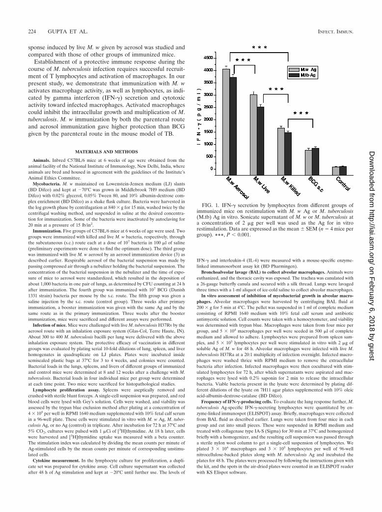
FIG. 1. IFN-� secretion by lymphocytes from different groups ofimmunized mice on restimulation with M. w Ag or M. tuberculosis(M.tb) Ag in vitro. Sonicate supernatant of M. w or M. tuberculosis ata concentration of 2 �g per well was used as the Ag for in vitrorestimulation. Data are expressed as the mean � SEM (n � 4 mice pergroup). ***, P � 0.001.
224 GUPTA ET AL. INFECT. IMMUN.
on February 6, 2018 by guest
http://iai.asm.org/
Dow
nloaded from

Cell-mediated cytotoxicity assay. The CD8� T-cell-mediated cytotoxicity ofdifferent groups of immunized mice toward infected and uninfected syngeneicmacrophages collected from BAL fluid was studied by flow cytometry. Thismethod is considered a promising alternative to the use of radioactive isotopesfor these analyses (9). Briefly, CD8� T cells were isolated from lymphocytes withCD8a antibody conjugated with magnetic nanoparticles (BD Biosciences, Phar-mingen). Target cells were labeled with the fluorescent cell membrane markerPKH-67 (Sigma). Effector (CD8� T cells) and target (M. tuberculosis-infected oruninfected macrophages) cells were cocultured at a 2:1 ratio for 8 h in a six-welltissue culture plate. The nuclear marker 7-aminoactinomycin D (MolecularProbes; Invitrogen) was added to distinguish between live and dead cells. Sam-ples were run in a flow cytometer (BD LSR). A minimum of 10,000 events werecollected and analyzed with WinMDI software.
Statistical analysis. Results are shown as the mean � the standard error of themean (SEM). Comparisons between groups were performed by analysis of vari-ance. P values of �0.05 were considered statistically significant.
RESULTS
Immune response to M. w. As IFN-� is a crucial componentof antimycobacterial immunity, the induction of this cytokineby in vitro restimulation of lymphocytes was assessed. It wasobserved that M. w aerosol immunization induced significantlyhigher IFN-� compared to immunization by the s.c. route. LiveM. w by the s.c. route, in turn, induced a significantly higherIFN-� response compared to killed M. w (Fig. 1). IL-4 secre-tion was also checked. None of the immunized groups showeda detectable amount of IL-4 (data not shown).
Cell-mediated cytotoxicity. It has been shown convincinglythat CD8� T cells have a role in protection against M. tuber-culosis. Hence, cell-mediated cytotoxicity of CD8� T cells iso-lated from different groups of immunized mice were studied inAg-pulsed and unpulsed syngeneic macrophages from BALfluid. The percentage of dead target cells was significantly
higher in M. w aerosol-immunized mice compared to the groupof mice immunized by the s.c. route with live or killed M. w(Fig. 2). The cell-mediated cytotoxicity of M. w in the aerosolgroup and the live M. w s.c. group was significantly higher thanthat in BCG-immunized mice and control mice. These resultsprovide evidence that M. w immunization also activates aCD8� T-cell response.
Intracellular growth inhibition activity of macrophages.Multiple mechanisms are important for inhibition of intracel-lular mycobacteria. To further study the lung-specific immuneresponse, the intracellular growth inhibition activity of alveolarmacrophages was studied. There was an about 80% reductionin the number of viable bacteria in alveolar macrophages col-lected from the M. w aerosol-immunized group compared tothe control group (Fig. 3). The percent reduction in this groupwas significantly higher than in the other three groups. Anabout 55% reduction in intracellular bacteria was observed inthe live M. w s.c. group, which was higher than in the BCGgroup (P � 0.05), where an about 38% reduction was observed.
Cocultures of alveolar macrophages and lymphocytes wereanalyzed for nitric oxide and reactive oxygen, as these areimplicated in the killing of intracellular bacteria. It was ob-served that after 72 h of coculture, reactive oxygen and nitricoxide levels were significantly higher in the M. w aerosol-immunized group than in the control group (data not shown).
Local lung immune response. BAL fluid collected from M. waerosol-immunized mice, as well as control mice, was analyzedfor secretory immunoglobulin A (IgA) antibody. A significantly
FIG. 2. Cell-mediated cytotoxicity of CD8� T cells isolated fromdifferent groups of immunized or unimmunized mice toward M. tuber-culosis Ag-pulsed syngeneic macrophages from BAL fluid. Target cellswere labeled with PKH-67. The nuclear marker 7-aminoactinomycin Dwas added to distinguish between live and dead cells. Cells were takenfrom four mice per group for this study. The results shown here arerepresentative of two similar experiments. *, P � 0.05; ***, P � 0.001.
FIG. 3. Intracellular growth inhibition activity of alveolar macro-phages. Macrophages collected from the BAL fluid of all groups ofimmunized or control mice were infected in vitro with M. tuberculosisH37Ra and cocultured with lymphocytes for 72 h. The number ofviable bacilli present inside macrophages after 72 h were determinedby CFU counting. Cells were taken from four mice per group for thisstudy. The results shown here are representative of two similar exper-iments. ***, P � 0.001.
VOL. 77, 2009 MYCOBACTERIUM w PROTECTION AGAINST PULMONARY TB 225
on February 6, 2018 by guest
http://iai.asm.org/
Dow
nloaded from
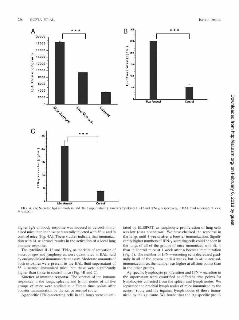
higher IgA antibody response was induced in aerosol-immu-nized mice than in those parenterally injected with M. w and incontrol mice (Fig. 4A). These studies indicate that immuniza-tion with M. w aerosol results in the activation of a local lungimmune response.
The cytokines IL-12 and IFN-�, as markers of activation ofmacrophages and lymphocytes, were quantitated in BAL fluidby enzyme-linked immunosorbent assay. Moderate amounts ofboth cytokines were present in the BAL fluid supernatant ofM. w aerosol-immunized mice, but these were significantlyhigher than those in control mice (Fig. 4B and C).
Kinetics of immune response. The kinetics of the immuneresponses in the lungs, spleens, and lymph nodes of all fivegroups of mice were studied at different time points afterbooster immunization by the s.c. or aerosol route.
Ag-specific IFN-�-secreting cells in the lungs were quanti-
tated by ELISPOT, as lymphocyte proliferation of lung cellswas low (data not shown). We have checked the response inthe lungs until 4 weeks after a booster immunization. Signifi-cantly higher numbers of IFN-�-secreting cells could be seen inthe lungs of all of the groups of mice immunized with M. wthan in control mice at 1 week after a booster immunization(Fig. 5). The number of IFN-�-secreting cells decreased grad-ually in all of the groups until 4 weeks, but in M. w aerosol-immunized mice, the number was higher at all time points thanin the other groups.
Ag-specific lymphocyte proliferation and IFN-� secretion inthe supernatant were quantified at different time points forlymphocytes collected from the spleen and lymph nodes. Weseparated the brachial lymph nodes of mice immunized by theaerosol route and the inguinal lymph nodes of those immu-nized by the s.c. route. We found that the Ag-specific prolif-
FIG. 4. (A) Secreted IgA antibody in BAL fluid supernatant. (B and C) Cytokines IL-12 and IFN-�, respectively, in BAL fluid supernatant. ***,P � 0.001.
226 GUPTA ET AL. INFECT. IMMUN.
on February 6, 2018 by guest
http://iai.asm.org/
Dow
nloaded from
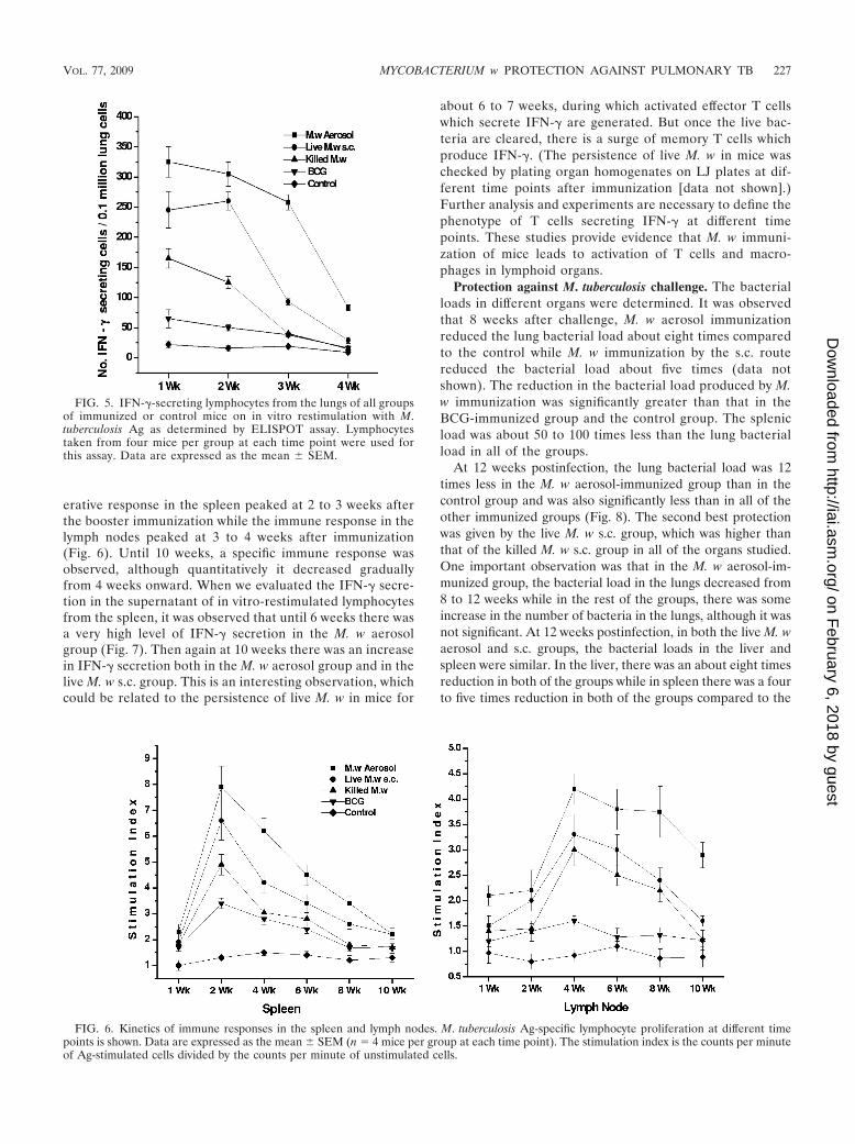
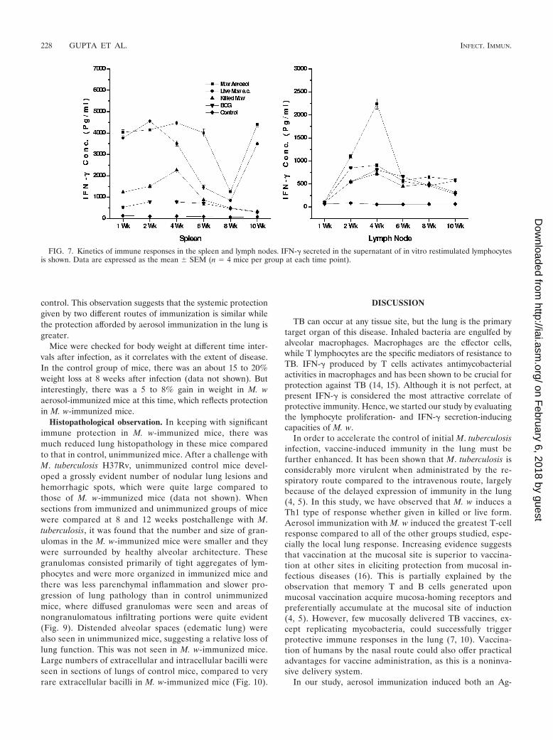
erative response in the spleen peaked at 2 to 3 weeks afterthe booster immunization while the immune response in thelymph nodes peaked at 3 to 4 weeks after immunization(Fig. 6). Until 10 weeks, a specific immune response wasobserved, although quantitatively it decreased graduallyfrom 4 weeks onward. When we evaluated the IFN-� secre-tion in the supernatant of in vitro-restimulated lymphocytesfrom the spleen, it was observed that until 6 weeks there wasa very high level of IFN-� secretion in the M. w aerosolgroup (Fig. 7). Then again at 10 weeks there was an increasein IFN-� secretion both in the M. w aerosol group and in thelive M. w s.c. group. This is an interesting observation, whichcould be related to the persistence of live M. w in mice for
about 6 to 7 weeks, during which activated effector T cellswhich secrete IFN-� are generated. But once the live bac-teria are cleared, there is a surge of memory T cells whichproduce IFN-�. (The persistence of live M. w in mice waschecked by plating organ homogenates on LJ plates at dif-ferent time points after immunization [data not shown].)Further analysis and experiments are necessary to define thephenotype of T cells secreting IFN-� at different timepoints. These studies provide evidence that M. w immuni-zation of mice leads to activation of T cells and macro-phages in lymphoid organs.
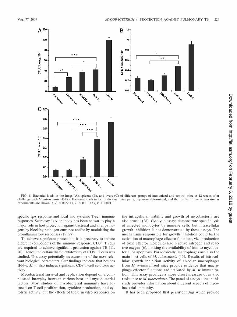
Protection against M. tuberculosis challenge. The bacterialloads in different organs were determined. It was observedthat 8 weeks after challenge, M. w aerosol immunizationreduced the lung bacterial load about eight times comparedto the control while M. w immunization by the s.c. routereduced the bacterial load about five times (data notshown). The reduction in the bacterial load produced by M.w immunization was significantly greater than that in theBCG-immunized group and the control group. The splenicload was about 50 to 100 times less than the lung bacterialload in all of the groups.
At 12 weeks postinfection, the lung bacterial load was 12times less in the M. w aerosol-immunized group than in thecontrol group and was also significantly less than in all of theother immunized groups (Fig. 8). The second best protectionwas given by the live M. w s.c. group, which was higher thanthat of the killed M. w s.c. group in all of the organs studied.One important observation was that in the M. w aerosol-im-munized group, the bacterial load in the lungs decreased from8 to 12 weeks while in the rest of the groups, there was someincrease in the number of bacteria in the lungs, although it wasnot significant. At 12 weeks postinfection, in both the live M. waerosol and s.c. groups, the bacterial loads in the liver andspleen were similar. In the liver, there was an about eight timesreduction in both of the groups while in spleen there was a fourto five times reduction in both of the groups compared to the
FIG. 5. IFN-�-secreting lymphocytes from the lungs of all groupsof immunized or control mice on in vitro restimulation with M.tuberculosis Ag as determined by ELISPOT assay. Lymphocytestaken from four mice per group at each time point were used forthis assay. Data are expressed as the mean � SEM.
FIG. 6. Kinetics of immune responses in the spleen and lymph nodes. M. tuberculosis Ag-specific lymphocyte proliferation at different timepoints is shown. Data are expressed as the mean � SEM (n � 4 mice per group at each time point). The stimulation index is the counts per minuteof Ag-stimulated cells divided by the counts per minute of unstimulated cells.
VOL. 77, 2009 MYCOBACTERIUM w PROTECTION AGAINST PULMONARY TB 227
on February 6, 2018 by guest
http://iai.asm.org/
Dow
nloaded from
control. This observation suggests that the systemic protectiongiven by two different routes of immunization is similar whilethe protection afforded by aerosol immunization in the lung isgreater.
Mice were checked for body weight at different time inter-vals after infection, as it correlates with the extent of disease.In the control group of mice, there was an about 15 to 20%weight loss at 8 weeks after infection (data not shown). Butinterestingly, there was a 5 to 8% gain in weight in M. waerosol-immunized mice at this time, which reflects protectionin M. w-immunized mice.
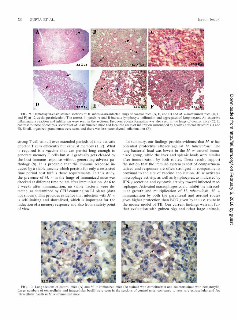
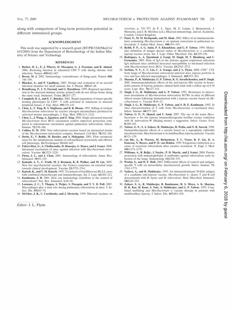
Histopathological observation. In keeping with significantimmune protection in M. w-immunized mice, there wasmuch reduced lung histopathology in these mice comparedto that in control, unimmunized mice. After a challenge withM. tuberculosis H37Rv, unimmunized control mice devel-oped a grossly evident number of nodular lung lesions andhemorrhagic spots, which were quite large compared tothose of M. w-immunized mice (data not shown). Whensections from immunized and unimmunized groups of micewere compared at 8 and 12 weeks postchallenge with M.tuberculosis, it was found that the number and size of gran-ulomas in the M. w-immunized mice were smaller and theywere surrounded by healthy alveolar architecture. Thesegranulomas consisted primarily of tight aggregates of lym-phocytes and were more organized in immunized mice andthere was less parenchymal inflammation and slower pro-gression of lung pathology than in control unimmunizedmice, where diffused granulomas were seen and areas ofnongranulomatous infiltrating portions were quite evident(Fig. 9). Distended alveolar spaces (edematic lung) werealso seen in unimmunized mice, suggesting a relative loss oflung function. This was not seen in M. w-immunized mice.Large numbers of extracellular and intracellular bacilli wereseen in sections of lungs of control mice, compared to veryrare extracellular bacilli in M. w-immunized mice (Fig. 10).
DISCUSSION
TB can occur at any tissue site, but the lung is the primarytarget organ of this disease. Inhaled bacteria are engulfed byalveolar macrophages. Macrophages are the effector cells,while T lymphocytes are the specific mediators of resistance toTB. IFN-� produced by T cells activates antimycobacterialactivities in macrophages and has been shown to be crucial forprotection against TB (14, 15). Although it is not perfect, atpresent IFN-� is considered the most attractive correlate ofprotective immunity. Hence, we started our study by evaluatingthe lymphocyte proliferation- and IFN-� secretion-inducingcapacities of M. w.
In order to accelerate the control of initial M. tuberculosisinfection, vaccine-induced immunity in the lung must befurther enhanced. It has been shown that M. tuberculosis isconsiderably more virulent when administrated by the re-spiratory route compared to the intravenous route, largelybecause of the delayed expression of immunity in the lung(4, 5). In this study, we have observed that M. w induces aTh1 type of response whether given in killed or live form.Aerosol immunization with M. w induced the greatest T-cellresponse compared to all of the other groups studied, espe-cially the local lung response. Increasing evidence suggeststhat vaccination at the mucosal site is superior to vaccina-tion at other sites in eliciting protection from mucosal in-fectious diseases (16). This is partially explained by theobservation that memory T and B cells generated uponmucosal vaccination acquire mucosa-homing receptors andpreferentially accumulate at the mucosal site of induction(4, 5). However, few mucosally delivered TB vaccines, ex-cept replicating mycobacteria, could successfully triggerprotective immune responses in the lung (7, 10). Vaccina-tion of humans by the nasal route could also offer practicaladvantages for vaccine administration, as this is a noninva-sive delivery system.
In our study, aerosol immunization induced both an Ag-
FIG. 7. Kinetics of immune responses in the spleen and lymph nodes. IFN-� secreted in the supernatant of in vitro restimulated lymphocytesis shown. Data are expressed as the mean � SEM (n � 4 mice per group at each time point).
228 GUPTA ET AL. INFECT. IMMUN.
on February 6, 2018 by guest
http://iai.asm.org/
Dow
nloaded from
specific IgA response and local and systemic T-cell immuneresponses. Secretory IgA antibody has been shown to play amajor role in host protection against bacterial and viral patho-gens by blocking pathogen entrance and/or by modulating theproinflammatory responses (19, 27).
To achieve significant protection, it is necessary to inducedifferent components of the immune response. CD8� T cellsare required to achieve significant protection against TB (11,20). Hence, the cell-mediated cytotoxicity of CD8� T cells wasstudied. This assay potentially measures one of the most rele-vant biological parameters. Our findings indicate that besidesIFN-�, M. w also induces significant CD8 T-cell cytotoxic ac-tivity.
Mycobacterial survival and replication depend on a com-plicated interplay between various host and mycobacterialfactors. Most studies of mycobacterial immunity have fo-cused on T-cell proliferation, cytokine production, and cy-tolytic activity, but the effects of these in vitro responses on
the intracellular viability and growth of mycobacteria arealso crucial (28). Cytolytic assays demonstrate specific lysisof infected monocytes by immune cells, but intracellulargrowth inhibition is not demonstrated by these assays. Themechanisms responsible for growth inhibition could be theactivation of macrophage effector functions, viz., productionof toxic effector molecules like reactive nitrogen and reac-tive oxygen (6), limiting the availability of iron to mycobac-teria, or apoptosis. Paradoxically, macrophages are also themain host cells of M. tuberculosis (15). Results of intracel-lular growth inhibition activity of alveolar macrophagesfrom M. w-immunized mice provide evidence that macro-phage effector functions are activated by M. w immuniza-tion. This assay provides a more direct measure of in vivoresistance to M. tuberculosis. The panel of assays done in thisstudy provides information about different aspects of myco-bacterial immunity.
It has been proposed that persistent Ags which provide
FIG. 8. Bacterial loads in the lungs (A), spleens (B), and livers (C) of different groups of immunized and control mice at 12 weeks afterchallenge with M. tuberculosis H37Rv. Bacterial loads in four individual mice per group were determined, and the results of one of two similarexperiments are shown. *, P � 0.05; **, P � 0.01; ***, P � 0.001.
VOL. 77, 2009 MYCOBACTERIUM w PROTECTION AGAINST PULMONARY TB 229
on February 6, 2018 by guest
http://iai.asm.org/
Dow
nloaded from
strong T-cell stimuli over extended periods of time activateeffector T cells efficiently but exhaust memory (1, 2). Whatis required is a vaccine that can persist long enough togenerate memory T cells but still gradually gets cleared bythe host immune response without generating adverse pa-thology (8). It is probable that the immune response in-duced by a viable vaccine which persists for only a restrictedtime period best fulfills these requirements. In this study,the presence of M. w in the lungs of immunized mice waschecked at different time points after immunization. At 6 to7 weeks after immunization, no viable bacteria were de-tected, as determined by CFU counting on LJ plates (datanot shown). This provides evidence that infection with M. wis self-limiting and short-lived, which is important for theinduction of a memory response and also from a safety pointof view.
In summary, our findings provide evidence that M. w haspotential protective efficacy against M. tuberculosis. Thelung bacterial load was lowest in the M. w aerosol-immu-nized group, while the liver and splenic loads were similarafter immunization by both routes. These results supportthe notion that the immune system is sort of compartmen-talized and responses are often strongest in compartmentsproximal to the site of vaccine application. M. w activatesmacrophage activity, as well as lymphocytes, as indicated byIFN-� secretion and cytotoxic activity toward infected mac-rophages. Activated macrophages could inhibit the intracel-lular growth and multiplication of M. tuberculosis. M. wimmunization by both the parenteral and aerosol routesgives higher protection than BCG given by the s.c. route inthe mouse model of TB. Our current findings warrant fur-ther evaluation with guinea pigs and other large animals,
FIG. 9. Hematoxylin-eosin-stained sections of M. tuberculosis-infected lungs of control mice (A, B, and C) and M. w-immunized mice (D, E,and F) at 12 weeks postinfection. The arrows in panels A and B indicate lymphocyte infiltration and aggregates of lymphocytes. An extensiveinflammatory reaction and infiltration were seen in the sections. Frequent edema formation was also seen in the lungs of control mice (C). Incontrast to those of controls, sections of M. w-immunized mice had localized areas of infiltration surrounded by healthy alveolar structure (D andE). Small, organized granulomas were seen, and there was less parenchymal inflammation (F).
FIG. 10. Lung sections of control mice (A) and M. w-immunized mice (B) stained with carbolfuchsin and counterstained with hematoxylin.Large numbers of extracellular and intracellular bacilli were seen in the sections of control mice, compared to very rare extracellular and fewintracellular bacilli in M. w-immunized mice.
230 GUPTA ET AL. INFECT. IMMUN.
on February 6, 2018 by guest
http://iai.asm.org/
Dow
nloaded from
along with comparison of long-term protection potential indifferent immunized groups.
ACKNOWLEDGMENT
This work was supported by a research grant (BT/PR/5340/Med/14/619/2005) from the Department of Biotechnology of the Indian Min-istry of Science and Technology.
REFERENCES
1. Barber, D. L., E. J. Wherry, D. Masopust, G. J. Freeman, and R. Ahmed.2006. Restoring function in exhausted CD8 T cells during chronic viralinfection. Nature 439:682–687.
2. Bevan, M. J. 2002. Immunology: remembrance of things past. Nature 420:748–749.
3. Bhaskar, S., and P. Upadhyay. 2003. Design and evaluation of an aerosolinfection chamber for small animals. Int. J. Pharm. 255:43–48.
4. Brandtzaeg, P., I. N. Farstad, and G. Haraldsen. 1999. Regional specializa-tion in the mucosal immune system: primed cells do not always home alongthe same track. Immunol. Today 20:267–277.
5. Campbell, D. J., and E. C. Butcher. 2002. Rapid acquisition of tissue-specifichoming phenotypes by CD4� T cells activated in cutaneous or mucosallymphoid tissues. J. Exp. Med. 195:135–141.
6. Chan, J., Y. Xing, R. S. Majliozzo, and B. R. Bloom. 1992. Killing of virulentMycobacterium tuberculosis by reactive nitrogen intermediates produced byactivated murine macrophages. J. Exp. Med. 175:1111–1122.
7. Chen, L., J. Wang, A. Zganiacz, and Z. Xing. 2004. Single intranasal mucosalMycobacterium bovis BCG vaccination confers improved protection com-pared to subcutaneous vaccination against pulmonary tuberculosis. Infect.Immun. 72:238–246.
8. Collins, D. M. 2000. New tuberculosis vaccines based on attenuated strainsof the Mycobacterium tuberculosis complex. Immunol. Cell Biol. 78:342–348.
9. Derby, E., V. Reddy, M. Baseler, and A. Malyguine. 2001. Flow cytometricassay for the simultaneous analysis of cell-mediated cytotoxicity and effectorcell phenotype. BioTechniques 31:660–665.
10. Falero-Diaz, G., S. Challacombe, D. Banerjee, G. Douce, and J. Ivanyi. 2000.Intranasal vaccination of mice against infection with Mycobacterium tuber-culosis. Vaccine 18:3223–3229.
11. Flynn, J. L., and J. Chan. 2001. Immunology of tuberculosis. Annu. Rev.Immunol. 19:93–129.
12. Kamath, A. T., U. Fruth, M. J. Brennan, K. B. Walker, and M. Liu. 2005.New live mycobacterial vaccines: the Geneva consenses on essential stepstowards clinical development. Vaccine 23:3753–3761.
13. Katoch, K., and V. M. Katoch. 1995. Treatment of bacilliferrous BL/LL caseswith combined chemotherapy and immunotherapy. Int. J. Lepr. 63:202–212.
14. Kaufmann, S. H. 2001. How can immunology contribute to the control oftuberculosis? Nat. Rev. Immunol. 1:20–30.
15. Leemans, J. C., T. Thepen, S. Weijer, S. Florquin, and T. V. D. Poll. 2005.Macrophages play a dual role during pulmonary tuberculosis in mice. J. In-fect. Dis. 191:65–74.
16. McGhee, J. R., C. Czerkinsky, and J. Mestecky. 1999. Mucosal vaccines: an
overview, p. 741–757. In P. L. Ogra, M. E. Lamm, J. Bienenstock, J.Mestecky, and J. R. McGhee (ed.), Mucosal immunology, 2nd ed. Academic,London, United Kingdom.
17. Patel, N., M. M. Deshpande, and M. Shah. 2002. Effect of an immunomodu-lator containing Mycobacterium w on sputum conversion in pulmonary tu-berculosis. J. Indian Med. Assoc. 100:191–193.
18. Reddi, P. P., A. G. Amin, P. S. Khandekar, and G. P. Talwar. 1994. Molec-ular definition of unique species status of Mycobacterium w; a candidateleprosy vaccine strain. Int. J. Lepr. Other Mycobact. Dis. 62:229–236.
19. Rodríguez, A., A. Tjarnlund, J. Ivanji, M. Singh, M. T. Blomberg, and C.Fernandez. 2005. Role of IgA in the defense against respiratory infectionsIgA deficient mice exhibited increased susceptibility to intranasal infectionwith Mycobacterium bovis BCG. Vaccine 23:2565–2572.
20. Serbina, N. V., C. C. Liu, C. A. Scanga, and J. L. Flynn. 2000. CD8� CTLfrom lungs of Mycobacterium tuberculosis-infected mice express perforin invivo and lyse infected macrophages. J. Immunol. 165:353–363.
21. Sharma, P., R. Mukherjee, G. P. Talwar, K. G. Sarathchandra, and P. Singh.2005. Immunoprophylactic effects of the anti-leprosy Mw vaccine in house-hold contacts of leprosy patients: clinical field trials with a follow up of 8-10years. Lepr. Rev. 76:127–143.
22. Singh, I. G., R. Mukherjee, and G. P. Talwar. 1991. Resistance to intrave-nous inoculation of Mycobacterium tuberculosis H37Rv in mice of differentinbred strains following immunization with a leprosy vaccine based on My-cobacterium w. Vaccine 9:10–13.
23. Singh, I. G., R. Mukherjee, G. P. Talwar, and S. H. E. Kaufmann. 1992. Invitro characterization of T cells from Mycobacterium w-vaccinated mice.Infect. Immun. 60:257–263.
24. Talwar, G. P., N. Ahmed, and V. Saini. 2007. The use of the name Myco-bacterium w for the leprosy immunotherapeutic bacillus creates confusionwith M. tuberculosis-W (Beijing strain): a suggestion. Infect. Genet. Evol.8:100–101.
25. Talwar, G. P., S. A. Zaheer, R. Mukherjee, R. Walia, and N. R. Suresh. 1990.Immunotherapeutic effects of a vaccine based on a saprophytic cultivablemycobacterium, Mycobacterium w in multibacillary leprosy patients. Vaccine8:121–129.
26. van Rie, A., R. Warren, M. Richardson, T. C. Victor, R. P. Gie, D. A.Enarson, N. Beyers, and P. D. van Helden. 1999. Exogenous reinfection as acause of recurrent tuberculosis after curative treatment. N. Engl. J. Med.341:1174–1179.
27. Williams, A., R. Reljic., I. Naylor., P. D. Marsh., and J. Ivanyi. 2004. Passiveprotection with immunoglobulin A antibodies against tuberculous early in-fection of the lungs. Immunology 111:328–333.
28. Worku, S., and D. F. Hoft. 2003. Differential effects of control and antigen-specific T cells on intracellular mycobacterial growth. Infect. Immun. 71:1763–1773.
29. Yadava, A., and R. Mukherjee. 1993. An immunodominant 30-kDa antigenof a candidate anti-leprosy vaccine, Mycobacterium w, shares T and B celldeterminants with M. leprae and M. tuberculosis. Med. Microbiol. Immunol.182:243–253.
30. Zaheer, S. A., R. Mukherjee, B. Ramkumar, R. S. Misra, A. K. Sharma,H. K. Kar, H. Kaur, S. Nair, A. Mukherjee, and G. P. Talwar. 1993. Com-bined multidrug and Mycobacterium w vaccine therapy in patients withmultibacillary leprosy. J. Infect. Dis. 167:401–410.
Editor: J. L. Flynn
VOL. 77, 2009 MYCOBACTERIUM w PROTECTION AGAINST PULMONARY TB 231
on February 6, 2018 by guest
http://iai.asm.org/
Dow
nloaded from