Embed Size (px)
Citation preview

of October 21, 2018.This information is current as
Arginase IIMediated by Induction of Macrophage
IsHelicobacter pyloriImmune Evasion by
P. Gobert, Rupesh Chaturvedi and Keith T. WilsonAlainThibaut de Sablet, Kshipra Singh, M. Blanca Piazuelo,
Nuruddeen D. Lewis, Mohammad Asim, Daniel P. Barry,
http://www.jimmunol.org/content/186/6/3632doi: 10.4049/jimmunol.1003431February 2011;
2011; 186:3632-3641; Prepublished online 4J Immunol
MaterialSupplementary
1.DC1http://www.jimmunol.org/content/suppl/2011/02/04/jimmunol.100343
Referenceshttp://www.jimmunol.org/content/186/6/3632.full#ref-list-1
, 27 of which you can access for free at: cites 55 articlesThis article
average*
4 weeks from acceptance to publicationFast Publication! •
Every submission reviewed by practicing scientistsNo Triage! •
from submission to initial decisionRapid Reviews! 30 days* •
Submit online. ?The JIWhy
Subscriptionhttp://jimmunol.org/subscription
is online at: The Journal of ImmunologyInformation about subscribing to
Permissionshttp://www.aai.org/About/Publications/JI/copyright.htmlSubmit copyright permission requests at:
Email Alertshttp://jimmunol.org/alertsReceive free email-alerts when new articles cite this article. Sign up at:
Print ISSN: 0022-1767 Online ISSN: 1550-6606. All rights reserved.1451 Rockville Pike, Suite 650, Rockville, MD 20852The American Association of Immunologists, Inc.,
is published twice each month byThe Journal of Immunology
by guest on October 21, 2018
http://ww
w.jim
munol.org/
Dow
nloaded from
by guest on October 21, 2018
http://ww
w.jim
munol.org/
Dow
nloaded from

The Journal of Immunology
Immune Evasion by Helicobacter pylori Is Mediated byInduction of Macrophage Arginase II
Nuruddeen D. Lewis,*,† Mohammad Asim,*,‡ Daniel P. Barry,*,‡ Thibaut de Sablet,*,‡
Kshipra Singh,*,‡ M. Blanca Piazuelo,* Alain P. Gobert,*,‡,x Rupesh Chaturvedi,*,‡
and Keith T. Wilson*,†,‡
Helicobacter pylori infection persists for the life of the host due to the failure of the immune response to eradicate the
bacterium. Determining how H. pylori escapes the immune response in its gastric niche is clinically important. We have
demonstrated in vitro that macrophage NO production can kill H. pylori, but induction of macrophage arginase II (Arg2)
inhibits inducible NO synthase (iNOS) translation, causes apoptosis, and restricts bacterial killing. Using a chronic H. pylori
infection model, we determined whether Arg2 impairs host defense in vivo. In C57BL/6 mice, expression of Arg2, but not
arginase I, was abundant and localized to gastric macrophages. Arg22/2 mice had increased histologic gastritis and decreased
bacterial colonization compared with wild-type (WT) mice. Increased gastritis scores correlated with decreased colonization in
individual Arg22/2 mice but not in WT mice. When mice infected with H. pylori were compared, Arg22/2 mice had more
gastric macrophages, more of these cells were iNOS+, and these cells expressed higher levels of iNOS protein, as determined by
flow cytometry and immunofluorescence microscopy. There was enhanced nitrotyrosine staining in infected Arg22/2 versus
WT mice, indicating increased NO generation. Infected Arg22/2 mice exhibited decreased macrophage apoptosis, as well as
enhanced IFN-g, IL-17a, and IL-12p40 expression, and reduced IL-10 levels consistent with a more vigorous Th1/Th17 re-
sponse. These studies demonstrate that Arg2 contributes to the immune evasion of H. pylori by limiting macrophage iNOS
protein expression and NO production, mediating macrophage apoptosis, and restraining proinflammatory cytokine
responses. The Journal of Immunology, 2011, 186: 3632–3641.
Helicobacter pylori is a Gram-negative, microaerophilicbacterium that selectively colonizes the human stomach.All infected individuals exhibit chronic active gastritis,
and a substantial proportion of subjects develop peptic ulcer dis-ease or gastric adenocarcinoma (1). H. pylori infects ∼50% of theworld’s population, and, more importantly, the associated gastriccancer is the second leading cause of cancer-related deathworldwide (2). The infection is usually acquired in childhood andpersists for the life of the host despite eliciting a seemingly vig-
orous immune response (3). Understanding the mechanisms bywhich H. pylori avoids being eliminated by the immune system isclinically relevant because antibiotic-based eradication regimensare expensive and not always effective, with success rates that canbe ,50% in some regions of the world (4).Although H. pylori is typically considered to be noninvasive
because most of the bacteria reside in the mucous layer of thestomach in contact with the epithelium, studies have demonstratedthat the bacterium and its products can be in direct contact withlamina propria immune cells (5–7). Consequently, infection withH. pylori results in a large influx of immune cells that includeneutrophils, macrophages, dendritic cells, and lymphocytes, andan associated innate and adaptive immune response (8). Althoughthis has been shown to include both Th1 and Th17 components,one hallmark of the response is that there is also a downregulationof effective immunity that appears to involve recruitment of reg-ulatory T cells (Tregs) and B cells (3). Vaccination studies,adoptive transfer of Th1-selected lymphocytes, and efforts tosuppress Treg responses have been successful at reducing orclearing the infection in mice (9–12). These studies have providedevidence that the cellular immune response is not vigorous enoughto lead to eradication of the infection. One important aspect thatremains to be fully elucidated is the role of the innate immuneresponse in the impairment of the host response. We propose thatthere may be an inability of effector cells to eliminate the infectionwhen given the opportunity to do so.One primordial mechanism for antimicrobial host defense is the
generation of high levels of NO derived from the enzyme inducibleNO synthase (iNOS) (13). Our laboratory and others have dem-onstrated that H. pylori induces the expression and activity ofiNOS in macrophages both in vivo and in vitro (14–17). Further,
*Division of Gastroenterology, Department of Medicine, Vanderbilt University Med-ical Center, Nashville, TN 37232; †Department of Cancer Biology, Vanderbilt Uni-versity Medical Center, Nashville, TN 37232; ‡Veterans Affairs Tennessee ValleyHealthcare System, Nashville, TN 37212; and xInstitut National de la RechercheAgronomique, Unite de Microbiologie UR454, Saint-Genes-Champanelle, France63122
Received for publication October 15, 2010. Accepted for publication January 10,2011.
This work was supported by grants from the Office of Medical Research, Departmentof Veterans Affairs (to K.T.W.) and by National Institutes of Health GrantsR01DK053620, R01AT004821, P01CA028842, and P01CA116087 (to K.T.W.).N.D.L. was supported by T32CA009592 from the National Cancer Institute andF31GM083500 from the National Institute of General Medical Sciences. Flow Cy-tometry Core Services were performed through the Vanderbilt University MedicalCenter Digestive Disease Research Center supported by National Institutes of HealthGrant P30DK058404. A.P.G. and T.S. were supported in part by the Philippe Foun-dation.
Address correspondence and reprint requests to Dr. Keith T. Wilson, Division ofGastroenterology, Department of Medicine, Vanderbilt University School of Medi-cine, 1030C MRB IV, 2215B Garland Avenue, Nashville, TN 37232. E-mail address:[email protected]
The online version of this article contains supplemental material.
Abbreviations used in this article: 7-AAD, 7-aminoactinomycin D; Arg1, arginase I;Arg2, arginase II; iNOS, inducible NO synthase; Treg, regulatory T cell; WT, wild-type.
www.jimmunol.org/cgi/doi/10.4049/jimmunol.1003431
by guest on October 21, 2018
http://ww
w.jim
munol.org/
Dow
nloaded from

we have reported that macrophages cocultured with H. pylori cankill the bacterium by an NO-dependent mechanism (15, 18).However, this killing is incomplete in vitro, and, moreover, thereis clearly a failure of this mechanism in vivo despite the expres-sion of iNOS in the infected mucosa (14, 17). This reasoning hasled our laboratory to consider the possibility that iNOS-mediatedhost defense to H. pylori is suboptimal. We have reported that thegeneration of NO by macrophages in response to H. pylori isentirely dependent on the availability of its substrate, L-arginine,which enhances expression of iNOS protein, without altering theinduction of mRNA expression (15, 18). Specifically, we foundthat addition of increasing levels of extracellular L-arginine resultsin a proportionate increase in NO production even at concen-trations well above the circulating levels in humans and mice of0.1 mM and above the Km of the iNOS enzyme for L-arginine,which is in the range of 10 mM (19). In order for macrophages toproduce bactericidal amounts of NO when cocultured with H.pylori in our model system, concentrations of L-arginine in themedium that exceeded 0.1 mM were needed. Importantly, in-fection of iNOS2/2 mice with H. pylori results in similar levels ofbacterial colonization as those in wild-type (WT) mice (20), fur-ther suggesting a defect in iNOS-dependent host defense.Arginase enzymes are the endogenous antagonists to iNOS be-
cause they compete for the same L-arginine substrate by metabo-lizing it to urea and L-ornithine (21). The latter is used by ornithinedecarboxylase to produce the polyamines putrescine, spermidine,and spermine. There are two isoforms of arginase: arginase I (Arg1)is abundant in liver and is important for the urea cycle, and arginaseII (Arg2) is abundant in kidney and localizes to mitochondria (22).We have reported that Arg2, but not Arg1, is upregulated in H.pylori-stimulated macrophages (15). Induction of arginase activityby pathogens has been reported to modulate macrophage NOproduction in the case of both Arg1 and Arg2, which can restricteffective immunity (23–25). These effects have been attributed tosubstrate competition. However, we have recently reported thatinduction of macrophage Arg2 by H. pylori inhibits iNOS trans-lation, NO production, and bacterial killing in vitro (15). Further-more, in mice infected with H. pylori for 48 h, treatment with anarginase inhibitor resulted in increased iNOS protein levels andNO production in gastric macrophages (15). We have also reportedthat the downstream effects of arginase metabolism can be detri-mental to iNOS-mediated host defense against H. pylori. The poly-amine spermine inhibits L-arginine uptake in macrophages, there-by blocking NO production and bacterial killing (26). Moreover,back-conversion of spermine into spermidine by the enzyme sper-mine oxidase, which is also induced byH. pylori, releases hydrogenperoxide, which causes apoptosis (27, 28). Consequently, we havefound that inhibition of arginase blocks H. pylori-induced macro-phage apoptosis in our in vitro studies (29).We hypothesized that Arg2 expression is detrimental to host
defense against H. pylori in vivo by restricting macrophage NOproduction and inducing macrophage apoptosis. We now reportthat chronic infection of Arg22/2 mice with H. pylori results indecreased bacterial colonization and increased gastritis comparedwith that in infected WT mice. Importantly, we found that in-creased gastritis correlated with decreased bacterial colonizationin individual Arg22/2 mice but not in WT mice. Moreover, weshow that in Arg22/2 mice infected with H. pylori, there are moreiNOS+ gastric macrophages that express increased levels of iNOS,enhanced proinflammatory cytokine responses, and decreasedlevels of macrophage apoptosis. Our data suggest that upregula-tion of macrophage Arg2 is detrimental to the host responseagainst H. pylori and demonstrate a mechanism by which H. py-lori evades host immunity.
Materials and MethodsReagents
All reagents used for RNA extraction were from Invitrogen (Carlsbad, CA).Real-time PCR reagents were from Bio-Rad (Hercules, CA). Isolation ofDNA was performed using the DNeasy Blood & Tissue kit (Qiagen,Valencia, CA). All other chemicals were from Sigma-Aldrich (St. Louis,MO).
Bacteria
H. pylori SS1, a mouse-adapted human strain, was grown on Brucellablood agar plates under microaerobic conditions as described (16, 28).Prior to infection, bacteria were grown in a Brucella broth culture for 16–20 h. Concentrations of bacteria were determined by OD at 600 nm (OD of1 = 109 CFU/ml) (16).
Mice
All animal experiments were approved by the Institutional Animal Careand Use Committee at Vanderbilt University (Nashville, TN). C57BL/6Arg22/2 and WT male mice were used at 6–8 wk of age as described(15). Mice were gavaged with 5 3 108 H. pylori SS1 in 100 ml Brucellabroth or with broth vehicle control alone. Some mice were sacrificed at2 d postinoculation, and stomachs were excised and used for isolation ofgastric macrophages, which was performed as described (14, 18). Othermice were sacrificed at 4 mo postinoculation, and colonization and gastritiswere assessed. H. pylori levels were measured by quantitative DNA PCRfor H. pylori ureB, standardized to mouse 18S rRNA copy number (14).Histologic gastritis was quantified by a pathologist (M.B.P.) using a scaleof 0–3 for acute inflammation and for chronic inflammation in the regionsof the gastric antrum and body with the scores added together for a totalscore of 0–12 (14, 30).
Apoptosis detection by annexin V staining
Gastric macrophages were isolated and stained with annexin V conjugatedto FITC and 7-aminoactinomycin D (7-AAD; BD Biosciences, San Jose,CA) as described (31, 32).
Real-time PCR
RNAwas extracted using the RNeasy Mini Kit from Qiagen. PCR methodswere performed as described (15, 18, 29, 33). Primer sequences used wereas follows: IL-10, 59-CCAAGCCTTATCGGAAATGA-39 (forward) and59-TCACTCTTCACCTGCTCCAC-39 (reverse); IL-12p40, 59-GATTCAG-ACTCCAGGGGACA-39 (forward) and 59-CATCTTCTTCAGGCGTGT-CA-39 (reverse); IL-17a, 59-GCTCCAGAAGGCCCTCAGA-39 (forward)and 59-CTTTCCCTCCGCATTGACA-39 (reverse); Foxp3, 59-AGAGC-CCTCACAACCAGCTA-39 (forward) and 59-CCAGATGTTGTGGGTG-AGTG-39 (reverse); IL-6, 59-AGTTGCCTTCTTGGGACTGA-39 (forward)and 59-TCCACGATTTCCCAGAGAAC-39 (reverse); and IL-23p19, 59-CA-TGGGGCTATCAGGGAGTA-39 (forward) and 59-AATAATGTGCCCCG-TATCCA-39 (reverse). Primer sequences for IFN-g (33), Arg1 (29), Arg2(15), b-actin (18), and iNOS (18) were as described.
Immunofluorescence staining for F4/80, Arg2, and iNOS, andimmunoperoxidase staining for nitrotyrosine and cleavedcaspase-3
Immunofluorescence staining for F4/80 and DAPI was performed as de-scribed (14). iNOS was detected with a rabbit polyclonal Ab (1:100 di-lution; BD Biosciences, San Diego, CA). Arg2 was detected with a goatpolyclonal Ab (1:200 dilution; Santa Cruz Biotechnology, Santa Cruz,CA). Immunoperoxidase staining was performed as described (14).Nitrotyrosine was detected with a mouse monoclonal anti-vertebratenitrotyrosine Ab (1:100 dilution; Millipore, Billerica, MA). Cleavedcaspase-3 was detected with a rabbit polyclonal Ab (1:200 dilution; CellSignaling Technology, Danvers, MA). Quantification of cleaved caspase-3staining was performed by counting all positively-stained cells among theinflammatory infiltrate and dividing this number by the total number ofinflammatory cells to calculate the percentage of positive cells. Cellcounting was performed at 3600 magnification under oil immersion.
Gastric macrophages
Immune cells were isolated from the glandular stomach by enzymaticdigestion as described (14, 18, 31). Briefly, mice were sacrificed, and thestomach was removed. The forestomach (nonglandular portion) was ex-cised and discarded. The glandular portion of the stomach was washed, cut
The Journal of Immunology 3633
by guest on October 21, 2018
http://ww
w.jim
munol.org/
Dow
nloaded from

into 2-mm pieces, and digested for 20 min with 1 mg/ml dispase, 0.25mg/ml collagenase A, and 25 U/ml DNase (Roche Diagnostics, Indian-apolis, IN) at 37˚C while shaking. The suspension was passed through a70-mm cell strainer (BD Biosciences, San Diego, CA) and cells harvestedby centrifugation. Cells were stained for F4/80 and iNOS and analyzed byflow cytometry. F4/80+ and F4/80+ iNOS+ cell counts were standardized tothe weight in grams of the glandular stomach.
Statistical analysis
Quantitative data are shown as the mean 6 SE. Statistical analysis wasperformed with Prism version 5.0c (GraphPad Software, San Diego, CA).Where two groups were compared, Student t test was used. Data with morethan two groups were analyzed by ANOVA and the Student–Newman–Keuls post hoc multiple comparisons test. A p value,0.05 was consideredstatistically significant. The relationship between gastritis and colonizationwas determined using Spearman’s correlation test; the correlation co-efficient, r, is shown along with the p value.
ResultsH. pylori infection induces Arg2, not Arg1, in gastric laminapropria F4/80+ macrophages
Mice were infected for 4 mo, a time point at which we havedemonstrated consistent development of chronic active gastritis
with strain SS1 (14). Arg2 mRNA expression was increased by.9–fold in H. pylori-infected WT mice when assessed by real-time PCR, whereas Arg1 was not induced (Fig. 1A, 1B). Fur-thermore, Arg1 was not upregulated in Arg22/2 mice (Fig. 1B).Additionally, iNOS mRNA expression increased in infected WTmice, and this was further enhanced in Arg22/2 mice (Fig. 1C).To determine which cell type expressed Arg2 in the stomach, we
immunostained formalin-fixed, paraffin-embedded gastric tissuesections from WT and Arg22/2 mice (Fig. 1D). This stainingdemonstrated increased Arg2 levels in H. pylori-infected tissuesfrom WT mice that localized to F4/80+ macrophages. Arg2 stain-ing was not present in macrophages in the Arg22/2 tissues.
Chronically infected Arg22/2 mice have increased gastritisand decreased H. pylori colonization
To determine the role of Arg2 during H. pylori infection, weinfected WT and Arg22/2 mice for 4 mo and assessed gastritislevels and bacterial colonization. With H. pylori infection, therewas a significant increase in overall gastritis in WT mice that wasfurther increased in the Arg22/2 mice (Fig. 2A). In addition toenhanced gastritis levels, we found a significant decrease in H.
FIGURE 1. H. pylori infection induces iNOS and Arg2, but not Arg1, in gastric tissues. C57BL/6 WT and Arg22/2 mice were infected with H. pylori
strain SS1. At 4 mo postinfection, mice were sacrificed, gastric tissues were removed, and RNA was extracted. A–C, Arg2 (A), Arg1 (B), and iNOS (C)
mRNA levels were measured by real-time PCR from the antral portion of the stomach. All real-time PCR data were standardized to b-actin and presented as
fold increase versus uninfected WT mice. Data are the mean 6 SEM, n = 6 in the uninfected mice and n = 10 in each of the infected mice groups. ***p ,0.001 compared with WT uninfected mice. xp , 0.05, xxxp , 0.001 compared with WT infected mice. D, Photomicrographs, displayed at original
magnifications 3200 and 3600 for the insets, demonstrating Arg2 immunofluorescence staining in F4/80+ macrophages. F4/80 was detected with tet-
ramethyl rhodamine isothiocyanate (red), and Arg2 was detected with FITC. Data are representative of three to four mice per group.
3634 ARG2 INHIBITS MACROPHAGE FUNCTION IN H. PYLORI-INFECTED MICE
by guest on October 21, 2018
http://ww
w.jim
munol.org/
Dow
nloaded from

pylori colonization levels in Arg22/2 mice (Fig. 2B). Notably,increased gastritis correlated significantly with decreased bacterialcolonization in Arg22/2 mice (p = 0.006, r = –0.491), but therewas no such effect in WT mice (p = 0.636, r = –0.088).Representative photomicrographs of H&E-stained gastric sec-
tions are shown for uninfected and infected WTand Arg22/2 mice(Fig. 2D). These demonstrate that in the transition zone betweenthe body and the antrum of the stomach where H. pylori-inducedinflammation is typically most severe, there was more exten-sive acute and chronic inflammatory cell infiltration in Arg22/2
mice. There was no spontaneous inflammation in the uninfectedArg22/2 mice. Additionally, we observed gross thickening of thegastric mucosa in the transition zone of Arg22/2 mice that wasindicative of increased gastric inflammation (Fig. 2E).
Chronic infection of H. pylori induces proinflammatorycytokine production that is further enhanced in Arg22/2 mice
To assess alterations in cytokine production during H. pylori in-fection and determine differences in the immune response, weanalyzed mRNA expression of various cytokines in uninfected and
infected WT and Arg22/2 mice. IFN-g, IL-12p40, and IL-17awere increased in infected WT mice and were further upregulat-ed in Arg22/2 mice (Fig. 3A, 3B, 3C, respectively). IL-10 ex-pression, a hallmark of the counterregulatory response to Th1 andTh17 responses in H. pylori infection (11, 34), was increased ininfected WT but not in Arg22/2 mice (Fig. 3D). IL-23p19, IL-6,and Foxp3 were each modestly increased upon infection with H.pylori, but there was no difference between the WT and Arg22/2
mice (Supplemental Fig. 1).
Arg22/2 macrophages are more abundant, express more iNOS,and have increased nitrotyrosine staining compared with WTmacrophages during H. pylori infection
We have previously demonstrated that H. pylori stimulationinduces Arg2 expression in macrophages, and this expressionattenuates iNOS translation in vitro (15). Furthermore, we haveshown that Arg22/2 gastric macrophages isolated 48 h post-inoculation with H. pylori express more iNOS protein and producemore NO compared with WT macrophages (15). Therefore, todetermine if such an effect occurred during chronic infection with
FIGURE 2. Increased gastritis and decreased bacterial colonization in Arg22/2 mice. C57BL/6 WT and Arg22/2 mice were infected with H. pylori for
4 mo, sacrificed, and their stomachs were removed. A, H&E-stained slides were prepared from a strip of the glandular stomach containing both the antrum
and body. Acute and chronic inflammation were each scored 0–3 in the antrum and body, and the scores were added for a total score of 0–12. **p, 0.01. B,
DNAwas extracted from the body of the stomach, and bacterial colonization was quantified by real-time PCR for the H. pylori gene ureB normalized to 18S
rRNA. **p , 0.01. C, Gastritis scores and bacterial colonization were plotted for individual WT and Arg22/2 mice to determine if a correlation exists
between gastritis and bacterial colonization. Linear regression lines are shown. For WT mice, Spearman’s correlation coefficient r = –0.088 and p = 0.636.
For Arg22/2 mice, Spearman’s correlation coefficient r = –0.491 and p = 0.006. D, Representative H&E-stained sections are shown for both uninfected and
infected WT and Arg22/2 mice. Photomicrographs are depicted at original magnification3200 (original magnification3600 for the inset). E, Photographs
were taken to demonstrate the gross anatomy of the glandular portion of the stomach. The red rectangle highlights the transition zone (TZ) of the stomach.
The Journal of Immunology 3635
by guest on October 21, 2018
http://ww
w.jim
munol.org/
Dow
nloaded from

H. pylori, we analyzed levels of the macrophage surface markerF4/80 and iNOS protein expression by immunofluorescence inWT and Arg22/2 gastric tissues 4 mo postinoculation (Fig. 4),along with the appropriate isotype controls (Supplemental Fig. 2).Consistent with our recent report (14), there was increased mac-rophage staining in H. pylori-infected versus uninfected tissues inWT mice. The abundance of this F4/80 staining was significantlyincreased in the infected Arg22/2 mice. Similarly, with H. pyloriinfection, iNOS staining was increased in WT mice but sub-stantially potentiated in the Arg22/2 mice (Fig. 4). When themerged images were assessed, the iNOS staining was found tolocalize predominately to the F4/80+ cells, and there were moreiNOS+ macrophages in the Arg22/2 mice (Fig. 4). This stainingwas present in the lamina propria, with trails of iNOS+ macro-phages migrating toward the lumen, as well as in the submucosalregion.To confirm our observation that there are more iNOS+ macro-
phages in infected Arg22/2 mice compared with that in infectedWT mice, we isolated gastric immune cells and analyzed F4/80and iNOS expression by flow cytometry. In accordance with ourimmunofluorescence data, we found a significant increase in boththe quantity of macrophages (Fig. 5A) and iNOS+ macrophages(Fig. 5B) in infected Arg22/2 mice compared with that in WTmice. Representative flow cytometric dot plots are also showndemonstrating an increased percentage of F4/80+ iNOS+ cells ininfected Arg22/2 mice versus WT mice (Fig. 5C). Importantly, theArg22/2 gastric macrophages expressed more iNOS protein thanthat expressed by the cells from the WT mice (Fig. 5D, 5E).Because we found that gastric macrophages isolated from H.
pylori-infected Arg22/2 mice express higher iNOS protein levelsthan those of gastric macrophages from infected WT mice, wesought to determine if this resulted in increased NO production.To assess this, we used nitrotyrosine staining as a marker of NO
synthesis in the mucosa. Nitrotyrosine formation occurs whentyrosine residues react with peroxynitrite, which is formed by thereaction of NO with superoxide (O2
–), and this has been used asa marker of the production of reactive nitrogen species (23, 35–37). We stained tissues from chronically infected WTand Arg22/2
mice and found that inflammatory cells in Arg22/2 mice had in-creased nitrotyrosine staining compared with that in WT mice(Fig. 5F). This staining was most intense in mononuclear immunecells of the lamina propria near the luminal surface, similarin location to the macrophages that we identified in Fig. 1D andFig. 4.
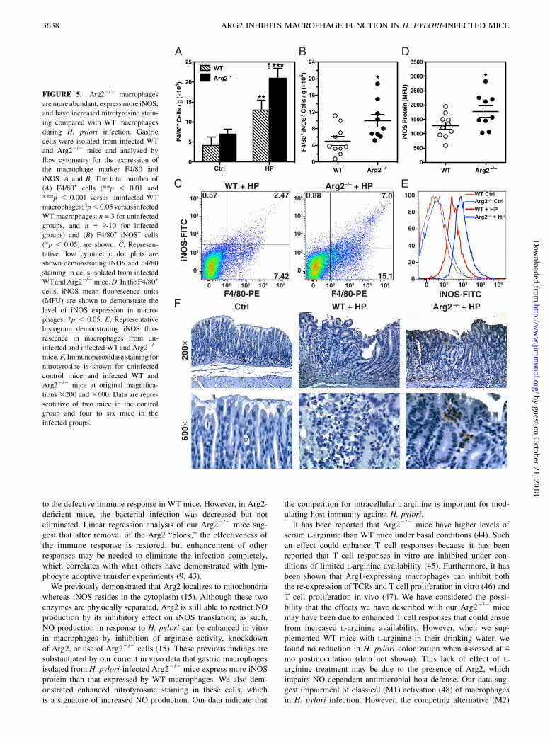
H. pylori infection increases macrophage apoptosis that isabolished in Arg22/2 mice
To determine whether increased survival of macrophages couldbe a mechanism responsible for increased iNOS+ macrophagesin Arg22/2 mice, we isolated gastric macrophages and measuredapoptosis by annexin V staining. We have previously shown thatH. pylori induces macrophage apoptosis in vitro that is Arg2 de-pendent (29), therefore we sought to confirm this in vivo. Topursue this, we used a 48-h model of infection, as we have pre-viously demonstrated that there is both maximum macrophageinfiltration into the stomach and maximum apoptosis at this timepoint after inoculation with H. pylori (31). Additionally, we havedemonstrated that Arg2 restricts NO production in gastric mac-rophages at this time point (15). Consistent with our in vitro data,we found that H. pylori infection induced macrophage apoptosisand that this was abolished in Arg22/2 macrophages (Fig. 6A).Representative flow cytometric dot plots demonstrating annexin Vand 7-AAD staining in infected WT and Arg22/2 macrophagesare shown in Fig. 6B.We also sought to corroborate this finding in our chronic in-
fection model by immunostaining tissues for cleaved caspase-3,a marker for apoptosis in H. pylori gastritis tissues (38). Ininfected WT mice, there was abundant staining in the mono-nuclear inflammatory cells with marked staining of cells withapoptotic bodies (Fig. 6C). In contrast, cleaved caspase-3 stainingin Arg22/2 mice was less intense and less frequent, which cor-relates with our findings with the annexin V staining of isolatedgastric macrophages in Fig. 6A. Additionally, quantification of thisstaining revealed a decrease in cleaved caspase-3 staining amongthe inflammatory cells in the Arg22/2 mice compared with that inWT mice (Fig. 6D).
DiscussionH. pylori infection induces a vigorous immune response, and inthe murine model there is a rapid influx of macrophages (31, 39)and neutrophils (39) 48 h postinfection, followed by infiltrationof lymphocytes 10 d postinfection (39). This produces a smolder-ing gastritis that persists as long as the bacteria reside in thegastric niche. Despite this robust immune response, the bacteriumtypically persists for the life of the host. It is generally assumedthat the immune response is not vigorous enough to eliminate theinfection, due to demonstration of clearance of the bacterium inadoptive transfer experiments and IL-102/2 mice, both of whichexhibit enhanced gastric inflammation (9, 34). We sought to de-termine if the macrophage response is inhibited by H. pylori. Inthe current report, we demonstrate that H. pylori upregulatesmacrophage Arg2, thereby restricting iNOS protein levels and NOproduction and enhancing macrophage apoptosis. Consequently,this restricts host defense against H. pylori. This is the first report,to our knowledge, to demonstrate that macrophage Arg2 expres-sion has a deleterious impact on the effectiveness of host immu-nity by impairing the inflammatory response in vivo.
FIGURE 3. Chronic infection with H. pylori induces proinflammatory
cytokine production that is further enhanced in Arg22/2 mice. mRNAwas
extracted from the gastric antrum of uninfected and infected WT and
Arg22/2 mice at 4 mo postinoculation, converted to cDNA, and real-time
PCR was performed for IFN-g (A), IL-12p40 (B), IL-17a (C), and IL-10
(D). Data were standardized to b-actin and presented as fold increase
versus uninfected WT mice. For uninfected mice, n = 3–6 per group, and
for infected mice, n = 5–10 per group. *p , 0.05, **p , 0.01, ***p ,0.001 compared with uninfected WT mice. xp , 0.05, xxp , 0.01 com-
pared with infected WT mice.
3636 ARG2 INHIBITS MACROPHAGE FUNCTION IN H. PYLORI-INFECTED MICE
by guest on October 21, 2018
http://ww
w.jim
munol.org/
Dow
nloaded from
In this study, we have demonstrated that induction of Arg2during chronic H. pylori infection restricts macrophage iNOSprotein levels, limits the proinflammatory immune response, andincreases bacterial colonization. These data confirm our recentstudies showing that inhibition of macrophage arginase in vitroenhances iNOS translation and NO production and, consequently,causes more bacterial killing (15). We have now shown that in-fection with H. pylori causes upregulation of Arg2 that localizes tolamina propria and submucosal macrophages. We have previouslyreported that Arg2 expression is induced in RAW 264.7 cells andperitoneal macrophages stimulated ex vivo with H. pylori (15, 29).Furthermore, we have shown that Arg2 gene expression is up-regulated in human gastric tissues infected with H. pylori (29).Simultaneous induction of both iNOS and arginase in macro-phages is uncommon, as studies have demonstrated that inductionof one usually leads to the inhibition of the other (40, 41). Nev-ertheless, several pathogens have devised strategies to upregulatearginase to suppress iNOS-dependent host defense. For example,downregulation of NO production by macrophages has been at-tributed to induction of Arg1 by the parasites Leishmania major(24) and Toxoplasma gondii (23) and the bacterium Mycobacte-rium tuberculosis (23) and to induction of Arg2 by the parasiteTrypanosoma brucei brucei (42) and the bacteria Chlamydia
psittaci and Chlamydia pneumoniae (25). We now demonstratethat H. pylori upregulates Arg2 in vivo leading to an impairedmacrophage immune response.Our data suggest that the enhanced inflammation induced by H.
pylori has no benefit for reducing bacterial colonization undernormal circumstances. When we analyzed the gastritis scores andcolonization levels in WT mice, we found that there was no cor-relation between these two parameters, and the linear regressionline was almost flat. This was surprising because it is generallyassumed that in H. pylori infection, enhanced inflammation willdecrease bacterial colonization (9, 10, 34). In fact, persistence ofthe bacterial infection is primarily thought to be due to an immuneresponse that is not vigorous enough (8). However, our datademonstrate that even in WT mice with very high levels of gas-tritis, there was no noticeable ability of this response to reducebacterial colonization. These data suggest that there is a defect inthe immune response against H. pylori and that effector mecha-nisms responsible for clearance of the bacterial infection areinhibited. In contrast, mice deficient in Arg2 showed a beneficialinverse correlation between gastritis and bacterial colonization,producing a negative linear regression line, thus demonstratingthat mice with high levels of inflammation had less bacterialcolonization. These data suggest that Arg2 induction contributes
FIGURE 4. Arg22/2 mice have increased iNOS+ macrophages during chronic infection with H. pylori. Representative immunofluorescence staining
from uninfected and infected WT and Arg22/2 mice is shown. The macrophage marker F4/80 was detected with tetramethyl rhodamine isothiocyanate
(red), iNOS was detected with FITC (green), and nuclei were stained with DAPI (blue); colocalization is shown in merged images by the yellow color.
Photomicrographs are shown at original magnification 3200 (original magnification 3600 for the inset).
The Journal of Immunology 3637
by guest on October 21, 2018
http://ww
w.jim
munol.org/
Dow
nloaded from
to the defective immune response in WT mice. However, in Arg2-deficient mice, the bacterial infection was decreased but noteliminated. Linear regression analysis of our Arg22/2 mice sug-gest that after removal of the Arg2 “block,” the effectiveness ofthe immune response is restored, but enhancement of otherresponses may be needed to eliminate the infection completely,which correlates with what others have demonstrated with lym-phocyte adoptive transfer experiments (9, 43).We previously demonstrated that Arg2 localizes to mitochondria
whereas iNOS resides in the cytoplasm (15). Although these twoenzymes are physically separated, Arg2 is still able to restrict NOproduction by its inhibitory effect on iNOS translation; as such,NO production in response to H. pylori can be enhanced in vitroin macrophages by inhibition of arginase activity, knockdownof Arg2, or use of Arg22/2 cells (15). These previous findings aresubstantiated by our current in vivo data that gastric macrophagesisolated from H. pylori-infected Arg22/2 mice express more iNOSprotein than that expressed by WT macrophages. We also dem-onstrated enhanced nitrotyrosine staining in these cells, whichis a signature of increased NO production. Our data indicate that
the competition for intracellular L-arginine is important for mod-ulating host immunity against H. pylori.It has been reported that Arg22/2 mice have higher levels of
serum L-arginine than WT mice under basal conditions (44). Suchan effect could enhance T cell responses because it has beenreported that T cell responses in vitro are inhibited under con-ditions of limited L-arginine availability (45). Furthermore, it hasbeen shown that Arg1-expressing macrophages can inhibit boththe re-expression of TCRs and T cell proliferation in vitro (46) andT cell proliferation in vivo (47). We have considered the possi-bility that the effects we have described with our Arg22/2 micemay have been due to enhanced T cell responses that could ensuefrom increased L-arginine availability. However, when we sup-plemented WT mice with L-arginine in their drinking water, wefound no reduction in H. pylori colonization when assessed at 4mo postinoculation (data not shown). This lack of effect of L-
arginine treatment may be due to the presence of Arg2, whichimpairs NO-dependent antimicrobial host defense. Our data sug-gest impairment of classical (M1) activation (48) of macrophagesin H. pylori infection. However, the competing alternative (M2)
FIGURE 5. Arg22/2 macrophages
aremore abundant, expressmore iNOS,
and have increased nitrotyrosine stain-
ing compared with WT macrophages
during H. pylori infection. Gastric
cells were isolated from infected WT
and Arg22/2 mice and analyzed by
flow cytometry for the expression of
the macrophage marker F4/80 and
iNOS. A and B, The total number of
(A) F4/80+ cells (**p , 0.01 and
***p , 0.001 versus uninfected WT
macrophages; xp, 0.05 versus infected
WT macrophages; n = 3 for uninfected
groups, and n = 9-10 for infected
groups) and (B) F4/80+ iNOS+ cells
(*p , 0.05) are shown. C, Represen-
tative flow cytometric dot plots are
shown demonstrating iNOS and F4/80
staining in cells isolated from infected
WTandArg22/2mice.D, In the F4/80+
cells, iNOS mean fluorescence units
(MFU) are shown to demonstrate the
level of iNOS expression in macro-
phages. *p , 0.05. E, Representative
histogram demonstrating iNOS fluo-
rescence in macrophages from un-
infected and infected WT and Arg22/2
mice.F, Immunoperoxidase staining for
nitrotyrosine is shown for uninfected
control mice and infected WT and
Arg22/2 mice at original magnifica-
tions 3200 and 3600. Data are repre-
sentative of two mice in the control
group and four to six mice in the
infected groups.
3638 ARG2 INHIBITS MACROPHAGE FUNCTION IN H. PYLORI-INFECTED MICE
by guest on October 21, 2018
http://ww
w.jim
munol.org/
Dow
nloaded from
activation nomenclature has been used to refer to cells with in-duction of Arg1 rather than Arg2 (48). We have shown that up-regulation of Arg1 does not occur in H. pylori infection, andintriguingly, Arg2 induction has recently been associated with M1responses in a murine model of atherosclerosis (49). Anotherfactor in the use of L-arginine in host defense is that its uptake intomacrophages is required to allow generation of NO (18, 50). Wehave reported that while the L-arginine transporter, cationic aminoacid transporter 2, is upregulated in gastric macrophages uponinfection with H. pylori, L-arginine uptake is actually inhibited bythe polyamine spermine, which is generated downstream of Arg2(14). We are currently investigating the role of macrophage cat-ionic amino acid transporter 2 during chronic H. pylori infection.Additionally, we have found that induction of Arg2 enhances H.
pylori-induced apoptosis in gastric macrophages, consistent withour previous in vitro findings (29). This apoptosis was associatedwith decreased abundance of macrophages in the gastric mucosain WT mice compared with that in Arg22/2 mice. Another con-tributing factor to the increase in macrophages in the Arg22/2
mice may be increased stimulation of mononuclear cell in-filtration, because enhanced Th1 and Th17 responses, as we havedetected, have been correlated with increased inflammatory cellsin the gastric mucosa (9, 10, 34). Taken together, our data suggestthat the limited number of macrophages associated with Arg2induction results in diminished gastric inflammation and proin-flammatory cytokine production. Consequently, the inhibition ofH. pylori-induced macrophage apoptosis that we achieved by us-ing mice deficient in Arg2 caused an increase in the number ofsurviving infiltrating macrophages and an associated increase in
gross and histologic gastritis (increased neutrophils and lympho-cytes) and proinflammatory cytokine production. Other reportshave demonstrated that macrophages mediate gastric inflam-mation during H. pylori infection, as depletion of macrophagesfrom mice with clodronate-loaded liposomes resulted in reducedlevels of histologic gastritis (51). It should be noted that the re-duction in macrophages had no effect on H. pylori colonizationlevels in that study (51), which is consistent with our currentfindings that WT mice with higher gastritis scores exhibited noreduction in colonization and further supports the concept thataltered macrophage immune function is a hallmark of H. pyloriinfection, as loss of cells would not be expected to have an effecton host defense if the cells are already defective.In summary, our data indicate that induction of Arg2 by H. pylori
is a mechanism by which the pathogen escapes the host innateimmune response and contributes to the immunopathogenesis ofthe infection. However, we also recognize that another possibilityis that the Arg2 component of the innate immune response inmacrophages may serve to protect the host from unrestrained in-flammation, and as such Arg2 could prevent overabundant nitro-sative stress and its associated mutagenic potential that wouldderive from unrestricted NO production (52). However, it shouldbe noted that the increased nitrotyrosine staining in the Arg22/2
mice that we observed did not appear to involve epithelial cells inour model, indicating that Arg2 may be dispensable in protectingepithelial cells from nitrosative stress. Because we have reportedthat Arg2 is upregulated in H. pylori gastritis tissues from humansubjects, insights into the importance of Arg2 could be gainedfrom molecular epidemiology studies of Arg2 levels in human
FIGURE 6. Arg22/2 macrophages undergo less apoptosis than WT macrophages during H. pylori infection. A, Gastric macrophages from WT and
Arg22/2 mice were isolated 48 h after inoculation with H. pylori SS1 and stained for flow cytometry with annexin V–FITC and 7-AAD. The percentages of
annexin-V+ 7-AAD– cells are shown. n = 4 mice per group. **p , 0.01 compared with uninfected WT mice. xxp , 0.01 compared with infected WT mice.
B, Representative flow cytometric dot plots are shown demonstrating staining for annexin V and 7-AAD in gastric macrophages isolated from infected WT
and Arg22/2 mice. C, Photomicrographs (original magnification 3600) of slides from chronically infected WT and Arg22/2 mice stained for cleaved
caspase-3 (brown immunoperoxidase stain). The arrowheads are used to highlight apoptotic bodies. Data are representative of seven to eight mice per
group. D, Quantification of cleaved caspase-3 staining. The percentages of positively stained inflammatory cells are shown.
The Journal of Immunology 3639
by guest on October 21, 2018
http://ww
w.jim
munol.org/
Dow
nloaded from

subject groups, such as in persons from Latin America whereregions of low versus high risk of gastric cancer have been de-scribed, despite similarly high prevalence rates of H. pylori (53–55). Studies related to this issue may be a promising area forfuture investigation.
DisclosuresThe authors have no financial conflicts of interest.
References1. Peek, R. M., Jr., and M. J. Blaser. 2002. Helicobacter pylori and gastrointestinal
tract adenocarcinomas. Nat. Rev. Cancer 2: 28–37.2. Atherton, J. C., and M. J. Blaser. 2009. Coadaptation of Helicobacter pylori and
humans: ancient history, modern implications. J. Clin. Invest. 119: 2475–2487.3. Peek, R. M., Jr., C. Fiske, and K. T. Wilson. 2010. Role of innate immunity in
Helicobacter pylori-induced gastric malignancy. Physiol. Rev. 90: 831–858.4. Megraud, F. 2004. H pylori antibiotic resistance: prevalence, importance, and
advances in testing. Gut 53: 1374–1384.5. Aspholm, M., F. O. Olfat, J. Norden, B. Sonden, C. Lundberg, R. Sjostrom,
S. Altraja, S. Odenbreit, R. Haas, T. Wadstrom, et al. 2006. SabA is the H. pylorihemagglutinin and is polymorphic in binding to sialylated glycans. PLoS Pathog.2: e110.
6. Necchi, V., M. E. Candusso, F. Tava, O. Luinetti, U. Ventura, R. Fiocca, V. Ricci,and E. Solcia. 2007. Intracellular, intercellular, and stromal invasion of gastricmucosa, preneoplastic lesions, and cancer by Helicobacter pylori. Gastroenter-ology 132: 1009–1023.
7. Oh, J. D., S. M. Karam, and J. I. Gordon. 2005. Intracellular Helicobacter pyloriin gastric epithelial progenitors. Proc. Natl. Acad. Sci. USA 102: 5186–5191.
8. Wilson, K. T., and J. E. Crabtree. 2007. Immunology of Helicobacter pylori:insights into the failure of the immune response and perspectives on vaccinestudies. Gastroenterology 133: 288–308.
9. Eaton, K. A., and M. E. Mefford. 2001. Cure of Helicobacter pylori infectionand resolution of gastritis by adoptive transfer of splenocytes in mice. Infect.Immun. 69: 1025–1031.
10. DeLyria, E. S., R. W. Redline, and T. G. Blanchard. 2009. Vaccination of miceagainst H pylori induces a strong Th-17 response and immunity that is neutrophildependent. Gastroenterology 136: 247–256.
11. Kao, J. Y., M. Zhang, M. J. Miller, J. C. Mills, B. Wang, M. Liu, K. A. Eaton,W. Zou, B. E. Berndt, T. S. Cole, et al. 2010. Helicobacter pylori immune escapeis mediated by dendritic cell-induced Treg skewing and Th17 suppression inmice. Gastroenterology 138: 1046–1054.
12. Rad, R., L. Brenner, S. Bauer, S. Schwendy, L. Layland, C. P. da Costa,W. Reindl, A. Dossumbekova, M. Friedrich, D. Saur, et al. 2006. CD25+/Foxp3+T cells regulate gastric inflammation and Helicobacter pylori colonizationin vivo. Gastroenterology 131: 525–537.
13. Bogdan, C. 2001. Nitric oxide and the immune response. Nat. Immunol. 2: 907–916.
14. Chaturvedi, R., M. Asim, S. Hoge, N. D. Lewis, K. Singh, D. P. Barry, T. deSablet, M. B. Piazuelo, A. R. Sarvaria, Y. Cheng, et al. 2010. Polyamines impairimmunity to Helicobacter pylori by inhibiting L-arginine uptake required fornitric oxide production. Gastroenterology 139: 1686–1698, 1698, e1–e6.
15. Lewis, N. D., M. Asim, D. P. Barry, K. Singh, T. de Sablet, J.-L. Boucher,A. P. Gobert, R. Chaturvedi, and K. T. Wilson. 2010. Arginase II restricts hostdefense to Helicobacter pylori by attenuating inducible nitric oxide synthasetranslation in macrophages. J. Immunol. 184: 2572–2582.
16. Wilson, K. T., K. S. Ramanujam, H. L. Mobley, R. F. Musselman, S. P. James,and S. J. Meltzer. 1996. Helicobacter pylori stimulates inducible nitric oxidesynthase expression and activity in a murine macrophage cell line. Gastroen-terology 111: 1524–1533.
17. Fu, S., K. S. Ramanujam, A. Wong, G. T. Fantry, C. B. Drachenberg, S. P. James,S. J. Meltzer, and K. T. Wilson. 1999. Increased expression and cellular local-ization of inducible nitric oxide synthase and cyclooxygenase 2 in Helicobacterpylori gastritis. Gastroenterology 116: 1319–1329.
18. Chaturvedi, R., M. Asim, N. D. Lewis, H. M. Algood, T. L. Cover, P. Y. Kim, andK. T. Wilson. 2007. L-arginine availability regulates inducible nitric oxidesynthase-dependent host defense against Helicobacter pylori. Infect. Immun. 75:4305–4315.
19. Stuehr, D. J., H. J. Cho, N. S. Kwon, M. F. Weise, and C. F. Nathan. 1991.Purification and characterization of the cytokine-induced macrophage nitricoxide synthase: an FAD- and FMN-containing flavoprotein. Proc. Natl. Acad.Sci. USA 88: 7773–7777.
20. Garhart, C. A., F. P. Heinzel, S. J. Czinn, and J. G. Nedrud. 2003. Vaccine-induced reduction of Helicobacter pylori colonization in mice is interleukin-12dependent but gamma interferon and inducible nitric oxide synthase in-dependent. Infect. Immun. 71: 910–921.
21. Mori, M. 2007. Regulation of nitric oxide synthesis and apoptosis by arginaseand arginine recycling. J. Nutr. 137(6, Suppl 2)1616S–1620S.
22. Wu, G., and S. M. Morris, Jr. 1998. Arginine metabolism: nitric oxide and be-yond. Biochem. J. 336: 1–17.
23. El Kasmi, K. C., J. E. Qualls, J. T. Pesce, A. M. Smith, R. W. Thompson,M. Henao-Tamayo, R. J. Basaraba, T. Konig, U. Schleicher, M.-S. Koo, et al.2008. Toll-like receptor-induced arginase 1 in macrophages thwarts effectiveimmunity against intracellular pathogens. Nat. Immunol. 9: 1399–1406.
24. Iniesta, V., J. Carcelen, I. Molano, P. M. V. Peixoto, E. Redondo, P. Parra,M. Mangas, I. Monroy, M. L. Campo, C. G. Nieto, and I. Corraliza. 2005. Ar-ginase I induction during Leishmania major infection mediates the developmentof disease. Infect. Immun. 73: 6085–6090.
25. Huang, J., F. J. DeGraves, S. D. Lenz, D. Gao, P. Feng, D. Li, T. Schlapp, andB. Kaltenboeck. 2002. The quantity of nitric oxide released by macrophagesregulates Chlamydia-induced disease. Proc. Natl. Acad. Sci. USA 99: 3914–3919.
26. Bussiere, F. I., R. Chaturvedi, Y. Cheng, A. P. Gobert, M. Asim, D. R. Blumberg,H. Xu, P. Y. Kim, A. Hacker, R. A. Casero, Jr., and K. T. Wilson. 2005. Sperminecauses loss of innate immune response to Helicobacter pylori by inhibition ofinducible nitric-oxide synthase translation. J. Biol. Chem. 280: 2409–2412.
27. Xu, H., R. Chaturvedi, Y. Cheng, F. I. Bussiere, M. Asim, M. D. Yao, D. Potosky,S. J. Meltzer, J. G. Rhee, S. S. Kim, et al. 2004. Spermine oxidation induced byHelicobacter pylori results in apoptosis and DNA damage: implications forgastric carcinogenesis. Cancer Res. 64: 8521–8525.
28. Chaturvedi, R., Y. Cheng, M. Asim, F. I. Bussiere, H. Xu, A. P. Gobert,A. Hacker, R. A. Casero, Jr., and K. T. Wilson. 2004. Induction of polyamineoxidase 1 by Helicobacter pylori causes macrophage apoptosis by hydrogenperoxide release and mitochondrial membrane depolarization. J. Biol. Chem.279: 40161–40173.
29. Gobert, A. P., Y. Cheng, J. Y. Wang, J. L. Boucher, R. K. Iyer, S. D. Cederbaum,R. A. Casero, Jr., J. C. Newton, and K. T. Wilson. 2002. Helicobacter pyloriinduces macrophage apoptosis by activation of arginase II. J. Immunol. 168:4692–4700.
30. Dixon, M. F., R. M. Genta, J. H. Yardley, and P. Correa. 1996. Classification andgrading of gastritis. The updated Sydney System. International Workshop on theHistopathology of Gastritis, Houston 1994. Am. J. Surg. Pathol. 20: 1161–1181.
31. Asim, M., R. Chaturvedi, S. Hoge, N. D. Lewis, K. Singh, D. P. Barry,H. S. Algood, T. de Sablet, A. P. Gobert, and K. T. Wilson. 2010. Helicobacterpylori induces ERK-dependent formation of a phospho-c-Fos c-Jun activatorprotein-1 complex that causes apoptosis in macrophages. J. Biol. Chem. 285:20343–20357.
32. Bussiere, F. I., R. Chaturvedi, M. Asim, K. L. Hoek, Y. Cheng, J. Gainor,A. Scholz, W. N. Khan, and K. T. Wilson. 2006. Low multiplicity of infection ofHelicobacter pylori suppresses apoptosis of B lymphocytes. Cancer Res. 66:6834–6842.
33. Gobert, A. P., Y. Cheng, M. Akhtar, B. D. Mersey, D. R. Blumberg, R. K. Cross,R. Chaturvedi, C. B. Drachenberg, J. L. Boucher, A. Hacker, et al. 2004. Pro-tective role of arginase in a mouse model of colitis. J. Immunol. 173: 2109–2117.
34. Matsumoto, Y., T. G. Blanchard, M. L. Drakes, M. Basu, R. W. Redline,A. D. Levine, and S. J. Czinn. 2005. Eradication of Helicobacter pylori andresolution of gastritis in the gastric mucosa of IL-10-deficient mice. Helicobacter10: 407–415.
35. Erdman, S. E., V. P. Rao, T. Poutahidis, A. B. Rogers, C. L. Taylor,E. A. Jackson, Z. Ge, C. W. Lee, D. B. Schauer, G. N. Wogan, et al. 2009. Nitricoxide and TNF-alpha trigger colonic inflammation and carcinogenesis in Heli-cobacter hepaticus-infected, Rag2-deficient mice. Proc. Natl. Acad. Sci. USA106: 1027–1032.
36. Meira, L. B., J. M. Bugni, S. L. Green, C. W. Lee, B. Pang, D. Borenshtein,B. H. Rickman, A. B. Rogers, C. A. Moroski-Erkul, J. L. McFaline, et al. 2008.DNA damage induced by chronic inflammation contributes to colon carcino-genesis in mice. J. Clin. Invest. 118: 2516–2525.
37. Gaur, U., S. C. Roberts, R. P. Dalvi, I. Corraliza, B. Ullman, and M. E. Wilson.2007. An effect of parasite-encoded arginase on the outcome of murine cuta-neous leishmaniasis. J. Immunol. 179: 8446–8453.
38. Ashktorab, H., M. Neapolitano, C. Bomma, C. Allen, A. Ahmed, A. Dubois,T. Naab, and D. T. Smoot. 2002. In vivo and in vitro activation of caspase-8 and-3 associated with Helicobacter pylori infection. Microbes Infect. 4: 713–722.
39. Algood, H. M., J. Gallo-Romero, K. T. Wilson, R. M. Peek, Jr., and T. L. Cover.2007. Host response to Helicobacter pylori infection before initiation of theadaptive immune response. FEMS Immunol. Med. Microbiol. 51: 577–586.
40. Munder, M., K. Eichmann, and M. Modolell. 1998. Alternative metabolic statesin murine macrophages reflected by the nitric oxide synthase/arginase balance:competitive regulation by CD4+ T cells correlates with Th1/Th2 phenotype. J.Immunol. 160: 5347–5354.
41. Corraliza, I. M., G. Soler, K. Eichmann, and M. Modolell. 1995. Arginase in-duction by suppressors of nitric oxide synthesis (IL-4, IL-10 and PGE2) inmurine bone-marrow-derived macrophages. Biochem. Biophys. Res. Commun.206: 667–673.
42. Duleu, S., P. Vincendeau, P. Courtois, S. Semballa, I. Lagroye, S. Daulouede,J. L. Boucher, K. T. Wilson, B. Veyret, and A. P. Gobert. 2004. Mouse strainsusceptibility to trypanosome infection: an arginase-dependent effect. J. Immu-nol. 172: 6298–6303.
43. Eaton, K. A., M. Mefford, and T. Thevenot. 2001. The role of T cell subsets andcytokines in the pathogenesis of Helicobacter pylori gastritis in mice. J.Immunol. 166: 7456–7461.
44. Shi, O., S. M. Morris, Jr., H. Zoghbi, C. W. Porter, and W. E. O’Brien. 2001.Generation of a mouse model for arginase II deficiency by targeted disruption ofthe arginase II gene. Mol. Cell. Biol. 21: 811–813.
45. Bronte, V., and P. Zanovello. 2005. Regulation of immune responses by L-arginine metabolism. Nat. Rev. Immunol. 5: 641–654.
46. Rodriguez, P. C., D. G. Quiceno, J. Zabaleta, B. Ortiz, A. H. Zea, M. B. Piazuelo,A. Delgado, P. Correa, J. Brayer, E. M. Sotomayor, et al. 2004. Arginase Iproduction in the tumor microenvironment by mature myeloid cells inhibits T-cell receptor expression and antigen-specific T-cell responses. Cancer Res. 64:5839–5849.
3640 ARG2 INHIBITS MACROPHAGE FUNCTION IN H. PYLORI-INFECTED MICE
by guest on October 21, 2018
http://ww
w.jim
munol.org/
Dow
nloaded from

47. Pesce, J. T., T. R. Ramalingam, M. M. Mentink-Kane, M. S. Wilson, K. C. ElKasmi, A. M. Smith, R. W. Thompson, A. W. Cheever, P. J. Murray, andT. A. Wynn. 2009. Arginase-1-expressing macrophages suppress Th2 cytokine-driven inflammation and fibrosis. PLoS Pathog. 5: e1000371.
48. Gordon,S.2003.Alternative activation ofmacrophages.Nat.Rev. Immunol.3:23–35.49. Khallou-Laschet, J., A. Varthaman, G. Fornasa, C. Compain, A.-T. Gaston,
M. Clement, M. Dussiot, O. Levillain, S. Graff-Dubois, A. Nicoletti, andG. Caligiuri. 2010. Macrophage plasticity in experimental atherosclerosis. PLoSONE 5: e8852.
50. Closs, E. I., F. Z. Basha, A. Habermeier, and U. Forstermann. 1997. Interferenceof L-arginine analogues with L-arginine transport mediated by the y+ carrierhCAT-2B. Nitric Oxide 1: 65–73.
51. Kaparakis, M., A. K. Walduck, J. D. Price, J. S. Pedersen, N. van Rooijen,M. J. Pearse, O. L. Wijburg, and R. A. Strugnell. 2008. Macrophages are
mediators of gastritis in acute Helicobacter pylori infection in C57BL/6 mice.Infect. Immun. 76: 2235–2239.
52. Nam, K. T., S. Y. Oh, B. Ahn, Y. B. Kim, D. D. Jang, K. H. Yang, K. B. Hahm,and D. Y. Kim. 2004. Decreased Helicobacter pylori associated gastric carci-nogenesis in mice lacking inducible nitric oxide synthase. Gut 53: 1250–1255.
53. Correa, P., C. Cuello, E. Duque, L. C. Burbano, F. T. Garcia, O. Bolanos,C. Brown, and W. Haenszel. 1976. Gastric cancer in Colombia. III. Naturalhistory of precursor lesions. J. Natl. Cancer Inst. 57: 1027–1035.
54. Haenszel, W., P. Correa, C. Cuello, N. Guzman, L. C. Burbano, H. Lores, andJ. Munoz. 1976. Gastric cancer in Colombia. II. Case-control epidemiologicstudy of precursor lesions. J. Natl. Cancer Inst. 57: 1021–1026.
55. Cuello, C., P. Correa, W. Haenszel, G. Gordillo, C. Brown, M. Archer, andS. Tannenbaum. 1976. Gastric cancer in Colombia. I. Cancer risk and suspectenvironmental agents. J. Natl. Cancer Inst. 57: 1015–1020.
The Journal of Immunology 3641
by guest on October 21, 2018
http://ww
w.jim
munol.org/
Dow
nloaded from