Embed Size (px)
Citation preview

Dementia is an emerging global public health challenge for our generation: over 35 million people are affected by this condition worldwide and the global estimated financial cost of dementia in 2010 was in excess of US$600 billion1. The most prevalent cause of dementia is Alzheimer disease (AD), which is a fatal neurode-generative disorder that is characterized by progressive cognitive and functional impairment and memory loss. Most cases of AD are late-onset and sporadic, with no proven evidence for a Mendelian pattern of inheritance. The prevalence of the disease increases with life expec-tancy, and it affects more than one-third of people over the age of 90 (REF. 2). There are no treatments to cure or halt the progression of AD; the currently approved pharmacotherapies provide only modest and transient symptomatic benefit. Validated biomarkers for early diagnosis of the disease also do not exist.
The amyloid cascade hypothesis has been the major pathogenic concept in the field of AD research for the past few decades. It states that the pathological sequence of events leading to AD are the accumula-tion of the amyloid-β peptide (Aβ), followed by the deposition of neurofibrillary tangles (NFTs), which are composed of the microtubule-associated protein tau, and the onset of synaptic and neuronal dysfunc-tion and loss3. AD pathology is also characterized by an inflammatory response, which is primarily driven by the brain’s intrinsic myeloid cells (known as micro-glia) and escalates with disease progression. For more than a decade, there have been data indicating that the immune system may have a role in AD; however, the
importance of inflammation to AD pathogenesis has only very recently been appreciated, and inflamma-tion is now thought to contribute to and exacerbate AD pathology4–12. We propose that a better understanding of the role of inflammation in the pathogenesis of AD will deliver new therapeutic targets and attractive biomarkers for this disease that are relevant for diagnostics.
The overall aim of this article is to review our cur-rent knowledge of the contribution of the immune sys-tem to AD pathogenesis. We first summarize the current consensus view of AD pathogenesis and then integrate immune actions into the existing knowledge of patho-genic events in AD. Finally, we refine the concept of neu-roinflammation in AD by specifying the reactive cells, their products and their signalling pathways that are associated with the disease, without any preconception about whether these immune actions are deleterious or helpful. These novel immune-related insights broaden our overall understanding of AD pathogenesis and may ultimately lead to novel therapeutic targets for controlling the disease process.
The amyloid cascade hypothesisThe two primary pathological hallmarks of AD are Aβ plaques, which are extracellular deposits of Aβ (which is derived from the β-amyloid precursor protein (APP)), and NFTs, which are primarily composed of hyperphos-phorylated tau. Although the pathophysiology of AD is still unknown, much evidence indicates that Aβ and tau species make an important contribution to disease progression. Indeed, according to the amyloid cascade
Myeloid cellsThe subset of leukocytes that are not lymphocytes. They include granulocytes, monocytes, macrophages and dendritic cells.
Immune attack: the role of inflammation in Alzheimer diseaseFrank L. Heppner1,2, Richard M. Ransohoff 3 and Burkhard Becher 4
Abstract | The past two decades of research into the pathogenesis of Alzheimer disease (AD) have been driven largely by the amyloid hypothesis; the neuroinflammation that is associated with AD has been assumed to be merely a response to pathophysiological events. However, new data from preclinical and clinical studies have established that immune system-mediated actions in fact contribute to and drive AD pathogenesis. These insights have suggested both novel and well-defined potential therapeutic targets for AD, including microglia and several cytokines. In addition, as inflammation in AD primarily concerns the innate immune system — unlike in ‘typical’ neuroinflammatory diseases such as multiple sclerosis and encephalitides — the concept of neuroinflammation in AD may need refinement.
1Department of Neuropathology, Charitéplatz 1, Charité - Universitätsmedizin Berlin, D-10117 Berlin, Germany.2Cluster of Excellence, NeuroCure, Charitéplatz 1, D-10117 Berlin, Germany.3Biogen, 225 Binney Street, Cambridge, Massachusetts 02142, USA.4Institute of Experimental Immunology, University of Zürich, Winterthurerstrasse 190, CH-8057 Zürich, Switzerland.Correspondence to F.L.H., R.M.R. or B.B. e-mails: [email protected]; [email protected]; [email protected]:10.1038/nrn3880
R E V I E W S
358 | JUNE 2015 | VOLUME 16 www.nature.com/reviews/neuro
© 2015 Macmillan Publishers Limited. All rights reserved

Familial ADAn uncommon form of AD that usually occurs before the age of 65 and is inherited in an autosomal dominant fashion.
hypothesis, Aβ accumulation and deposition in the brain — resulting from the aberrant processing of APP or dysfunctional clearance of the Aβ peptide — are the initiating events in AD3 (FIG. 1a).
Several lines of evidence support the amyloid cas-cade hypothesis. Individuals with Down syndrome have a third copy (or part of a third copy) of chromo-some 21, on which APP is located, and such individu-als frequently develop the typical histopathological and clinical signs of AD even at young ages, thus linking the mani festation of AD in older individuals to APP process-ing. Furthermore, mutations in APP have been found in families with a history of early-onset AD. Indeed, all known mutations linked to familial AD affect the gen-eration or aggregation propensity of Aβ (note that most cases of familial AD are caused by dominant mutations in the genes that encode the presenilin proteins, which
form part of the γ-secretase complex that processes APP (FIG. 1a)). Finally, APP variants that protect against AD have been reported13. Thus, the genetic evidence strongly supports the hypothesis that abnormal pro-duction or accumulation of Aβ is a pathogenic event in both familial AD and sporadic AD3,14. The fact that transgenic mice harbouring human APP mutations develop Aβ pathology that is similar to the pathology that is observed in patients with AD, and that cell lines carrying APP mutations overexpress Aβ (for reviews, see REFS 15,16), further corroborates this idea.
Importantly, various (soluble and insoluble) species and aggregation states of Aβ coexist, including monomers, oligomers, protofibrils, fibrils and Aβ plaques, and recent insights into their biology show that they probably have varying levels of pathogenic impact. This is not only of therapeutic but also of diagnostic significance, as different
Figure 1 | Pathological events in Alzheimer disease and microglial priming. a | The increase in production and/or reduced clearance of amyloid-β (Aβ), which is derived from the β-amyloid precursor protein (APP), is throught to be a central event in Alzheimer disease (AD). Cleavage of APP occurs either in a non-amyloidogenic (‘physiological’) or in an amyloidogenic (‘pathological’) fashion; only the latter results in the production of amyloid-β (Aβ). In the non-amyloidogenic pathway, APP is cleaved first by α-secretase and then by γ-secretase, whereas in the amyloidogenic pathway, γ-secretase cleavage of APP is preceded by β-secretase cleavage, releasing Aβ into the extracellular compartment16. The cleavage site used by γ-secretase in the amyloidogenic pathway determines whether the predominant Aβ40 or the more aggregation-prone and neurotoxic Aβ42 species of the peptide is generated. Aβ monomers may then go on to form oligomers or other arrays, depending on mutations in the Aβ coding region of APP and post-translational modifications16,204. The arrow thickness indicates the likelihood of conversion of Aβ species or arrays. b | The presence of Aβ (as well as other pathological protein deposits, alterations in the CNS, systemic or local inflammation, and mutations in genes encoding innate immune molecules) can ‘prime’ microglial cells; that is, Aβ makes these cells susceptible to a secondary stimulus and/or promotes their activation. Priming results in various functional microglia phenotypes (indicated by different colours), presumably accompanied with no or only minor morphological alterations and/or no (major) cell-surface marker differences. In AD, Aβ sustains chronic activation of primed microglia (due to the peptide’s accumulation), which results in a constant production of inflammatory cytokines and chemokines by these cells; in turn, the cytokines and chemokines maintain activation of the primed cells. This process results in a vicious circle, which ultimately impairs microglia (although this impairment is reversible for some time); moreover, it affects surrounding CNS resident cells (astrocytes, oligodendrocytes and neurons), possibly aggravating tau pathology (denoted by the dashed line and a question mark), and finally causing neurodegeneration and neuron loss. If these processes perpetuate over a prolonged period, it forces microglia into a senescent, ‘burn-out’-like (dystrophic) phenotype, which is thought to be irreversible.
Nature Reviews | Neuroscience
a
b
Microglial cell• CNS pathology, such as Aβ pathology• Systemic or local inflammation• Mutant innate immune molecule Primed
microglial cell
Microglial ‘burn out’
Inflammatory cytokines and chemokines
Tau pathology?
• Neurodegeneration• Neuron loss
Non-amyloidogenic pathway Amyloidogenic pathway
Aβ oligomers
Aβ protofibrils Aβ fibrils Plaques
α-secretaseγ-secretase
p3
β-secretase
Aβ
Aβ monomer
R E V I E W S
NATURE REVIEWS | NEUROSCIENCE VOLUME 16 | JUNE 2015 | 359
© 2015 Macmillan Publishers Limited. All rights reserved

Microglia activationA term used to describe a functional activation of microglial cells, for example, in response to a defined stimulus in pathophysiological settings or during development; however, it is often used to describe a change in the morphological appearance of microglia that does not necessarily correspond to the functional status of these cells. Reactive microglia is a term used when microglia respond to pathological changes and deviate from the normal steady-state.
EncephalitidesAcute inflammatory diseases of the brain, typically consisting of tissue-invading leukocytes (mainly T cells).
Aβ species are, at least by today’s conventional diagnostic measures, not equally well detectable. Consequently, Aβ plaques — which are typically used as a neuropathological measure in the evaluation of AD brains — are only one of many ways in which Aβ presents, and may not always and necessarily correlate with the clinical signs of AD such as cognitive decline16,17, whereas other, pathogenically more relevant Aβ species remain undetected.
The overwhelming evidence for the pathogenic rel-evance of Aβ — or at least of certain species thereof — in AD has motivated the design of interventional strategies to clear excess Aβ, prevent its formation or remove it. This particular focus may explain, at least in part, why AD-associated alterations other than those at the cen-tre of the APP or tau processing machinery have been largely ignored by researchers and have been considered pathogenically irrelevant. This explains also why immune system-related events in AD have only recently become a central topic of pathogenic and, ultimately, possible therapeutic relevance. It has been proposed that the original amyloid cascade hypothesis should be slightly modified to incorporate a more central role for tau in the pathogenesis of AD18, and further modification is justified to incorporate a role for neuroinflammation.
Does neuroinflammation occur in AD?Neuroinflammation was assumed to occur only at late to end stages of AD and possibly to represent merely an epiphenomenon. In particular, glial cell activation was thought to accompany but not significantly contribute to amyloid pathology (for reviews, see REFS 11,19). However, the spectrum of glial cell actions and other immune-related changes in AD had not been fully dissected, and is still far from being well understood.
Recently, preclinical, genetic and bioinformatic data have shown that activation of the immune sys-tem accompanies AD pathology and contributes to the pathogenesis of this disease20. As has often been the case in AD research, genetics has led the way in forging these links. The identification of associations between AD and mutations in genes encoding triggering receptor expressed on myeloid cells 2 (TREM2)21,22 and myeloid cell surface antigen CD33 (REF. 23) proved to be con-ceptually transformational, as it was the first time that the link between immune alterations and AD patho-genesis was supported beyond the purely descriptive level. The discovery of risk variants of genes encoding immune system molecules prompted a reassessment of previously reported findings that levels of inflammatory cytokines, chemokines and other immune mediators are increased in the tissues and body fluids of individuals with AD or prodromal forms of this disease24,25.
Recent studies have not only identified various novel alterations in immune system molecules, pathways and genes in AD but have shifted our understanding of the timing of immune system changes in the course of this disease. According to the amyloid cascade hypothesis3, immune-system activation — ultimately mediated mainly by glial cells such as microglia and astrocytes — follows Aβ deposition. However, correlative analyses of the clini-cal symptoms that precede AD (that is, mild cognitive
impairment (MCI)) and the presence of inflammatory changes (for example, in the cerebrospinal fluid (CSF)) have indicated a much earlier involvement of the immune system24,25. Moreover, one study26 showed that systemic immune challenge by the viral mimic polyriboinosinic–polyribocytidilic acid ‘sporadically’ triggered and drove the development of AD-like neuropathology comprising Aβ plaques and tau aggregation, microglia activation and reactive gliosis in wild-type mice, suggesting that immune actions can precede AD-like pathology and are sufficient to cause it. The modulation of the neurodegenerative disease course by specific immune molecules in pre-clinical experimental approaches and the upregulation of inflammatory genes in arrays on tissues derived from patients with degenerative CNS diseases also point to a relationship between inflammation and neurodegenera-tive disorders (including AD), and implicate immune actions early in the pathogenic process5–7,9–11,27–31. These observations imply that immune processes may — at least at a given time point — drive AD pathology inde-pendently of Aβ deposition and sustain increased Aβ levels, thus exacerbating pathology and culminating in a vicious, pathophysiological cycle (FIG. 1b).
Neuroinflammatory responses can be induced by both CNS-intrinsic factors and systemic influences (fac-tors from outside the CNS). Systemic inflammation29,32 may result from chronic diseases — such as psoriasis, which recently has been shown to be associated with an increased risk of developing dementia (including AD-linked dementia)33,34 — or from obesity and (obesity-associated) type 2 diabetes, in which CNS inflammation and microglia activation have been described as impor-tant components35,36. CNS-intrinsic neuroinflammatory conditions (for example, traumatic brain injury37 and degeneration of the locus coeruleus38) have also been found to facilitate the development of AD pathology.
Refining neuroinflammationThe immune system activation that is observed in AD is often labelled ‘neuroinflammation’. We know that there is virtually no disorder of the CNS in which the immune system — or parts thereof — is not involved. It has become widely accepted that pathological changes within all tissues are sensed by the immune system. In particular, tissue-resident immune cells sense altera-tions in the tissue through so-called damage-associated molecular patterns (DAMPs)39, which in AD comprise misfolded proteins and amyloid (such as Aβ plaques)11. Traditionally defined neuroinflammatory diseases (such as multiple sclerosis (MS) or encephalitides) used to be distinguished from neurodegenerative diseases (such as AD or Parkinson disease (PD)) by virtue of the kind of inflammation they evoked. For example, tissue invasion of blood-derived leukocytes of the adaptive immune system — namely, T and B lymphocytes — is a prominent feature of MS and encephalitides, but as far as we know it is not a prominent feature of AD or PD12,40–43. Indeed, the inflammatory reaction observed in AD is driven primarily (but perhaps not entirely) by CNS-resident immune cells —namely, microglia, perivascular myeloid cells44 and other reactive elements
R E V I E W S
360 | JUNE 2015 | VOLUME 16 www.nature.com/reviews/neuro
© 2015 Macmillan Publishers Limited. All rights reserved
such as astrocytes — and reflects by and large the tissue reaction to pathological events that occur in the disease course. Irrespective of the ongoing discussion about whether neuroinflammation has not only a pathogenically relevant role but also a disease-initiating role in neuro-degenerative disorders, the contribution of microglia and astroglia in degenerative diseases of the CNS such as AD is held to be a naturally occuring and concomitant part of AD pathology, be it benign, reparative or detrimental. By contrast, immune activation in traditional neuroin-flammatory diseases (such as MS and encephalitides) is widely accepted to be disease-promoting.
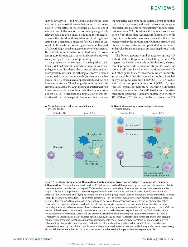
We propose that the feature that distinguishes tradi-tionally defined neuroinflammatory diseases from neu-rodegenerative disorders is the nature of inflammation; more precisely, whether the pathological process is driven by cardinal adaptive immune cells (as seen in encepha-litides), or CNS-resident and/or potentially blood-derived innate immune cells. Thus, it might be more useful to dis-criminate diseases of the CNS as being characterized by an innate immune element or by an adaptive immune com-ponent (FIG. 2). The translational implication of this dis-tinction will be that therapeutic development can focus on
the respective type of immune system contribution that is active in the disease, and it will be irrelevant or even insufficient for patients or translationally minded scien-tists to separate CNS disorders with immune involvement per se from those that lack neuroinflammation. With respect to the translation of treatments, it will also not matter whether the immune contribution is primary and disease-causing (such as in encephalitides), or secondary and involved in maintaining or exacerbating disease (such as in AD).
The following points could be used to contrast AD with MS at the pathogenic level. First, the genetics of MS suggest that T cells have a role in this disease45, whereas recent genome-wide association studies (GWASs) of sporadic AD cases have found associations between AD and other genes that are involved in innate immunity, as indicated by AD-linked mutations in the microglial or myeloid genes encoding TREM2 (REFS 21,22), CD33 (REFS 23,46), complement receptor 1 (CR1)47,48, mye-loid cell-expressed membrane-spanning 4-domains subfamily A member 6A (MS4A6A) and putative membrane-spanning 4-domains subfamily A member 4E (MS4A4E)49,50. Second, the disease process in MS
Nature Reviews | Neuroscience
Monocyte
Outside CNS Inside CNS
Perivascular macrophage
?
Inside CNS
B lymphocyte
T lymphocyteMicroglial cell
a Neurodegenerative disease: innate immune system-driven
b Neuroinflammatory disease: adaptive immune system-driven
Astrocyte
Outside CNS
Monocyte-derived inflammatory phagocyte
Figure 2 | Distinguishing neuroinflammation: innate immune-driven versus adaptive immune-driven neuro-inflammation. Neuroinflammation in various CNS disorders can be differentiated by the nature of inflammation; that is, diseases may be classified according to if CNS-resident and/or potentially blood-derived innate immune cells are the major pathogenic component (as in neurodegenerative diseases such as Alzheimer disease) (a), or if predominantly adaptive immune cells (B and T lymphocytes) drive the pathological process (as seen in encephalitides or multiple sclerosis (MS)) (b). The main contribution — apart from astrocytes — of the innate immune system in neurodegenerative diseases occurs within the CNS through resident microglia and perivascular macrophages, whereas the involvement of other blood-derived myeloid cells such as dendritic cells and monocytes appear to have no major impact on the course of neurodegeneration. Whether — and if so, to what extent — monocytes are recruited from the periphery to the CNS in the course of the disease is not entirely clear (denoted by the dashed arrow and a question mark) (a). Traditionally defined neuroinflammatory diseases such as MS are primarily driven by cells of the adaptive immune system such as T and B lymphocytes; various subtypes of myeloid cells have, however, also important pathogenic implications; blood-derived monocytes represent in fact the most numerous infiltrate into the CNS where they transform into monocyte-derived inflammatory phagocytes (macrophages or dendritic cells) and are thought to mediate much of the tissue damage observed (denoted by the thick arrow). As in neurodegenerative diseases, astrocytes and microglia also react to pathology, although it is not clear whether the type of response is similar to what happens in neurodegeneration (b).
R E V I E W S
NATURE REVIEWS | NEUROSCIENCE VOLUME 16 | JUNE 2015 | 361
© 2015 Macmillan Publishers Limited. All rights reserved

Quantitative trait lociStretches of DNA that contain or are linked to the genes that underlie a quantitative trait. Quantitative traits refer to certain phenotypes.
begins with T cell autoimmunity, as indicated by find-ings in informative animal models51, whereas the disease process in AD may begin with abnormal protein pro-cessing and, in its early stages, involve aberrant innate immunity11. Third, the expression of quantitative trait loci that are implicated in AD are expressed in monocytes, whereas those that are involved in MS are mainly found in T lymphocytes52.
Activation of the immune system in AD can act at least as pacemaker, perpetuating and accelerating the course of the disease; although at present it is not gen-erally thought to be the trigger of the disease process, it cannot be excluded that immune actions, at least in part, may also have a detrimental role in initiating the disease process4,7,10–12,26,28,29,31,53.
Neuroinflammatory mechanisms in ADMicroglia promote neuroinflammation in AD. Microglia are CNS-resident myeloid cells of embryonic haemato-poietic origin. Like most tissue macrophages, micro-glia survey the brain for pathogens and support CNS homeostasis and plasticity; for example, by guarding and remodelling synapses (for a review, see REF. 54). Microglia are equipped to sense so-called danger signals, such as protein aggregates in AD, and to respond to changes in neuronal health by adopting a set of morphological and functional attributes; such cells are termed ‘reactive’ or ‘primed’ (for a review, see REF. 4).
Among all non-neuronal CNS cells, microglia are the most intimately associated with the tissue changes that are observed in AD: in brain tissue taken at autopsy from individuals with AD, macrophages derived from microglia and, possibly, from infiltrating monocytes sur-round Aβ plaques, and the morphology of parenchymal microglia indicates that these cells are responding to challenge.
Soluble Aβ oligomers and Aβ fibrils can bind to vari-ous receptors that are expressed by microglia, including CD14, CD36, CD47, α6β1 integrin, class A scavenger receptor, receptor for advanced glycosylation end products (RAGE) and toll-like receptors (TLRs)55–62. Binding of Aβ to, for example, CD36 or TLR4 results in the production of inflammatory cytokines and chemokines in vitro59,63. In vivo, interleukin-1 (IL-1) (REF. 64), IL-6, granulocyte-macrophage colony-stimulating factor (GM-CSF)65, IL-12 and IL-23 (REF. 66), and tumour necrosis factor (TNF)67 — all markers of inflammation — are detectable or upreg-ulated in animal models of AD or in the brains or CSF from humans with AD (for details, see REF. 4). In studies in transgenic mouse models of AD, TNF release by microglia in response to Aβ was triggered by an interaction of CD40 with CD40L or by TLR4 engagement68–70.
Besides the production of inflammatory mediators upon binding of Aβ to various microglia receptors, Aβ has been shown to be cleared by microglia in vitro through receptor-mediated phagocytosis and degrada-tion. However, the relevance of these reports is uncer-tain, as it remains unclear whether microglia themselves phagocytose Aβ fibrils in a pathophysiological AD setting in vivo (for a review, see REF. 4). Nevertheless, microglia possess the machinery to degrade the more
‘digestible’ (and pathogenically more relevant71) soluble Aβ species via extracellular proteases such as neprilysin and insulin-degrading enzyme (IDE) (for a review, see REF. 72).
Aside from a few visionary investigators43,73,74, most researchers consider it axiomatic that the amoeboid mor-phology of microglia in AD equates to a toxic microglial phenotype that is accompanied by the production of solu-ble inflammatory factors, and this morphology is often habitually — but erroneously — interpreted as a sign of microglial ‘activation’. Remarkably, however, there is now strong evidence for a progressive, Aβ-dependent impair-ment of microglial function, as shown by a decrease in the phagocytosis of beads and in a reduced capacity to extend processes towards a tissue lesion in a mouse model of AD75. These findings are in line with another study showing that microglia from transgenic AD mice had reductions in the levels of Aβ-binding scavenger receptor and Aβ-degrading enzyme76. Importantly, efficient phago-cytosis has recently been shown to involve a component of the autophagy pathway, namely beclin 1, the levels of which were found to be markedly reduced in microglia derived from people with AD77. The idea that microglial impairment might represent the functional correlate of their amoeboid phenotype in AD carries considerable relevance for understanding AD pathogenesis, especially in light of the finding that inefficient clearance of Aβ — including that mediated through microglial proteases — is a major pathogenic factor in sporadic AD78. It is therefore noteworthy but not surprising that transient depletion (using suicide-gene technology) of microglia that have acquired a dysfunctional phenotype has no impact on Aβ burden in an animal model of AD79.
Microglial impairment might paradoxically be sus-tained by inflammatory cytokines such as TNF, IL-1, IL-12 and IL-23 (REFS 64,66,67). This idea suggests that AD pathology could be accelerated through this negative feedback loop. Ultimately, prolonged microglia impair-ment will also be accompanied by a loss of trophic func-tions and the elimination of protective properties. As the microglia-specific deficiency of brain-derived neuro-trophic factor (BDNF) has recently identified microglia-produced BDNF to be key for achieving a motor-learning task by promoting learning-related synapse formation80, a lack of functional microglia providing trophic factors such as BDNF may further impact neuronal integrity in the course of AD.
Aβ oligomers impair neuronal function in part because they can interact with neuronal membranes in a receptor-independent fashion81,82. Thus, it is tempting to speculate that microglial dysfunction in the early stages of AD may also be due to an interaction with such Aβ species, as these hydrophobic amyloid moieties can bind any cell membrane83 (FIG. 3).
In summary, it is necessary to view the transformed microglial cell as being indicative of a loss of tissue homeostasis. Because microglia carry out critical physi-ological tasks in the healthy brain84,85, a phenotypically transformed microglial cell should raise suspicions that their intrinsic tasks are not being performed efficiently and that the loss of microglial integrity may contribute
R E V I E W S
362 | JUNE 2015 | VOLUME 16 www.nature.com/reviews/neuro
© 2015 Macmillan Publishers Limited. All rights reserved

to tissue pathology as readily as do other impaired ele-ments of the CNS. The idea of neuroinflammation in AD therefore goes beyond the degree of mere alterations in microglial morphology and instead implies changes in this cell’s phenotype and function, and the relevant consequences.
Rare structural variants of genes encoding the immune receptors TREM2 (REFS 21,22,86,87), CD33 (REFS 23,46) and CR1 (REF. 47), all of which are expressed on microglia and other myeloid cells, have been found to be associ-ated with a higher risk of AD. These findings support the concept of altered microglial function in AD. As TREM2 has previously been shown to be involved in regulating microglial phagocytosis88,89, it was surprising to learn that the chief impact of TREM2 in vivo in various AD mouse models is to promote the survival of activated microglia and their peripherally derived myeloid coun-terparts90,91, and to engage these cells with Aβ plaques90–92 through sensing lipids associated with Aβ accumulation and neuronal loss90. Interestingly, a TREM2 deficiency in APPPS1 mice, which express mutant forms of human APP and presenilin 1 (PS1) (and hence model AD), was
shown to ameliorate hippocampal Aβ accumulation91, whereas 5XFAD mice (an AD-like mouse model express-ing human mutant variants of APP and PS1), in which Aβ deposition develops less rapidly than in APPPS1 mice, showed an increase in hippocampal Aβ in the absence of TREM2 (REF. 90). Future studies will need not only to address the reasons for the differences in Aβ pathology in the various AD-like mouse models lacking TREM2, but also to distinguish between potential differences in phe-notypes resulting from TREM2 deficiency versus TREM2 mutations. Accordingly, genetic variation in the gene encoding TYRO protein tyrosine kinase-binding protein (TYROBP; also known as DAP12) — the TREM2 adap-tor protein — has been shown to be associated with late-onset AD20. Together, these findings suggest that impaired TREM2 function and, consequently, altered myeloid cell (monocyte or microglia) function, have a role in AD pathogenesis.
CD33 encodes a cell-surface protein of the sialic acid-binding Ig-like lectin (SIGLEC) family93 and provides another example of a myeloid-cell- or microglial-cell-expressed gene in which variants have been associated
Figure 3 | Dynamic, multifaceted interactions with amyloid-β mediate microglial phenotypes in Alzheimer disease. In both in vitro and in vivo experiments, microglia exhibit receptor-dependent interactions58,61,205,206 with various forms of amyloid-β (Aβ; from monomers to oligomers, protofibrils, fibrils and plaques) as well as non-receptor mediated interactions (particularly with oligomers)81. Aβ species can stimulate changes in microglial function or production of inflammatory mediators through signalling receptors207, by inducing production of such mediators by other cells such as astrocytes208 and through post-phagocytic processes within microglia, including lysosomal injury, which acidifies the cytosol and contributes to activating the NLRP3 (NACHT, LRR and PYD domains-containing protein 3) inflammasome149. Receptor-mediated interactions between microglia and Aβ monomers do not show evidence for altered microglial function aside from induction of a ‘primed’ state, typified by heightened responses to subsequent DAMP (damage-associated molecular pattern) or cytokine stimuli. Microglia function, as monitored by motility in response to laser lesion and phagocytic activity of latex beads, is severely impaired in APPPS1 Alzheimer disease (AD) mice75, known to also exhibit oligomeric Aβ209. This functionally compromised state affects the microglial response to downstream Aβ species such as fibrils and plaques, culminating in a morphology and irreversible phenotype that is termed ‘dystrophic’ — corresponding to a ‘burn out’ of microglia (see also FIG. 1b). The arrow thickness indicates the likelihood of conversion of Aβ species or arrays.
Nature Reviews | Neuroscience
Receptor-mediated interaction
Receptor-mediated interaction
Receptor-mediated interaction
Non-receptor-mediated interaction
• Relatively non-toxic• May prime
inflammatory signalling
• Toxic to microglia, driving them to a dystrophic phenotype
• Reduces phagocytic capacity
• Elicits cytokine production
• May enhance oligomer toxicity to microglia, resulting in dystrophic phenotype
• Elicits cytokine production
• May activate inflammasomes
Aβ oligomers Protofibrils FibrilsAβ monomers
Microglial cell
R E V I E W S
NATURE REVIEWS | NEUROSCIENCE VOLUME 16 | JUNE 2015 | 363
© 2015 Macmillan Publishers Limited. All rights reserved

Cerebral amyloidosisThis term describes all forms of CNS diseases that feature the deposition of proteins (so-called proteinopathies).
HypertrophyThe increase in the volume of an organ, tissue or cell.
Apolipoprotein E(APOE). A class of apolipoprotein that is required for the catabolism of triglyceride-rich lipoprotein constituents. In the CNS, APOE is generated primarily by astrocytes, and transports cholesterol to neurons via APOE receptors, whereas in the periphery, APOE is mainly produced by the liver and macrophages, and mediates cholesterol metabolism.
Neurovascular unit(NVU). This consists of vascular cells such as brain endothelial cells, pericytes and vascular smooth muscle cells, glial cells such as astrocytes, microglia and oligodendroglia, and neurons. It links neural activity to blood flow and controls the exchange of biologically relevant protein interactions between brain and the periphery.
with an increased risk of developing AD23,46. One study showed that CD33 expression was upregulated on micro-glia from post-mortem samples of human AD brains, and that a CD33 variant, namely the protective CD33 single-nucleotide polymorphism (SNP) rs3865444, was associated with reductions in both CD33 expression and insoluble Aβ levels in the AD brain94. By contrast, another study found that monocytes derived from car-riers of this risk genotype showed a decrease in vitro in phagocytosis of Aβ fibrils23.
GWAS-based discoveries of alterations in genes that regulate innate immune system functions in people with AD, along with insights into the contribution of the immune system to AD from a variety of experimen-tal approaches, provide good evidence for a pathogenic role of CNS-resident myeloid cells such as microglia in the course of the disease. It is thus conceivable that mutations in innate immune molecules compromise microglia function, similar to the way in which Aβ impairs microglial performance over time75; micro-glia dysfunction, irrespective of whether it is caused by genetic alterations or by prolonged exposure to the Aβ-rich AD environment, will ultimately — if not inter-rupted — culminate in a state of these cells that has been termed ‘dystrophic’73,95. In this state, microglia lack their beneficial functions and have acquired a detrimental, senescent-like phenotype, which could justifiably be termed a cellular ‘burn out’ of microglia that resembles an irreversible end stage (FIG. 1b).
Myeloid cells other than resident microglia. The CNS has a rich complement of non-microglial myeloid cells including meningeal and choroid plexus macrophages, as well as perivascular macrophages. Of these, perivas-cular macrophages seem to have a particularly crucial role in the physiological removal of Aβ and protection from amyloid pathology96,97. Perivascular macrophages are, in contrast to microglia, continuously replaced from progeny of the circulating monocyte pool during adulthood98. Preclinical studies using animal models of AD based on amyloid deposition96,97,99,100 have shown that all CNS myeloid cells are, in principle, capable of promoting Aβ clearance and, thereby, of limiting the deposition of vascular amyloid, thus influencing dis-ease outcome. It remains uncertain whether infiltrating myeloid cells — monocyte-derived macrophages that typically do not contribute substantially in numerical terms to parenchymal macrophages in the course of AD100 or in other proteinopathies such as prion dis-ease101 — can, in principle, modulate AD pathology when experimentally instructed to enter the AD brain, as reports are not conclusive44.
Astrocytes promote neuroinflammation in AD. Astrocytes are CNS-resident cells of neuroectodermal origin that can — like microglia — respond to patho-logical stimuli through reactive gliosis102,103. Also simi-larly to myeloid cells, astrocytes surround Aβ plaques104, and studies using transgenic mice exhibiting cerebral amyloidosis105 have shown that their activation occurs early in the course of pathogenesis. Although reactive
astrocytes typically upregulate their expression of glial fibrillary acidic protein (GFAP), they do not form ‘glial scars’ in the brain of individuals with AD as they do in CNS diseases such as MS or in stroke102. In transgenic mouse models of AD, astrocytes underwent atrophy that preceded the Aβ plaque-related astrogliosis (except those astrocytes surrounding plaques, which show hypertrophy), and this eventually resulted in deficient glutamatergic transmission, possibly contributing to the cognitive impairment106,107. Furthermore, reduc-ing astrocyte activation through a viral vector driving the expression of VIVIT, a peptide that interferes with the immune or inflammatory calcineurin–nuclear fac-tor of activated T cells (NFAT) signalling pathway, in an Aβ-overexpressing mouse model of AD ameliorated AD-like pathology108. Several studies have shown that reactive astrocytes that are localized near plaques take up and degrade Aβ109–111. Furthermore, astrocyte activation followed by the release of apolipoprotein E (APOE) from astrocytes has been shown to be crucial for the ability of microglia to remove fibrillar Aβ in an animal model of AD112. Moreover, astrocytes, like microglia, increase their expression of Aβ-degrading enzymes when exposed to native Aβ extracts ex vivo113–115. However, the atrophy of astrocytes (as shown in a mouse model of AD106,107) may also result in a reduced proteolytic clearance of Aβ. Hence, AD pathogenesis may entail altered astrocyte function, which may be considered an integral part of the neuroinflammatory response.
Neuroinflammatory actions of other CNS cells in AD. Besides microglia and astrocytes, other CNS-resident cells such as endothelial cells, oligodendrocytes and neurons can contribute to neuroinflammation. Oligodendrocytes and myelin are known targets of immune reactions in neurological disorders such as MS. Despite the fact that AD research lacks more detailed studies in this context, there is evidence for changes in oligodendrocytes and myelin abnormalities in AD white matter (reviewed in REFS 115–117). As oligodendrocytes have been shown to express the complement components C1q, C1s, C2, C3, C4, C5, C6, C7, C8 and C9 (REF. 118), and complement-activated oligodendrocytes are found in various neurodegenerative conditions with a neuroin-flammatory component including AD119, these cells may contribute to neuroinflammation by providing increased levels of complement in the AD brain (for details on the complement system in AD see REF. 115).
Neurons are equipped with a variety of molecules that protect against inflammation, of which fractalkine120,121, the complement defense protein CD59 (REFS 122,123) and CD200 (REF. 124) have been shown to be decreased in pathology-affected regions of the AD brain. Thus, neurons can potentially contribute to the neuroinflam-matory aspect of AD perhaps mainly by reducing their expression of inhibitors of inflammation in part due to cell death or dysfunction.
Endothelial cells, besides being a key element of the neurovascular unit that contributes to the transport of Aβ species between the brain and the periphery (for a review, see REF. 125), have also been described
R E V I E W S
364 | JUNE 2015 | VOLUME 16 www.nature.com/reviews/neuro
© 2015 Macmillan Publishers Limited. All rights reserved

to influence inflammation in AD. Brain endothelial cells produce immune molecules such as IL-6, IL-1β and CCL2 in vitro upon exposure to Aβ peptides and in human AD brains126. Moreover, the vasoconstrictor endothelin 1 is upregulated upon Aβ binding to RAGE — the latter confers pleiotropic effects in the AD setting — which ultimately also regulates vascular inflammation in patients with AD127. For a more detailed review on endothelial actions in AD, including mechanisms influ-encing the efflux of amyloid and/or neurotoxic species as well as other proteins such as growth factors, and the expression of adhesion molecules enabling influx of immune cells to the brain, see REFS 125,128,129.
Therapeutic neuroinflammatory AD targetsWhich cells should be modulated? To date, most thera-peutic efforts have been directed towards developing purely symptomatic treatments or agents that target Aβ or tau; however, the strategy of reducing inflam-mation in AD has recently attracted more interest
(for reviews, see REFS 11,130). Based on our current knowledge, innate immune cells such as microglia and macrophages are the prime targets for modulating neuroinflammation.
Resident microglia in AD and other pathological conditions have been shown to produce either induc-ible nitric oxide synthase (NOS2) or arginase131. As NOS2 expression was suggestive of M1 and arginase production of M2 phenotypes proposed for some tis-sue macrophages (BOX 1), microglia were categorized similarly18,131. However, research into macrophage biol-ogy over the past 15 years has led to an abandonment of the M1 versus M2 concept for macrophages132. Indeed, it has been suggested that macrophages should not be classified according to arginine metabolism133, at least in part because macrophages and microglia commonly express both arginase and NOS2. Hence, we propose to follow suit and relinquish this framework for micro-glia. Regardless of how microglial phenotypes may ulti-mately be classified, these cells produce a large diversity
Box 1 | M1 and M2 macrophages: the rise and fall of an idea
In recent years, the concept of macrophage polarization has been widely applied, both to tissue macrophages and monocyte-derived macrophages, largely extrapolating results obtained in vitro to the situation in vivo. The concept incorporates two precepts: first, that there is a two-dimensional spectrum comprising all macrophage activation states; and, second, that the termini of this spectrum can usefully be modelled by inflammatory macrophages (termed M1 or classically activated) at one end, and reparative (termed M2 or alternatively activated) macrophages at the other. This concept has recently been reviewed and reappraised132. This paradigm began with classical findings that resistance to infection was associated with macrophage activation192. Seminal reports193,194 showed that interferon-γ (IFNγ) and IL-4 elicited different reactions from macrophages in vitro. At the same time, two different subsets of T helper (T
H) cell, producing IFNγ and IL-4, respectively, were shown to orchestrate
responses to differing categories of infectious pathogens195. It was thus attractive to propose that M1 macrophages versus M2
macrophages responded to the products of different T cell subsets (IFNγ produced by T
H1 cells versus IL-4 produced by T
H2 cells, respectively), and
that TH1 cells and M1 macrophages operated through coordinated
teamwork, as did TH2 cells and M2 macrophages196. By analogy, microglia
were suggested to adopt M1 or M2 states131 and this idea rapidly gained
ascendency. Through application of genome-wide transcriptional profiling and epigenetics, the molecular mechanisms that govern T cell polarization have been identified197–199. However, these same approaches were not able to delineate the molecular pathways underlying the expected two types of macrophage responses to stimuli200,201. Indeed, the more stimuli were studied, the more ‘polarized states’ for macrophages were described202. The different expression profiles associated with each polarization state stubbornly resisted being aligned along a spectrum whose extremes were defined by the M1 and M2 transcriptomes203. Ultimately, this leads to the realization that stable subset commitment is an authentic attribute of T
H cells and is useful for understanding their
biology. By contrast, mononuclear phagocytes seem to have a near-infinite plasticity that lends itself ideally to dealing with an endless variety of environmental challenges132. Importantly, if macrophages cannot be categorized according to the M1 versus M2 paradigm, then it is also time to abandon this constricting strait-jacket in research into the functions of microglia. A recent perspective article authored jointly by 25 senior investigators133 reviewed the history of the concept of macrophage activation, proposed a new nomenclature and indicated that a simple bipolar scheme is not sufficient. CSF1, macrophage colony-stimulating factor 1.
1962 1983 1986 1992 2000 2004 2009 2011 2012 2014
Nature Reviews | Neuroscience
Proposal of the concept of macrophage activation
Description of TH1 cells (which produce IFNγ) and TH2 cells (which produce IL-4)
Selective macrophage-activation responses are found to IL-4 that are distinct from those to IFNγ
Selective macrophage-activation responses are found to IFNγ
M1 and M2 macrophages are proposed to be linked to TH1 and TH2 cells, respectively
Additional M2 (a-c) subsets are proposed, based on responses to various stimuli
Cytokines are found to polarize microglia to M1- and M2-like phenotypes in vitro
M1 and M2 macrophage signalling pathways are found to overlap, suggesting that stable polarized states are not identifiable
The growth factors CSF1 and CSF2 are found to drive M1- or M2-like polarization, respectively
Network analyses of stimulus-dependent human macrophage transcriptomes revealed nine distinct states of polariza-tion. Portrayed in a three-dimensional format, these states failed to align along a linear spectrum but rather were distrib-uted as points within an apparent sphere
Call for new view on macrophage polarization: “Our opinion is that macrophages do not form stable subsets but respond to a combina-tion of factors present in the tissue; we have, rather than subsets of macrophages, pathways that interact to form complex, even mixed, phenotypes.”132
R E V I E W S
NATURE REVIEWS | NEUROSCIENCE VOLUME 16 | JUNE 2015 | 365
© 2015 Macmillan Publishers Limited. All rights reserved

Adaptive immune systemThe immune system that forms the basis for acquired immunity and immunological memory and involves T and B lymphocytes.
Mast cellsThese are resident granulocytes of several types of tissues containing many granules rich in histamine and heparin.
BiologicalsMedicinal products that are manufactured in or extracted from biological sources (they may also be termed biopharmaceutical or biologic medical products); they are distinct from chemically synthesized pharmaceutical products.
NLRP3 inflammasomeInflammasomes comprise a sensor molecule from the NOD-like receptor (NLR) family or the pyrin and HIN domain-containing protein (PYHIN) family, the adaptor protein ASC and caspase 1. The NALP3 inflammasome is expressed in myeloid cells, senses a wide range of aggregated molecules, and promotes the maturation of the inflammatory cytokines interleukin-1 (IL-1) and IL-18.
of mediators, some of which may be attractive targets for modulating neuroinflammation in AD and thus, potentially, for ameliorating the disease course.
Although astrocytes may also seem to be suitable and attractive targets in this regard, the current limitations in our understanding of astrocyte biology and metho-dological options to modulate these cells restrict the potential for astrocyte manipulation in the near future. By contrast, cells of the adaptive immune system, such as B and T cells, appear not to be significant components of the neuroinflammatory reaction in AD according to our current knowledge12,40–43, and thus are not major therapeu-tic targets. Reports of an involvement of other immune cells such as mast cells are so far mostly descriptive (reviewed in REF. 134) and await experimental confirma-tion. Nevertheless, a tyrosine kinase inhibitor that inhibits mast cell differentiation and degranulation showed some benefit as an adjunct therapy to the current standard of care in a small Phase II trial in people with AD135.
When should modulation occur? As the phenotypes and functional properties of microglia obviously change dur-ing the course of AD, interventional approaches aimed at modulating neuroinflammation crucially depend on when and where to interfere. This may explain why despite the epidemiological link between the use of non-steroidal anti-inflammatory drugs (NSAIDs) and reduced risk of developing AD136,137, prospective clinical trials have failed to demonstrate a positive effect of traditional non-selective NSAIDs or of selective cyclooxy genase 2 (COX2) inhibitors in the treatment or prevention of AD130,138–141. However, in one large prevention trial, participants showed cognitive improvement for a prolonged period of time after termination of NSAID treatment142; thus, the effec-tiveness of NSAID as well as of COX2 inhibitor treatment in presymptomatic individuals is currently inconclusive130. This demonstrates the importance of stringent assessment of both the time point (before or after the onset of symp-toms or biomarker changes) of initiation and the duration of anti-inflammatory treatment in AD. Moreover, more-specific immune actions may need to be targeted that are not affected by NSAIDs or COX2 inhibitors.
The lack of success in ameliorating AD by the use of broad anti-inflammatory drugs should thus not diminish the enthusiasm and efforts for research in this direction, as recent insights into pathogenically relevant immune actions will enable a more precise targeting of well-defined immune pathways and molecules, even when consid-ering the existing limitations of AD clinical trials that suffer from the lack of well-defined outcome measures and precise diagnostic assays, and long durations.
Which immune molecules should be targeted? Based on preclinical data or findings in AD patients, various immune molecules or signalling pathways have been found to be promising therapeutic targets both from a scientific and a venture capital viewpoint143. Below we discuss the most promising or better validated targets (aside from those mentioned in previous sections). TABLE 1 provides a more-complete summary of potential immune targets for AD.
IL-12 and IL-23 are both therapeutically attractive immune targets for AD, as they have been shown to be increased in the CSF of AD and/or MCI patients66,144. Moreover, levels of the common subunit of IL-12 and IL-23, namely p40, were higher in the plasma of MCI and AD patients in an unbiased approach performed by the Alzheimer’s Disease Neuroimaging Initiative consor-tium145, IL‑12 gene expression was augmented in post-mortem brain tissue of individuals with AD compared with control individuals8, and polymorphisms of the gene encoding the IL-23 receptor were associated with AD in a northern Han Chinese population146. In addition, IL-12 and IL-23 were found to be released by a subpopulation of activated CD11c-positive microglia in a preclinical model of AD66. Ablation of IL-12, IL-23, p40 or the IL-12 receptor β1 (through which both IL-12 and IL-23 signal) in an Aβ-overexpressing transgenic mouse model of AD resulted in a substantial reduction in cerebral Aβ burden. Administration of neutralizing p40 antibodies before or after the onset of amyloid accumulation in this mouse model likewise markedly reduced the AD-like pathol-ogy, including cognitive and behavioural changes66. Neutralizing p40 — through antibody administration or through the use of small interfering RNA — in another mouse model of AD had similar effects147. Together, these findings point to IL-12 and IL-23 signalling as a tangible target for interventional approaches in AD130,143,148.
Biologicals that inhibit IL-12 and/or IL-23 have under-gone clinical validation in trials for other diseases and, in some cases, have already been approved by the US Food and Drug Administration (FDA) (for example, neutral-izing p40 antibodies for the treatment of psoriasis); this means that a drug with a known risk profile specifically targeting IL-12 and IL-23 is, in principle, available for a first clinical trial in patients with AD. Although the non-CNS (that is, peripheral) actions of IL-12 and IL-23 are typically mediated via T and natural killer (NK) cells, in the AD CNS setting, IL-12 and IL-23 seem to act through a novel mechanism that is independent from T and NK cells66. In this latter scenario, it seems that IL-12 and IL-23 act directly on astrocytes, which express the respective receptors66 (FIG. 4). Thus, existing biologicals that inhibit IL-12 and/or IL-23 would be assumed to be equally effective in AD as in IL-12 and IL-23 T cell-mediated peripheral diseases such as psoriasis, in which blockade of the IL-12–IL-23 signalling pathway, irrespective of the cellular source of origin, is regarded as the crucial therapeutic event.
Another attractive approach to modulate immune responses in AD centres on the NLRP3 inflammasome (NACHT, LRR and PYD domains-containing pro-tein 3 inflammasome). In vitro, this inflammasome can receive an activation signal when Aβ fibrils damage lysosomal membranes of microglia, leading to cytosol acidification149. NLRP3 is involved in regulating the catalytic activity of caspase 1, the enzyme that medi-ates the cleavage of the precursors of IL-1β and IL-18 into bioactive cytokines. Elevated levels of active cas-pase 1 have been detected in brain tissue from patients with AD and AD mice, whereas mutations in the genes encoding NLRP3 or caspase 1 reduced AD-like
R E V I E W S
366 | JUNE 2015 | VOLUME 16 www.nature.com/reviews/neuro
© 2015 Macmillan Publishers Limited. All rights reserved

pathology in a mouse model of AD, and this was accom-panied by a change in microglial phenotype150. There are currently no FDA-approved drugs that exclusively and specifically target NLRP3, but the recent identifica-tion of a small molecule inhibitor of NLRP3 (REF. 151) and of CD36 as a key upstream regulator of NLRP3 activation60 may circumvent this problem. However, in vivo data on the role of CD36 in animal models of AD are still lacking. CD36 mediates microglia and macrophage responses to Aβ in vitro and in vivo, thus playing a key part in the inflammatory events associ-ated with AD63, without compromising the NLRP3 inflammasome activity that is required for host defence against pathogens. Importantly, however, peroxisome proliferator-activated receptor-γ (PPARγ) agonists, such as pioglitazone, induce Aβ clearance in a mouse model of AD by stimulating microglial uptake of Aβ in a CD36-mediated manner152. This observation suggests that downregulating CD36 may reduce inflammation
but may also diminish the clearance of Aβ — another reason why in vivo experiments on CD36 function in animal models of AD are required.
In this context, the retinoid bexarotene, which selec-tively activates retinoid X receptors (RXRs), also needs to be mentioned. Bexarotene increased the microglia-mediated uptake of soluble Aβ in an APOE-dependent manner in an AD mouse model, resulting in reduced pathology153. Several studies confirmed the effects of bex-arotene on clearance of soluble Aβ, but some of the initially reported effects of bexarotene on AD-like pathology could not be fully reproduced at a quantitative level154–157.
A deficiency in CC chemokine receptor type 2 (CCR2), which is expressed by monocytes, amplified and accelerated mortality in a transgenic mouse model of AD96 by impairing the population maintenance of perivascular macrophages100, leading to Aβ deposition in cerebral vasculature. Whether overexpression of CCR2 has a positive effect on AD pathology, and thus qualifies
Table 1 | Attractive immune targets for manipulating AD pathology at various levels of validation
Immune target or signalling pathway
Function Therapeutic manipulation Refs
TREM2 or TYROBP Promotes Aβ uptake and/or sustains microglia or myeloid cell response to Aβ through sensing lipids that are associated with Aβ
Unclear whether targets should be upregulated or inhibited (to date, the collected in vivo data have been inconsistent)
20–22,89–92
CD33 Inhibits Aβ phagocytosis Inhibition or downregulation 23,46
CR1 Modulates the effect of APOE ε4 on brain fibrillar amyloid burden
Upregulation or activation 47,48
PPARγ or RXR Induces Aβ clearance Upregulation or activation 152,153
NLRP3 (or inflammasome-associated molecules IL-1β and caspase 1)
Regulates caspase 1 and IL-1β activation, and Aβ clearance
Inhibition or downregulation 150
CD36 Upstream regulator of NLRP3 activation, and binds Aβ
Presumably inhibition or downregulation (absence of in vivo data)
60,63
CD14 Co-receptor (along with TLR4 and protein MD2) for DAMPs, including Aβ
Inhibition or downregulation 62,207
IL-12 and/or IL-23 Part of the Aβ-driven inflammatory response
Inhibition or downregulation 8,66,145–147
IL-6 Part of the Aβ-driven inflammatory response
Presumably upregulation or activation
210,211
TNF–TNFR Part of the Aβ-driven inflammatory response
Presumably inhibition (inconsistent data)
212–215
CX3CR1 Enables homeostatic neuronal–microglia crosstalk
Upregulation or activation 121,158,159
P2X7R Microglial-expressed member of purinergic ionotropic receptors
Inhibition or downregulation 216,217
SCARA1 Myeloid cell-expressed scavenger receptor for soluble Aβ
Upregulation or activation 160
TGFβ1 Member of the transforming growth factor-β family of cytokines
Unclear (overproduction reduces Aβ burden; inhibition in myeloid cells reduces Aβ burden)
167–170
Aβ, amyloid-β; AD, Alzheimer disease; APOE, apolipoprotein E; CR1, complement receptor 1; CX3CR1, CX3C chemokine receptor 1; DAMPs, damage-associated molecular patterns; IL, interleukin; MD2, NLRP3, NACHT, LRR and PYD domains- containing protein 3; PPARγ, peroxisome proliferator-activated receptor-γ; RXR, retinoic acid receptor RXR; SCARA1, macrophage scavenger receptor types I and II; TGFβ1, transforming growth factor β1; TLR4, Toll-like receptor 4; TNF, tumour necrosis factor; TNFR, TNF receptor; TREM2, triggering receptor expressed on myeloid cells 2; TYROBP, TYRO protein tyrosine kinase-binding protein.
R E V I E W S
NATURE REVIEWS | NEUROSCIENCE VOLUME 16 | JUNE 2015 | 367
© 2015 Macmillan Publishers Limited. All rights reserved

CCR2 to be of potential therapeutic benefit, remains to be established. Similarly, modulation of the expression of microglial CX3C chemokine receptor 1 (CX3CR1; also known as the fractalkine receptor) had positive effects in mouse models of amyloid deposition while dramatically worsening tau pathology121,158,159. Likewise, macrophage scavenger receptor types I and II (SCARA1) has recently been shown to be involved in clearing soluble amyloid-β by myeloid cells in vivo and in vitro160, thus offering yet another druggable immune target.
Also of interest are studies linking complement, microglia and AD pathology161–163. Microglia have a role in complement-mediated synaptic pruning164,165 dur-ing postnatal development, and a reactivation of this mechanism could drive the progression of neurodegen-erative diseases associated with synapse loss166. In this context, CR1 needs to be mentioned, which has been shown to modulate the impact of the APOE ε4 allele on brain fibrillar amyloid burden and is associated with a higher risk of developing AD47, thus qualifying for being a potential immune target in AD143.
Finally, transforming growth factor β1 (TGFβ1) is an important regulatory cytokine that inhibits microglia activation and whose levels in plasma, CSF and brain are elevated in AD145,167–170. Transgenic overexpression of TGFβ1 decreased Aβ burden in an AD mouse model by promoting microglial Aβ clearance168. However, block-ing TGFβ1 and downstream SMAD2–SMAD3 signal-ling specifically in CD11c-positive myeloid cells also reduced Aβ-like pathology in a genetic mouse model of AD169, but this effect seemed to be due to an increased influx of activated peripheral myeloid cells rather than a modulation of resident microglia activation. This finding emphasizes the need to distinguish the role of CNS-resident microglia from that of blood-derived mononuclear cells that may enter the CNS over the dis-ease course or that are being introduced experimentally to the brains of AD mouse models. Despite some pro-gress in dissecting the various roles of resident versus peripherally derived myeloid cells in AD44,100,171, it is still unknown whether — and if so, by what means — peripheral myeloid cells may be superior in fighting AD pathology at least when experimentally forced to enter the AD brain. Similarly, in the context of Aβ vaccination, the role of resident microglia, despite some insights, has not been fully elucidated in vivo172–179 — a topic that is discussed in more detail elsewhere180.
The concept of instructing innate immune cells to facilitate the resolution of inflammatory responses through anti-inflammatory mediators is supported by various recent in vivo data: aspirin-triggered lipoxin A4 (LXA4) was shown to reduce nuclear factor-κB (NF-κB) activation, pro-inflammatory cytokines and chemokines in mice exhibiting AD-like pathology, while it increased the levels of IL-10 — yet another promi-nent anti-inflammatory player — and TGFβ1 (REF. 181). Similarly, IL-10, expressed via adeno-associated virus (AAV) delivery, reduced microgliosis and astroglio-sis, and improved cognitive performance in AD-like mice carrying mutated forms of human APP and PS1 (REF. 182). Results using a different AAV delivery system for IL-10 produced an opposite result183, leaving the part of this complex cytokine in AD pathogenesis uncertain. Treatment of Tg2576 AD-like mice expressing mutated human APP with the anti-inflammatory small molecule HPB242 resulted also in amelioration of AD pathol-ogy184, supporting the idea that lowering inflammation confers beneficial effect in AD. However, anti-inflamma-tion is not always favourable: hippocampal expression of the anti-inflammatory IL-4 or, as mentioned above, IL-10 (REF. 183) in AD mice worsened amyloid pathol-ogy185, indicating that a deeper understanding of how and when to manipulate the immune response will be crucial for obtaining favourable outcomes.
Conclusion and outlookRecent data clearly show that immune activation in AD has the capacity to facilitate and trigger the pathophysi-ology of AD. The immune system may thus provide exciting novel and realistic routes for the diagnosis and treatment of AD. However, interfering with neuroin-flammatory pathways and molecules requires precise
Figure 4 | Proposed Aβ-dependent CNS specific non-adaptive IL-12 and IL-23 actions in AD. a | In healthy brains, microglia do not express detectable levels of interleukin-12 (IL-12) or IL-23, and astrocytes are largely unresponsive to these cytokines. b | In Alzheimer disease (AD), exposure to Aβ leads to expression of both of these cytokines in microglia and reactive astrogliosis is accompanied by the expression of the respective receptors by astrocytes66. It is not clear whether the astroglial expression of IL-12 and IL-23 receptors is mediated by other cytokines or whether Aβ per se can also induce IL-12 and IL-23 receptor expression on astrocytes (denoted by the dashed arrows). This rise in IL-12 and IL-23, and their receptors, leads to exacerbation of AD pathology, including increased deposition of Aβ and ultimately to cognitive impairment, presumably through neuronal damage, whereas inhibition of the IL-12–IL-23 signalling pathway ameloriates pathology. However, what remains to be resolved is whether the IL-12- and IL-23-mediated effects in the CNS are conferred by astrocytes and/or by microglia, and whether IL-12 or rather IL-23 acts as the major player in this context.
Microglial cell
Neuronal damage
Astrocyte
Neuronal homeostasis
a Healthy brain
b Alzheimer disease
IL-23
IL-12
IL-12 receptorIL-23 receptor
?
?
Aβ species
Nature Reviews | Neuroscience
R E V I E W S
368 | JUNE 2015 | VOLUME 16 www.nature.com/reviews/neuro
© 2015 Macmillan Publishers Limited. All rights reserved

knowledge about the underlying immune events — which may change during the disease course. Taking into account the heterogeneity of AD, implying that not neces-sarily all individuals with AD exhibit neuro inflammation, or at all time points in the course of the disease, patients need to be stratified to identify those who may benefit most from anti-inflammatory interventions.
Ultimately, combination therapy consisting of both a drug targeting Aβ and/or tau, and a medication modu-lating inflammation may be a way to substantially delay progression of the disease. In this respect, repurposing strategies may be useful, for several, obvious reasons. Examples of potentially repurposed drugs are neutral-izing antibodies against p40, which are FDA-approved for the treatment of psoriasis66, and the NSAID deriva-tive CHF5074, which was initially developed as a γ-secretase modulator and has now been reclassified as a novel, first-in-class microglial modulator for the
treatment of AD, based on the finding that it reduces both amyloid burden and microglial activation186,187. Interim results from an ongoing Phase II trial in MCI patients suggest that CHF5074 confers positive effects by reducing biomarkers of neuroinflammation130,188,189.
A detailed knowledge of neuroimmune pathways and their molecular underpinnings in AD may also lead to a better understanding and treatment of degen-erative and/or proteinopathic CNS diseases other than AD. In addition, it will be equally interesting to learn how systemic immune components from young, healthy individuals that have been shown to positively modu-late cognitive performance190 may also have beneficial effects (for example, upon blood transfer191) in patho-physiological conditions, including neurodegenerative disorders such as AD. This would broaden the spectrum of opportunities to modulate CNS diseases by immune factors.
1. Wimo, A. & Prince, M. World Alzheimer Report 2010: The Global Economic Impact of Dementia (Alzheimer’s Disease International (ADI), 2010).
2. Querfurth, H. W. & LaFerla, F. M. Alzheimer’s disease. N. Engl. J. Med. 362, 329–344 (2010).
3. Hardy, J. & Selkoe, D. J. The amyloid hypothesis of Alzheimer’s disease: progress and problems on the road to therapeutics. Science 297, 353–356 (2002).
4. Prokop, S., Miller, K. R. & Heppner, F. L. Microglia actions in Alzheimer’s disease. Acta Neuropathol. 126, 461–477 (2013).
5. Gandy, S. & Heppner, F. L. Microglia as dynamic and essential components of the amyloid hypothesis. Neuron 78, 575–577 (2013).
6. Perry, V. H. & Holmes, C. Microglial priming in neurodegenerative disease. Nature Rev. Neurol. 10, 217–224 (2014).
7. Cunningham, C. Microglia and neurodegeneration: the role of systemic inflammation. Glia 61, 71–90 (2013).
8. Sudduth, T. L., Schmitt, F. A., Nelson, P. T. & Wilcock, D. M. Neuroinflammatory phenotype in early Alzheimer’s disease. Neurobiol. Aging 34, 1051–1059 (2013).
9. Krstic, D. & Knuesel, I. Deciphering the mechanism underlying late-onset Alzheimer disease. Nature Rev. Neurol. 9, 25–34 (2013).
10. Hickman, S. E. & El Khoury, J. TREM2 and the neuroimmunology of Alzheimer’s disease. Biochem. Pharmacol. 88, 495–498 (2014).
11. Heneka, M. T., Kummer, M. P. & Latz, E. Innate immune activation in neurodegenerative disease. Nature Rev. Immunol. 14, 463–477 (2014).
12. Akiyama, H. et al. Inflammation and Alzheimer’s disease. Neurobiol. Aging 21, 383–421 (2000).
13. Jonsson, T. et al. A mutation in APP protects against Alzheimer’s disease and age-related cognitive decline. Nature 488, 96–99 (2012).
14. Scheuner, D. et al. Secreted amyloid β-protein similar to that in the senile plaques of Alzheimer’s disease is increased in vivo by the presenilin 1 and 2 and APP mutations linked to familial Alzheimer’s disease. Nature Med. 2, 864–870 (1996).This report showed that all common forms of familial AD could be integrated into a unified pathogenic scheme.
15. LaFerla, F. M. & Green, K. N. Animal models of Alzheimer disease. Cold Spring Harb. Perspect. Med. 2, a006320 (2012).
16. Haass, C., Kaether, C., Thinakaran, G. & Sisodia, S. Trafficking and proteolytic processing of APP. Cold Spring Harb. Perspect. Med. 2, a006270 (2012).
17. Holmes, C. et al. Long-term effects of Aβ42 immunisation in Alzheimer’s disease: follow-up of a randomised, placebo-controlled phase I trial. Lancet 372, 216–223 (2008).
18. Giacobini, E. & Gold, G. Alzheimer disease therapy — moving from amyloid-β to tau. Nature Rev. Neurol. 9, 677–686 (2013).
19. Wyss-Coray, T. Inflammation in Alzheimer disease: driving force, bystander or beneficial response? Nature Med. 12, 1005–1015 (2006).
20. Zhang, B. et al. Integrated systems approach identifies genetic nodes and networks in late-onset Alzheimer’s disease. Cell 153, 707–720 (2013).This tour‑de‑force of bioinformatics illuminated the heretofore cryptic relevance of inflammation‑system genes for AD pathogenesis.
21. Jonsson, T. et al. Variant of TREM2 associated with the risk of Alzheimer’s disease. N. Engl. J. Med. 368, 107–116 (2013).
22. Guerreiro, R. et al. TREM2 variants in Alzheimer’s disease. N. Engl. J. Med. 368, 117–127 (2013).These two back‑to‑back studies identified rare variants of a single myeloid cell receptor as conferring surprisingly high risk for late‑onset sporadic AD.
23. Bradshaw, E. M. et al. CD33 Alzheimer’s disease locus: altered monocyte function and amyloid biology. Nature Neurosci. 16, 848–850 (2013).
24. Tarkowski, E., Andreasen, N., Tarkowski, A. & Blennow, K. Intrathecal inflammation precedes development of Alzheimer’s disease. J. Neurol. Neurosurg. Psychiatry 74, 1200–1205 (2003).
25. Brosseron, F., Krauthausen, M., Kummer, M. & Heneka, M. T. Body fluid cytokine levels in mild cognitive impairment and Alzheimer’s disease: a comparative overview. Mol. Neurobiol. 50, 534–544 (2014).
26. Krstic, D. et al. Systemic immune challenges trigger and drive Alzheimer-like neuropathology in mice. J. Neuroinflamm. 9, 151 (2012).This study shows that unspecific immune activation maternally and postnatally in wild‑type mice can induce full‑blown AD pathology, thus causally linking (early) immune stimulation and the development of AD pathology.
27. Perry, V. H. Contribution of systemic inflammation to chronic neurodegeneration. Acta Neuropathol. 120, 277–286 (2010).
28. Cunningham, C. et al. Systemic inflammation induces acute behavioral and cognitive changes and accelerates neurodegenerative disease. Biol. Psychiatry 65, 304–312 (2009).
29. Holmes, C. et al. Systemic inflammation and disease progression in Alzheimer disease. Neurology 73, 768–774 (2009).This mechanistic clinical research study showed that AD patients with frequent, mild intercurrent infections deteriorated more rapidly, supporting the influence of systemic inflammation on disease course.
30. Cribbs, D. H. et al. Extensive innate immune gene activation accompanies brain aging, increasing vulnerability to cognitive decline and neurodegeneration: a microarray study. J. Neuroinflamm. 9, 179 (2012).
31. Schwartz, M. & Shechter, R. Systemic inflammatory cells fight off neurodegenerative disease. Nature Rev. Neurol. 6, 405–410 (2010).
32. Kyrkanides, S. et al. Osteoarthritis accelerates and exacerbates Alzheimer’s disease pathology in mice. J. Neuroinflamm. 8, 112 (2011).
33. Abuabara, K. et al. Cause-specific mortality in patients with severe psoriasis: a population-based cohort study in the U.K. Br. J. Dermatol. 163, 586–592 (2010).
34. Gisondi, P. et al. Mild cognitive impairment in patients with moderate to severe chronic plaque psoriasis. Dermatology 228, 78–85 (2014).
35. Thaler, J. P. et al. Obesity is associated with hypothalamic injury in rodents and humans. J. Clin. Invest. 122, 153–162 (2012).
36. Takeda, S. et al. Diabetes-accelerated memory dysfunction via cerebrovascular inflammation and Aβ deposition in an Alzheimer mouse model with diabetes. Proc. Natl Acad. Sci. USA 107, 7036–7041 (2010).
37. Mayeux, R. et al. Genetic susceptibility and head injury as risk factors for Alzheimer’s disease among community-dwelling elderly persons and their first-degree relatives. Ann. Neurol. 33, 494–501 (1993).
38. Heneka, M. T. et al. Locus ceruleus degeneration promotes Alzheimer pathogenesis in amyloid precursor protein 23 transgenic mice. J. Neurosci. 26, 1343–1354 (2006).
39. Rubartelli, A. & Lotze, M. T. Inside, outside, upside down: damage-associated molecular-pattern molecules (DAMPs) and redox. Trends Immunol. 28, 429–436 (2007).
40. Bornemann, K. D. et al. Aβ-induced inflammatory processes in microglia cells of APP23 transgenic mice. Am. J. Pathol. 158, 63–73 (2001).
41. Stalder, A. K. et al. Invasion of hematopoietic cells into the brain of amyloid precursor protein transgenic mice. J. Neurosci. 25, 11125–11132 (2005).
42. Eikelenboom, P. et al. Neuroinflammation in Alzheimer’s disease and prion disease. Glia 40, 232–239 (2002).
43. Streit, W. J. Microglia and Alzheimer’s disease pathogenesis. J. Neurosci. Res. 77, 1–8 (2004).
44. Prinz, M., Priller, J., Sisodia, S. S. & Ransohoff, R. M. Heterogeneity of CNS myeloid cells and their roles in neurodegeneration. Nature Neurosci. 14, 1227–1235 (2011).
45. International Multiple Sclerosis Genetics Consortium et al. Genetic risk and a primary role for cell-mediated immune mechanisms in multiple sclerosis. Nature 476, 214–219 (2011).
46. Naj, A. C. et al. Common variants at MS4A4/MS4A6E, CD2AP, CD33 and EPHA1 are associated with late-onset Alzheimer’s disease. Nature Genet. 43, 436–441 (2011).
47. Thambisetty, M. et al. Effect of complement CR1 on brain amyloid burden during aging and its modification by APOE genotype. Biol. Psychiatry 73, 422–428 (2013).
48. Lambert, J. C. et al. Genome-wide association study identifies variants at CLU and CR1 associated with Alzheimer’s disease. Nature Genet. 41, 1094–1099 (2009).
49. Hollingworth, P. et al. Common variants at ABCA7, MS4A6A/MS4A4E, EPHA1, CD33 and CD2AP are associated with Alzheimer’s disease. Nature Genet. 43, 429–435 (2011).
R E V I E W S
NATURE REVIEWS | NEUROSCIENCE VOLUME 16 | JUNE 2015 | 369
© 2015 Macmillan Publishers Limited. All rights reserved

50. Liang, Y. & Tedder, T. F. Identification of a CD20-, FcεRIβ-, and HTm4-related gene family: sixteen new MS4A family members expressed in human and mouse. Genomics 72, 119–127 (2001).
51. Ransohoff, R. M. Animal models of multiple sclerosis: the good, the bad and the bottom line. Nature Neurosci. 15, 1074–1077 (2012).
52. Raj, T. et al. Polarization of the effects of autoimmune and neurodegenerative risk alleles in leukocytes. Science 344, 519–523 (2014).
53. Iwashyna, T. J., Ely, E. W., Smith, D. M. & Langa, K. M. Long-term cognitive impairment and functional disability among survivors of severe sepsis. JAMA 304, 1787–1794 (2010).
54. Prinz, M. & Priller, J. Microglia and brain macrophages in the molecular age: from origin to neuropsychiatric disease. Nature Rev. Neurosci. 15, 300–312 (2014).
55. Du Yan, S. et al. Amyloid-β peptide-receptor for advanced glycation endproduct interaction elicits neuronal expression of macrophage-colony stimulating factor: a proinflammatory pathway in Alzheimer disease. Proc. Natl Acad. Sci. USA 94, 5296–5301 (1997).
56. El Khoury, J. et al. Scavenger receptor-mediated adhesion of microglia to β-amyloid fibrils. Nature 382, 716–719 (1996).
57. Bamberger, M. E., Harris, M. E., McDonald, D. R., Husemann, J. & Landreth, G. E. A cell surface receptor complex for fibrillar β-amyloid mediates microglial activation. J. Neurosci. 23, 2665–2674 (2003).
58. Paresce, D. M., Ghosh, R. N. & Maxfield, F. R. Microglial cells internalize aggregates of the Alzheimer’s disease amyloid β-protein via a scavenger receptor. Neuron 17, 553–565 (1996).
59. Stewart, C. R. et al. CD36 ligands promote sterile inflammation through assembly of a Toll-like receptor 4 and 6 heterodimer. Nature Immunol. 11, 155–161 (2010).
60. Sheedy, F. J. et al. CD36 coordinates NLRP3 inflammasome activation by facilitating intracellular nucleation of soluble ligands into particulate ligands in sterile inflammation. Nature Immunol. 14, 812–820 (2013).
61. Koenigsknecht, J. & Landreth, G. Microglial phagocytosis of fibrillar β-amyloid through a β1 integrin-dependent mechanism. J. Neurosci. 24, 9838–9846 (2004).
62. Fassbender, K. et al. The LPS receptor (CD14) links innate immunity with Alzheimer’s disease. FASEB J. 18, 203–205 (2004).
63. El Khoury, J. B. et al. CD36 mediates the innate host response to β-amyloid. J. Exp. Med. 197, 1657–1666 (2003).
64. Griffin, W. S. et al. Brain interleukin 1 and S-100 immunoreactivity are elevated in Down syndrome and Alzheimer disease. Proc. Natl Acad. Sci. USA 86, 7611–7615 (1989).
65. Patel, N. S. et al. Inflammatory cytokine levels correlate with amyloid load in transgenic mouse models of Alzheimer’s disease. J. Neuroinflamm. 2, 9 (2005).
66. Vom Berg, J. et al. Inhibition of IL-12/IL-23 signaling reduces Alzheimer’s disease-like pathology and cognitive decline. Nature Med. 18, 1812–1819 (2012).This study showed that genetic as well as pharmacological blocking of the IL‑12–IL‑23 pathway substantially ameliorated AD pathology in an AD mouse model and provided the first hints that IL‑12 and IL‑23 are upregulted in the CSF of patients with AD, thus offering a druggable immune target made for repurposing existing IL‑12 and IL‑23 inhibitors.
67. Fillit, H. et al. Elevated circulating tumor necrosis factor levels in Alzheimer’s disease. Neurosci. Lett. 129, 318–320 (1991).
68. Tan, J. et al. Role of CD40 ligand in amyloidosis in transgenic Alzheimer’s mice. Nature Neurosci. 5, 1288–1293 (2002).
69. Tan, J. et al. Microglial activation resulting from CD40–CD40L interaction after β-amyloid stimulation. Science 286, 2352–2355 (1999).
70. Jin, J. J., Kim, H. D., Maxwell, J. A., Li, L. & Fukuchi, K. Toll-like receptor 4-dependent upregulation of cytokines in a transgenic mouse model of Alzheimer’s disease. J. Neuroinflamm. 5, 23 (2008).
71. Haass, C. & Selkoe, D. J. Soluble protein oligomers in neurodegeneration: lessons from the Alzheimer’s
amyloid β-peptide. Nature Rev. Mol. Cell Biol. 8, 101–112 (2007).
72. Lee, C. Y. & Landreth, G. E. The role of microglia in amyloid clearance from the AD brain. J. Neural Transm. 117, 949–960 (2010).
73. Streit, W. J., Sammons, N. W., Kuhns, A. J. & Sparks, D. L. Dystrophic microglia in the aging human brain. Glia 45, 208–212 (2004).
74. Perry, V. H., Nicoll, J. A. & Holmes, C. Microglia in neurodegenerative disease. Nature Rev. Neurol. 6, 193–201 (2010).
75. Krabbe, G. et al. Functional impairment of microglia coincides with beta-amyloid deposition in mice with Alzheimer-like pathology. PLoS ONE 8, e60921 (2013).
76. Hickman, S. E., Allison, E. K. & El Khoury, J. Microglial dysfunction and defective β-amyloid clearance pathways in aging Alzheimer’s disease mice. J. Neurosci. 28, 8354–8360 (2008).
77. Lucin, K. M. et al. Microglial beclin 1 regulates retromer trafficking and phagocytosis and is impaired in Alzheimer’s disease. Neuron 79, 873–886 (2013).This paper shows that beclin 1 is altered in microglia derived from AD patients and provides the first molecular explanations of some aspects of microglial impairment in AD.
78. Mawuenyega, K. G. et al. Decreased clearance of CNS β-amyloid in Alzheimer’s disease. Science 330, 1774 (2010).
79. Grathwohl, S. A. et al. Formation and maintenance of Alzheimer’s disease β-amyloid plaques in the absence of microglia. Nature Neurosci. 12, 1361–1363 (2009).
80. Parkhurst, C. N. et al. Microglia promote learning-dependent synapse formation through brain-derived neurotrophic factor. Cell 155, 1596–1609 (2013).This paper demonstrates that microglia have an important role in learning and memory by providing neurotrophic factors, such as BDNF.
81. Hong, S. et al. Soluble Aβ oligomers are rapidly sequestered from brain ISF in vivo and bind GM1 ganglioside on cellular membranes. Neuron 82, 308–319 (2014).
82. Selkoe, D. J. SnapShot: pathobiology of Alzheimer’s disease. Cell 154, 468–468 e1 (2013).
83. Verdier, Y., Zarandi, M. & Penke, B. Amyloid β–peptide interactions with neuronal and glial cell plasma membrane: binding sites and implications for Alzheimer’s disease. J. Pept. Sci. 10, 229–248 (2004).
84. Wake, H., Moorhouse, A. J. & Nabekura, J. Functions of microglia in the central nervous system — beyond the immune response. Neuron Glia Biol. 7, 47–53 (2011).
85. Salter, M. W. & Beggs, S. Sublime microglia: expanding roles for the guardians of the CNS. Cell 158, 15–24 (2014).
86. Frank, S. et al. TREM2 is upregulated in amyloid plaque-associated microglia in aged APP23 transgenic mice. Glia 56, 1438–1447 (2008).
87. Hickman, S. E. et al. The microglial sensome revealed by direct RNA sequencing. Nature Neurosci. 16, 1896–1905 (2013).
88. Hsieh, C. L. et al. A role for TREM2 ligands in the phagocytosis of apoptotic neuronal cells by microglia. J. Neurochem. 109, 1144–1156 (2009).
89. Kleinberger, G. et al. TREM2 mutations implicated in neurodegeneration impair cell surface transport and phagocytosis. Sci. Transl Med. 6, 243ra86 (2014).
90. Wang, Y. et al. TREM2 lipid sensing sustains the microglial response in an Alzheimer’s disease model. Cell 160, 1061–1071 (2015).
91. Jay, T. R. et al. TREM2 deficiency eliminates TREM2+ inflammatory macrophages and ameliorates pathology in Alzheimer’s disease mouse models. J. Exp. Med. 212, 287–295 (2015).These two publications show the importance and function of TREM2 in AD mouse models; that is, to promote the survival of activated microglia and myeloid cells, to recruit these cells to Aβ plaques through sensing for Aβ‑associated lipids, and to modulate hippocampal Aβ burden.
92. Ulrich, J. D. et al. Altered microglial response to Aβ plaques in APPPS1-21 mice heterozygous for TREM2. Mol. Neurodegener. 9, 20 (2014).
93. Lajaunias, F., Dayer, J. M. & Chizzolini, C. Constitutive repressor activity of CD33 on human monocytes requires sialic acid recognition and phosphoinositide 3-kinase-mediated intracellular signaling. Eur. J. Immunol. 35, 243–251 (2005).
94. Griciuc, A. et al. Alzheimer’s disease risk gene CD33 inhibits microglial uptake of amyloid beta. Neuron 78, 631–643 (2013).
95. Miller, K. R. & Streit, W. J. The effects of aging, injury and disease on microglial function: a case for cellular senescence. Neuron Glia Biol. 3, 245–253 (2007).
96. El Khoury, J. et al. Ccr2 deficiency impairs microglial accumulation and accelerates progression of Alzheimer-like disease. Nature Med. 13, 432–438 (2007).
97. Hawkes, C. A. & McLaurin, J. Selective targeting of perivascular macrophages for clearance of β-amyloid in cerebral amyloid angiopathy. Proc. Natl Acad. Sci. USA 106, 1261–1266 (2009).
98. Hickey, W. F. & Kimura, H. Perivascular microglial cells of the CNS are bone marrow-derived and present antigen in vivo. Science 239, 290–292 (1988).
99. Lai, A. Y. & McLaurin, J. Clearance of amyloid-β peptides by microglia and macrophages: the issue of what, when and where. Future Neurol. 7, 165–176 (2012).
100. Mildner, A. et al. Distinct and non-redundant roles of microglia and myeloid subsets in mouse models of Alzheimer’s disease. J. Neurosci. 31, 11159–11171 (2011).
101. Gomez-Nicola, D., Schetters, S. T. & Perry, V. H. Differential role of CCR2 in the dynamics of microglia and perivascular macrophages during prion disease. Glia 62, 1041–1052 (2014).
102. Burda, J. E. & Sofroniew, M. V. Reactive gliosis and the multicellular response to CNS damage and disease. Neuron 81, 229–248 (2014).
103. Sofroniew, M. V. & Vinters, H. V. Astrocytes: biology and pathology. Acta Neuropathol. 119, 7–35 (2010).
104. Medeiros, R. & LaFerla, F. M. Astrocytes: conductors of the Alzheimer disease neuroinflammatory symphony. Exp. Neurol. 239, 133–138 (2013).
105. Heneka, M. T. et al. Focal glial activation coincides with increased BACE1 activation and precedes amyloid plaque deposition in APP[V717I] transgenic mice. J. Neuroinflamm. 2, 22 (2005).
106. Olabarria, M., Noristani, H. N., Verkhratsky, A. & Rodriguez, J. J. Age-dependent decrease in glutamine synthetase expression in the hippocampal astroglia of the triple transgenic Alzheimer’s disease mouse model: mechanism for deficient glutamatergic transmission? Mol. Neurodegener. 6, 55 (2011).
107. Olabarria, M., Noristani, H. N., Verkhratsky, A. & Rodriguez, J. J. Concomitant astroglial atrophy and astrogliosis in a triple transgenic animal model of Alzheimer’s disease. Glia 58, 831–838 (2010).
108. Furman, J. L. et al. Targeting astrocytes ameliorates neurologic changes in a mouse model of Alzheimer’s disease. J. Neurosci. 32, 16129–16140 (2012).
109. Nagele, R. G., D’Andrea, M. R., Lee, H., Venkataraman, V. & Wang, H. Y. Astrocytes accumulate Aβ42 and give rise to astrocytic amyloid plaques in Alzheimer disease brains. Brain Res. 971, 197–209 (2003).
110. Wyss-Coray, T. et al. Adult mouse astrocytes degrade amyloid-β in vitro and in situ. Nature Med. 9, 453–457 (2003).This paper demonstrates that astrocytes can have an important impact on catabolising Aβ.
111. Koistinaho, M. et al. Apolipoprotein E promotes astrocyte colocalization and degradation of deposited amyloid-β peptides. Nature Med. 10, 719–726 (2004).
112. Terwel, D. et al. Critical role of astroglial apolipoprotein E and liver X receptor-α expression for microglial Aβ phagocytosis. J. Neurosci. 31, 7049–7059 (2011).
113. Pihlaja, R. et al. Multiple cellular and molecular mechanisms are involved in human Aβ clearance by transplanted adult astrocytes. Glia 59, 1643–1657 (2011).
114. Saido, T. & Leissring, M. A. Proteolytic degradation of amyloid β-protein. Cold Spring Harb. Perspect. Med. 2, a006379 (2012).
115. Wyss-Coray, T. & Rogers, J. Inflammation in Alzheimer disease — a brief review of the basic science and clinical literature. Cold Spring Harb. Perspect. Med. 2, a006346 (2012).
116. Roth, A. D., Ramirez, G., Alarcon, R. & Von Bernhardi, R. Oligodendrocytes damage in Alzheimer’s disease: beta amyloid toxicity and inflammation. Biol. Res. 38, 381–387 (2005).
117. Mitew, S. et al. Focal demyelination in Alzheimer’s disease and transgenic mouse models. Acta Neuropathol. 119, 567–577 (2010).
118. Hosokawa, M., Klegeris, A., Maguire, J. & McGeer, P. L. Expression of complement messenger RNAs and proteins by human oligodendroglial cells. Glia 42, 417–423 (2003).
R E V I E W S
370 | JUNE 2015 | VOLUME 16 www.nature.com/reviews/neuro
© 2015 Macmillan Publishers Limited. All rights reserved

119. Yamada, T., Akiyama, H. & McGeer, P. L. Complement-activated oligodendroglia: a new pathogenic entity identified by immunostaining with antibodies to human complement proteins C3d and C4d. Neurosci. Lett. 112, 161–166 (1990).
120. Harrison, J. K. et al. Role for neuronally derived fractalkine in mediating interactions between neurons and CX3CR1-expressing microglia. Proc. Natl Acad. Sci. USA 95, 10896–10901 (1998).
121. Lee, S. et al. CX3CR1 deficiency alters microglial activation and reduces beta-amyloid deposition in two Alzheimer’s disease mouse models. Am. J. Pathol. 177, 2549–2562 (2010).
122. Singhrao, S. K., Muller, C. T., Gilbert, S. J., Duance, V. C. & Archer, C. W. An immunofluorescence method for postembedded tissue in the acrylic resin Technovit 9100 New using fluorescein isothiocyanate secondary detection. Microsc. Res. Tech. 72, 501–506 (2009).
123. Yang, L. B., Li, R., Meri, S., Rogers, J. & Shen, Y. Deficiency of complement defense protein CD59 may contribute to neurodegeneration in Alzheimer’s disease. J. Neurosci. 20, 7505–7509 (2000).
124. Walker, D. G., Dalsing-Hernandez, J. E., Campbell, N. A. & Lue, L. F. Decreased expression of CD200 and CD200 receptor in Alzheimer’s disease: a potential mechanism leading to chronic inflammation. Exp. Neurol. 215, 5–19 (2009).
125. Sagare, A. P., Bell, R. D. & Zlokovic, B. V. Neurovascular dysfunction and faulty amyloid β-peptide clearance in Alzheimer disease. Cold Spring Harb. Perspect. Med. 2, a011452 (2012).
126. Vukic, V. et al. Expression of inflammatory genes induced by beta-amyloid peptides in human brain endothelial cells and in Alzheimer’s brain is mediated by the JNK–AP1 signaling pathway. Neurobiol. Dis. 34, 95–106 (2009).
127. Palmer, J. C., Barker, R., Kehoe, P. G. & Love, S. Endothelin-1 is elevated in Alzheimer’s disease and upregulated by amyloid-β. J. Alzheimers Dis. 29, 853–861 (2012).
128. Grammas, P. Neurovascular dysfunction, inflammation and endothelial activation: implications for the pathogenesis of Alzheimer’s disease. J. Neuroinflamm. 8, 26 (2011).
129. Lyros, E., Bakogiannis, C., Liu, Y. & Fassbender, K. Molecular links between endothelial dysfunction and neurodegeneration in Alzheimer’s disease. Curr. Alzheimer Res. 11, 18–26 (2014).
130. Chiang, K. & Koo, E. H. Emerging therapeutics for Alzheimer’s disease. Annu. Rev. Pharmacol. Toxicol. 54, 381–405 (2014).
131. Michelucci, A., Heurtaux, T., Grandbarbe, L., Morga, E. & Heuschling, P. Characterization of the microglial phenotype under specific pro-inflammatory and anti-inflammatory conditions: effects of oligomeric and fibrillar amyloid-β. J. Neuroimmunol. 210, 3–12 (2009).
132. Martinez, F. O. & Gordon, S. The M1 and M2 paradigm of macrophage activation: time for reassessment. F1000Prime Rep. 6, 13 (2014).
133. Murray, P. J. et al. Macrophage activation and polarization: nomenclature and experimental guidelines. Immunity 41, 14–20 (2014).
134. Anand, P., Singh, B., Jaggi, A. S. & Singh, N. Mast cells: an expanding pathophysiological role from allergy to other disorders. Naunyn Schmiedebergs Arch. Pharmacol. 385, 657–670 (2012).
135. Piette, F. et al. Masitinib as an adjunct therapy for mild-to-moderate Alzheimer’s disease: a randomised, placebo-controlled phase 2 trial. Alzheimers Res. Ther. 3, 16 (2011).
136. Breitner, J. C. & Zandi, P. P. Do nonsteroidal antiinflammatory drugs reduce the risk of Alzheimer’s disease? N. Engl. J. Med. 345, 1567–1568 (2001).
137. in t’ Veld, B. A. et al. Nonsteroidal antiinflammatory drugs and the risk of Alzheimer’s disease. N. Engl. J. Med. 345, 1515–1521 (2001).
138. Aisen, P. S. The potential of anti-inflammatory drugs for the treatment of Alzheimer’s disease. Lancet Neurol. 1, 279–284 (2002).
139. Group, A. R. et al. Cognitive function over time in the Alzheimer’s Disease Anti-inflammatory Prevention Trial (ADAPT): results of a randomized, controlled trial of naproxen and celecoxib. Arch. Neurol. 65, 896–905 (2008).
140. Group, A. R. et al. Naproxen and celecoxib do not prevent AD in early results from a randomized controlled trial. Neurology 68, 1800–1808 (2007).
141. Aisen, P. S. et al. Effects of rofecoxib or naproxen versus placebo on Alzheimer disease progression: a randomized controlled trial. JAMA 289, 2819–2826 (2003).
142. Breitner, J. C. et al. Extended results of the Alzheimer’s disease anti-inflammatory prevention trial. Alzheimers Dement. 7, 402–411 (2011).
143. Lo, A. W., Ho, C., Cummings, J. & Kosik, K. S. Parallel discovery of Alzheimer’s therapeutics. Sci. Transl Med. 6, 241cm5 (2014).
144. Guerreiro, R. J. et al. Peripheral inflammatory cytokines as biomarkers in Alzheimer’s disease and mild cognitive impairment. Neurodegener. Dis. 4, 406–412 (2007).
145. Hu, W. T. et al. Plasma multianalyte profiling in mild cognitive impairment and Alzheimer disease. Neurology 79, 897–905 (2012).Using an unbiased proteome approach, this study provides evidence for changes in immune‑relevant factors in the plasma of MCI and AD patients.
146. Liu, Y. et al. Interleukin-23 receptor polymorphisms are associated with Alzheimer’s disease in Han Chinese. J. Neuroimmunol. 271, 43–48 (2014).
147. Tan, M. S. et al. IL12/23 p40 inhibition ameliorates Alzheimer’s disease-associated neuropathology and spatial memory in SAMP8 mice. J. Alzheimers Dis. 38, 633–646 (2014).
148. Griffin, W. S. Neuroinflammatory cytokine signaling and Alzheimer’s disease. N. Engl. J. Med. 368, 770–771 (2013).
149. Halle, A. et al. The NALP3 inflammasome is involved in the innate immune response to amyloid-β. Nature Immunol. 9, 857–865 (2008).
150. Heneka, M. T. et al. NLRP3 is activated in Alzheimer’s disease and contributes to pathology in APP/PS1 mice. Nature 493, 674–678 (2013).This study shows that NLRP3 activation occurs in microglia in patients with AD and provides evidence that inhibition of NLRP3 reduces AD pathology in vivo.
151. Coll, R. C. et al. A small-molecule inhibitor of the NLRP3 inflammasome for the treatment of inflammatory diseases. Nature Med. 21, 248–255 (2015).
152. Yamanaka, M. et al. PPARγ/RXRα-induced and CD36-mediated microglial amyloid-β phagocytosis results in cognitive improvement in amyloid precursor protein/presenilin 1 mice. J. Neurosci. 32, 17321–17331 (2012).
153. Cramer, P. E. et al. ApoE-directed therapeutics rapidly clear β-amyloid and reverse deficits in AD mouse models. Science 335, 1503–1506 (2012).
154. Veeraraghavalu, K. et al. Comment on “ApoE-directed therapeutics rapidly clear β-amyloid and reverse deficits in AD mouse models”. Science 340, 924-f (2013).
155. Tesseur, I. et al. Comment on “ApoE-directed therapeutics rapidly clear β-amyloid and reverse deficits in AD mouse models”. Science 340, 924-e (2013).
156. Price, A. R. et al. Comment on “ApoE-directed therapeutics rapidly clear β-amyloid and reverse deficits in AD mouse models”. Science 340, 924-d (2013).
157. Fitz, N. F., Cronican, A. A., Lefterov, I. & Koldamova, R. Comment on “ApoE-directed therapeutics rapidly clear β-amyloid and reverse deficits in AD mouse models”. Science 340, 924-c (2013).
158. Fuhrmann, M. et al. Microglial Cx3cr1 knockout prevents neuron loss in a mouse model of Alzheimer’s disease. Nature Neurosci. 13, 411–413 (2010).
159. Nash, K. R. et al. Fractalkine overexpression suppresses tau pathology in a mouse model of tauopathy. Neurobiol. Aging 34, 1540–1548 (2013).
160. Frenkel, D. et al. Scara1 deficiency impairs clearance of soluble amyloid-β by mononuclear phagocytes and accelerates Alzheimer’s-like disease progression. Nature Commun. 4, 2030 (2013).
161. Wyss-Coray, T. et al. Prominent neurodegeneration and increased plaque formation in complement-inhibited Alzheimer’s mice. Proc. Natl Acad. Sci. USA 99, 10837–10842 (2002).
162. Fonseca, M. I., Zhou, J., Botto, M. & Tenner, A. J. Absence of C1q leads to less neuropathology in transgenic mouse models of Alzheimer’s disease. J. Neurosci. 24, 6457–6465 (2004).
163. Maier, M. et al. Complement C3 deficiency leads to accelerated amyloid β plaque deposition and neurodegeneration and modulation of the microglia/macrophage phenotype in amyloid precursor protein transgenic mice. J. Neurosci. 28, 6333–6341 (2008).
164. Paolicelli, R. C. et al. Synaptic pruning by microglia is necessary for normal brain development. Science 333, 1456–1458 (2011).
165. Schafer, D. P. et al. Microglia sculpt postnatal neural circuits in an activity and complement-dependent manner. Neuron 74, 691–705 (2012).
166. Stephan, A. H., Barres, B. A. & Stevens, B. The complement system: an unexpected role in synaptic pruning during development and disease. Annu. Rev. Neurosci. 35, 369–389 (2012).
167. van der Wal, E. A., Gómez-Pinilla, F. & Cotman, C. W. Transforming growth factor-beta 1 is in plaques in Alzheimer and Down pathologies. Neuroreport 4, 69–72 (1993).
168. Wyss-Coray, T. et al. TGF-β1 promotes microglial amyloid-β clearance and reduces plaque burden in transgenic mice. Nature Med. 7, 612–618 (2001).
169. Town, T. et al. Blocking TGF-β–Smad2/3 innate immune signaling mitigates Alzheimer-like pathology. Nature Med. 14, 681–687 (2008).This paper demonstrates that inhibition of TGFβ in myeloid cells can reduce AD pathology in mice.
170. Swardfager, W. et al. A meta-analysis of cytokines in Alzheimer’s disease. Biol. Psychiatry 68, 930–941 (2010).
171. Simard, A. R., Soulet, D., Gowing, G., Julien, J. P. & Rivest, S. Bone marrow-derived microglia play a critical role in restricting senile plaque formation in Alzheimer’s disease. Neuron 49, 489–502 (2006).
172. Bard, F. et al. Peripherally administered antibodies against amyloid β-peptide enter the central nervous system and reduce pathology in a mouse model of alzheimer disease. Nature Med. 6, 916–919 (2000).
173. Garcia-Alloza, M. et al. A limited role for microglia in antibody mediated plaque clearance in APP mice. Neurobiol. Dis. 28, 286–292 (2007).
174. Golde, T. E., Das, P. & Levites, Y. Quantitative and mechanistic studies of Aβ immunotherapy. CNS Neurol. Disord. Drug Targets 8, 31–49 (2009).
175. Koenigsknecht-Talboo, J. et al. Rapid microglial response around amyloid pathology after systemic anti-Abeta antibody administration in PDAPP mice. J. Neurosci. 28, 14156–14164 (2008).
176. Wilcock, D. M. et al. Intracranially administered anti-Aβ antibodies reduce β-amyloid deposition by mechanisms both independent of and associated with microglial activation. J. Neurosci. 23, 3745–3751 (2003).
177. Wilcock, D. M. et al. Microglial activation facilitates Aβ plaque removal following intracranial anti-Aβ antibody administration. Neurobiol. Dis. 15, 11–20 (2004).
178. Wilcock, D. M. et al. Passive amyloid immunotherapy clears amyloid and transiently activates microglia in a transgenic mouse model of amyloid deposition. J. Neurosci. 24, 6144–6151 (2004).
179. Wang, A., Das, P., Switzer, R. C. 3rd, Golde, T. E. & Jankowsky, J. L. Robust amyloid clearance in a mouse model of Alzheimer’s disease provides novel insights into the mechanism of amyloid-β immunotherapy. J. Neurosci. 31, 4124–4136 (2011).
180. Citron, M. Alzheimer’s disease: strategies for disease modification. Nature Rev. Drug Discov. 9, 387–398 (2010).
181. Medeiros, R. et al. Aspirin-triggered lipoxin A4 stimulates alternative activation of microglia and reduces Alzheimer disease-like pathology in mice. Am. J. Pathol. 182, 1780–1789 (2013).
182. Kiyota, T. et al. AAV serotype 2/1-mediated gene delivery of anti-inflammatory interleukin-10 enhances neurogenesis and cognitive function in APP+PS1 mice. Gene Ther. 19, 724–733 (2012).
183. Chakrabarty, P. et al. IL-10 alters immunoproteostasis in APP mice, increasing plaque burden and worsening cognitive behavior. Neuron 85, 519–533 (2015).
184. Jin, P. et al. Anti-inflammatory and anti-amyloidogenic effects of a small molecule, 2,4-bis(p-hydroxyphenyl)-2-butenal in Tg2576 Alzheimer’s disease mice model. J. Neuroinflamm. 10, 2 (2013).
185. Chakrabarty, P. et al. Hippocampal expression of murine IL-4 results in exacerbation of amyloid deposition. Mol. Neurodegener. 7, 36 (2012).
186. Imbimbo, B. P. et al. CHF5074, a novel γ-secretase modulator, attenuates brain β-amyloid pathology and learning deficit in a mouse model of Alzheimer’s disease. Br. J. Pharmacol. 156, 982–993 (2009).
187. Sivilia, S. et al. Multi-target action of the novel anti-Alzheimer compound CHF5074: in vivo study of long term treatment in Tg2576 mice. BMC Neurosci. 14, 44 (2013).
R E V I E W S
NATURE REVIEWS | NEUROSCIENCE VOLUME 16 | JUNE 2015 | 371
© 2015 Macmillan Publishers Limited. All rights reserved

188. CERESPIR. CERESPIR Incorporated is pleased with positive interim Phase 2 results for CHF 5074 in patients with mild cognitive impairment, presented by Chiesi at the AAIC 2013 Meeting in Boston. CERESPIR [online], http://www.cerespir.com/cerespir-incorporated-is-pleased-with-positive-interim-phase-2-results-for-chf-5074-in-patients-with-mild-cognitive-impairment-presented-by-chiesi-at-the-aaic-2013-meeting-in-boston/ (2013).
189. Ross, J. et al. CHF5074 reduces biomarkers of neuroinflammation in patients with mild cognitive impairment: a 12-week, double-blind, placebo-controlled study. Curr. Alzheimer Res. 10, 742–753 (2013).
190. Villeda, S. A. et al. The ageing systemic milieu negatively regulates neurogenesis and cognitive function. Nature 477, 90–94 (2011).
191. Villeda, S. A. et al. Young blood reverses age-related impairments in cognitive function and synaptic plasticity in mice. Nature Med. 20, 659–663 (2014).These two studies show that changes in blood‑borne factors including immune molecules such as the chemokine CCL11 are linked to impaired neurogenesis and decline in cognitive performance during ageing, which can be rescued by transfer of young blood to aged mice.
192. Mackaness, G. B. Cellular resistance to infection. J. Exp. Med. 116, 381–406 (1962).
193. Nathan, C. F., Murray, H. W., Wiebe, M. E. & Rubin, B. Y. Identification of interferon-γ as the lymphokine that activates human macrophage oxidative metabolism and antimicrobial activity. J. Exp. Med. 158, 670–689 (1983).
194. Stein, M., Keshav, S., Harris, N. & Gordon, S. Interleukin 4 potently enhances murine macrophage mannose receptor activity: a marker of alternative immunologic macrophage activation. J. Exp. Med. 176, 287–292 (1992).
195. Mosmann, T. R., Cherwinski, H., Bond, M. W., Giedlin, M. A. & Coffman, R. L. Two types of murine helper T cell clone. I. Definition according to profiles of lymphokine activities and secreted proteins. J. Immunol. 136, 2348–2357 (1986).
196. Mills, C. D., Kincaid, K., Alt, J. M., Heilman, M. J. & Hill, A. M. M-1/M-2 macrophages and the Th1/Th2 paradigm. J. Immunol. 164, 6166–6173 (2000).
197. Vahedi, G., Kanno, Y., Sartorelli, V. & O’Shea, J. J. Transcription factors and CD4 T cells seeking identity: masters, minions, setters and spikers. Immunology 139, 294–298 (2013).
198. Hirahara, K. et al. Helper T-cell differentiation and plasticity: insights from epigenetics. Immunology 134, 235–245 (2011).
199. Josefowicz, S. Z. Regulators of chromatin state and transcription in CD4 T-cell polarization. Immunology 139, 299–308 (2013).
200. Lawrence, T. & Natoli, G. Transcriptional regulation of macrophage polarization: enabling diversity with identity. Nature Rev. Immunol. 11, 750–761 (2011).
201. Lacey, D. C. et al. Defining GM-CSF- and macrophage-CSF-dependent macrophage responses by in vitro models. J. Immunol. 188, 5752–5765 (2012).
202. Xue, J. et al. Transcriptome-based network analysis reveals a spectrum model of human macrophage activation. Immunity 40, 274–288 (2014).
203. Chiu, I. M. et al. A neurodegeneration-specific gene-expression signature of acutely isolated microglia from an amyotrophic lateral sclerosis mouse model. Cell Rep. 4, 385–401 (2013).
204. Mucke, L. & Selkoe, D. J. Neurotoxicity of amyloid β-protein: synaptic and network dysfunction. Cold Spring Harb. Perspect. Med. 2, a006338 (2012).
205. Cui, Y. H. et al. Up-regulation of FPR2, a chemotactic receptor for amyloid β 1–42 (Aβ42), in murine microglial cells by TNFα. Neurobiol. Dis. 10, 366–377 (2002).
206. Lotz, M. et al. Amyloid beta peptide 1–40 enhances the action of Toll-like receptor-2 and -4 agonists but antagonizes Toll-like receptor-9-induced inflammation in primary mouse microglial cell cultures. J. Neurochem. 94, 289–298 (2005).
207. Reed-Geaghan, E. G., Savage, J. C., Hise, A. G. & Landreth, G. E. CD14 and Toll-like receptors 2 and 4 are required for fibrillar Aβ-stimulated microglial activation. J. Neurosci. 29, 11982–11992 (2009).
208. Hu, J., Akama, K. T., Krafft, G. A., Chromy, B. A. & Van Eldik, L. J. Amyloid-β peptide activates cultured astrocytes: morphological alterations, cytokine induction and nitric oxide release. Brain Res. 785, 195–206 (1998).
209. Larson, M. et al. The complex PrPc–Fyn couples human oligomeric Aβ with pathological tau changes in Alzheimer’s disease. J. Neurosci. 32, 16857-71a (2012).
210. Papassotiropoulos, A. et al. A genetic variation of the inflammatory cytokine interleukin-6 delays the initial onset and reduces the risk for sporadic Alzheimer’s disease. Ann. Neurol. 45, 666–668 (1999).
211. Chakrabarty, P. et al. Massive gliosis induced by interleukin-6 suppresses Aβ deposition in vivo: evidence against inflammation as a driving force for amyloid deposition. FASEB J. 24, 548–559 (2010).
212. Chakrabarty, P., Herring, A., Ceballos-Diaz, C., Das, P. & Golde, T. E. Hippocampal expression of murine TNFα results in attenuation of amyloid deposition in vivo. Mol. Neurodegener. 6, 16 (2011).
213. Li, R. et al. Tumor necrosis factor death receptor signaling cascade is required for amyloid-β protein-induced neuron death. J. Neurosci. 24, 1760–1771 (2004).
214. Cheng, X., Yang, L., He, P., Li, R. & Shen, Y. Differential activation of tumor necrosis factor receptors distinguishes between brains from Alzheimer’s disease and non-demented patients. J. Alzheimers Dis. 19, 621–630 (2010).
215. Tobinick, E. L. & Gross, H. Rapid cognitive improvement in Alzheimer’s disease following perispinal etanercept administration. J. Neuroinflamm. 5, 2 (2008).
216. Ryu, J. K. & McLarnon, J. G. Block of purinergic P2X7 receptor is neuroprotective in an animal model of Alzheimer’s disease. Neuroreport 19, 1715–1719 (2008).
217. Diaz-Hernandez, J. I. et al. In vivo P2X7 inhibition reduces amyloid plaques in Alzheimer’s disease through GSK3β and secretases. Neurobiol. Aging 33, 1816–1828 (2012).
AcknowledgementsThis work was supported by the Deutsche Forschung-sgemeinschaft (SFB TRR 43, NeuroCure Exc 257 and HE 3130/6-1 to FLH), the Federal Ministry of Education and Research (DLR/BMBF; Kompetenznetz Degenerative Demenzen), the European Union ITN-NeuroKine project, and a Collaborative Research Grant of the Berlin Institute of Health (BIH) to F.L.H. Work in the Ransohoff lab has been supported by the U. S. National Institutes of Health, the National Multiple Sclerosis Society, the Williams Family Fund for MS Research and the Guthy Jackson Charitable Foundation. Work in the Becher lab is supported by grants from the Swiss National Science Foundation (316030_150768, 310030_146130 and CRSII3_136203), European Union FP7 project TargetBraIn, NeuroKine and ATECT, and the university research priority project translational cancer research.
Competing interests statementThe authors declare competing interests: see Web version for details.
R E V I E W S
372 | JUNE 2015 | VOLUME 16 www.nature.com/reviews/neuro
© 2015 Macmillan Publishers Limited. All rights reserved


