Embed Size (px)
Citation preview

9/5/2014
Imaging of the KNEE
• Plain film radiography • MRI • Ultrasound
9/5/2014
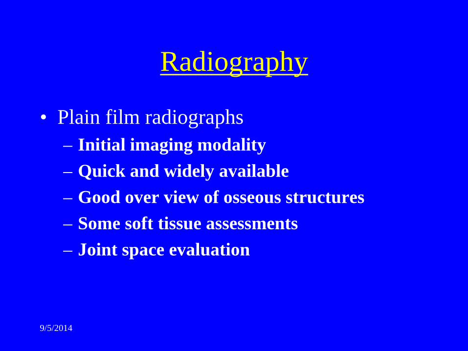
Radiography
• Plain film radiographs – Initial imaging modality – Quick and widely available – Good over view of osseous structures – Some soft tissue assessments – Joint space evaluation
9/5/2014
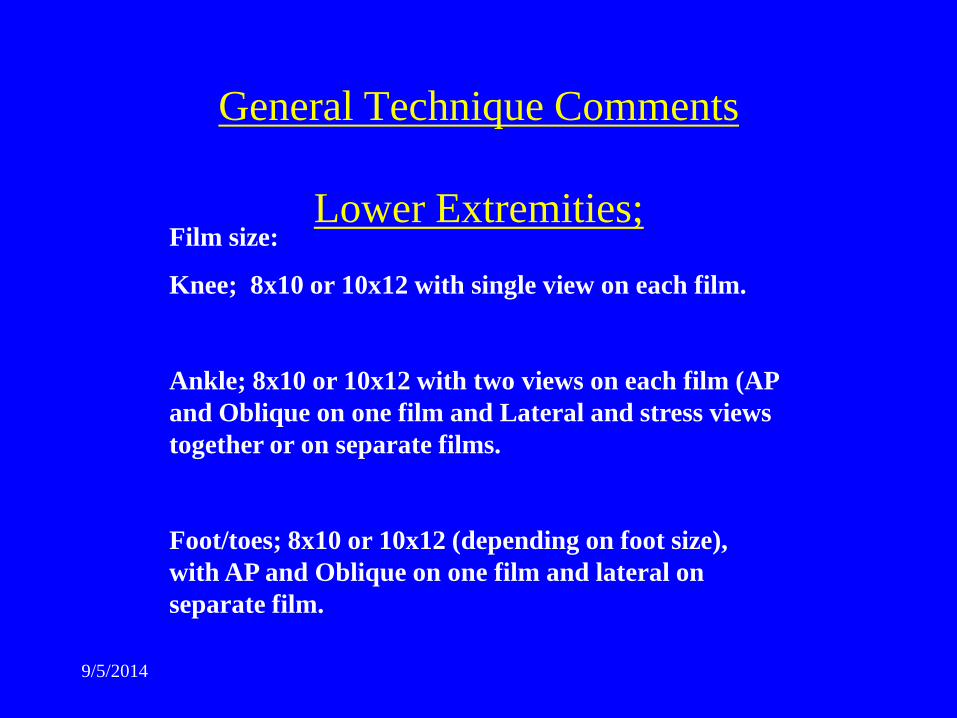
General Technique Comments
Lower Extremities; Film size:
Knee; 8x10 or 10x12 with single view on each film.
Ankle; 8x10 or 10x12 with two views on each film (AP and Oblique on one film and Lateral and stress views together or on separate films.
Foot/toes; 8x10 or 10x12 (depending on foot size), with AP and Oblique on one film and lateral on separate film.

9/5/2014
Knee
• Basic 3 View Series: – AP – Lateral – Tunnel (intercondylar)
• Optional views – Sunrise (Tangential) – Obliques

9/5/2014
AP Knee: FFD: 40”, kVp: 60, mA: 100
Sec/mAs: Dependant on measurement of part
Tube Tilt: 5 degrees ceph.
Patient position: AP, standing or recumbent with knee extended with patella on top.
Central Ray: 1cm inferior to inferior pole of patella.
Measure: through central ray
Breathing: hold

9/5/2014
TABLE TOP TECHNIQUE
Cassette is on the table top. “Nonbucky” technique.
Nonbucky technique will require less x-ray ( 1/2 the mAs) compared to a “bucky” technique.
The grid absorbs some good, as well as scatter x-ray and thus requires more x-ray to make the exposure when a bucky technique is used.

9/5/2014
Lateral Knee FFD: 40”, kVp: 60, mA: 100
Sec/mAs: Dependant on measurement of part.
Tube Tilt: 0 - 5 degrees cephalad.
Patient Position: Affected side against bucky/table top. Knee flexed at 35-45 degrees. To place the patella is proper position.
Central Ray: At the femoral-tibial joint line.
Measure: Through the central ray.
Breathing: Hold

9/5/2014
Lateral Knee:
Patient positioned with affected side against bucky. Knee is flexed.
For knee to be in true lateral position. Elevate and “block” the heel with a foam wedge.
If patient is standing, then strict, true lateral positioning must be obtained.

9/5/2014
Tunnel Knee:
FFD: 40”, kVp: 60, mA: 100
Sec/mAs: Dependant on measurement of part.
Tube Tilt: 0 or 45 depending on lower leg position.
Patient Position: PA, standing or recumbent (prone or kneeling). Knee flexed
Central Ray: Through knee at popliteal fossa.
Measure: Through the central ray
Breathing: Hold

9/5/2014
Tunnel Knee:
Kneeling and standing: 0 tube tilt. Knee flexed with femur at ~25 degree angle with central ray.
Nonbucky technique: Double the mAs or Time for the AP knee projection.
Kneeling
Standing
NOTE: Bucky technique and will require double the mAs of the nonbucky technique.

9/5/2014
Tunnel Knee, Prone method
Knee flexed to 45 degrees with matching caudal 45 degree tube tilt.
Match cassette placement to steep tube tilt.

9/5/2014
Tangential (sunrise) Knee
FFD: 40”, kVp: 60, mA: 100
Sec/mAs: Dependant on measurement of part.
Tube Tilt: 10 degree ceph.
Patient Position: prone with knee flexed maximally.
Central Ray: inferior pole of patella, between femur and patella
Measure: Through the central ray
Breathing: Hold

9/5/2014 http://radiology.rsnajnls.org/cgi/reprint/137/1/57.pdf

9/5/2014

9/5/2014

9/5/2014
Tangential Knee:
Knee is flexed maximally,
patient may need to hold knee flexed with a strap or belt around ankle and held by patient.
Optional method: Seated method with patient sitting at the end of table with cassette on knee. Tube tilt up through patellofemoral joint.
9/5/2014
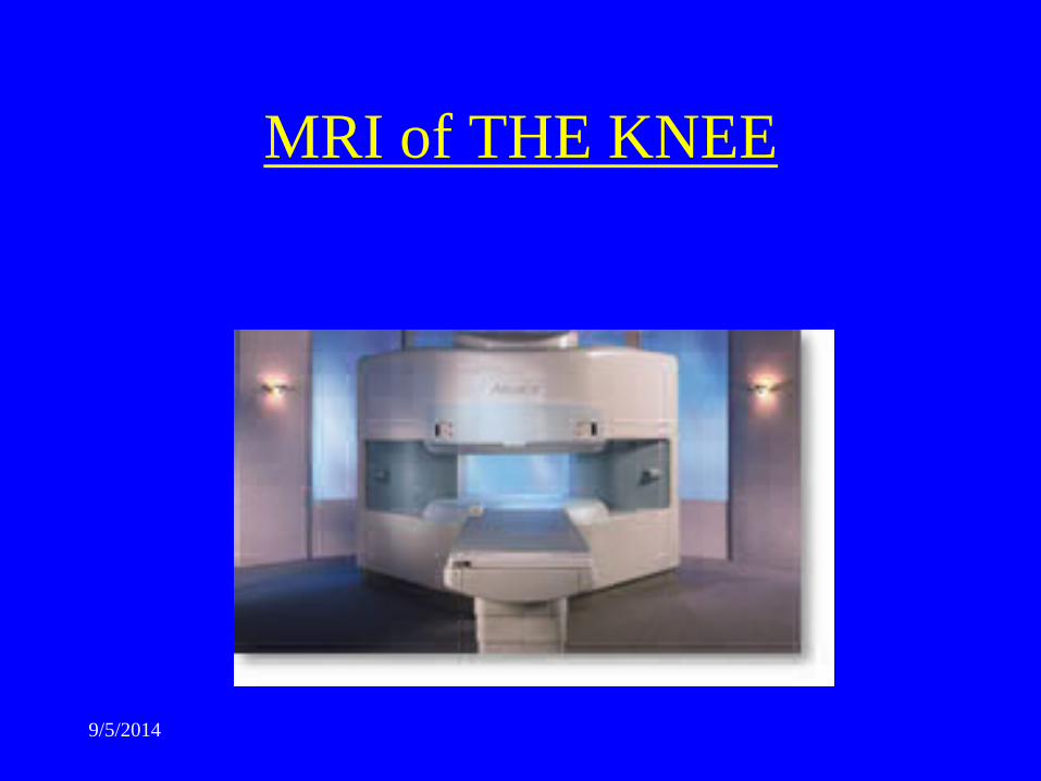
MRI of THE KNEE

9/5/2014
Knee MRI • Indications;
• Trauma; acute with or without effusion. • Bone bruise, ligament, tendon, muscle, meniscal
injuries. • Joint locking - popping • Degenerative joint disease • Non-traumatic effusions of joint • Stress/repetitive stress injury • Abnormal x-rays; e.g. differentiate bone island from
tumor.
9/5/2014
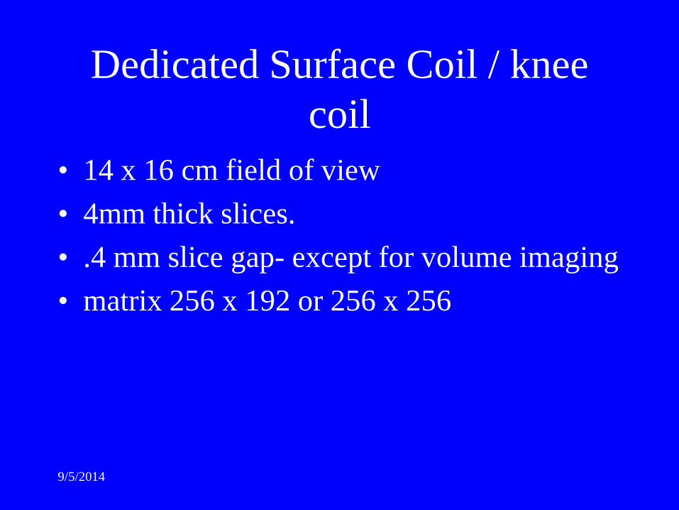
Dedicated Surface Coil / knee coil
• 14 x 16 cm field of view • 4mm thick slices. • .4 mm slice gap- except for volume imaging • matrix 256 x 192 or 256 x 256
9/5/2014
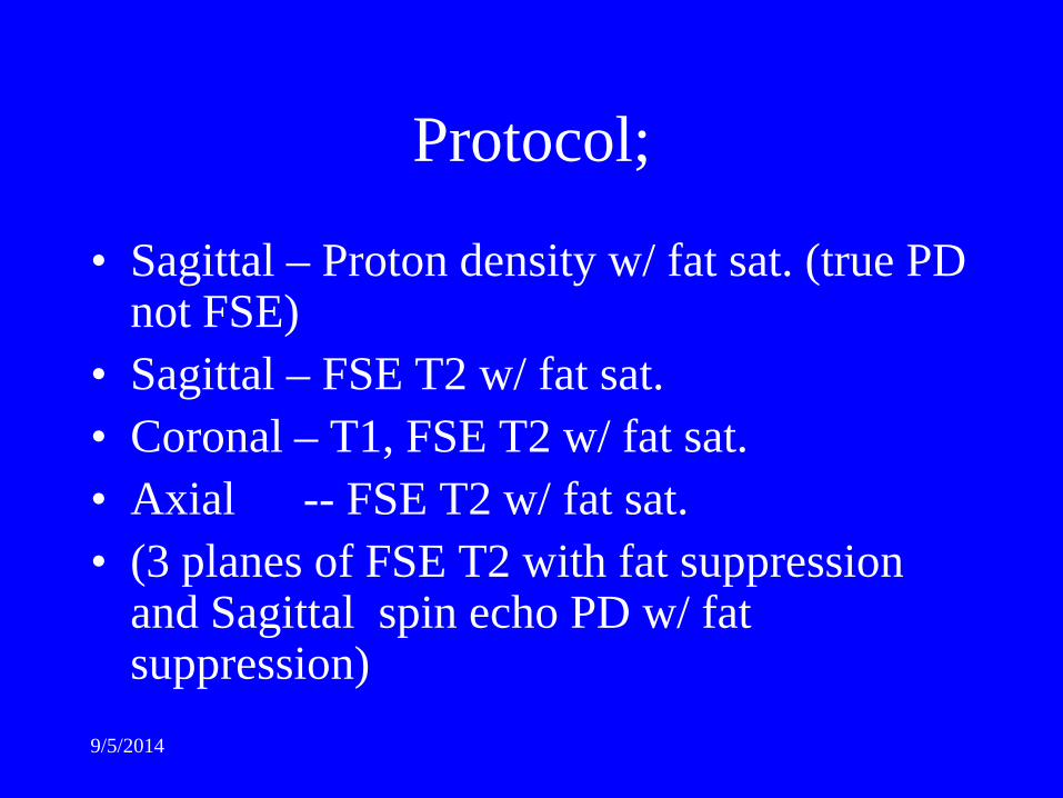
Protocol;
• Sagittal – Proton density w/ fat sat. (true PD not FSE)
• Sagittal – FSE T2 w/ fat sat. • Coronal – T1, FSE T2 w/ fat sat. • Axial -- FSE T2 w/ fat sat. • (3 planes of FSE T2 with fat suppression
and Sagittal spin echo PD w/ fat suppression)
9/5/2014
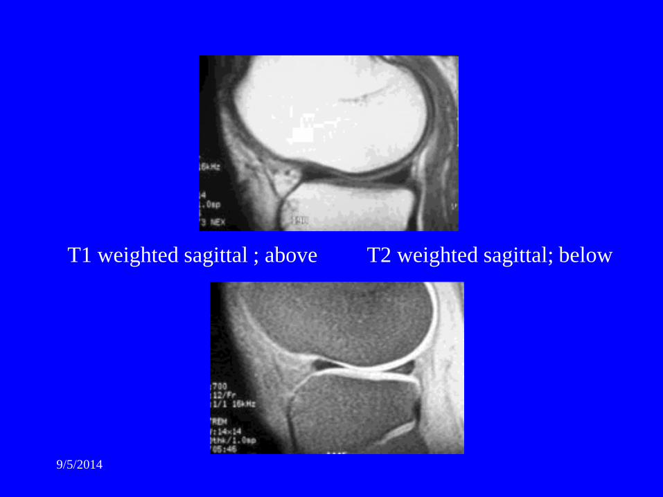
T1 weighted sagittal ; above T2 weighted sagittal; below

9/5/2014
Coronal T1

9/5/2014
Axial T2 - patellar cart. and trochlear cart.
• Retinaculum, collateral, and 2nd/3rd look at cruciates.

9/5/2014
Relaxed state of knee positioning, not forced
• Knee 5 degrees external rotation for ACL on Sagittal
• Menisci seen best in sagittal plane • Short TE to effectively see meniscal tears. • e.g, conventional spin echo T1, PD, and gradient
echo • Best 4 mm thick sagittal SE-PD w/ fat Sat.

9/5/2014
Cruciate Ligaments
• Sagittal – 4 mm thick FSE T2 w/ fat suppression.
• No need for coronal sequences for mensical eval. Sagittal images find 99% of meniscal tears, not coronal.
• Coronal T2 images - FSE T2 w/ fat suppression
9/5/2014
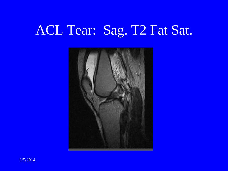
ACL Tear: Sag. T2 Fat Sat.
9/5/2014
Fast Spin Echo PD are unacceptable for view menisci.
• 215 consecutive knees – compared PD and FSE-PD
• results; FSE-PD missed 42 tears that were seen on PD sequences
• PD sequences sensitivity of meniscal tears = 90-95%
• FSE-PD sequences = sensitivity of 80%.

9/5/2014
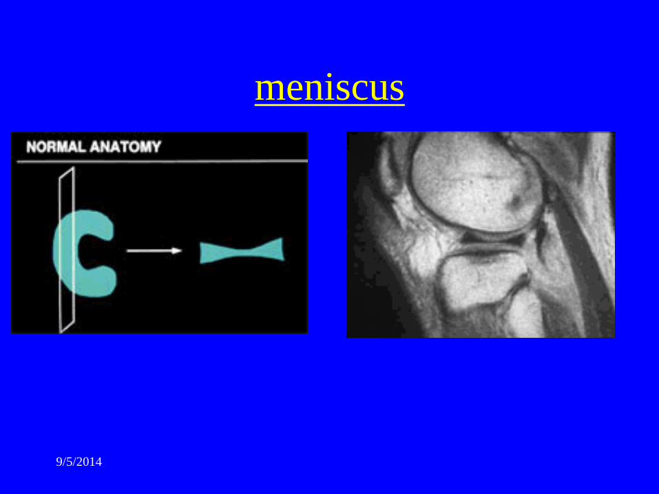
Normal Menisci
• “C” shaped fibrocartilage • Posterior horn of medial meniscus usually
larger than the anterior horn. • Anterior and posterior horns of lateral
meniscus are equal in size. • Posterior horn of either meniscus should
never be smaller than the anterior horn, if it is normal.
9/5/2014
meniscus

9/5/2014
Normal meniscus is devoid of signal on all sequences.
• (exception is children and young adults, due to vascularity of meniscus)
• (Intermediate signal in mensicus, posterior horn near capsular attachments)

9/5/2014
Tears
• Meniscal Tears • Grading systems = several • Most important – signal that disrupts the
articular surface of the meniscus • & • Intrasubstance signal from myxoid
degeneration
9/5/2014
Myxoid degeneration
• Wear and tear or aging? • Not clinically symptomatic • Not tx surgically or clinically • Etiology uncertain • Report it so others know that we saw it and
decided it was not a tear.

9/5/2014
Meniscus Tear Grading

9/5/2014
Meniscus Tear Grading

9/5/2014
If signal comes close to the articular surface but does not quite reach it, then not a tear.

9/5/2014
ACL tears – associated with peripheral meniscus tears of the
medial or lateral meniscus.
• And associated with posterior horn of lateral meniscus tears.

9/5/2014
Oblique/ horizontal Tears;
• M.C. type of tear and involves under surface of posterior horn of medial meniscus
• Commonly degenerative nature instead of trauma.

9/5/2014
Bucket Handle tears:
• Vertical, longitudinal tear – free edge of meniscus is pinched/caught
between fem condyle and tibial artic surface on, for example, valgus injury, lateral meniscus is trapped and free edge pulled from body of meniscus, resulting in bucket handle tear.
• Is 10% of meniscus tears

9/5/2014
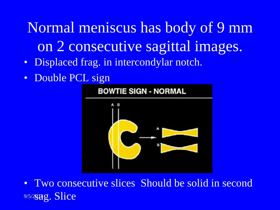
Normal meniscus has body of 9 mm on 2 consecutive sagittal images.
• Displaced frag. in intercondylar notch. • Double PCL sign
• Two consecutive slices Should be solid in second sag. Slice
9/5/2014
Normal meniscus has body of 9 mm on 2 consecutive sagittal images.
• Displaced frag. in intercondylar notch. • Double PCL sign
• Two consecutive slices Should be solid in second sag. Slice

9/5/2014

9/5/2014
Bucket Handle Tear

9/5/2014
Bucket Handle Tear
Double PCL sign

9/5/2014
Bucket Handle Tear
Anteriorly flipped posterior horn fragment

9/5/2014
Medial Flipped meniscus;
• Seen with MRI but overlooked at arthroscope • Flap tear of medial men. with the flap of men.
flipped into the medial gutter underneath the meniscus. Body segments look thinner than normal on MRI.
• Medial men. Will have a piece missing from the undersurface.
• Flipped flap, seen on coronal images.

9/5/2014
Medial Flipped Meniscus

9/5/2014
Radial or Free edge tears;
• Absent bowtie sign – small gap in bow-tie
• Free edge tears are common
• Unusual source of symptoms, unless large.

9/5/2014
Free Edge Tear
Left; The first sagittal image shows a normal body segment and the next one (see right) shows a two-millimeter gap. It is not big enough to be a bucket-handle, plus the blunted anterior fragment or edge is a giveaway that this is clearly chronic and abnormal. This patient had a parrot-beak tear at surgery.

9/5/2014
Cysts;
• Can been seen w/o tear to the articular surface of meniscus.
• Fluid expressed out to adjacent soft tissues on weightbearing, called parameniscal cyst.
• W/o tear, cyst can be missed at arthroscope. • Surgeon may decompress meniscal cyst by
extra-articular approach, rather then via arthroscope.

9/5/2014
Meniscal cysts, don’t show markedly high T2 signal (Intrasubstance degen)
• Parameniscal cyst is usually very high T2 signal.
• Meniscal cyst – signal is like intrasubstance degen. If mass effect(swollen) meniscus then meniscal cyst. If no mass effect and normal sized meniscus, then intrasubstance degeneration.

9/5/2014
Meniscal Cyst
9/5/2014
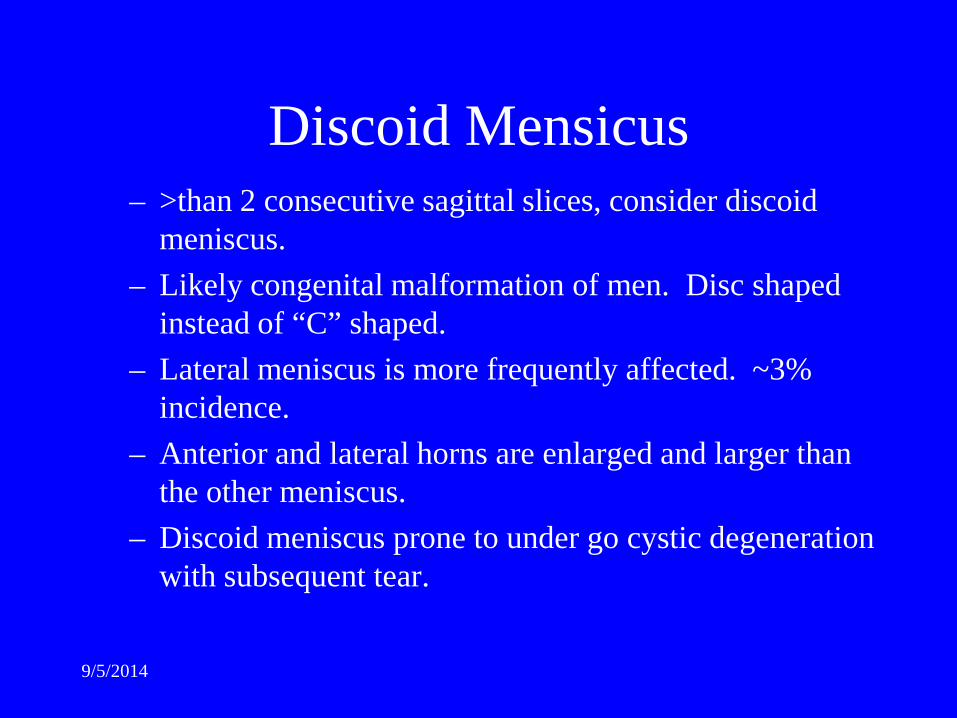
Discoid Mensicus – >than 2 consecutive sagittal slices, consider discoid
meniscus. – Likely congenital malformation of men. Disc shaped
instead of “C” shaped. – Lateral meniscus is more frequently affected. ~3%
incidence. – Anterior and lateral horns are enlarged and larger than
the other meniscus. – Discoid meniscus prone to under go cystic degeneration
with subsequent tear.

9/5/2014
Discoid Meniscus

9/5/2014
Abnormalities w/ absent bow tie sign.
• Bucket handle tear • Radial tear • Medially flip flap tear • Meniscal cyst

9/5/2014
Pitfalls in Absent Bow-tie Sign;
• Children or small adults • Post op. • Severe osteo-arthritis (worn down free
edge, leaving thin body segment of men.) • Older patients ( >65 yoa).

9/5/2014
Pitfalls
• Conditions that cause an appearance similar to a pathology or mimic the pathologic process or can hide pathology and cause reviewer to falsely interpret images.
• Must be aware of pitfalls to prevent false positives or false negatives.

9/5/2014
Speckled anterior horn of lateral mensicus.
• Mimics a macerated or torn anterior horn. • Caused by fibers of ACL inserting into
meniscus • Up to 60% of normal patients
9/5/2014
Pitfalls involving the posterior horn of Lateral meniscus
• Meniscofemoral ligament insertion • Pulsation artifact from the popliteal artery • Magic angle phenomenon • Popliteus tendon. • Concomitant ACL tear

9/5/2014
Transverse ligament
– Insertion on anterior horn of meniscus. – Runs across anterior aspect of the knee in
Hoffa’s fat pad from anterior horn to anterior horn.
– Function – not known-, not present in every knee.
– Insertion of anterior horn of lateral mensicus – often has appearance of tear. D.D. by following it across knee on sequential sagittal images.
9/5/2014
Meniscofemoral ligament insertion:
• (Posterior Horn lateral meniscus pitfall) • Insertion of meniscofemoral ligament of Humphry
(anterior to PC) or Wrisberg (posterior to PCL) can give appearance of a meniscal tear.
• Meniscofemoral lig. is present in 75% of knees. • Originates on med. femoral condyle and runs obliquely
across the knee in the intercondylar notch. Anterior (lig of Humphry) or posterior (lig. Of Wrisberg) to the PCL and inserts into the posterior horn of the lateral mensicus.
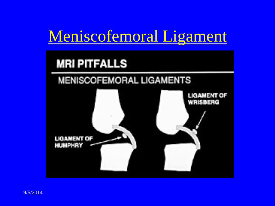
9/5/2014
Meniscofemoral Ligament

9/5/2014
Meniscofemoral ligament insertion: continued.
• ? of pseudotear follow the insertion of one of the meniscofemoral ligaments through the intercondylar notch to the PCL on sequential sagittal images.
• 2-3% of knees both meniscofemoral ligaments are present (both Humphry and Wrisberg).
• Function is not known and injury to it has not been described.

9/5/2014
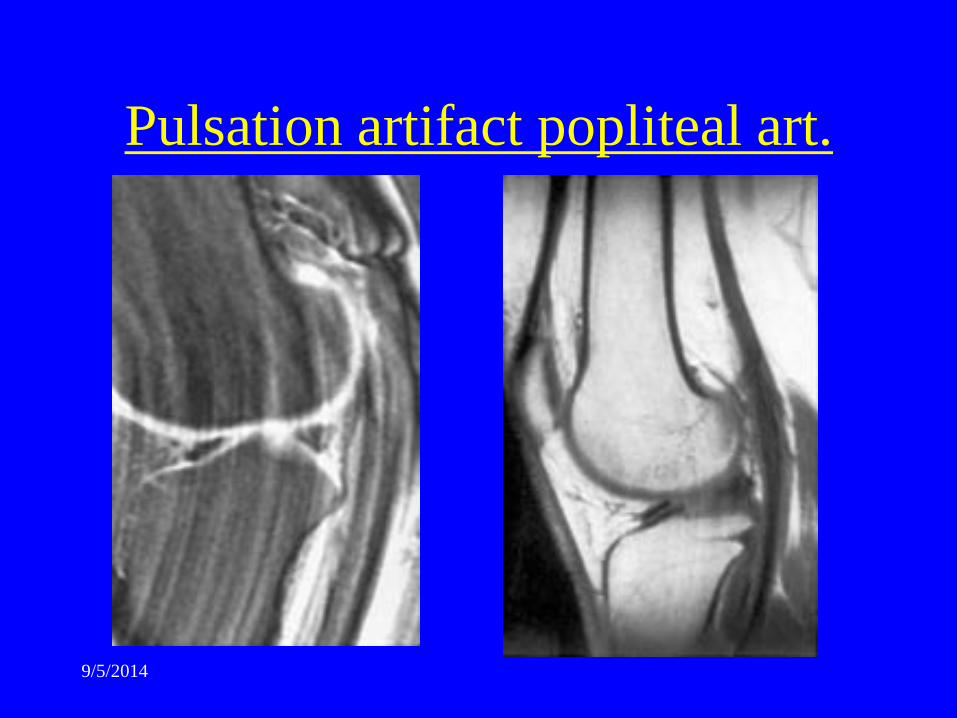
Pulsation of Popliteal Artery
• Popliteal artery just posterior to the posterior horn of lateral meniscus.
• Pulsations artifacts project over posterior horn of lateral men. And cause areas of increased signal and mimic vertical tear.
• Corrected by swap of phase and frequency directions to make pulsations superior to inferior rather than anterior/ posterior.
9/5/2014
Pulsation artifact popliteal art.
9/5/2014
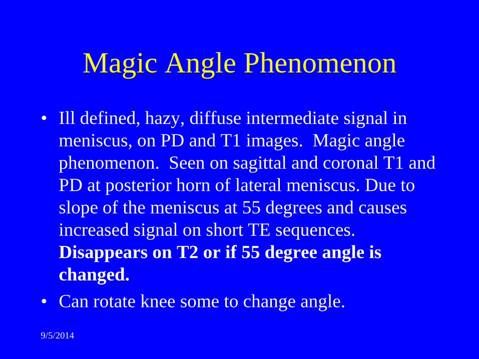
Magic Angle Phenomenon
• Ill defined, hazy, diffuse intermediate signal in meniscus, on PD and T1 images. Magic angle phenomenon. Seen on sagittal and coronal T1 and PD at posterior horn of lateral meniscus. Due to slope of the meniscus at 55 degrees and causes increased signal on short TE sequences. Disappears on T2 or if 55 degree angle is changed.
• Can rotate knee some to change angle.

9/5/2014
Magic Angle Phenomenon

9/5/2014
Popliteus Tendon Pseudotear
• Tendon originates on the lateral femoral condyle and extends inferiorly between the posterior horn of the lateral mensicus and the joint capsule.
• Where tendon passes between the meniscus and the capsule it can give appearance of mensicus tear.
• Don’t confuse tear with normal tendon. • Vertical tear of posterior horn of lateral
meniscus often occurs with ACL tears.

9/5/2014
Popliteus Tendon vs. Tear
Pseudotear; Above.
True posterior horn lateral meniscus tears; right top and bottom. (if tear then posterior horn is smaller than anterior horn.
Popliteus Tendon
9/5/2014
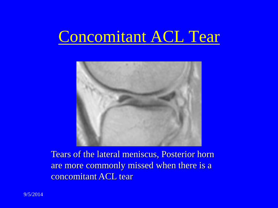
Concomitant ACL Tear
Tears of the lateral meniscus, Posterior horn are more commonly missed when there is a concomitant ACL tear

9/5/2014
Ligaments:

9/5/2014
ACL
– Straight taut fibers that parallel roof of the intercondylar notch.
– Typically striated with some high signal within it, especially at the insertion on the tibia. T2 sagittal recommended for evaluation of ACL.
– MRI accuracy of ACL tear is 95-100%. – Torn ACL is obvious – no normal appearing fibers of
ACL can be identified. – When it tears it explodes leaving nothing with which a
surgeon can do a primary repair.

9/5/2014
Normal ACL
Child ACL tear
Adult ACL tear

9/5/2014
ACL
• Occasionally ACL tear seen with fibers intact but flatter than normal Angle of ACL (fibers should be parallel with intercondylar roof). Partial tear of ACL is treated non-operatively. Little info. Concerning accuracy of DX of partial tears in imaging literature.

9/5/2014
Chronic ACL tears.
Angle of ACL is too flat, not parallel with roof of intercondylar fossa.
9/5/2014
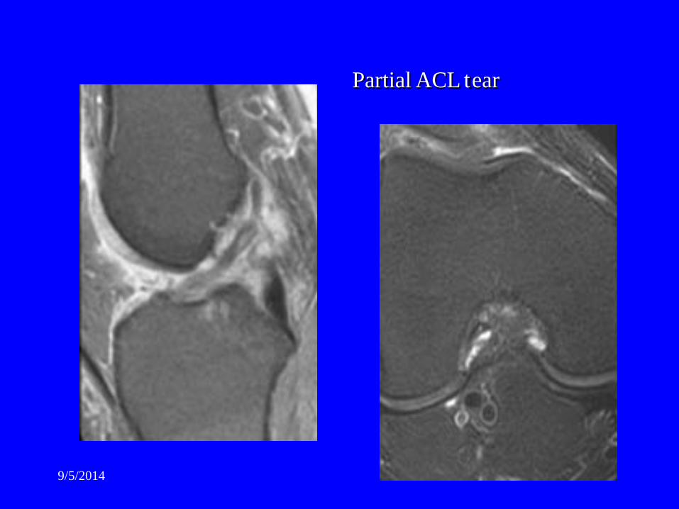
Partial ACL tear

9/5/2014
Pitfall
• ACL cyst. Unknown cause, ACL is distended with muscinous fluid
• normal ACL fibers are not seen and appear disrupted.
• Patient has no instability and are asymptomatic or have feeling of swelling or fullness in the knee and are unable to fully flex the knee, because of he mass effect.
• Seen in 1% of knees.
9/5/2014
ACL repair
• Reconstructed via tendon graft from the patellar tendon. (occasionally an donor allograft can be used but there are not enough from donors to go around) Dr. Jannsen lecture 11/15/03). 1/3 of patients do ok without repair of ACL. However, the amount of DJD that occurs in knees without repair will result in knee replacement later.

9/5/2014
ACL repair
• Tendon graft is dead tissue and the body uses the graft as a lattice work and slowly incorporates and replaces it with living tissue. You actually grow a new tendon on and through the graft. Japanese have done DNA studies on post-surg patients and have shown that the donor DNA is replaced by the hosts DNA over time.
9/5/2014
ACL repair
• Prosthetic/synthetic ligament repairs are not as successful because you never grow a living ligament and the synthetic ligament will eventually fail, due to the many cycles of motion the knee goes through (bend a wire until is breaks).

9/5/2014
Post Op ACL reconstruction.
• ACL graft should be present as taut strux. Some increased signal on T2 sag.
• If graft is disrupted or absent – it has failed. • Tibial tunnel (graft tunnel) should be
parallel to roof of femoral intercondylar notch. If too sharp, graft can impinge by femur on extension. If too flat graft can be too lax and lead to instability.

9/5/2014
Arthrofibrosis • One of most common reasons for pain post-op is presence
of arthrofibrosis (scar) in Hoffa’s fat pad. May require re-operation. – Several appearances can be seen. – Cyclops lesion-- Round mass of scar in Hoffa’s fat pad.
Can decrease knee extension and needs resection. – Linear scar that extends to inferior pole of the patella
can restrict patellar motion and cause pain.
9/5/2014
PCL • Low signal, gently curving structure in the
intercondylar notch. • Infrequently torn and even less frequently
surgically repaired. • PCL tears missed because don’t get high signal on
T2. Can be high signal on STIR. • Uncommon – but can avulse from tibial insertion
– easily diagnosed.

9/5/2014
PCL • If torn, most surgeons will not repair and
won’t inspect at arthroscopy.

9/5/2014
• Cunningham • Tweitmeyer

9/5/2014
MCL • Origin – medial distal femur, inserts medial
proximal tibia. Fibers interlaced with joint capsule at the level of the joint.
• Medical meniscus is directly attached • Extrasynovial – not seen or repaired
arthroscopically • Accuracy of MRI not established but agreed
that MRI is highly accurate in depicting the MCL.
9/5/2014
MCL Injuries • 3 –grades of injury. Clinically correspond to 3
appearances of MCL w/ coronal T2 image. • Grade I- sprain – Increased signal of soft tissue
medial to MCL (increased signal outside of MCL from jt.)
• Grade II – severe sprain – Partial tear – Increased signal in soft tissue medial to MCL and also increased signal or partial disruption of MCL.
• Grade III – complete tear – disruption of MCL

9/5/2014
MCL
Grade II MCL sprain

9/5/2014
MCL
Grade II MCL sprain with meniscocapsular separation. Fluid between MCL and medial meniscus.
Also note bone bruise lateral femoral condyle

9/5/2014
MCL Repair:
• MCL is seldom repaired even if completely disrupted, unless multiple other ligaments are torn.
• Casting works very well for good result healing.
• Bracing can work but the patient must wear the brace 24-7 for good result.

9/5/2014
MCL repair, continued
• Grade I and II sprains usually treated conservatively with bracing and continue athletic activity as pain allows.
• High signal medial to the MCL may occur from causes unrelated to MCL sprain, Such as Sub Q edema.

9/5/2014
Mensicocapsular Separation; • Easily dx on coronal T2 by noting fluid between the MCL
and the medial meniscus. Can be overlooked on T1 Coronal.
• Brace is not acceptable for treatment of Mensicocapsular separation.
• The vascular interface of between the MCL and meniscus can become avascular with continued activity – resulting in meniscus not healing.
• Meniscocapsular separation – needs immobilized or surgery.
9/5/2014
LCL complex; • Composed of many structures;
• 3 are well evaluated with MRI • From posterior to anterior; • Biceps femoris tendon • Fibulocollateral ligament (true LCL) • Ilitotibial band (ITB)

9/5/2014
LCL tears are less common than MCL tears
• However, Nearly emergency situation because associated with injury to the “posterior lateral corner”. Posterior lateral corner injuries result in instability if not treated within 3-4 days of injury (surgical).

9/5/2014
LCL tears are seen with;
• 1. Popliteus tendon tears • 2. Arcuate ligament tears (posterior lateral
capsular thickening) • 3. ACL or PCL tears.

9/5/2014
Popliteus Tendon Tears;
• Isolated or associated with other injuries of the posterior lateral corner.
• Tears demonstrated by; – Lax popliteus tendon – Increased signal in and around popliteus muscle and – Increased fluid in tendon sheath. (normal pop. tendon not to confuse with vertical men. tear.)

9/5/2014
Faulty Extensor Mechanisms Acute trauma, overuse injuries, developmental alterations may lead to dysfunction of the extensor mechanism of the knee, structures involved;
Quadriceps muscle group,
Quadriceps tendon,
Patella,
Patellar tendon,
Patellar retinaculum,
Adjacent soft tissues
9/5/2014
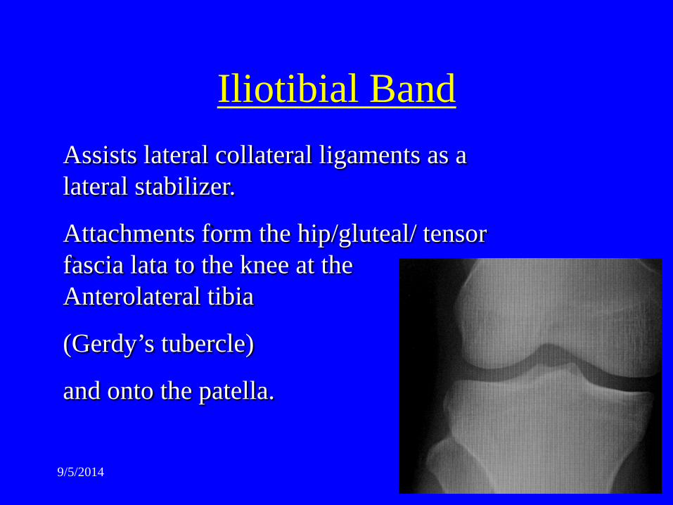
Iliotibial Band Assists lateral collateral ligaments as a lateral stabilizer.
Attachments form the hip/gluteal/ tensor fascia lata to the knee at the Anterolateral tibia
(Gerdy’s tubercle)
and onto the patella.
9/5/2014
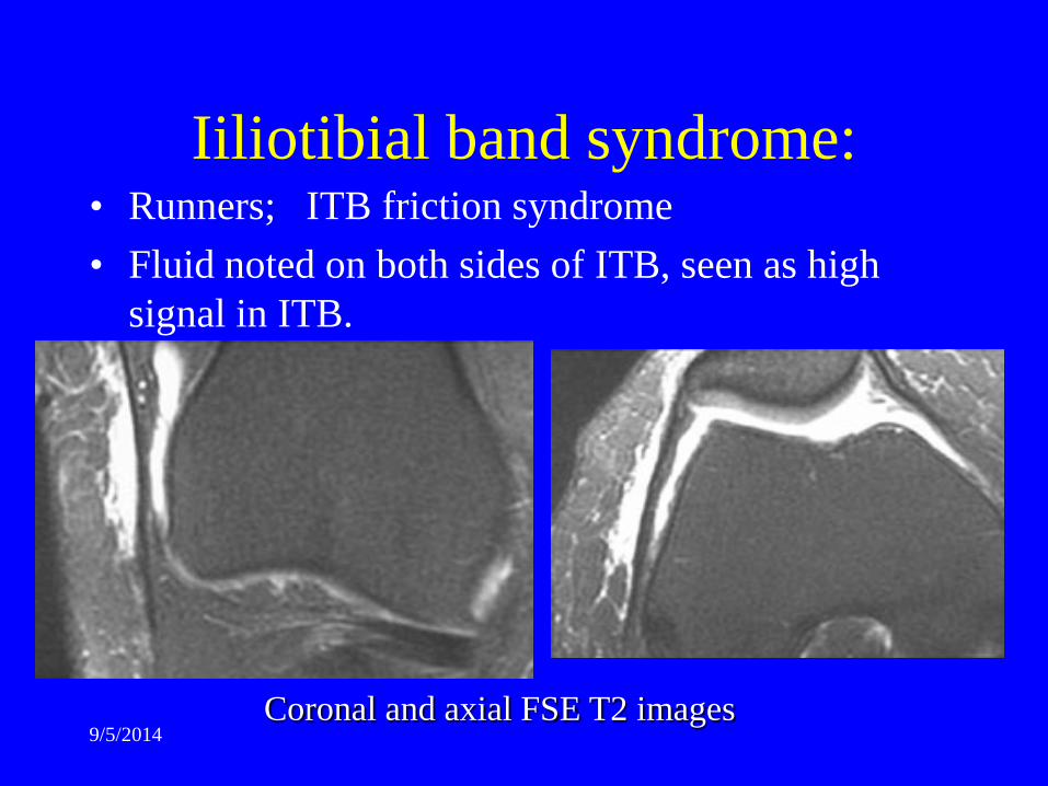
Iiliotibial band syndrome: • Runners; ITB friction syndrome • Fluid noted on both sides of ITB, seen as high
signal in ITB.
Coronal and axial FSE T2 images
9/5/2014
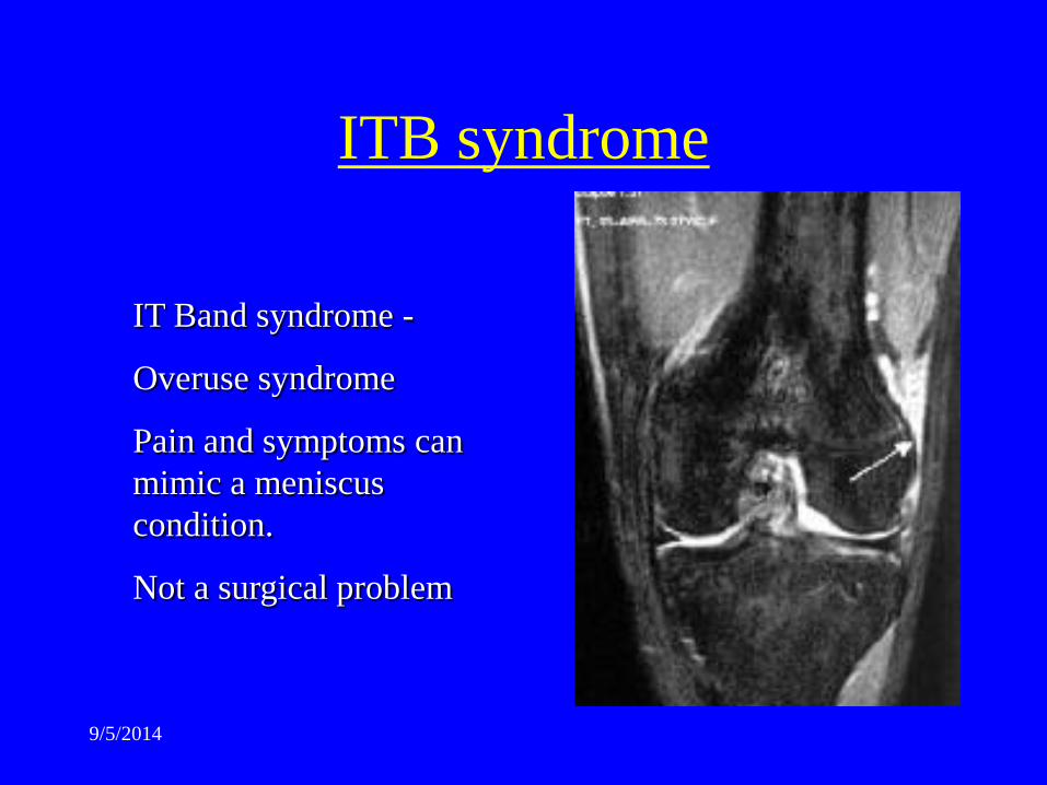
ITB syndrome
IT Band syndrome -
Overuse syndrome
Pain and symptoms can mimic a meniscus condition.
Not a surgical problem

9/5/2014
Synovial Plicae; • Thin fibrous band seen on axial
images. • Extends from medial joint
capsule toward the medial facet of the patella
• Normal structure- remnant of embryologic development of knee.
• Superior, Inferior, and Medial patella plica.

9/5/2014
Plica

9/5/2014
Synovial Plicae; • Medial plica only one associated with symptoms,
may become thickened and stiff and trapped between patella and the femur.
• Causes pain and clicking and locking, similar to torn meniscus.
• No established measurement for thickness on MRI “just judged” to be thickened from experience.
• Plica syndrome- not common – dx – tx is surgical removal of the medial plica.

9/5/2014
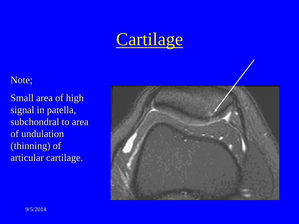
Cartilage
FSE T2 or Fast STIR
evaluate cartilage surfaces in two or three planes.
Cartilage defects
OCD, Trauma, DJD, CPPD.
9/5/2014
Cartilage
Note;
Small area of high signal in patella, subchondral to area of undulation (thinning) of articular cartilage.

9/5/2014
Patella • Lateral dislocation – rapidly reduces on its own. • ~50% of patients are aware of what really occurred. • Pts get referred for MRI to r/o internal derangement. • MRI reveals contusion on anterior lateral portion of
lateral femoral condyle. • Patellar contusion (kiss contusion) at medial facet. • Medial retinaculum is always injured by frank tear
may not be found.

9/5/2014
Patella
Position of patella in trochlear notch
cartilage thickness
patellar osseous changes; fx, spur, bruise.
9/5/2014
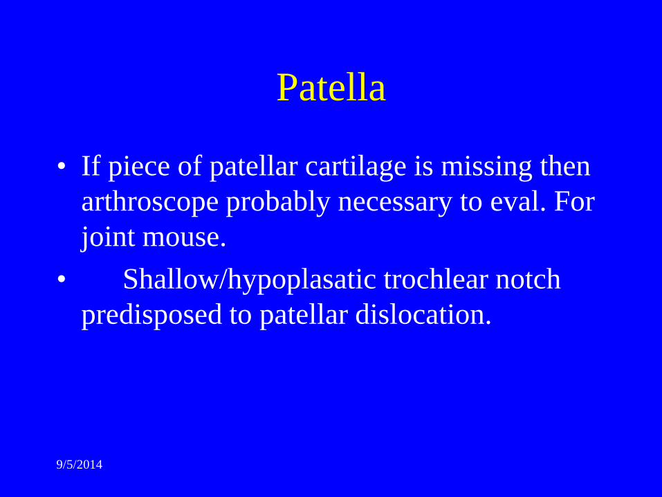
Patella
• If piece of patellar cartilage is missing then arthroscope probably necessary to eval. For joint mouse.
• Shallow/hypoplasatic trochlear notch predisposed to patellar dislocation.
9/5/2014
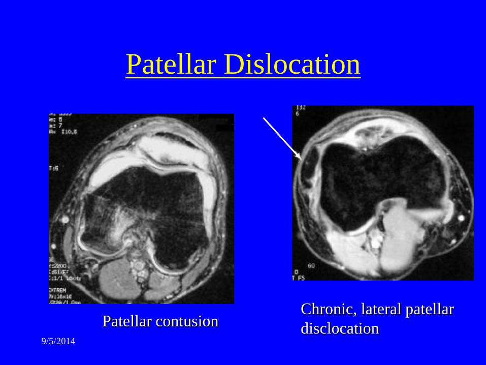
Patellar Dislocation
Patellar contusion Chronic, lateral patellar disclocation
9/5/2014
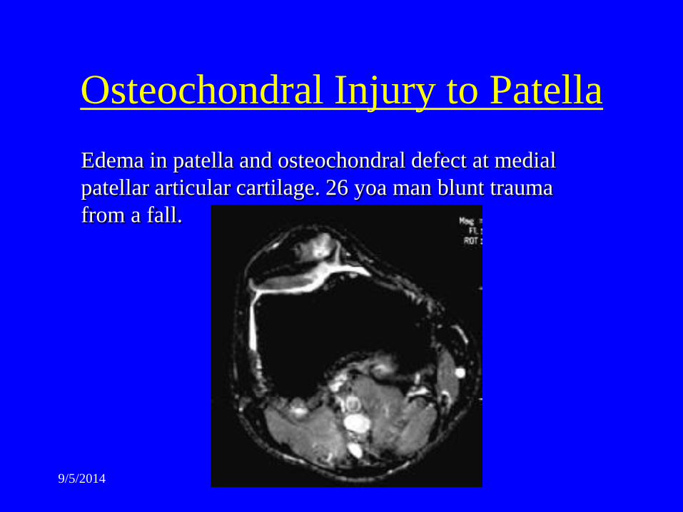
Osteochondral Injury to Patella Edema in patella and osteochondral defect at medial patellar articular cartilage. 26 yoa man blunt trauma from a fall.

9/5/2014
Bipartite Patella Bipartite patella is a rare cause of anterior knee pain. In symptomatic bipartite patellae, the pain may result from chronic stress injury.

9/5/2014
Patellar Tendon: • Jumpers Knee: Increased signal
from myxoid degeneration at the superior margin of the patellar tendon. ? tx surgical resection of myxoid tissue to heal. – Increased pain at superior
patellar tendon on resisted extension from 90 degrees of knee flexion.
– Fat pad syndrome (anterior knee pain) will have decreased pain on resisted extension of knee from 90 degrees of flexion.
9/5/2014
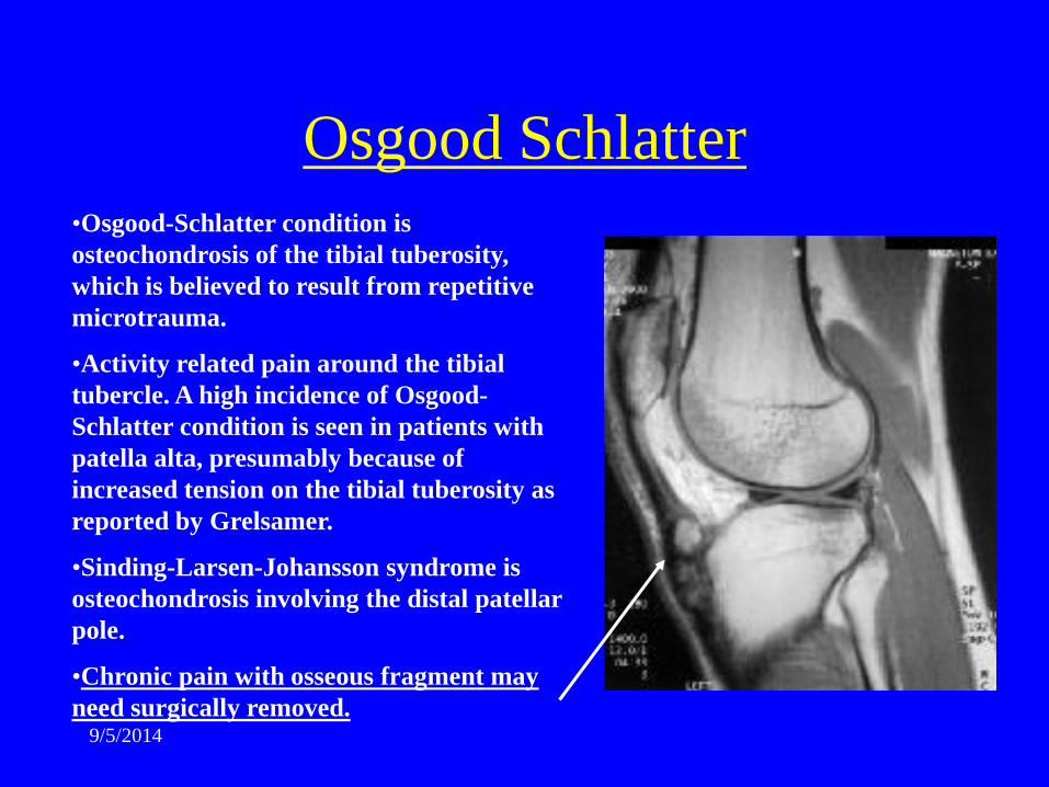
Osgood Schlatter •Osgood-Schlatter condition is osteochondrosis of the tibial tuberosity, which is believed to result from repetitive microtrauma.
•Activity related pain around the tibial tubercle. A high incidence of Osgood-Schlatter condition is seen in patients with patella alta, presumably because of increased tension on the tibial tuberosity as reported by Grelsamer.
•Sinding-Larsen-Johansson syndrome is osteochondrosis involving the distal patellar pole.
•Chronic pain with osseous fragment may need surgically removed.
9/5/2014
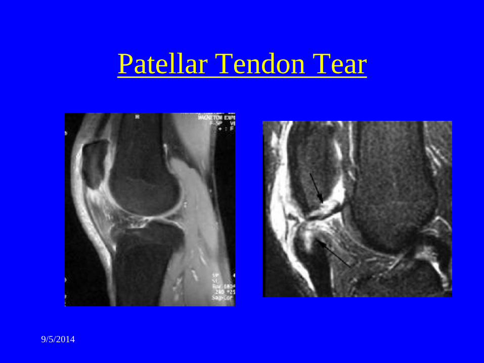
Patellar Tendon Tear
9/5/2014

9/5/2014
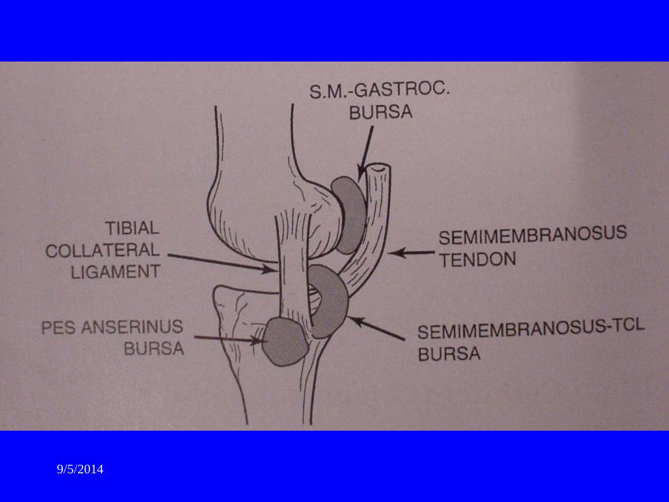
Bursae: 5 of significance
Popliteal bursa
Pes Anserinus bursa
Prepatellar bursa
Semimembanosus bursa
Tibial Collateral ligament bursa

9/5/2014
Posterior bursa;
1. Popliteal (Baker’s cyst) between the medial head of gastroc and the semimembranosus.
• Should be no more than 5 cc of fluid.

9/5/2014
Anterior bursae; 2. Pre-patellar bursa – housemaid’s knee
9/5/2014
Pes Anserinus bursa ( pes anserine means goose’s foot) • Anteromedial tibia just below joint line. • Pes Tendons – Gracilis, Sartoris, and Semitendonosus • Bursae lies beneath the tendons – when inflamed, extends
proximally towards the joint. • Pts can have clicking, popping, and painful knee, like a
mensicus tear.
Anterior bursae;

9/5/2014
Anterior bursae;
9/5/2014
Anterior bursae;
4. Semimembranosus/Tibial collateral ligament bursa can mimic meniscal tear – fluid signal drapes over semimembranosus
tendon on coronal and sag. images. – Arises at level of meniscus and extends
inferiorly. – D.D. with parameniscal cyst. – Bursa has no
communication with meniscus.

9/5/2014
Anterior bursae;

9/5/2014
Anterior bursae;
5. Tibial collateral ligament bursa deep to the medial collateral lig. and extends vertically above and below the joint line.
(space between the superficial and deep MCL layers) sjg opinion.

9/5/2014
Bones:

9/5/2014
Contusions:
• Geographic ( more well defined margin of contusion) more likely to progress to Osteochondritis Dessicans (OCD).
• Reticular pattern, ill-defined, less likely to progress to OCD.

9/5/2014
Contusion Patterns/ locations;
• ACL tear: posterior lateral aspect of tibial plateau
• And possible kiss contusion of the anterior lateral femoral condyle (over anterior horn of lateral meniscus).

9/5/2014
2nd contusion pattern with ACL tear;
• posterior medial tibial plateau and medial femoral condyle, posterior to MCL. Must have meniscocapsular separation of medial menicus as well.

9/5/2014
Soft tissues:

9/5/2014
Strains of muscles • Grade I – microscopic damage, no loss of
function. • Grade II – partial tearing with some loss of
function – anything between grade I and grade III • Grade III –complete disruption of muscle with
loss of function of the unit.
9/5/2014
• Plantaris tendon; “Tennis Leg” calf pain like medial gastroc injury or d.d. with DVT. – Swelling in popliteal fossa, – purplish skin discoloration from hemorrhage.
• Hamstring Ruptures;
Strains of muscles

9/5/2014