Embed Size (px)
Citation preview

169RADIATION THERAPIST, Fall 2016, Volume 25, Number 2
CEDirected Reading
Image-guided adaptive radiation therapy (IGART) is an evolving approach to cancer treatment and represents the latest effort to fulfill a longstanding goal for radiation oncology: escalated therapeutic doses of ionizing radiation that are delivered precisely in time and anatomic space to tumor tissue while sparing healthy, nontarget tissues to maximize the therapeutic ratio. This article introduces and reviews the concepts, history, radiobiology, targeting, technological, planning, delivery, and plan-adaptation principles and approaches to IGART, with a focus on the increasingly important roles of functional imaging modalities in the IGART process.
This article is a Directed Reading. Your access to Directed Reading quizzes for continuing education credit is determined by your membership status and CE preference.
After completing this article, the reader should be able to:Describe ways tumor and nontarget anatomic volumes can change over time.List the steps, goals, and component technologies of image-guided adaptive radiation
therapy (IGART).Explain the roles of various imaging modalities in IGART planning, monitoring, and
plan adaptation.Identify time scales at which offline, online, and dynamic treatment plan adaptation
occur.Describe the existing and emerging roles of functional imaging and biomarkers in IGART.Compare IGART approaches in the treatment of different types of cancers.
Bryant Furlow, BA
Image-Guided Adaptive Radiation Therapy
Radiation therapy plays an important role in cancer treat-ment for many patients, but its curative potential has been
constrained by dose-limiting toxici-ties that can follow irradiation of healthy, nontarget tissues, and insuffi-cient dose delivery to tumor tissue. Many technological advances over recent decades have helped reduce the incidence and magnitude of these problems by better tailoring radiation delivery to target volumes and better sparing adjacent nontarget organs and tissues. One persistent challenge has been that tumors, nontarget tissues, and organs move during treatment, and their contours, positions, and sizes can change during the course of treatment. These changes can affect delivered radiation dose distributions in ways that cause significant devia-tions from treatment plans.1
Image-guided adaptive radiation therapy (IGART) is a recent and evolv-ing integrative approach to further minimize radiation toxicities by detect-ing and adapting treatment plans to compensate for sources of external-beam radiation therapy targeting variances.2 The goal of IGART is to adjust and improve the therapeutic ratio continu-ally throughout a patient’s treatment course.3 It represents a synthesis of image-guided radiation therapy, which usually is confined to setup corrections, and adaptive radiation therapy, which refers to the technological capture of feedback data so that treatment plans can be modified to compensate for ana-tomic change.1 IGART also includes retrospectively reconstructed dose delivery history as feedback data in plan reoptimization.1
Conventional radiation therapy relies on initial imaging for radiation

170 RADIATION THERAPIST, Fall 2016, Volume 25, Number 2
CEDirected Reading
Image-Guided Adaptive Radiation Therapy
(CT), magnetic resonance (MR) imaging, and func-tional and other imaging modalities all have undergone technological advances over the past decade, helping to usher in an era of clinical IGART.5 Functional positron emission tomography (PET)-CT imaging plays a role in IGART treatments employing modulated radiation therapy, intensity-modulated radiation therapy (IMRT) and volumetric modulated radiation therapy (VMAT).3
Although IGART will continue to evolve, it already is playing an increasing role in cancer treatment.2,8 IGART is not a single technology or a seamless, inte-grated set of technologies; instead, it is a paradigm or approach to radiation therapy that employs a collec-tion of tools—planning, targeting, gating, delivery, and adaptation software and equipment—to address clinical challenges presented by a patient’s particular disease. This toolkit allows options and approaches for achieving the therapeutic goals of IGART: improved tumor control and patient safety with a reduced risk of acute or late radiotoxicities.5,6,8,9 However, components of IGART technologies, such as volumetric imaging, image registration, image data postprocessing, and rapid radiation therapy adaptation and replanning algorithms, are being integrated into commercially available radiation therapy systems, and IGART-ready systems will become less expensive and widely avail-able in coming years.1,6
IGART is “a generic term” encompassing several ways to use medical images to assist radiation therapy, and some of these technologies are being researched.8 Researchers have summarized several “fundamental aims” for IGART, including2,3,8,10: Radiation therapy planning. Anatomic and functional target delineation. Identification of tumor biology and the molecular
biology of adjacent tissues to inform modulated treatment planning.
Patient positioning because the patient’s loca-tion relative to planned radiation beam paths can reduce delivery errors associated with interfrac-tion changes in anatomic positioning and target volumes.
Detection of intrafraction motion with the aim to compensate for it.
Determination of how much radiation has been delivered to the target.
therapy simulation and treatment planning, and fol-low-up imaging during patient setup before dose deliv-ery to reduce targeting errors associated with incorrect patient positioning.4 In contrast, IGART involves fol-low-up imaging and dose-delivery assessments through-out a course of radiation therapy to correct for setup and anatomical sources of targeting errors and to deter-mine whether treatment plan modification is required before each subsequent dose is delivered.4 These goals are achieved through imaging during and between dose deliveries. IGART is “the ongoing use of imaging to monitor, update, and adjust the treatment process,” and dynamic IGART is “the use of streams of imaging data to automatically control the dose delivery in real-time.” 5 IGART can be described as “a closed-loop [radiation therapy] process in which treatment plans are modified using systematic feedback of measurements over time.” 2 The ongoing adjustment of radiation therapy planning volumes and margins allows radiation dose escalation without increased radiation toxicity to the patient.2,6,7
Technologically, IGART is situated at the intersec-tions of advanced imaging, gating, planning, and radia-tion delivery technologies and software systems, but it is not a new concept. Essentially, IGART represents a fulfillment of longstanding goals for radiation therapy: the conformal delivery of toxic levels of ionizing radia-tion to tumor tissue while largely sparing healthy, non-target tissue, and thereby maximizing the therapeutic ratio. These goals have driven radiation therapy-plan-ning imaging for much of the past century.8 Emerging technological advances mean those goals now can be achieved better than ever before, accommodating both anatomic and tumor motion caused by physiologic pro-cesses, such as respiration, and slower changes in target and nontarget volumes associated with radiation thera-py and other cancer treatments, either directly (tumor shrinkage or deformation caused by radiation therapy) or indirectly (a patient’s weight loss).6
Imaging is central to this process. The role of imag-ing in radiation therapy was long restricted to the use of 2-D—and later, 3-D—diagnostic radiographic images acquired for treatment planning.5 However, volumetric imaging now is incorporated into radiation treatment planning and delivery, and multimodality imaging is integral to treatment planning, patient setup, and the delivery of radiation therapy.5 Computed tomography

171RADIATION THERAPIST, Fall 2016, Volume 25, Number 2
CEDirected Reading
Furlow
Radiation therapy outcomes monitoring (eg, tumor deformation, shrinkage, or evidence of intrinsic or acquired radioresistance). PET imag-ing can detect interfraction gross tumor volume (GTV) changes sooner than CT imaging can. PET and PET-CT also play a clinical role in response assessment in many cancers.
In the future, clinical IGART practice likely will incorporate the use of numerous molecular and metabol-ic biomarkers of treatment response, in addition to ana-tomic measurements, to further and more rapidly inform radiation therapy plan reoptimization.2 Elements of this approach already are evident with the use of functional imaging such as PET in IGART treatment.
HistoryIGART has emerged because of innovations in radia-
tion therapy delivery and medical imaging, particularly the availability of modulated treatment delivery, cone-beam CT (CBCT), and other onboard volumetric imag-ing capabilities.1 The technological demands of contem-porary IGART required advances in computer process-ing speeds, imaging equipment design, and image data processing algorithms that emerged in the 1990s. The electronic portal image device (EPID) and standalone CT scanners offered early feedback systems for adaptive redelineation of tumor volumes, for example.1 Three-dimensional conformal radiation therapy (3-D CRT) was an early example of IGART because the beam portal could be adjusted based on feedback from online portal imaging.1 The emergence of IMRT, VMAT, stereotactic treatment methods, and real-time gating, motion-harmo-nized 4-D radiation therapy (using 4-D CT) to accom-modate tumor movement caused by respiratory motion, allowed more precisely delivered radiation beams and smaller safety margins around irradiated volumes.1,6
Although IGART is not a single technology, it devel-oped and matured during the IMRT era of treatment delivery and sometimes is described as a historical outgrowth of—or complement to—IMRT, which also emerged in the 1990s.1, 8 It uses imaging-data-informed algorithms to generate precise 3-D tumor-irradiation plans to deliver multiple external radiation beams of different shapes, orientations, and intensities, using adjustable leaf collimators and allowing steep radiation gradients across anatomy.3,8 It is particularly well suited
for the treatment of complex tumor contours and might therefore be more sensitive than other treatment tech-niques to interfraction tumor deformation, particularly in light of its steep dose gradients.3
Radiation therapy planning volumes can be affected by small differences in patient position, organ motion, or changes in a patient’s body mass. Modulated treatment methods better achieve external-beam radiation therapy’s longstanding conformal goals for radiation dose delivery across anatomic volumes, and growing evidence sug-gests that it improves the therapeutic ratio.1,6 However, modulated treatment methods’ improved delivery of the correct dose to the correct place is not sufficient alone to meet the goals of radiation therapy because of the other intrafractional and interfractional sources of targeting error, such as organ motion and tumor change over time.1 These sources of targeting imprecision also must be taken into account for IMRT to maximize its therapeutic potential—a problem that first led to the formal proposal of adaptive radiation therapy in the late 1990s.1
Before IGART was implemented, the same treat-ment plan and initial patient-positioning simulation radiographs typically were used throughout a patient’s radiation therapy treatment course.1,6 This is still the case in many facilities. The emphasis was placed on attempts to replicate simulation-stage patient position-ing and immobilization, rather than accommodating and correcting for this and other sources of targeting error.1 Xing et al described the differences between anatomies and planning volumes at simulation and sub-sequent deliveries of radiation dose treatment fractions as “one of the weakest links in the quality chain of the current radiation therapy practice.” 1
IGART can address this weakness. It allows modu-lated treatment methods and conformal delivery of radiation to accommodate anatomic and tumor con-tours at the time of initial treatment planning and simulation. It also allows radiation beam targeting to be adjusted repeatedly (reoptimized) throughout the course of treatment to accommodate anatomic and tumor changes over time, allowing better sparing of radiosensitive nontarget organs.1,8 Over the past decade, IGART began to replace a linear process of simulation, treatment planning, and delivery of a set treatment plan, with a model of iterated reoptimization of the ini-tial treatment plan over time.1,6

172 RADIATION THERAPIST, Fall 2016, Volume 25, Number 2
CEDirected Reading
Image-Guided Adaptive Radiation Therapy
The repeated offline and online imaging examina-tions provide feedback data so radiation delivery can be adapted to changing conditions.2,7 It now also is possible to acquire volumetric feedback data online and dynami-cally to assess and reoptimize a patient’s radiation therapy model or plan repeatedly.1,6 Planning tumor volumes and margins can be adjusted between and during radiation dose-fraction delivery, and because delivered radiation doses also are calculated with imaging feedback data, plan adaptations can correct for past treatment fractions’ failure to deliver planned radiation doses.1 Simulation and planning are the first, rather than the final, word in IGART plan implementation, providing “an overall esti-mate of the treatment specifics and dosimetry” and
IGART software tools essentially are algorithms for reiterated evaluation of imaging feedback about tumor and anatomic changes over time, and the use of those feedback data for plan adjustment (ie, reoptimization of the treatment plan). Two overall categories of these software tools have been described by researchers1: Adapting to changing geometry algorithms –
quantify interfraction changes (tumor or organ deformation) immediately before each treatment fraction.
Adapting to changing geometry and delivered dose algorithms – adapt or update treatment plans to accommodate anatomic deformations and cumulative radiation dose distributions for previ-ous fractions.
IGART-related technologies and software are undergoing rapid development, refinement, and clini-cal testing. However, IGART still is far from achieving its full promise to precisely “personalize” radiation therapy treatments. The anticipated ability of IGART to render immobilization strategies and radiation therapy planning-volume safety margins largely unnec-essary has not yet arrived. The field is evolving rapidly and the goals of IGART, including real-time, intrafrac-tional plan adaptation, widely are considered feasible and achievable in the near future.10,11
Radiobiology and IGART ConceptsReducing irradiation of nontarget tissues in conven-
tional radiation therapy has relied on closely replicating patients’ positioning from initial simulation imaging in radiation treatments. In contrast, IGART involves imaging-based delineation of planning volumes, assess-ment of changes in those volumes, and plan reoptimiza-tion to accommodate detected changes on 3 distinct time scales: off line (between delivery of treatment fractions), online (right before a radiation fraction is delivered), and in real time during irradiation (dynamic IGART).6,8 Although the conventional approach relies on implementing the initial plan as precisely as possible, IGART involves using feedback data to reoptimize the treatment plan repeatedly. This is why adaptive radia-tion therapy has been described as a “feedback control strategy” that incorporates new information about potential sources of error during and between delivery of treatment fractions (see Figure 1).2,6,7
Acquire volumetric images
Deliver treatment
Perform image registration and autocontouring
Reconstruct dose delivered
Calculate dose and assess images
Next treatment session
Decision PointIs replanning needed?
NoContinue with current plan
YesReoptimize plan
Figure 1. Image-guided adaptive radiation therapy (IGART) process for each new treatment session or fraction. The radiation oncologist decides whether replanning and reoptimization are needed or whether treatment can be delivered as planned. If adjustments are needed, the previous steps are repeated until the physician is satisfied and instructs treatment delivery. © 2013 ASRT.
173RADIATION THERAPIST, Fall 2016, Volume 25, Number 2
CEDirected Reading
Furlow
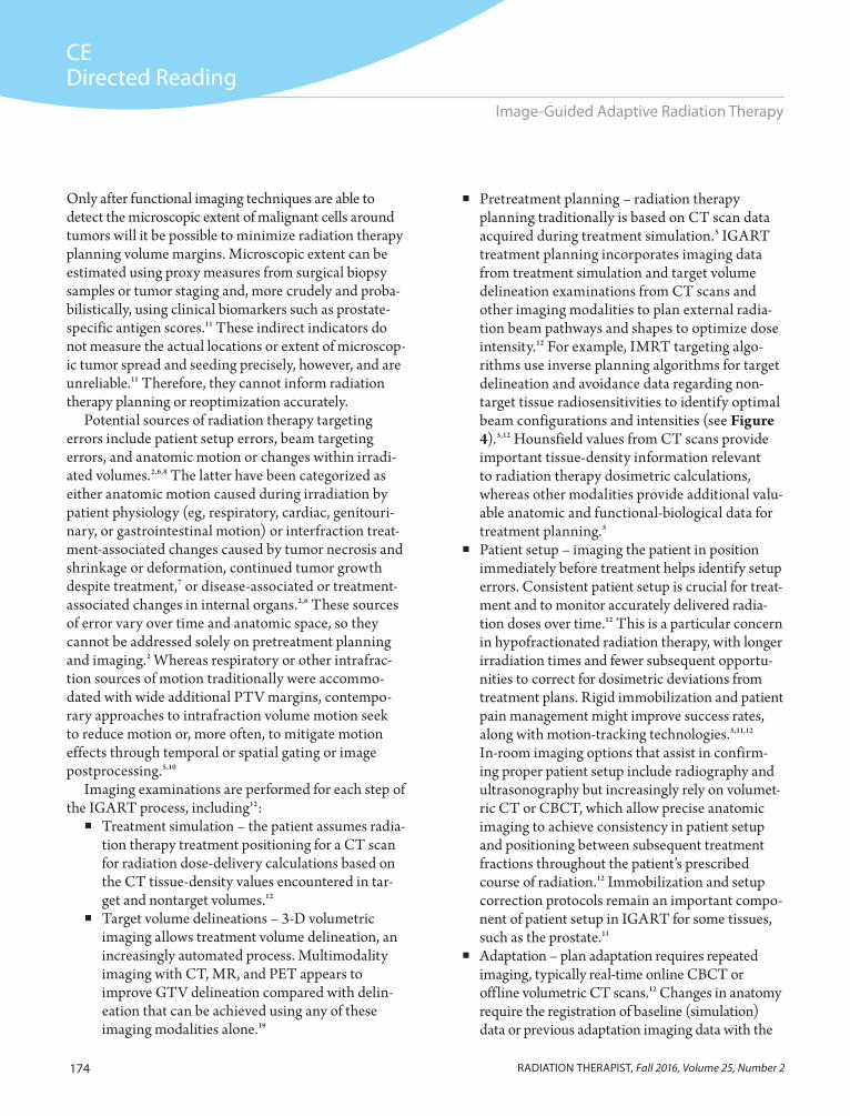
geometric uncertainties in treatment setup and patient movement (see Figure 3). Because traditional, nonadap-tive radiation therapy does not correct for setup errors, patient motion, or anatomic changes, PTV margins before IGART had to be fairly wide, causing unavoidable, dose-limiting irradiation of nontarget tissue around the tumor.12
With advances in precision functional imaging of met-abolic and molecular processes, anatomic delineations of treatment volumes increasingly will be informed by so-called biologic planning volumes.13-18 This is an impor-tant consideration because anatomic imaging modalities do not visualize tumor cells harbored in the seemingly healthy tissue surrounding a macroscopic tumor.11 Therefore, the true biologic extent of a tumor can be larg-er than is suggested by the visualized macroscopic tumor.
“a reference and starting point for subsequent replan-ning.” 1 Plan reoptimization increasingly is automated and rapid, and researchers anticipate that online IGART soon will become routine.1 Component technologies still in development include validated deformable registration algorithms for automatically calculating cumulative radi-ation doses, more rapid CBCT segmentation algorithms, and dose calculations.1
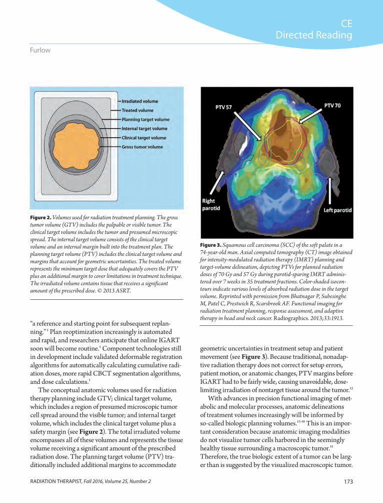
The conceptual anatomic volumes used for radiation therapy planning include GTV; clinical target volume, which includes a region of presumed microscopic tumor cell spread around the visible tumor; and internal target volume, which includes the clinical target volume plus a safety margin (see Figure 2). The total irradiated volume encompasses all of these volumes and represents the tissue volume receiving a significant amount of the prescribed radiation dose. The planning target volume (PTV) tra-ditionally included additional margins to accommodate
Figure 2. Volumes used for radiation treatment planning. The gross tumor volume (GTV) includes the palpable or visible tumor. The clinical target volume includes the tumor and presumed microscopic spread. The internal target volume consists of the clinical target volume and an internal margin built into the treatment plan. The planning target volume (PTV) includes the clinical target volume and margins that account for geometric uncertainties. The treated volume represents the minimum target dose that adequately covers the PTV plus an additional margin to cover limitations in treatment technique. The irradiated volume contains tissue that receives a significant amount of the prescribed dose. © 2013 ASRT.
Figure 3. Squamous cell carcinoma (SCC) of the soft palate in a 74-year-old man. Axial computed tomography (CT) image obtained for intensity-modulated radiation therapy (IMRT) planning and target-volume delineation, depicting PTVs for planned radiation doses of 70 Gy and 57 Gy during parotid-sparing IMRT adminis-tered over 7 weeks in 35 treatment fractions. Color-shaded isocon-tours indicate various levels of absorbed radiation dose in the target volume. Reprinted with permission from Bhatnagar P, Subesinghe M, Patel C, Prestwich R, Scarsbrook AF. Functional imaging for radiation treatment planning, response assessment, and adaptive therapy in head and neck cancer. Radiographics. 2013;33:1913.
174 RADIATION THERAPIST, Fall 2016, Volume 25, Number 2
CEDirected Reading
Image-Guided Adaptive Radiation Therapy
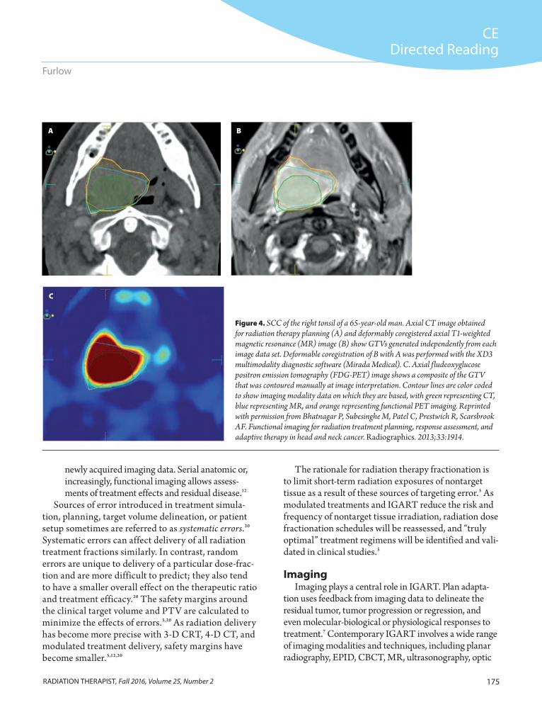
Pretreatment planning – radiation therapy planning traditionally is based on CT scan data acquired during treatment simulation.5 IGART treatment planning incorporates imaging data from treatment simulation and target volume delineation examinations from CT scans and other imaging modalities to plan external radia-tion beam pathways and shapes to optimize dose intensity.12 For example, IMRT targeting algo-rithms use inverse planning algorithms for target delineation and avoidance data regarding non-target tissue radiosensitivities to identify optimal beam configurations and intensities (see Figure 4).5,12 Hounsfield values from CT scans provide important tissue-density information relevant to radiation therapy dosimetric calculations, whereas other modalities provide additional valu-able anatomic and functional-biological data for treatment planning.5
Patient setup – imaging the patient in position immediately before treatment helps identify setup errors. Consistent patient setup is crucial for treat-ment and to monitor accurately delivered radia-tion doses over time.12 This is a particular concern in hypofractionated radiation therapy, with longer irradiation times and fewer subsequent opportu-nities to correct for dosimetric deviations from treatment plans. Rigid immobilization and patient pain management might improve success rates, along with motion-tracking technologies.5,11,12 In-room imaging options that assist in confirm-ing proper patient setup include radiography and ultrasonography but increasingly rely on volumet-ric CT or CBCT, which allow precise anatomic imaging to achieve consistency in patient setup and positioning between subsequent treatment fractions throughout the patient’s prescribed course of radiation.12 Immobilization and setup correction protocols remain an important compo-nent of patient setup in IGART for some tissues, such as the prostate.11
Adaptation – plan adaptation requires repeated imaging, typically real-time online CBCT or offline volumetric CT scans.12 Changes in anatomy require the registration of baseline (simulation) data or previous adaptation imaging data with the
Only after functional imaging techniques are able to detect the microscopic extent of malignant cells around tumors will it be possible to minimize radiation therapy planning volume margins. Microscopic extent can be estimated using proxy measures from surgical biopsy samples or tumor staging and, more crudely and proba-bilistically, using clinical biomarkers such as prostate-specific antigen scores.11 These indirect indicators do not measure the actual locations or extent of microscop-ic tumor spread and seeding precisely, however, and are unreliable.11 Therefore, they cannot inform radiation therapy planning or reoptimization accurately.
Potential sources of radiation therapy targeting errors include patient setup errors, beam targeting errors, and anatomic motion or changes within irradi-ated volumes.2,6,8 The latter have been categorized as either anatomic motion caused during irradiation by patient physiology (eg, respiratory, cardiac, genitouri-nary, or gastrointestinal motion) or interfraction treat-ment-associated changes caused by tumor necrosis and shrinkage or deformation, continued tumor growth despite treatment,7 or disease-associated or treatment-associated changes in internal organs.2,6 These sources of error vary over time and anatomic space, so they cannot be addressed solely on pretreatment planning and imaging.2 Whereas respiratory or other intrafrac-tion sources of motion traditionally were accommo-dated with wide additional PTV margins, contempo-rary approaches to intrafraction volume motion seek to reduce motion or, more often, to mitigate motion effects through temporal or spatial gating or image postprocessing.5,10
Imaging examinations are performed for each step of the IGART process, including12: Treatment simulation – the patient assumes radia-
tion therapy treatment positioning for a CT scan for radiation dose-delivery calculations based on the CT tissue-density values encountered in tar-get and nontarget volumes.12
Target volume delineations – 3-D volumetric imaging allows treatment volume delineation, an increasingly automated process. Multimodality imaging with CT, MR, and PET appears to improve GTV delineation compared with delin-eation that can be achieved using any of these imaging modalities alone.19
175RADIATION THERAPIST, Fall 2016, Volume 25, Number 2
CEDirected Reading
Furlow
The rationale for radiation therapy fractionation is to limit short-term radiation exposures of nontarget tissue as a result of these sources of targeting error.5 As modulated treatments and IGART reduce the risk and frequency of nontarget tissue irradiation, radiation dose fractionation schedules will be reassessed, and “truly optimal” treatment regimens will be identified and vali-dated in clinical studies.5
Imaging Imaging plays a central role in IGART. Plan adapta-
tion uses feedback from imaging data to delineate the residual tumor, tumor progression or regression, and even molecular-biological or physiological responses to treatment.7 Contemporary IGART involves a wide range of imaging modalities and techniques, including planar radiography, EPID, CBCT, MR, ultrasonography, optic
newly acquired imaging data. Serial anatomic or, increasingly, functional imaging allows assess-ments of treatment effects and residual disease.12
Sources of error introduced in treatment simula-tion, planning, target volume delineation, or patient setup sometimes are referred to as systematic errors.20 Systematic errors can affect delivery of all radiation treatment fractions similarly. In contrast, random errors are unique to delivery of a particular dose-frac-tion and are more difficult to predict; they also tend to have a smaller overall effect on the therapeutic ratio and treatment efficacy.20 The safety margins around the clinical target volume and PTV are calculated to minimize the effects of errors.5,20 As radiation delivery has become more precise with 3-D CRT, 4-D CT, and modulated treatment delivery, safety margins have become smaller.5,12,20
Figure 4. SCC of the right tonsil of a 65-year-old man. Axial CT image obtained for radiation therapy planning (A) and deformably coregistered axial T1-weighted magnetic resonance (MR) image (B) show GTVs generated independently from each image data set. Deformable coregistration of B with A was performed with the XD3 multimodality diagnostic software (Mirada Medical). C. Axial fludeoxyglucose positron emission tomography (FDG-PET) image shows a composite of the GTV that was contoured manually at image interpretation. Contour lines are color coded to show imaging modality data on which they are based, with green representing CT, blue representing MR, and orange representing functional PET imaging. Reprinted with permission from Bhatnagar P, Subesinghe M, Patel C, Prestwich R, Scarsbrook AF. Functional imaging for radiation treatment planning, response assessment, and adaptive therapy in head and neck cancer. Radiographics. 2013;33:1914.
A B
C
176 RADIATION THERAPIST, Fall 2016, Volume 25, Number 2
CEDirected Reading
Image-Guided Adaptive Radiation Therapy
imaging, and functional imaging with single-photon emission CT (SPECT), PET, and PET-CT.5,6,8 The role of functional imaging and the use of biomarkers in treat-ment planning is growing.
Plan AdaptationPlan-adaptation imaging data can be acquired and
registered for treatment reoptimization off line, online, or in real time. Each of these involves different time intervals between planning, treating, imaging, and plan adaptation (see Table 1). The specific approaches considered part of IGART vary among researchers, but broadly construed, IGART represents any approach to radiation therapy that involves repeated reassessment and replanning based on feedback in the form of imag-ing data during the course of treatment.1
Offline plan adaptation refers to the acquisition of imaging feedback data separate from radiation therapy—typically a day or more removed from treat-ment.6 Offline imaging typically is performed with a conventional or in-room volumetric (helical or multide-tector) CT scanner to assess tumor response between treatment fractions and changes in patient anatomy.6 With this data, GTV contours can be compared with previous imaging data to help recalculate GTV as part of plan adaptation.6 Significant changes frequently are evident between initial treatment planning and simula-tion images and midtreatment images. Those differenc-es inform dosimetry calculations (monitoring delivered radiation) and radiation therapy adaptation for the sub-sequent treatment.6 Offline imaging and adaptation can be built in to the IGART protocol, and it might be indi-cated when substantial clinical changes are observed in the patient, or if differences are noted between initial planning CT images and CBCT images acquired online or during pretreatment setup imaging.6
Online image acquisition and plan adaptation are performed during treatment in the radiation therapy room and occur minutes or less before radiation ther-apy.1,6 Similar to off line imaging, online imaging most frequently involves CT scanning equipment.6
Dynamic adaptation refers to near-simultaneous imaging and radiation therapy, and it is the newest com-ponent of IGART.1,5,6 The time interval between imaging, adaptation, and irradiation is a few seconds.6 Real-time adaptation is indicated when a target tumor occurs in the
chest or abdomen and is affected by respiratory or other sources of intrafraction motion.10 Means of addressing intrafraction motion include conventional approaches (eg, patient breath-hold or compression of the abdomen), or IGART approaches (eg, respiratory gating or real-time image-based plan adaptation).6,10 Real-time IGART imaging and treatment adaptation still is a nascent field, but imaging components for dynamic IGART, which are undergoing research, development, and clinical testing, include volumetric MR, CT, x-ray, optical, and electro-magnetic transponder systems.6 A CyberKnife robotic treatment system also is available for real-time tumor-motion adaptation.5,6
Regardless of the time between imaging and treat-ment, image data must be compared with previously acquired images. This is the heart of IGART. Image regis-tration and coregistration (fusion) of multimodality imag-ing examinations using algorithmic processes is required.
ModalitiesMajor contemporary imaging technologies have dif-
ferent and overlapping roles or applications in IGART, as well as particular strengths and weaknesses (see Tables 2 and 3). Two-dimensional digital radiography can be employed for patient positioning and tumor target localization for treatment planning, particularly if there are no issues with tumor or organ motion.4 However, volumetric imaging allows precise visualiza-tion of tumor or organ deformation or motion.4
Volumetric CT plays central roles in offline simulation and planning and subsequent IGART imaging for plan
Table 1
Offline, Online, and Real-time Imaging Feedback for IGART
6
Type Time Interval Between Imaging and Irradiation
Typical Imaging Equipment
Offline 1 day or more Conventional or in-room volumetric imaging
Online Minutes or less In-room volumetric imaging
Real-time Seconds or less 4-D CT or other imaging modalities

177RADIATION THERAPIST, Fall 2016, Volume 25, Number 2
CEDirected Reading
Furlow
Table 2
Major Ionizing Imaging and Feedback Techniques21,a
Technology Clinical Applications Advantages Disadvantages
Stereoscopic kilovolt (kV) x-ray
kV-pretreatment planar images
Intrafraction imaging for motion management
Implanted marker alignment
6-D translational and rotational correction
Appropriate when bony landmarks are used as surrogates
Capable of real-time tracking
Compatible with other systems for hybrid guidance and motion management
High radiation dose
Blocked views at certain gantry angles
Might only provide “snapshot” images for evaluation
Not capable of 3-D volumetric data acquisition
Megavoltage (MV) and kV cone-beam computed tomography (CBCT)
3-D volumetric image acquisi-tion, with 3-D matching to planning CT
X-ray source/detector
O-ring x-ray source rotation and flat panel detector for imaging data collection
Slow acquisition provides average posi-tion of internal anatomy for respiratory motion
Monitoring patient setup and anatomical changes during treatment
Ongoing monitoring of tumor response throughout treatment course
Opportunity for online replanning
Same isocenter for MV CT and linear accelerator
kV has better contrast resolution than does MV
kV has more artifacts present on high-density materials
Degradation of image quality from patient scatter—especially for larger patients on kV imaging
Radiation dose can be excessive with routine use
No real-time organ motion information
In-room CT or CT-on-rails
Pretreatment volumetric imaging for setup and adaptive plan reoptimization
3-D, 4-D image information, or both
Online replanning
Diagnostic-quality CT images
Excessive radiation dose if used routinely
No real-time organ motion-tracking information
Assumes fixed isocenter relationship between the independent CT scanner and linear accelerator
MV helical CT Pretreatment volumetric imag-ing for setup and adaptive replanning
Pretreatment MV projection imaging for patient setup
Ability to monitor ongoing treatment response
Widely applicable to anatomical sites
Excessive radiation dose if used routinely
Soft-tissue discrimination inferior to kV CT
aNot all equipment has the same applications and capabilities. Adapted with permission from De Los Santos J, Popple R, Agazaryan N, et al. Image-guided radiation therapy (IGRT) technologies for radiation therapy localization and delivery. Int J Radiat Oncol Biol Phys. 2013;87(1):33-45. doi:10.1016/j.ijrobp.2013.02.021.
reoptimization, frequently using in-room or online CT scanners that can be positioned over the patient during radiation therapy or that are integrated with radiation therapy linear accelerators.20 Some radiographic imaging modalities used with IGART include stereoscopic kilo-voltage (kV) x-ray imaging, in-room conventional volu-metric CT, and online kV or megavoltage (MV) CBCT (MV CBCT) systems that have been integrated with beam-source equipment.1,5,12,20
EPID images are projection images using the treatment beam and have long been preferred for ensuring patient
setup accuracy between treatment fractions.1,5 EPID allows precise geometric concordance between visualization and treatment delivery.5 However, portal imaging and in-room planar radiography rely on bony anatomic landmarks or implanted fiducial markers and do not reliably detect soft-tissue deformations.1,5
In-room CT was developed to visualize more sensi-tively soft tissues for patient setup, improving accuracy.5 CT also allows 3-D volumetric imaging and treatment dose calculation.5 Radiation shielding in radiation ther-apy rooms is sufficient for the addition of CT imaging

178 RADIATION THERAPIST, Fall 2016, Volume 25, Number 2
CEDirected Reading
Image-Guided Adaptive Radiation Therapy
contrast and signal-to-noise ratio for soft-tissue imaging.5 However, concerns persist regarding the use of CBCT in treatment planning because of streaking and blurring artifacts not seen in fan-beam CT and scatter-related Hounsfield number errors that can distort radiation dose calculations.5 Research is underway to address these issues and to reduce CBCT radiation doses.5 CBCT sometimes is used for patient setup and can detect rota-tional errors in patient positioning.1 However, it can less readily detect tumor or organ deformation.1,4,5
Helical tomotherapy delivers IMRT while perform-ing helical MV CT, streamlining rapid patient setup and repositioning.5 After setup confirmation, image data can be used online for treatment plan adaptation.5 This treatment is limited to radiation therapy for small
systems.5 Different in-room CT imaging systems include conventional CT-on-rails, kV CBCT (with an additional kV x-ray source and detector attached to the treatment machine), and MV CBCT portal-imaging designs.5 CT-on-rails systems are conventional CT equipment positioned to allow the treatment couch to serve for imaging and treatment.5 The patient can be positioned once on the couch, and the couch is maneuvered beneath the CT scanner for pretreatment imaging and then moved into place for treatment.5
CBCT conducts high-resolution (0.5 mm) volumetric imaging in one gantry rotation.5 MV CBCT can use the linear accelerator treatment beam using an EPID or an additional kV x-ray tube-and-detector array.5 When com-pared with MV CBCT, kV CBCT offers superior image
Table 3
Major Nonionizing Imaging and Feedback Techniques21,a
Technology Clinical Applications Advantages Disadvantages
Ultrasonography Pretreatment volumetric imaging for setup
Limited anatomical applications
Nonionizing alignment and tracking method, with visualization of nearby radiosensitive organs
4-D capabilities offer real-time intrafraction monitoring
Interuser variability
Limited real-time target tracking
Applicability to specific treatment sites, primarily prostate and lung
Optical surface imaging
Patient setup
Real-time intrafraction motion monitoring
Indicates anatomic changes
Nonionizing superficial alignment and tracking method
Real-time monitoring and gating capabilities
No internal anatomic or target information
Applications limited to specific anatomical surfaces; not ideal for flat surfaces
Electromagnetic transponders localization
Pretreatment setup and target localization
Real-time intrafraction fiducial marker tracking
Nonionizing radiation
High accuracy of tracking
Temporospatial target information
No spatial information of nearby radiosensitive organs
Invasive transponder implantation
Limited applications in compatible anatomic sites and because of array capabilities
Typically causes imaging artifacts on MR imaging
Magnetic resonance (MR)
Pretreatment volumetric imaging for setup
Should be capable of adap-tive plan reoptimization
Nonionizing alignment and visualization of nearby radiosensitive organs
3-D image information and online replanning
Superior soft tissue information
Nonuniform magnetic field causes image distortions
Susceptible to artifacts and motion
aNot all equipment has the same applications and capabilities.
Adapted with permission from De Los Santos J, Popple R, Agazaryan N, et al. Image-guided radiation therapy (IGRT) technologies for radiation therapy localization and delivery. Int J Radiat Oncol Biol Phys. 2013;87(1):33-45. doi:10.1016/j.ijrobp.2013.02.021.delivery.
179RADIATION THERAPIST, Fall 2016, Volume 25, Number 2
CEDirected Reading
Furlow
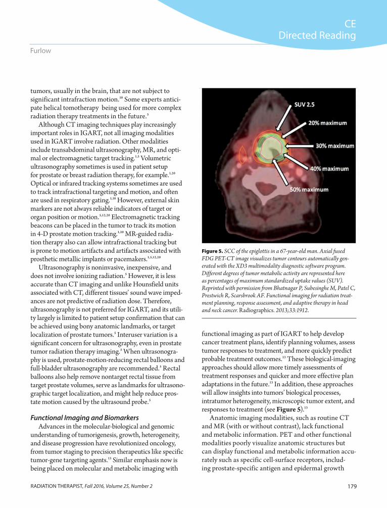
functional imaging as part of IGART to help develop cancer treatment plans, identify planning volumes, assess tumor responses to treatment, and more quickly predict probable treatment outcomes.13 These biological-imaging approaches should allow more timely assessments of treatment responses and quicker and more effective plan adaptations in the future.13 In addition, these approaches will allow insights into tumors’ biological processes, intratumor heterogeneity, microscopic tumor extent, and responses to treatment (see Figure 5).13
Anatomic imaging modalities, such as routine CT and MR (with or without contrast), lack functional and metabolic information. PET and other functional modalities poorly visualize anatomic structures but can display functional and metabolic information accu-rately such as specific cell-surface receptors, includ-ing prostate-specific antigen and epidermal growth
tumors, usually in the brain, that are not subject to significant intrafraction motion.10 Some experts antici-pate helical tomotherapy being used for more complex radiation therapy treatments in the future.5
Although CT imaging techniques play increasingly important roles in IGART, not all imaging modalities used in IGART involve radiation. Other modalities include transabdominal ultrasonography, MR, and opti-mal or electromagnetic target tracking.1,5 Volumetric ultrasonography sometimes is used in patient setup for prostate or breast radiation therapy, for example.1,20 Optical or infrared tracking systems sometimes are used to track intrafractional targeting and motion, and often are used in respiratory gating.1,10 However, external skin markers are not always reliable indicators of target or organ position or motion.5,12,20 Electromagnetic tracking beacons can be placed in the tumor to track its motion in 4-D prostate motion tracking.1,20 MR-guided radia-tion therapy also can allow intrafractional tracking but is prone to motion artifacts and artifacts associated with prosthetic metallic implants or pacemakers.1,5,12,20
Ultrasonography is noninvasive, inexpensive, and does not involve ionizing radiation.5 However, it is less accurate than CT imaging and unlike Hounsfield units associated with CT, different tissues’ sound wave imped-ances are not predictive of radiation dose. Therefore, ultrasonography is not preferred for IGART, and its utili-ty largely is limited to patient setup confirmation that can be achieved using bony anatomic landmarks, or target localization of prostate tumors.5 Interuser variation is a significant concern for ultrasonography, even in prostate tumor radiation therapy imaging.5 When ultrasonogra-phy is used, prostate-motion-reducing rectal balloons and full-bladder ultrasonography are recommended.5 Rectal balloons also help remove nontarget rectal tissue from target prostate volumes, serve as landmarks for ultrasono-graphic target localization, and might help reduce pros-tate motion caused by the ultrasound probe.5
Functional Imaging and BiomarkersAdvances in the molecular-biological and genomic
understanding of tumorigenesis, growth, heterogeneity, and disease progression have revolutionized oncology, from tumor staging to precision therapeutics like specific tumor-gene targeting agents.13 Similar emphasis now is being placed on molecular and metabolic imaging with
Figure 5. SCC of the epiglottis in a 67-year-old man. Axial fused FDG PET-CT image visualizes tumor contours automatically gen-erated with the XD3 multimodality diagnostic software program. Different degrees of tumor metabolic activity are represented here as percentages of maximum standardized uptake values (SUV). Reprinted with permission from Bhatnagar P, Subesinghe M, Patel C, Prestwich R, Scarsbrook AF. Functional imaging for radiation treat-ment planning, response assessment, and adaptive therapy in head and neck cancer. Radiographics. 2013;33:1912.

180 RADIATION THERAPIST, Fall 2016, Volume 25, Number 2
CEDirected Reading
Image-Guided Adaptive Radiation Therapy
spectroscopy might play increasing roles in defining target and nontarget volumes for CRT planning and IGART alongside anatomic imaging.5,12 FDG PET-CT already is used widely for the detection, diagnosis, and staging of tumors, and treatment monitoring. It also is employed for modulated treatment planning and target volume delinea-tion because target delineation is crucial for modulated therapy’s steep radiation dose gradients.3 MR data sets can be interrogated with different algorithms to reveal numerous facets of a tumor’s physiology.13 MR spectros-copy allows sensitive visualization of cell metabolism and metabolic byproducts and is undergoing investigation as a possible way to differentiate malignant from benign tumors and the metabolic extent of tumors. However, this and other MR-based functional imaging approaches remain investigational and are not in widespread clinical use for adaptive radiation therapy.3 FDG PET-MR also is under investigation for metabolic volume delineations (see Figure 6).3
factor receptors (EGFRs). EGFRs can be targeted with cetuximab to increase radiation therapy effective-ness.3,13 Combining anatomic and functional imaging modalities to yield information-rich composite images therefore makes sense.5 Functional imaging with PET-CT uses different tracer agents, allowing the visualiza-tion of facets of tumor histology that can help guide treatment planning with targeted biologic agents.3 Functional imaging also allows sensitive visualization of molecular-biological processes involved in a tumor’s development of resistance to radiation, including tumor hypoxia, cell proliferation, and apoptosis (cell death) and could allow early detection of acquired radioresistance.3
Fludeoxyglucose (FDG) is a radiolabeled form of glu-cose that is accumulated differentially in metabolically active cells.3,22 Once inside the cell, FDG is metabolized into FDG-6-phosphate, which cannot be further broken down and accumulates inside cells.22 Tumor cells accumu-late greater concentrations of this form of FDG than do healthy cells because of their elevated glucose metabolic rate. FDG-PET and FDG PET-CT can demonstrate bio-logical processes that allow tumors to escape the cytotoxic (cell-killing) effects of cancer treatment, including intrin-sic (pretreatment) radioresistance and hypoxia.3
However, other disease processes, such as pul-monary tuberculosis, also can cause differential accumulation of FDG, and FDG is therefore not a tumor-specific imaging marker.22 Functional imaging can identify hypoxia and metabolic activity associated with hastened cellular proliferation at baseline, before treatment, and throughout treatment to monitor the emergence of radioresistance and to inform treatment adaptation (ie, dose escalation or cessation of radiation therapy in favor of other treatment options).3
Functional and anatomic image data sets are fused to create information-rich data sets and visualizations—a process that largely is automated now with integrated PET-CT and PET-MR systems.5 Such functional imag-ing approaches help to determine and track the biological extent of cancer over time and provide more precise and timely visualization of treatment effects.3 Serial functional imaging examinations at each step of the IGART process should allow more sensitive and detailed monitoring of treatment response and plan-adaptation needs than ana-tomic imaging alone.3 PET, SPECT, and functional MR
Figure 6. SCC in the right aspect of the tongue base in a 51-year-old man. Axial fused FDG PET-MR image demonstrates a metaboli-cally active primary tumor (arrow) and ipsilateral metastatic spread of the malignancy to a lymph node (arrowhead). Reprinted with permission from Bhatnagar P, Subesinghe M, Patel C, Prestwich R, Scarsbrook AF. Functional imaging for radiation treatment planning, response assessment, and adaptive therapy in head and neck cancer. Radiographics. 2013;33:1925.

181RADIATION THERAPIST, Fall 2016, Volume 25, Number 2
CEDirected Reading
Furlow
The metabolic uptake of FDG in FDG-PET imag-ing might even have prognostic value, predicting tumor responses and patient outcomes, but this remains an area of controversy.22 Proponents of FDG as a prognostic biomarker point to evidence that tumor uptake of FDG reflects the tumor’s metabolic activity before and after irradiation and that the difference might be a proxy mea-sure of tumor viability and aggressiveness.22 Preliminary studies of small numbers of patients suggest that lower tumor metabolic rate after radiation therapy, as assessed by FDG uptake, has been associated with superior rates of both local tumor control and patient survival.22
In addition to FDG, other radiopharmaceuticals used in PET imaging are undergoing development for different clinical-imaging and treatment-planning applications.13 For example, fluorine-18 fluoromisonidazole and fluoro-azomycin arabinoside accumulate in hypoxic tissues and are undergoing development for use with PET imaging to detect emerging regions of tumor hypoxia.13 3′-deoxy-3′-fluorothymidine can visualize tumor cell proliferation and also is undergoing development for use with PET.13 The comparative effectiveness research is scant, and no consen-sus has been reached about which radiopharmaceuticals are best for visualizing hypoxia.3
Ongoing research aims to identify other prognostic and predictive genomic biomarkers detected in blood tests or imaging examinations that might further person-alize the molecular-biological basis of radiation therapy planning and adaptation by more precisely identifying tumor contours or margins and treatment effects, poten-tially allowing the precise assessment of radioresistant foci emerging within a given tumor.13,16,17 Ultimately, these imaging biomarkers could help detect tumor responses to treatment earlier and predict the risk of dis-ease recurrence and late toxicities, which could inform patient survivorship care plans and post-treatment moni-toring needs.13,16 PET, SPECT, or MR imaging biomark-ers could help identify molecular or metabolic and cell-proliferation changes in tumor biology and intratumor responses to treatment, augmenting their already increas-ingly important role in IGART.13,17,18
Image Data ProcessingRepeated and multimodality imaging undertaken
for IGART planning, setup, monitoring, and adaptation requires 4 types of image data set processing:
Image segmentation. Image registration. Coregistration – fusion of data from different imag-
ing examinations or multimodality examinations. Visualization.Image segmentation identifies the surface contours
and boundaries of tumors and nontarget anatomies, whereas image registration identifies their positions and shapes using landmarks and other factors to allow identification of those anatomies in different images.5 Image segmentation partitions image data into ana-tomic features, distinguishing anatomic boundaries or surfaces of interest from adjacent anatomy so the sur-face contours or internal features of soft tissues, bones, or vasculature can be visualized. Segmentation involves several algorithmic approaches and increasingly is per-formed automatically, with relatively little manual input from the radiologic technologist. Segmentation reduces the file sizes, decreasing processing time.
Image registration anatomically aligns overlapping image data from serial or multimodality imaging exami-nations to help detect changes in the shape, size, or ori-entation of anatomic structures, or to detect differences in scale between sequential image data sets.5,23 Many registration algorithms compare maps of image inten-sity over anatomic space, between image data sets.1
Registration can be achieved using rigid or deformable registration algorithms to map anatomies between image data sets.5 Rigid registration uses landmarks to identify the patient’s positional differences between imaging examinations and uses this information to correct for the identification of anatomic positions.5 However, rigid registration poorly accommodates the anatomic changes that can occur within and around treatment volumes over the course of radiation therapy treatment.11 Deformable registration detects changes in specific anatomies relative to other anatomies to show physical changes that occur between imaging examinations.5 Typically, rigid image registration is used to correlate data sets from treatment planning images obtained from different types of imaging examinations, allowing image fusion to precisely quantify and locate tumor contours, cancer-involved regional lymph nodes, and nearby non-target organs and tissues.5
Deformable registration is used in planning images to assess the effect of respiratory or other sources of

182 RADIATION THERAPIST, Fall 2016, Volume 25, Number 2
CEDirected Reading
Image-Guided Adaptive Radiation Therapy
One challenge for IGART is the effect of repeated radiographic imaging on radiation dose distributions over time. Radiation doses associated with imaging long have been assumed not to approach those associated with radiation therapy. However, as radiation therapy doses to nontarget tissues continue to decline, radiographic imag-ing will become a more significant source of patients’ radiation doses, and it warrants consideration.1 This is particularly true for repeated CT scan acquisitions. For example, CBCT scans associated with patient setup can deliver radiation doses as high as 10 cGy per treatment day.1 Furthermore, the as low as reasonably achievable (ALARA) concept is observed to minimize the prob-ability of stochastic radiation risks, such as DNA damage or lifetime cancer risk.1 ALARA applies to radiographic radiation dose in IGART just as it does in other settings. Quantifying cumulative radiation doses associated with
physiologic motion on volumes or anatomies of interest and between delivered fractions of radiation treatment to identify anatomic changes resulting directly or indirectly from treatment.5 Deformable registration algorithms also allow more accurate tracking of delivered radiation doses throughout the treatment regimen.5 Several deformable-registration models have been developed, but clinical validation is ongoing and some challenges remain unre-solved, including certain types of relative organ displace-ment, such as sliding and mass loss.11 Deformable regis-tration should better track radiation dose distributions over time; volumetric CT scans between treatment frac-tions, for example, can be deformably registered to recal-culate delivered doses from the baseline or planning CT images forward.11 This allows cumulative dose-distribu-tion mapping of changing tumor and anatomic volumes over time and correction or compensation for deviations from the initial treatment plan and subsequent reopti-mized treatment plans.11
Segmentation and registration increasingly are per-formed automatically in near real time using software, but these tasks also can be completed manually in part.5 Nevertheless, automatic rigid registration of data from sequentially performed planning examinations largely has replaced manual registration between planning images using visual alignment of 2-D simulation and portal images.5
Fusion, or multimodality data coregistration, aligns anatomic data from different imaging modalities to produce information-rich fusion images. For example, PET-CT images contain physiologic PET image data and anatomic CT image data.5 Visualization, of course, refers to the end result of segmentation, registration, fusion, and postprocessing of image data to generate clinically useful images.
ALARA and Imaging Dose Management Imaging data is central to all steps of the IGART
process, and much research has been done on methods that do not use ionizing radiation for guidance and tracking to help limit patient dose. Because speed, accu-racy, and objectivity are so important, IGART relies heavily on automated processes.5 In addition, IGART involves monitoring and management of the ionizing radiation dose associated with radiation therapy and the associated radiographic imaging.5
Table 5
Dose from the Elekta kV CBCT 24
Parameter Head Chest
Mean dose at center (mGy) 29 16
Mean skin dose (mGy) 30 23
Effective dose (mSv) 3.0 3.0
Conversion factor (mSv/mGy cm2) 6.0 x 10
−516.0 x 10
−5
Abbreviations: mGy, milligray; mSv, millisievert. Reprinted with permission from Murphy MJ, Balter J, Balter S, et al. The management of imaging dose during image-guided radiotherapy: report of the AAPM Task Group 75. Med Phys. 2007;34(10):4057. doi:10.1118/1.2775667.
Table 4
Dose from kV CBCT 24
Parameter Head Chest
Maximum skin dose (mGy) 100.5 85.4
Mean skin dose (mGy) 68.5 57.0
Effective dose (mSv) 10.9 24.6
Conversion factor (mSv/mGy cm2) 6.0 x 10
−516.0 x 10
−5
Abbreviations: mGy, milligray; mSv, millisievert. Reprinted with permission from Murphy MJ, Balter J, Balter S, et al. The management of imaging dose during image-guided radiotherapy: report of the AAPM Task Group 75. Med Phys. 2007;34(10):4057. doi:10.1118/1.2775667.
183RADIATION THERAPIST, Fall 2016, Volume 25, Number 2
CEDirected Reading
Furlow
Simulation, Treatment Volume Delineation, and Treatment Planning
Simulation, an imaging procedure that helps to iden-tify a patient’s optimal position for treatment, provides baseline index or reference setup images of that optimal treatment position.4 The resulting images are used as references for patient positioning immediately before each treatment fraction. The differences represent one form of imaging feedback in IGART. Simulation has long been a separate procedure from treatment-planning imaging. However, beam’s-eye-view digitally reconstructed radiographs allow simulation refer-ence CT image data to be acquired during treatment planning rather than as a separate examination.1 This ensures excellent concordance between the patient’s treatment planning position and simulation positioning for treatment setup.1
Internal target volume was formalized as a plan-ning volume in part to accommodate tumor motion.10 Target volume delineations require the acquisition of image series representing the full range of physiological sources of motion, rather than a single planning image.Options include f luoroscopy, slow CT scans, 4-D CT, and MR, all of which include inhalation-phase and exhalation-phase breath-hold images.10
PET radiopharmaceuticals offer sensitive differen-tiation of malignant and healthy tissues for accurate delineation of tumor contours.3 Although scant evi-dence supports that automatic volume delineations outperform visual interpretation by experienced oper-ators, adjustments to the PET standardized-uptake values scale can change the visualization of tumor vol-ume significantly—a potential cause of inconsistent assessments between observers. A lack of consensus about the best target-contouring methods remains.3
repeated radiographic imaging using different techniques is a complex dosimetric challenge.1,5
The American Association of Physicists in Medicine formed Task Group 75 to report on doses from dif-ferent IGRT methods used in radiation therapy. For example, kV CBCT systems represent a significant portion of the IGRT methods employed. To evaluate patient dose correctly from a kV CBCT scan, differ-ent factors, or parameters, must be considered and accounted for to project the dose. Such factors include accounting for scatter radiation and the dose measured at the central axis. Dose for the central axis is measured in air kerma, represented in Gy, and if scatter is to be included, the CT dose index in air (CTDIa) and the weighted index (CTDIw) are necessary to account for scatter radiation values.24
According to the task group, although dose varies with different influencing factors, the effective doses for kV CBCT scans of the head can range from 3 mSv to 10.9 mSv, and for a chest scan the mean dose can be 16 mGy at the central axis and 23 mGy at the skin surface.24 To provide a more complete example of doses from the scans, the group compiled information into 2 tables. Table 4 provides measured doses for unspeci-fied kV CBCT equipment and Table 5 is specific to an Elekta Synergy kV CBCT (XVI) system.24
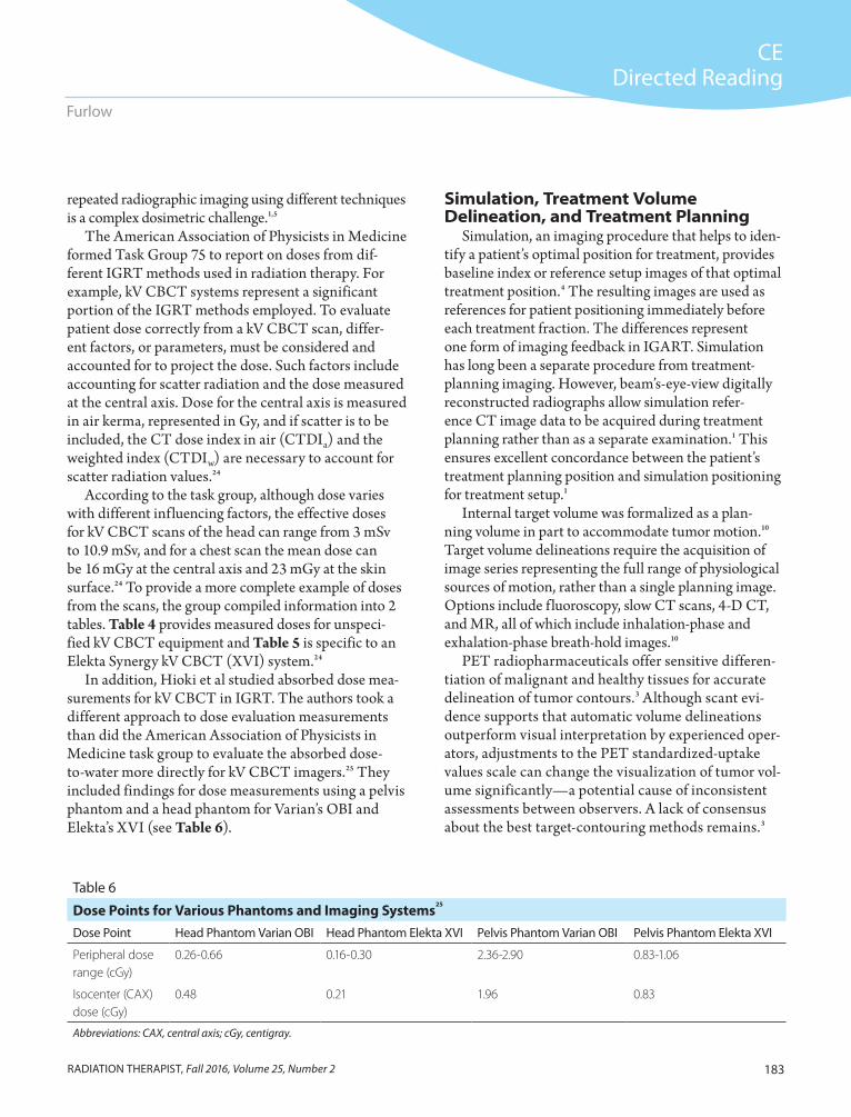
In addition, Hioki et al studied absorbed dose mea-surements for kV CBCT in IGRT. The authors took a different approach to dose evaluation measurements than did the American Association of Physicists in Medicine task group to evaluate the absorbed dose-to-water more directly for kV CBCT imagers.25 They included findings for dose measurements using a pelvis phantom and a head phantom for Varian’s OBI and Elekta’s XVI (see Table 6).
Table 6
Dose Points for Various Phantoms and Imaging Systems25
Dose Point Head Phantom Varian OBI Head Phantom Elekta XVI Pelvis Phantom Varian OBI Pelvis Phantom Elekta XVI
Peripheral dose range (cGy)
0.26-0.66 0.16-0.30 2.36-2.90 0.83-1.06
Isocenter (CAX) dose (cGy)
0.48 0.21 1.96 0.83
Abbreviations: CAX, central axis; cGy, centigray.
184 RADIATION THERAPIST, Fall 2016, Volume 25, Number 2
CEDirected Reading
Image-Guided Adaptive Radiation Therapy
comparable degrees of bladder filling, and patient coach-ing to help achieve regular respiration rates.11 Patient discomfort or pain can compromise a patient’s ability to achieve or hold a particular position; pain should be assessed throughout the treatment process and addressed with appropriate pain management when it is an issue. Dynamic dose-optimization techniques have been devel-oped for specific radiation therapy modalities, such as IMRT.11 Use of PET-CT imaging in the planning process allows plan-adaptation feedback with both anatomic deformation and biological data. These data offer comple-mentary but overlapping information about tumor chang-es over time; tumor deformation and positional changes between fractions can markedly change GTVs, in ways that can appear different in PET and CT image series.3
Dynamic AdaptationConventional radiation therapy maneuvers for respira-
tory motion control include10: Shallow breathing. Voluntary, coached patient breath-hold at a
specified part of the respiratory cycle.
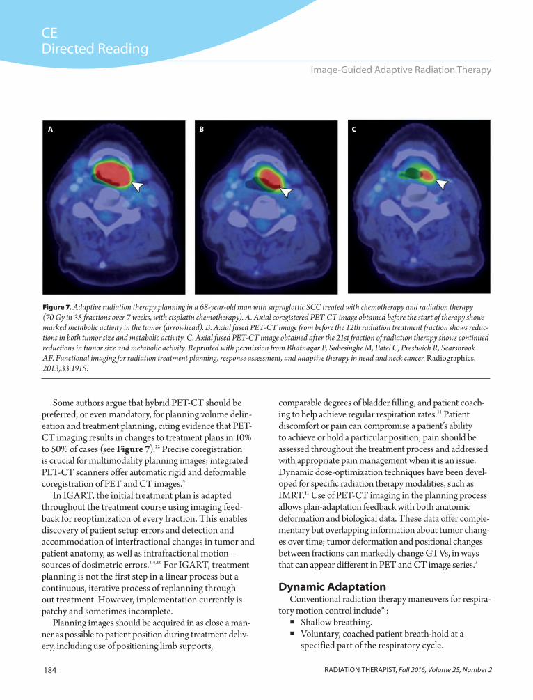
Some authors argue that hybrid PET-CT should be preferred, or even mandatory, for planning volume delin-eation and treatment planning, citing evidence that PET-CT imaging results in changes to treatment plans in 10% to 50% of cases (see Figure 7).22 Precise coregistration is crucial for multimodality planning images; integrated PET-CT scanners offer automatic rigid and deformable coregistration of PET and CT images.3
In IGART, the initial treatment plan is adapted throughout the treatment course using imaging feed-back for reoptimization of every fraction. This enables discovery of patient setup errors and detection and accommodation of interfractional changes in tumor and patient anatomy, as well as intrafractional motion—sources of dosimetric errors.1,4,10 For IGART, treatment planning is not the first step in a linear process but a continuous, iterative process of replanning through-out treatment. However, implementation currently is patchy and sometimes incomplete.
Planning images should be acquired in as close a man-ner as possible to patient position during treatment deliv-ery, including use of positioning limb supports,
Figure 7. Adaptive radiation therapy planning in a 68-year-old man with supraglottic SCC treated with chemotherapy and radiation therapy (70 Gy in 35 fractions over 7 weeks, with cisplatin chemotherapy). A. Axial coregistered PET-CT image obtained before the start of therapy shows marked metabolic activity in the tumor (arrowhead). B. Axial fused PET-CT image from before the 12th radiation treatment fraction shows reduc-tions in both tumor size and metabolic activity. C. Axial fused PET-CT image obtained after the 21st fraction of radiation therapy shows continued reductions in tumor size and metabolic activity. Reprinted with permission from Bhatnagar P, Subesinghe M, Patel C, Prestwich R, Scarsbrook AF. Functional imaging for radiation treatment planning, response assessment, and adaptive therapy in head and neck cancer. Radiographics. 2013;33:1915.
A B C
185RADIATION THERAPIST, Fall 2016, Volume 25, Number 2
CEDirected Reading
Furlow
Deep-inspiration breath-hold. Forced breath hold employing assisting devices
monitored by spirometry, such as an occlusion valve in the spirometer.
External immobilization or abdominal compres-sion, such as that used with stereotactic body radia-tion therapy.
In contrast, dynamic IGART involves the use of feed-back data during treatment to adapt radiation delivery to tumor motion in near real time. Dynamic IGART tech-niques might be necessary to treat tumors at anatomic locations where respiratory or other physiological sources of motion cannot be managed adequately with breath-control procedures (eg, tumors of the lungs, breasts, liver, pancreas, and kidneys).10
The most commonly encountered source of motion is respiration. Many thoracic and abdominal organs—even the prostate—are affected by respiratory motion.10 Therefore, much of the early research into managing the effects of intrafractional motion on radiation therapy focused on respiratory movement. However, other physi-ological processes, including bladder and rectum filling, also can cause organ motion.10
The ability to provide highly automated, real-time online imaging feedback and treatment plan reoptimiza-tion is a central, although incompletely realized, goal of IGART. Dynamic IGART is the most rapid and complex form of plan reoptimization, requiring precise image reg-istration and tracking of tumor motion that can approach speeds of several centimeters per second.5 One category of dynamic adaptation in clinical use is the calculation of tumor position using indirect surrogate markers, such as spirometric lung volume or optically tracked visual mark-ers on skin.1,5 Optical tracking systems are commercially available. However, not all patients’ respiration patterns are regular enough for this approach to be reliable.5 Electromagnetic tracking of implanted fiducial markers is undergoing clinical testing.5
Recent advances in computing speed have placed additional technological avenues toward dynamic adaptation within clinical reach.6 Several approaches or strategies for accommodating tumor motion during treatment exist. All require rapid and accurate target detection and tracking, and most involve complex plan-ning algorithms.5,10 Although some still are experimen-tal, such as moving the couch instead of the beam, a few
are in clinical use, including beam gating and 4-D CT, and beam tracking.5,10
With beam gating and 4-D CT, beam-on is triggered by the tumor’s arrival at a specified anatomic location.5,10 Beam gating frequently is used for lung and liver tumor radiation therapy.10 Internal gating systems use inter-nal landmarks or surrogate markers, such as fiducial implants, whereas external gating uses surface-motion markers and optical imaging systems.10 Four-dimensional CT is employed widely in treatment planning for gated radiation therapy. These planning scans involve acquisi-tion of up to 20 different images from throughout the respiratory cycle. Of these images, phase-specific images most relevant to internal target volume delineation are selected to develop a 4-D CT gated treatment plan, and these images are postprocessed, typically using maximum-intensity projection image postprocessing.10 These 4-D CT methods assume that a patient’s breath-ing patterns are regular and reproducible, so coaching is sometimes necessary.10
With beam tracking, the radiation beam source can be dynamically realigned to track and follow tumor motion.5,10 Beam tracking has been used in robotic radiosurgery such as the Synchrony Respiratory Tracking System used with the CyberKnife robotic lin-ear accelerator. These systems use implanted gold fidu-cial markers and employ radiographic planning images to calculate respiratory motion parameters.
Target detection and tracking provides real-time feed-back for treatment delivery adaptation. Open-loop beam alignment responses can be achieved via gating, beam-collimation change, or electromagnetic “steering” of the beam. In reality, even these direct tracking strategies include millisecond processing lag times that necessitate calculation of the near-future position of the tumor.5,10
Onboard CBCT imaging and kV fluoroscopy allows real-time observation of the tumor during treatment.5 The CyberKnife system employs pairs of kV x-ray sources and detectors to allow orthogonal visualization of the patient in radiographic mode.5
Clinical ApplicationsModulated radiation therapy treatments, such as
IMRT and volumetric modulated arc therapy (VMAT), are the most commonly used treatments for head and neck, lung, and breast cancers. Not every cancer patient

186 RADIATION THERAPIST, Fall 2016, Volume 25, Number 2
CEDirected Reading
Image-Guided Adaptive Radiation Therapy
intensities to several targets. Nontarget tissue contouring is undertaken for avoidance calculation—a crucial com-ponent of modulated treatment planning for head and neck cancers, in which nontarget tissue irradiation can lead to xerostomia (impaired saliva production), aspira-tion problems, and dysphagia, with serious effects on a patient’s quality of life. Nontarget parotid salivary gland shrinkage is common among patients undergoing head and neck tumor radiation therapy. This can be monitored between treatment fractions using pretreatment in-room CT or helical tomotherapy. Modulated treatment meth-ods can reduce the incidence and severity of xerostomia, probably through sparing of salivary glands.27
Setup errors for head and neck radiation therapy treatments are common, particularly regarding the larynx.26,27 However, intrafraction motion rarely is an issue in head and neck tumor radiation therapy because patients are immobilized with head rest supports, masks, and mouthpiece stents. Nevertheless, sporadic swallowing can be a source of intrafraction target and nontarget anatomy motion, and patients should be coached on this problem. Efforts are underway to cap-ture swallowing effects in PTV delineation.27
Interfraction changes in GTV and GTV position are assessed using pretreatment in-room CT or offline PET-CT between treatment fractions.3,27 IGART modulated treatment plan adaptation involves precisely registered treatment-target delineations and avoidance anatomy delineations for inverse planning. Deformable image reg-istration has been validated for use with head and neck tumor modulated treatment delivery; its use with auto-mated algorithms is replacing time-consuming manual contouring, which is prone to interobserver inconsisten-cies. Using deformable registration-based daily dose distri-bution mapping, cumulative dose distributions also can be assessed between fractions for modulated treatment plan adaptation to better spare salivary glands, for example.27
In the future, functional imaging-delineated details of biological processes within tumors likely will allow more precise dose escalations, such as in FDG-PET and PET-CT–guided hypoxia-guided radiation therapy plan adaptations.27
Lung CancerLung cancer is a leading cause of cancer deaths, and
radiation therapy is a key component of treatment for
requires or is likely to benefit from the use of IGART instead of conventional nonadaptive approaches to radiation therapy. Many tumors are treated using other radiation therapy approaches, and not all tumor types respond rapidly enough to radiation treatments for interfraction deformation to be a significant issue. Other cancer types, such as head and neck tumors, tend to exhibit marked deformation during the course of radiation therapy, including tumor shrinkage. Particularly with modulated treatment plans, these changes can degrade the conformal accuracy of initial radiation therapy plans, necessitating some degree of plan adaptation.3,12 Modulated treatment plans appear to be more sensitive to interfraction anatomic changes than other radiation therapy techniques, and non-IGART modulated treatment plans based on a single initial or baseline imaging examination likely increase the risk of marginal beam misses of target volumes and, therefore, the risk of adverse radiotoxicities for patients and underdosing the target.12,26 Therefore, the need for image guidance and plan adaptation varies depend-ing on tumor location, size, and stage.1 Exactly how IGART is approached also varies between patients and cancer types. For example, organ motion and the need for dynamic intrafractional imaging and adaptation is usually less of an issue for patients with oropharyngeal tumors than for those being treated for lung, liver, or bladder tumors.1,10 Nevertheless, IGART is well-suited for head and neck, breast, and lung cancers.
Head and Neck Cancer Modulated treatment delivery is the most common
approach for patients with head and neck tumors.27 It is well suited for complex tumor contours and close ana-tomic association with radiosensitive nontarget tissues seen in head and neck cancers. Initial target volumes are predictive of the rate of tumor volume change between fractions of radiation therapy, which can help identify patients who are most likely to benefit from IGART.27
Head and neck GTV represents the volumes with the highest tumor cell density, and therefore, the highest prescribed radiation dose.27 GTV contouring can employ CT images, MR images, or both and involves FDG-PET or PET-CT imaging.3,27 Modulated treatment delivery for head and neck cancer includes simultaneous integrated boost, which concurrently delivers different radiation
187RADIATION THERAPIST, Fall 2016, Volume 25, Number 2
CEDirected Reading
Furlow
Coached breathing, deep-inhalation breath-hold, and respiratory gating are options for minimizing intrafraction motion during breast radiation therapy.29 Simulation 4-D CT as part of pretreatment planning can identify whether respiratory gating can help spare heart tissues during treatment. Surgical clips can be used as markers or fiducial markers can be implanted during breast-conserving surgery to help define the lumpectomy site’s target margins for subsequent radia-tion therapy and might be useful in gated radiation therapy. However, fiducial markers tend to migrate.28
Modulated treatment delivery offers homogenous radiation doses but does not spare heart tissue as read-ily as does 3-D CRT with cardiac blocking.29 Whole-breast irradiation (45-50.4 Gy) sometimes is accom-panied by a boost dose of 10 Gy to 16 Gy at the tumor bed to increase the cumulative radiation dose in the tissues most likely to harbor remnant tumor cells.29
Interfraction changes and patient setup errors can be detected for IGART using CT-on-rails, helical tomo-therapy, kV CBCT, or MV CBCT.29 Three-dimensional ultrasonography also can be used for planning (fused with planning CT images) and subsequent daily pre-treatment target location and setup. MV and kV volu-metric CT imaging is used for interfraction plan adap-tations and delivered-dose distribution calculation.29
ConclusionConformal radiation therapy has been limited
in clinical practice by patient setup variations, changes in tumor and patient-organ size, volumetric contours, and relative positions, and by tumor and organ motion during treatment. The ultimate goal of IGART is to use feedback data from imaging exami-nations to reoptimize radiation therapy plans and beam parameters repeatedly throughout treatment. Technological advances in computing power, imag-ing, image-processing, and plan-adapting software, are hastening the day when IGART will be employed routinely in cancer treatment for selected patient populations. Automated replanning algorithm develop-ment and validation are crucial to progress IGART’s clinical implementation because of the time-consuming nature of repeated manual recontouring.
Functional imaging is improving target delineation and the identification of targeted dose escalation.12
most patients.28 Lung tumors are subject to considerable intrafraction motion associated with respiration, and irradiation of adjacent nontarget lung tissue can cause pneumonitis and other serious, dose-limiting acute radiotoxicities. Controlled patient respiration, 4-D CT, and beam gating all have been employed to reduce radiation therapy errors caused by respiratory motion in lung cancer treatment.28
Interfraction tumor shrinkage and deformation can be significant, with volume reductions of 1.2% per day, on average, between day 1 of radiation therapy and 60 days later.28 Adaptation usually is undertaken offline, using data from CT, helical tomotherapy, or PET-CT. The information value of PET-CT is illustrated by average midtreatment-regimen lung tumor volume reductions of 26% as assessed by CT and 44% as assessed by PET, in one study. PET data can be used to inform targeted radiation boost plan adaptations. MR and SPECT also have been used to assess interfraction tumor volume and position changes, and if the use of these modalities in interfraction IGART is foreseen, they should be included in the baseline planning imaging examinations carried out before a treatment regimen is initiated.28
The clinical rationale for IGART for lung cancer seems strong, but little empirical evidence exists showing that it improves patient outcomes when compared with other radiation therapy techniques.28
Breast CancerBreast-conserving surgery with adjuvant radiation
therapy to kill residual tumor cells is a standard treatment for early-stage breast cancer.29 Breast radiation therapy is plagued by tumor and nontarget anatomic motion during treatment and changes between treatment fractions.29
Treatment volumes can include the lumpectomy cav-ity or the entire breast. CT imaging is used in treatment planning to detect and delineate glandular breast tissue as part of clinical target volume.29 Breast PTV margin reduc-tion requires respiratory motion adaptation, management, or both. Four-dimensional CT gated radiation therapy reduces radiation doses to the heart, at least among patients undergoing treatment for left-breast tumors, and can be included in baseline planning imaging examina-tions. FDG PET-CT is undertaken to delineate planning volumes, including lymph nodes, and to track tumor met-abolic treatment responses between treatment fractions.29

188 RADIATION THERAPIST, Fall 2016, Volume 25, Number 2
CEDirected Reading
Image-Guided Adaptive Radiation Therapy
response assessment, and adaptive therapy in head and neck cancer. Radiographics. 2013;33:1909-1929. doi:10.1148/rg .337125163.
4. Xing L, Qian J, Choi K, Suh TS. Three- and four-dimensional morphological imaging for adaptive radiation therapy plan-ning. In: Li XA, ed. Adaptive Radiation Therapy. New York, NY: CRC Press; 2011:19-34.
5. Murphy MJ, Li T. Introduction to image-guided and adaptive radiation therapy. In: Timmerman R, Xing L, eds. Image-Guided and Adaptive Radiation Therapy. Philadelphia, PA: Wolters Kluwer/Lippincott Williams & Wilkins; 2015:3-40.
6. Keall PJ, Hsu A, Xing L. Image-guided adaptive radiotherapy. In: Hoppe R, Phillips TL, Roach M, eds. Leibel and Phillips Textbook of Radiation Oncology. 3rd ed. New York, NY: Saunders; 2010:213-223.
7. Glide-Hurst CK, Chetty IJ. Improving radiotherapy plan-ning, delivery accuracy, and normal tissue sparing using cutting edge technologies. J Thorac Dis. 2014;6(4):303-318. doi:10.3978/j.issn.2072-1439.2013.11.10.
8. Hoppe RT. Foreword. In: Timmerman R, Xing L, eds. Image-Guided and Adaptive Radiation Therapy. Philadelphia, PA: Wolters Kluwer/Lippincott Williams & Wilkins; 2015:ix-xii.
9. Court LE. 9.16: In-room image-guided radiation therapy. Comprehens Biomed Phys. 2014;9:401-430.
10. Cerviño LL, Jiang SB. Intrafraction variations and manage-ment technologies. In: Li XA, ed. Adaptive Radiation Therapy. New York, NY: CRC Press; 2011:203-216.
11. Sonke JJ, van Herk M. Errors, margins, localization and correction. In: Timmerman R, Xing L, eds. Image-Guided and Adaptive Radiation Therapy. Philadelphia, PA: Wolters Kluwer/Lippincott Williams & Wilkins; 2015:41-54.
12. Dong L, Yang J, Zhang Y. Imaging processing in adaptive radiotherapy. In: Brock KK, ed. Image Processing in Radiation Therapy. London, UK; CRC Press; 2013:3-20.
13. Prestwich RJ, Vaidyanathan S, Scarsbrook AF. Functional imaging biomarkers: potential to guide an individualised approach to radiotherapy. Clin Oncol (R Coll Radiol). 2015; 27(10):588-600. doi:10.1016/j.clon.2015.06.008.
14. Prestwich RJ, Sykes J, Carey B, Sen M, Dyker KE, Scarsbrook AF. Improving target definition for head and neck radiother-apy: a place for magnetic resonance imaging and 18-fluoride fluorodeoxyglucose positron emission tomography? Clin Oncol (R Coll Radiol). 2012;24(8):577-589. doi: 10.1016/j .clon.2012.04.002.
15. Geets X, Grégoire V, Lee JA. Implementation of hypoxia PET imaging in radiation therapy planning. Q J Nucl Med Mol Imaging. 2013;57(3):271-282.
16. Alonzi R. Functional radiotherapy targeting using focused dose escalation. Clin Oncol (R Coll Radiol). 2015;27(10):601-617. doi:10.1016/j.clon.2015.06.015.
Detailed biological imaging and the use of biomark-ers could allow even more timely and precise radiation therapy plan adaptation in the future. Hypoxic tumor regions identified with FDG-PET and PET-CT are candidate targets for integrated boosts or so-called hypoxia-guided radiation therapy.12 Some researchers in the field anticipate that IGART will become standard practice in radiation oncology in the years to come.1
Although the radiobiological rationale for IGART is sound, empirical confirmation of the tumor-control and survival benefits of IGART for patients will have to await completion of large, well-designed, prospective clinical trials.
Once IGART becomes more widely used, it likely will spark a reconsideration of fundamental issues in radiation therapy, such as fractionation of treatment regimens, which have been devised as a way to limit radiation doses to healthy nontarget tissues and organs. Modulated treatment delivery has allowed hypofrac-tionated radiation therapy that delivers total therapeutic radiation doses in fewer treatments. As IGART more precisely contours radiation doses to tumor tissue over time and across anatomic space, fractionation schedules likely will be reconsidered.5
New Mexico-based medical writer Bryant Furlow, BA, is a regular contributor to Radiologic Technology, Radiation Therapist, Lancet Oncology, Cancer Network, and several other medical and news publications. He reports on oncology, medical imaging, radiation therapy, and pulmonology.
Reprint requests may be mailed to the American Society of Radiologic Technologists, Publications Department, 15000 Central Ave NE, Albuquerque, NM 87123-3909, or emailed to [email protected].
© 2016 American Society of Radiologic Technologists
References 1. Xing L, Lee L, Timmerman R. Image-guided adaptive radia-
tion therapy and practical perspectives. In: Timmerman R, Xing L, eds. Image-Guided and Adaptive Radiation Therapy. Philadelphia, PA: Wolters Kluwer/Lippincott Williams & Wilkins; 2015:16-40.
2. Yan D. Adaptive radiotherapy: merging principle into clinical practice. Sem Radiat Oncol. 2010;20(2):79-83.
3. Bhatnagar P, Subesinghe M, Patel C, Prestwich R, Scarsbrook AF. Functional imaging for radiation treatment planning,

189RADIATION THERAPIST, Fall 2016, Volume 25, Number 2
CEDirected Reading
Furlow
17. Harris BH, Barberis A, West CM, Buffa FM. Gene expression signatures as biomarkers of tumour hypoxia. Clin Oncol (R Coll Radiol). 2015;27(10):547-560. doi:10.1016/j.clon.2015 .07.004.
18. West CM, Huddart RA. Biomarkers and imaging for precision radiotherapy. Clin Oncol (R Coll Radiol). 2015;27(10):545-546. doi:10.1016/j.clon.2015.06.021.
19. Bird D, Scarsbrook AF, Sykes J, et al. Multimodality imaging with CT, MR and FDG-PET for radiotherapy target volume delineation in oropharyngeal squamous cell carcinoma. BMC Cancer. 2015;15:844. doi:10.1186/s12885-015-1867-8.
20. Goyal S, Kataria T. Image guidance in radiation therapy: techniques and applications. Radiol Res Pract. 2014;2014: 705604. doi:10.1155/2014/705604.
21. De Los Santos J, Popple R, Agazaryan N, et al. Image-guided radiation therapy (IGRT) technologies for radiation ther-apy localization and delivery. Int J Radiat Oncol Biol Phys. 2013;87(1):33-45. doi:10.1016/j.ijrobp.2013.02.021.delivery.
22. Trovo M, Bradley J, Machtay M. Positron emission tomogra-phy/computed tomography and biologically conformal radia-tion therapy. In: Timmerman R, Xing L, eds. Image-Guided and Adaptive Radiation Therapy. Philadelphia, PA: Wolters Kluwer/Lippincott Williams & Wilkins; 2015:160-180.
23. Hill DL, Batchelor PG, Holden M, Hawkes DJ. Medical imag-ing registration. Phys Med Biol. 2001;46(3):R1-R45.
24. Murphy MJ, Balter J, Balter S, et al. The management of imag-ing dose during image-guided radiotherapy: report of the AAPM Task Group 75. Med Phys. 2007;34(10):4041-4063. doi:10.1118/1.2775667.
25. Hioki K, Araki F, Ohno T, Nakaguchi Y, Tomiyama Y. Absorbed dose measurements for kV-cone beam computed tomography in image-guided radiation therapy. Phys Med Biol. 2014;59(23):7297-7313. doi:10.1088/0031-9155/59/23/7297.
26. Samuelsson A, Mercke C, Johansson KA. Systematic set-up errors for IMRT in the head and neck region: effect on dose distribution. Radiother Oncol. 2003;66(3):301-311.
27. Dong L, Schwartz DL. Adaptive radiation therapy for head and neck cancer. In: Li XA, ed. Adaptive Radiation Therapy. New York, NY: CRC Press; 2011:257-274.
28. Lian J, Marks LB. Adaptive radiation therapy for lung cancer. In: Li XA, ed. Adaptive Radiation Therapy. New York, NY: CRC Press; 2011:293-312.
29. Kainz K, Li XA, White J. Adaptive radiation therapy for breast cancer. In: Li XA, ed. Adaptive Radiation Therapy. New York, NY: CRC Press; 2011:275-292.
190 RADIATION THERAPIST, Fall 2016, Volume 25, Number 2
Directed Reading Quiz
continued on next page
1. Image-guided adaptive radiation therapy (IGART) is the ongoing use of imaging to monitor, update, and adjust the treatment process.a. trueb. false
2. Which of the following are among IGART’s “fundamental aims,” according to researchers?
1. anatomic and functional target delineation2. intrafraction motion detection and
compensation3. determination of delivered radiation dose
a. 1 and 2b. 1 and 3c. 2 and 3d. 1, 2, and 3
3. ______ can detect interfraction gross tumor volume changes sooner than computed tomography (CT) can.a. Magnetic resonance (MR)b. Positron emission tomography (PET)c. Radiographyd. Ultrasonography
4. IGART developed and matured during the ______ era of treatment delivery.a. intensity-modulated radiation therapy (IMRT)b. 3-D conformal radiation therapy (CRT)c. helical tomotherapyd. beam-gated
5. ______ target volume margins before IGART had to be fairly wide.a. Clinical b. Gross c. Planning d. Internal
6. Which of the following can detect the microscopic extent of malignant cells?a. CTb. functional imaging c. MRd. ultrasonography
Read the preceding Directed Reading and choose the answer that is most correct based on the article.
To earn continuing education credit: Take this Directed Reading quiz online at www.asrt.org/drquiz. Or, transfer your responses to the answer sheet on Page 194 and mail to ASRT, PO Box 51870, Albuquerque, NM 87181-1870.
New and rejoining members are ineligible to take DRs from journal issues published prior to their most recent join date unless they have purchased access to the quiz from the ASRT. To purchase access to other quizzes, go to www.asrt.org/store.
*Your answer sheet for this Directed Reading must be received in the ASRT office on or before this date.
16811-022.25 Category A credits2.5 MDCB creditsExpires Oct. 31, 2019*
Image-Guided Adaptive Radiation Therapy

191RADIATION THERAPIST, Fall 2016, Volume 25, Number 2
Directed Reading Quiz
continued on next page
11. When compared with MV CBCT, ______ offers superior image contrast and signal-to-noise ratio for soft-tissue imaging.a. kV CBCTb. helical tomotherapyc. fan-beam CTd. PET-CT
12. According to the article, which of the following might play increasing roles in defining target and nontarget volumes for CRT planning and IGART alongside anatomic imaging?
1. PET2. single-photon emission CT 3. functional MR spectroscopy
a. 1 and 2b. 1 and 3c. 2 and 3d. 1, 2, and 3
13. Which of the following is true about deformable registration?a. It cannot detect changes in specific anatomies to
show physical changes that occur between imaging examinations.
b. It is used in planning images to identify anatom-ic changes resulting directly or indirectly from treatment.
c. It will not be able to track radiation dose distri-butions over time.
d. Initial challenges in deformable registration have been resolved, including certain types of organ displacement.
14. CBCT scans associated with patient setup can deliver radiation doses per treatment as high as:a. 10 cGyb. 3 mSvc. 10.9 mSvd. 16 mGy
7. Values obtained from which medical imaging modality provide important tissue-density information relevant to radiation therapy dosimetric calculations?a. CTb. functional imaging c. radiographsd. ultrasonography
8. Which of the following are characteristics of random errors?
1. They are unique to a particular dose- fraction.
2. They are difficult to predict.3. They have a smaller overall effect on the
therapeutic ratio and treatment efficacy.
a. 1 and 2b. 1 and 3c. 2 and 3d. 1, 2, and 3
9. Online image acquisition and plan adaptation are performed during treatment in the radiation therapy room and occur ______ before radiation therapy.a. minutes or lessb. 2 hoursc. a day or mored. 1 week
10. Which of the following is a disadvantage of cone-beam CT (CBCT), in-room CT, and megavoltage (MV) helical CT?a. applicability to limited disease sitesb. blocked views at certain gantry anglesc. excessive radiation dose if used routinelyd. no spatial information of nearby radiosensitive
organs

192 RADIATION THERAPIST, Fall 2016, Volume 25, Number 2
Directed Reading Quiz
15. Four-dimensional CT planning scans involve acquisition of up to ______ different images from throughout the respiratory cycle.a. 5b. 10c. 15d. 20
16. Adverse effects experienced by patients undergoing modulated treatment planning for head and neck cancers include all of the following except:a. aspiration problems.b. xerostomia.c. lymphedema.d. dysphagia.
17. Interfraction tumor shrinkage and deformation can be significant, with volume reductions of 1.2% per day, on average, between day 1 of radiation therapy and 60 days later in which type of cancer?a. breastb. headc. lungd. neck
18. Modulated treatment delivery offers homogenous radiation doses in breast cancer treatment but does not spare heart tissue as readily as:a. MV CBCT.b. kV CBCT.c. 4-D CT.d. 3-D CRT with cardiac blocking .