Embed Size (px)
Citation preview
Accepted Manuscript
Research Paper
Image analysis of lutrol/gelucire/olanzapine microspheres prepared by ultra-
sound-assisted spray congealing
Cristina Cavallari, Marisa Gonzalez-Rodriguez, Fabrizio Tarterini, Adamo Fini
PII: S0939-6411(14)00269-0
DOI: http://dx.doi.org/10.1016/j.ejpb.2014.08.014
Reference: EJPB 11702
To appear in: European Journal of Pharmaceutics and Biophar-
maceutics
Received Date: 1 July 2014
Accepted Date: 30 August 2014
Please cite this article as: C. Cavallari, M. Gonzalez-Rodriguez, F. Tarterini, A. Fini, Image analysis of lutrol/
gelucire/olanzapine microspheres prepared by ultrasound-assisted spray congealing, European Journal of
Pharmaceutics and Biopharmaceutics (2014), doi: http://dx.doi.org/10.1016/j.ejpb.2014.08.014
This is a PDF file of an unedited manuscript that has been accepted for publication. As a service to our customers
we are providing this early version of the manuscript. The manuscript will undergo copyediting, typesetting, and
review of the resulting proof before it is published in its final form. Please note that during the production process
errors may be discovered which could affect the content, and all legal disclaimers that apply to the journal pertain.
1
IMAGE ANALYSIS OF LUTROL/GELUCIRE/OLANZAPINE MICROSPHERES PREPARED BY ULTRASOUND-ASSISTED SPRAY CONGEALING Short Title IMAGE ANALYSIS OF MICROSPHERES PREPARED BY ULTRASOUND-ASSISTED SPRAY CONGEALING Cristina Cavallari1, Marisa Gonzalez-Rodriguez2, Fabrizio Tarterini3, Adamo Fini1 1 Department FABIT, University of Bologna, Bologna, Italy 2 Department of Pharmaceutical Chemistry and Technology, University of Seville, Seville, Spain 3 Department DIN, University of Bologna, Bologna, Italy Address for correspondence Prof. Adamo Fini Department FABIT, University of Bologna Via San Donato 15, 20127 Bologna (Italy) Tel. 00390512095655; Fax 00390512095652 E mail: [email protected]
2
Abstract Nine systems were prepared containing Gelucire 50/13 and various amounts (9-18-36-45% w/w) of
Lutrol F68 and F127 in the presence and in the absence of 10% w/w of olanzapine and formulated
as a solid dispersion in the form of microspheres by ultrasound (US)-assisted spray congealing.
Thermal analysis, using differential scanning calorimetry (DSC) and thermomicroscopy (HSM),
revealed the presence of particles of reduced size of olanzapine precipitated inside the
microspheres. The microspheres were also studied by means of electron microscopy (SEM) for their
shape and aspect, by some image analysis parameters (fractal dimension) and using Energy-
dispersive X-ray (X-EDS) and micro-Raman spectroscopy to qualitatively evaluate the composition
of different points of the surface. The surface of the microspheres displayed a non-homogeneous
distribution of the drug by the presence of wart-like protuberances, whose number increases as the
Lutrol content of the systems increases. The same systems in the absence of US, obtained after
cooling the molten mixtures, lack these structures and only a very few of them can be found. The
blooming of the surface was hypothesized as related to crystallization or phase de-mixing or lipid
component diffusion of the carrier mixture inside the cooling mass subjected to ultrasound
vibration. Ultrasounds accelerate the physical changes concerning carriers and drug, outlining the
importance of ultrasound to achieve stability for formulations of this type. The microspheres de-
aggregate on contact with the dissolution medium and release the drug with a bimodal mode
according to the Lutrol content.
Key words: solid dispersion; olanzapine; Gelucire 50/13; Lutrol F68/F127; microspheres;
ultrasound-assisted spray congealing; image analysis; fractal dimension; surface blooming.
3
Introduction
A major task of the pharmaceutical technologist is to control the release of the drug from a
formulation, intended in the broadest sense - to accelerate, retard, modulate, extend, and target the
release of an active ingredient. This requires an in-depth understanding of the nature of the active
substance to be formulated, as well as of the carriers and the technique of the preparation of the
formulation. As a consequence, knowledge of the new materials offered by the pharmaceutical
industry and their technological, as well as physical characteristics must be continuously updated.
The materials can provide interesting solutions to change the intrinsic nature of the active ingredient
(hydrophilic/hydrophobic) and to direct the system drug/carrier towards the desired goal.
Formulations containing newly structured excipients and carriers of unusual behaviour, such as
cyclodextrins, co-polymers, micellar systems, and liposomes, have thus recently been proposed.
Similarly, recent techniques, such as ultrasound-assisted compaction and atomization [1-6], have
been explored to modify the stability and behaviour at the release of the active agent.
The present paper is part of a work planned to study solid dispersions for the release of olanzapine
[7], and to collect more results concerning the application of ultrasound atomization to formulate
drug delivery systems [8-11].
The present systems were modelled into microspheres through the application of an ultrasound-
assisted spray congealing process to molten mixtures. The excipients (Gelucire 50/13 and Lutrol
F68 and F127) examined in this paper for the preparation of solid dispersions with olanzapine offer
a range of the desirable properties considered above, especially for their solubilizing or wetting
capacity towards the active principle in solution: the Lutrols, in fact, enable the formation of
polymer micelles in aqueous solution, and Gelucire 50/13 displays a high HLB value. The three
selected solid carriers prove suitable for the formation of solid dispersions by the melt method, both
for their low melting point and for the stability up to rather high temperatures, which does not
necessitate a strict control of the temperature during preparation of the solid dispersion. They also
allow the use of higher temperatures than those usually suggested to improve the solubility of the
4
active ingredient in the hot-carriers of this type. In a preparatory work [7], preliminary to the
present one, these individual carriers, as well as their binary and ternary mixtures, were proposed
for the preparation of solid dispersions, with the aim of selecting systems which could best dissolve
olanzapine in the molten state, and keep it dissolved or otherwise finely dispersed when the solid
dispersion had solidified. However, it was not possible to obtain microspheres by the present US-
assisted spray congealing technique [12] employing the Lutrols alone, while preparation was
possible when a Gelucire associated with a Lutrol was present in the system. Due to the excessive
presence in its composition of short chain fatty acids (C12 and C14), the initially proposed Gelucire
44/14 [7] proved unable to form microspheres. When the molten mixture was poured over the
sonotrode, the low viscosity and melting point [13] of the drops could not be "modelled" into solid
microspheres by the action of ultrasound; Gelucire 50/13, in which the fatty acid chains of the type
C16-C18 prevail, behaves better for the present purpose. Consequently, the systems initially
considered in the previous paper [7] have had to undergo a considerable modification, when their
ability to be formulated as microspheres was considered and only a limited comparison was
possible. Nine systems were thus prepared containing Gelucire 50/13 and various amounts (9, 18,
36, 45% w/w) of Lutrol F68 or F127 (in the presence or in the absence of 10% w/w olanzapine) and
formulated both as solid dispersions or in the form of microspheres by ultrasound-assisted spray
congealing. The various analyses carried out revealed interesting aspects that are briefly discussed
in terms of image analysis, release and stability of the final systems, arising from application of
ultrasound.
Experimental Part
Materials
Olanzapine was a gift of pharmaceutical grade (Montefarmaco OTC, Bollate-Milan, Italy): the
sample was crystallized for purification by cooling an anhydrous ethyl acetate solution that allows

5
crystallization of the unsolvated form of this drug: its thermogram fits that of a commercial sample
(m.p. 197°C). Lutrol F68, Lutrol F127 and Gelucire 50/13 (PEG-32glyceril palmito-stearate - m.p.
50°C; HLB 14) were obtained as gift samples from Gattefosse (Saint-Priest, France) at the highest
purity available.
Preparation of physical mixtures - Nine physical mixtures were prepared containing Gelucire
50/13, Lutrol F68 or Lutrol F127 in the relative percentages shown in Table 1; olanzapine was
added at the constant 10% w/w of the total amount. Some samples of these mixtures were also
prepared for comparison in the absence of olanzapine.
Preparation of the solid dispersions - Each mixture was heated on a hot plate and, due to thermal
stability of drug and carriers, heating could continue until complete dissolution of the drug, to
obtain a homogeneous starting system for the spray congealing process. When present, the drug was
added to the molten mixture, thus obtaining its dispersion into the carriers as a function of their
mutual affinity. The molten mass was divided into two parts.
One part was stored in a freezer at -20°C for two days; then milled, sieved and stored in a desiccator
over silica gel. Throughout the paper these systems are referred to as dispersions.
Another part was poured on the horn of the ultrasound device, previously heated at 70°C: the liquid
mass is divided by ultrasound energy into small droplets, which solidify in the form of
microspheres on cooling during free fall (1.5 m) down to a suitable container, collected and sieved,
and stored at ambient temperature in a desiccator. Throughout the paper these systems are referred
to as microspheres.
Dimensional Analysis - Dimensional analysis of the microspheres was performed using a vibrating
sieve Octagon Digital (Endecotts Limited, London, UK) to evaluate the influence of the drug
loading, but also of the nature of the carrier on the particle size distribution of the final
microspheres in the presence or in the absence of the active agent.
Scanning Electron Microscopy (SEM) - The morphological characteristics of the microspheres were
observed by Scanning Electron Microscopy.
6
Image analysis of the particles was carried out using a SEM (Philips XL30, Eindhoven,
Netherlands) at 10 kV accelerating voltage that used special software (Image® Pro Plus) to calculate
the coordinates (x, y) of the particle boundary through the digitization of the particle image obtained
by SEM. These coordinates are then used to calculate size parameters, such as projected area,
perimeter, mean diameter, and shape parameters, such as shape factor (s), aspect ratio (a) and
heterogeneity. The shape factor (s) provides information about the shape of the particles; for a
circular particle, the shape factor is 1, while in the other cases s is <1. In fact: s = 4π
[area/(perimeter)2]. The aspect ratio (a) is 1 for a round and square particle, while it is higher or
lower than one unit for elongated particles. All these parameters were calculated by analyzing at
least 20 particles for every sample (200 μm < x < 355 μm).
A second SEM (EVO50EP Carl Zeiss AG, Jena, Germany) was used to obtain photomicrographs of
the microspheres: the particles were observed without coating, working in VP mode at
approximately 90Pa in chamber and using a 20 kV accelerating voltage, before taking the image.
An X-EDS (Energy Dispersion Spectrometry) spectrum was taken from the surface of the particles
showing the main elemental composition of the area itself.
Differential Scanning Calorimetry (DSC) - Thermograms were obtained with Mettler equipment
(Greifensee, Switzerland: FP 80HT control unit, FP 85TA cell furnace and FP 89 control software).
Samples of about 10 mg were accurately weighed and analyzed in pierced Al crucibles in the range
of 40–300°C, at a heating rate of 10°C min−1. To compare the thermal behavior of the systems, the
temperature of the peak was preferred to the melting onset temperature, as in a previous paper [7].
The heating and cooling times were strictly respected to ensure reproducibility of the results in fresh
and aged systems.
Thermomicroscopy (HSM) - Hot-stage microscopy was carried out by means of a Mettler FP 82HT
hot plate (Greifensee, Switzerland), coupled to an Olympus BH-2 optical microscope, equipped
with a photographic recorder (Olympus C-35AD-4, Tokyo, Japan). A Mettler FP 80HT control unit
7
was used to control the heating rate of the hot plate in the range of 25–300 °C, with a scan rate of
10°C min−1.
Release Rate Studies - Release profiles were obtained using a USP XXIX paddle method (Turu-
Grau mod. D-6 apparatus). The dissolution medium was 1000 mL of bi-distilled water at 37°C at 50
rpm; an amount of microspheres (200 μm < x < 355 μm) equivalent to about 10 mg of olanzapine
was added to the dissolution flask. The drug concentration was continuously monitored
spectrophotometrically at 276 nm. Studies were conducted for a period of 2 h with the above fixed
parameters in triplicate. The average amount of olanzapine released was then calculated from the
recorded values and reported in per cent terms.
Results
The nine systems (Table 1) contained olanzapine at the same concentration (10% w/w) and different
Gelucire/Lutrol weight ratios (9-45%): system 9 was prepared using only Gelucire 50/13 as a
carrier. Two Lutrols, F68 and F127, were chosen for their high HLB, to offer a hydrophilic
environment to the poorly soluble olanzapine for a prompt release. These polymers are di-functional
tri-block copolymers with a central block of relatively hydrophobic (poly-propylene) oxide, while at
both sides are two blocks of relatively hydrophilic poly-ethylene oxide. The three chains are
different in the two Lutrols, the central chain in Lutrol F127 being almost twice as long as that of
Lutrol F68: this aspect could play an important role at the time of the release of the drug, since the
Lutrols are able to form polymer micelles in aqueous solution and Lutrol F127 can offer larger
internal pocket micelles, displaying a higher solubilising ability towards olanzapine.
Morphological Analysis
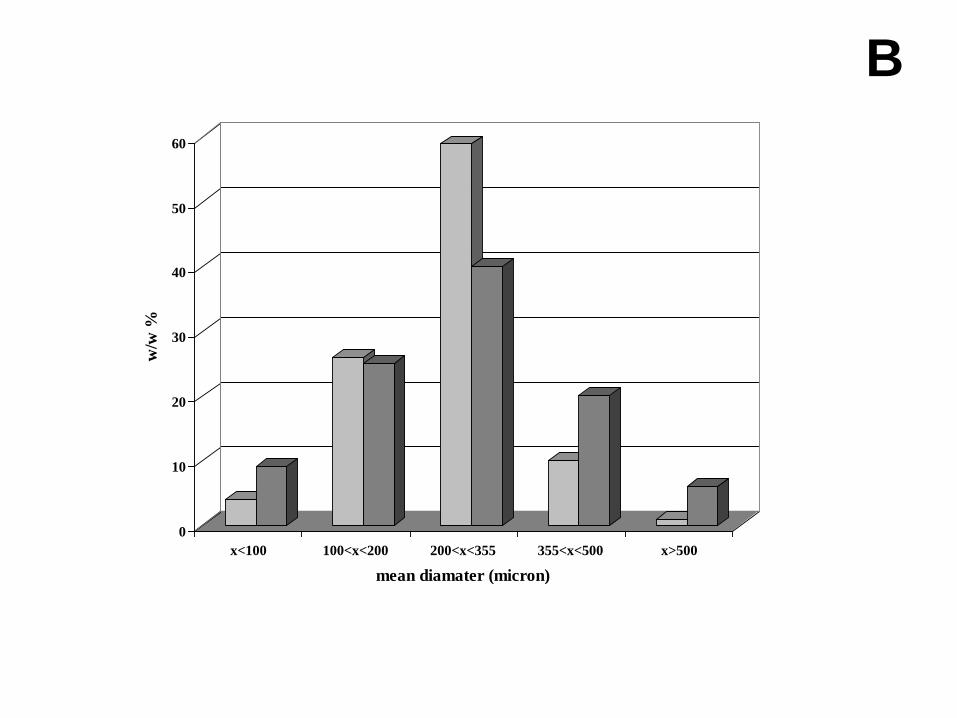
Size distribution - The graphs of Figure 1 compare the size distribution for two systems, the first
one containing only Gelucire (system 9) and the second one (system 3) Gelucire 50/13 and Lutrol
F68 in the weight ratio 3:2, in the absence and in the presence of olanzapine. There is a quasi
8
Gaussian distribution, with a high prevalence of the fraction 100 μm < x < 200 μm in the first case
(Figure 1A), while for microspheres also containing Lutrol F68 (Figure 1B) the range 200 μm < x <
355 μm is dominant. The presence of olanzapine in the mixtures does not affect the size distribution
for system 9, while it appears to increase the larger size fraction in the presence of Lutrol F68: in
this second case, the fraction 355 μm < x < 500 μm is practically doubled. The size distribution of
the microspheres is related to viscosity of the molten mixture when it is poured onto pre-heated
sonotrode: a lower viscosity generates better atomization and thus lower size microspheres.
The change from Lutrol F68 to Lutrol F127 has little effect on the distribution: while the main size
fraction 200 μm < x < 355 μm is maintained constant close to 50% for all the systems, the 100 μm <
x < 200 μm fraction increases up to 40% for system 7 and the largest fraction 355 μm < x < 500 μm
decreases down to 5% when the Lutrol F127 content is increased. Greater differences concerning
the main fraction could be observed at the highest Lutrol F127 concentration (these last results are
not reported).
SEM - All the studied systems produce microspheres when the molten mixtures undergo US-
assisted spray-congealing, independently of the nature of the Lutrol and composition.
The image analysis (see below) reveals the presence of defects on the microsphere surface, and the
X-EDS technique also suggests uneven composition of the surface. Actually each system is formed
by three components at different weight ratios with different consequences on the generation of
these defects on the microsphere surface. Therefore, systems with each possible combination
between carrier and drug were prepared and examined at SEM, in the presence and in the absence
of ultrasound vibration, to identify the possible cause of these defects: the physical mixtures were
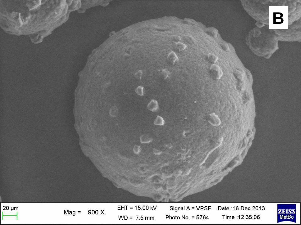
melted until a homogeneous mass was obtained (Figure 2). In Figure 2A, microspheres containing
only Gelucire 50/13, in the absence of olanzapine, show a smooth and regular surface; the addition
of Lutrol F127 at 50% w/w without olanzapine originates only small defects on the microsphere
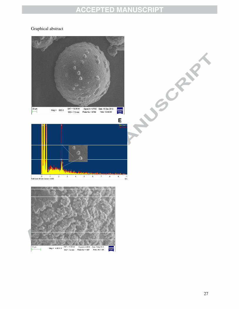
surface. Figure 2B shows a microsphere containing Gelucire 50/13 and olanzapine (as in system 9):
the surface offers the view of wart-like protuberances in the form of particles that the X-EDS
9
spectrum suggests contain S, that is a marker of olanzapine, at a lower concentration than the
smooth portion of the microsphere surface. Figure 2C displays the surface view of a ternary
microsphere (system 8), where these protuberances cover practically the whole surface and have a
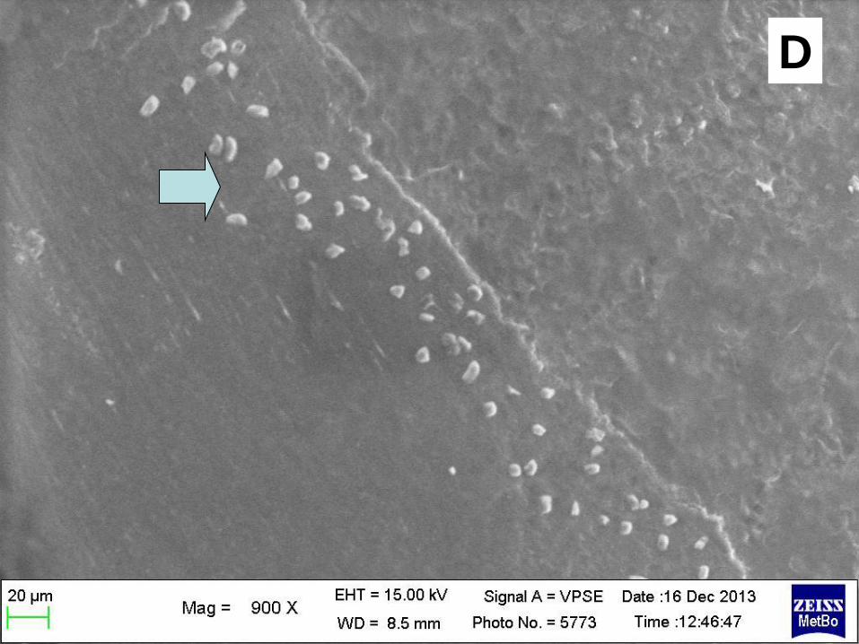
modified shape. While the solid dispersion samples of system 9, obtained in the absence of
ultrasound, lack any defined shape and do not show any presence of particles, in the presence of
Lutrol F127 (systems 5-8), a few features similar to those observed in Figure 2B can be observed.
The number of these protuberances increases when the Lutrol concentration is increased (Figure
2D). The differences between the same systems formulated as solid dispersions or as microspheres
appear related to the application of ultrasound that proved to be an important factor responsible for
the protuberances present on the surface, promoting the crystallization of the drug or the phase de-
mixing of the carriers. The same protuberances can be observed when the Lutrol concentration is
increased (Figure 2D), even in the absence of ultrasound. The concomitant presence of Lutrol inside
the starting mixture and the action of ultrasound to model the solid dispersion contribute to the
irregularity of the microspheres of the present systems, with rapid appearance of the features visible
on the surface, as a consequence of accelerated crystallization and phase de-mixing of drug and
carriers.
X-EDS - For an in-depth investigation of the microspheres that display unusual features on the
surface, the X-EDS technique was employed to evaluate the homogeneity of the microsphere
surface composition, through the study of characteristic spectra emitted by the elements of all the
components present in the studied system. In the present case, the formula of olanzapine
(C17H20N4S) suggests using S, as a marker of the presence of this molecule in the area under
examination, since it is absent in all the carriers.
Although the intensity of a peak in X-EDS analysis is not directly correlated with the concentration
of a given element on the surface (since it is a probabilistic analysis), in the present case it might
nevertheless be useful to indicate that olanzapine is more present at the level of the hollows
surrounding the wart-like protuberances. Here, the peak of S (indicating the presence of olanzapine)
10
obtained for the protuberance is less intense than the peak obtained at the level of the underlying
matrix.
Micro-Raman spectroscopy - Figure 3 shows the Raman spectra of the single components of the
microspheres (system 9) and of different zones of the same microsphere. With reference to peak
groups between 1400 and 1600 cm-1 that are present only in the olanzapine spectrum, it can be
observed that the intensity of these peaks is lower when the higher portions of protuberances are
considered, suggesting a lower content of the drug at this level and confirming its non-
homogeneous distribution on the microsphere surface. Moreover, the Raman spectra show a higher
content of the Lutrol at the level of the protuberance, its distinctive peaks at about 300, 850 and
1500 cm-1 being more intense. This fact further increases the complexity of the systems that
appeared to be formed by different Gelucire/Lutrol phases at different olanzapine concentrations.
Image Analysis – Full image analysis is reported only for systems 5-9, containing Lutrol F127; the
samples were sieved before the SEM analysis and a well-defined size fraction was examined: this is
reflected by the constant values of the size, obtained as diameters of the microsphere or as Feret’s
values (Table 2). The Feret's diameter of a particle is defined as the distance between two parallel
tangents to the perimeter of the projected area of the particle. Since for a single particle infinite
values of these diameters can be determined, the reported values actually represent averages. The
Feret’s values shown in the Table are constantly higher than the diameter of the microsphere,
suggesting somehow a difference from a perfect spherical shape of the particles.
Moreover, the unexpectedly high heterogeneity parameter offers some points of discussion about
the method of preparing microspheres by ultrasound-assisted spray congealing. As stated above,
one of the original components of the previous systems (Gelucire 44/14) [7] had to be changed,
because its low melting point did not guarantee the solidification of the droplet in the form of
microspheres. The use of Gelucire 50/13 allowed the formation of discrete solid particles in the
apparent form of microspheres. At SEM analysis, these microspheres proved to be not so perfectly
shaped as expected. The term heterogeneity should be as near-to-zero as possible to consider a
11
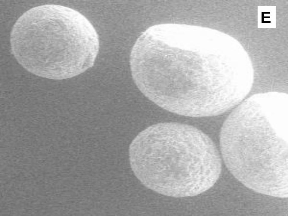
particle system homogeneous in terms of the shape. The values reported in Table 2 for this
parameter suggest that more than 50% of the particles differ from a perfect sphere: this can also be
noted in Figures 2E, where some deformed microspheres are shown.
The aspect ratio and roundness parameters also support this view. The aspect ratio parameter
measures the ratio between the longest and the shortest dimension of a particle: in the present case,
the values shown in Table 2 suggest that the microspheres of the different systems are not exactly
symmetrically shaped and, at the SEM magnification, appear as rather slightly pronounced
ellipsoids. This aspect was also confirmed by the roundness; this parameter is based on the ratio
between the maximum and minimum sizes for circles that are just sufficient to fit inside and to
enclose the shape of the particle. It is the measure of how closely the shape of the microsphere, as it
appears on the microscope plate, resembles that of a circle. In this case too, the values of Table 2
suggest a 10-20% difference from a perfect reference.
As a conclusion from these values, it appears that Gelucire 50/13, though able to form spherically
shaped particles, does not increase the melting point or the viscosity of the molten mixture at a level
enabling the production of microspheres on solidification without deformation or defects. A droplet
of low viscosity can easily modify its shape after the impact with the sonotrode during its fall; or
even during the contact with the collecting plane, if it is not completely solidified (see
Experimental).
Fractal dimension – The fractal dimension of the microsphere contour is shown in the last column
of Table 2 for some systems. The fractal dimension provides a measure of the complexity of a
particle profile or of a surface, when the scale of the measurement unit of these objects changes. A
fractal dimension value is usually not an integer value: it also indicates a measure of the space-
filling capacity of a pattern or its ability to occupy a superior dimension. For a particle perimeter or
profile, the fractal dimension ranges from 1 to 2: the closer the value is to 1, the smoother and more
regular the profile is. In the present case, the profile of the microsphere projection, that is a circle on
the microscope table, is measured with a step ranging from 1.30 to 3.80 μm. When ln(perimeter)

12
thus obtained is plotted against ln(step), a satisfactory linear relationship is observed: the fractal
dimension of the profile is obtained from the slope (Richardson plot) [14]. The values of the fractal
dimension shown in Table 2 for systems 5-9 indicate a rather smooth contour of the microspheres
and the photo of Figures 2F confirm the relative smoothness of the profile for some microspheres,
in agreement with the values of the fractal dimension. This appears in contrast with the aspect of the
surface (Figure 2C), when examined at high magnification: the range of the step values chosen for
the analysis is probably too large and unable to show the rugosity that can be seen from the
observation of the surface and that should also affect the profile.
Thermal Analysis
Thermomicroscopy (HSM) - The photos of Figures 4A-B show a sample of system 9 microspheres (
Gelucire 50/13 and 10% w/w olanzapine) heated at thermomicroscope up to almost complete
fusion. The ultrasound-assisted spray-congealing makes it possible to obtain uniformly coloured
microspheres, due to the homogeneous distribution of the yellow particles of olanzapine inside
them. At the optical microscope, the carrier is opaque when in the solid form and this prevents
observation of different details within the particles except for the colour. When the carrier begins to
soften and the external shape of the microspheres is deformed with heating, the molten carrier
becomes transparent and allows observation of discrete particles of the drug (Figure 4A). As the
temperature increases, the particles of olanzapine start to be dissolved in the molten mass of the
carrier and the thermograms of all the components of the systems proved to be thermally stable (at
least up to 150°C). In all the systems examined, the complete dissolution of the particles of
olanzapine above 120°C was observed, irrespective of the nature of the Lutrols and their
concentration in the mixed carrier, and no synergism could be observed for two-component carriers
concerning the dissolution of the drug into the molten carriers.
From the examination of the solubility parameters [7] both Lutrols and Gelucire 44/14 are expected
to be poor solvents for olanzapine at room temperature in the solid dispersions. On the contrary, it

13
was observed that the drug dissolves into these molten carriers without any problem. As a
consequence, precipitation of olanzapine on cooling could be anticipated also inside the present
systems, but was observed in the solid dispersion not subjected to ultrasound only when the carrier
was composed of Lutrols (Figure 4B). Most olanzapine remained dissolved even at room
temperature, when the carrier contained only Gelucire 44/14, in a metastable state, apparently
insensitive to aging. The same experiment, repeated using Gelucire 50/13, afforded similar results
in the absence of ultrasound. In the present case, on the contrary, all systems, despite the common
presence of the Gelucire, were shown at HSM to contain solid particles of olanzapine precipitated
on cooling. This fact, in agreement with the values of the solubility parameters, is attributed to the
”ultrasound effect” that the mixture underwent at the time of contact with the sonotrode, when
molten droplets turned into solid microspheres, and that favours crystallization. As a consequence,
the reduced and uniform size of the olanzapine particles suggests that the drug was initially
dissolved in the molten mixture, and that the applied ultrasound vibration accelerated the
crystallization, which for these systems normally occurs slowly with time (aging). The presence of
Lutrol, which was shown in the previous paper [7] to behave as a poor solvent for olanzapine (at
room temperature), increases the number of particles that can emerge from the molten phase; this
was particularly the case for Lutrol F127, that proved to be a poorer solvent than Lutrol F68.
DSC of individual carriers - The thermogram of Lutrol F68 has a single melting peak, centered at
56°C; the material is stable at heating up to 150°C, after which the baseline is raised, indicating
decomposition. The thermogram of Lutrol F127 has a single melting endotherm, more regular than
that of Lutrol F68: the peak is centered at 59°C; in this case too, the co-polymer shows thermal
stability up to about 160°C, above this value it begins to decompose. The thermogram of Gelucire
50/13 presents a rather large and asymmetric melting peak, which begins shortly after the ambient
temperature, justifying its definition as a semi-solid material and its nature as a multicomponent
mixture: in this case too, the material begins to decompose above 160°C. A common feature of the
thermograms of these compounds is the rapid decrease of the profile associated with the onset of the
14
melting endotherm right at the beginning of the heating; as a consequence, in order to identify
possible thermal effects of the composition or the experimental conditions, the endotherm peak
temperature was considered. Due to the deformation of the endotherm, using the peak temperatures
as a probe is of limited importance and considerable uncertainty must be associated with possible
conclusions.
DSC of the systems 1-9 in the form of microspheres - The thermogram of system 9 in the form of
microspheres shows the presence of a single melting endotherm of the carriers, where the onset of
the melting starts practically at room temperature, and the absence of the melting endotherm of
olanzapine (Figures 5A). Since HSM photos document the presence of the drug inside the
microsphere in the form of solid particles, this means that the drug dissolves into the molten carrier,
which acts as a solvent as the temperature increases during the heating for recording the
thermogram. Only small differences can be detected as a function of the composition.
The experimental conditions of the preparation appear to affect the thermal behaviour of the
systems. A certain effect is experienced by system 9, when it undergoes ultrasound-assisted spray
congealing that accelerates the organization of the solid state with respect to a simple cooling at -
20°C for the solid dispersion: this is reflected by the different peak temperatures.
These effects are present to a lesser extent when the systems contain Lutrol F127 and Gelucire
50/13 (system 8) (Figure 5B): the melting peaks are found closer to each other, at an intermediate
temperature between that of the melting peaks of Gelucire 50/13 and that of Lutrol F127. A
shoulder is present at the low temperature of the endotherm of the formulations. During the
preparation of the solid dispersion, all the components have had a chance to melt and possibly to
mutually dissolve, according to their solubility: in this case too, however, the melting endotherms
appear to be split. The cooling, the irradiation of ultrasound during the preparation of the
microspheres and the crystallization and solidification could produce phase de-mixing of the
carriers, and this could contribute to the deformation of endotherm and the splitting of the melting
endotherms of the individual components. It can be concluded that the systems thus proved to be
15
composed of immiscible solid phases of the carriers mutually saturated and saturated with respect to
olanzapine; and free olanzapine, as the HSM photos show.
The thermogram profile differs little when the nature of the Lutrol changes. The different
characteristics of the two Lutrols do not play different roles at the level of the solid dispersion and it
can be assumed that the same mechanism takes place in the formation of the dispersion. Only a shift
of the thermal parameters to the high temperatures can be observed in the case of Lutrol F127: the
similarly deformed melting endotherms suggest that in both cases the same phase de-mixing occurs
due to the poor miscibility between Gelucire 50/13 and both types of Lutrol. The differentiation of
the peaks appears less pronounced in the case of Lutrol F127 probably because this Lutrol has a
solubilizing capacity towards the Gelucire 50/13 compared to Lutrol F68.
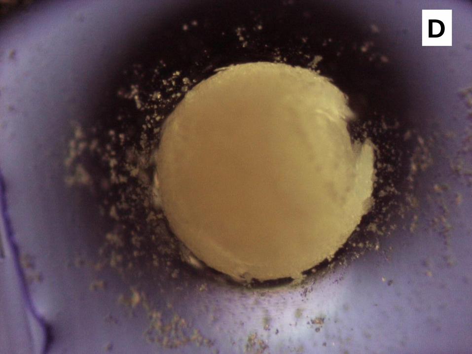
Behaviour in the presence of a dissolution medium - When in contact with the aqueous dissolution
medium, the microspheres undergo a de-aggregation into particles of reduced size and irregular
shape that also appears stratified. Figure 4C shows that microspheres containing Lutrol F127
(system 7) de-aggregate much more than those of system 3, containing Lutrol F68 (Figure 4D), and
this fact can further support the more rapid release of the drug from the first system (see below).
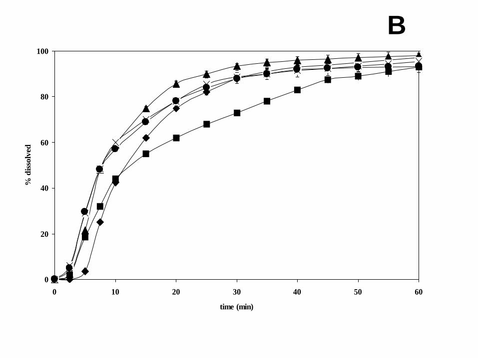
In vitro release profiles - The analysis of the release profiles (Figure 6) allows us to evaluate the in
vitro release of olanzapine from microspheres prepared with different concentrations of Lutrol and
Gelucire compared to the pure active ingredient.
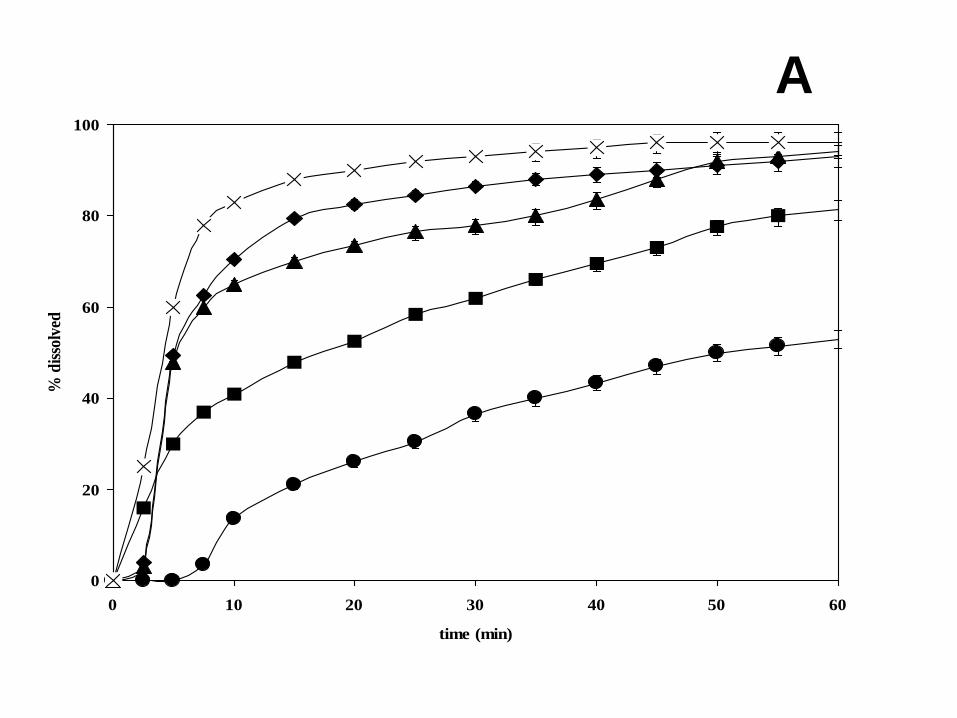
Figure 6A shows the release profiles of pure olanzapine compared to those of the microspheres
prepared with only Gelucire (system 9), with Gelucire/Lutrol F68 (system 3) and with
Gelucire/Lutrol F127 (system 7). Analysis of the graph shows that all of the prepared formulations
have an accelerated release with respect to the pure active ingredient. System 3 releases about 70%
in 5 minutes and about 90% in the first 10 minutes; while the pure olanzapine dissolves by only
15% in the first 10 minutes. The formulations also containing Lutrol compared to those containing
only Gelucire have a much more accelerated release: after 10 minutes, system 9, containing only
Gelucire, releases only 40%, while all formulations with Lutrol F68 (systems 1 and 3) release about
16
70% in 10 minutes. The possible reason for this difference between the two Lutrols can be found in
the ability of Lutrol F127 to gel and swell on contact with water (Figures 4C and 4D) and therefore
microspheres containing Lutrol F127 de-aggregate more rapidly, facilitating the release of the drug;
moreover, Lutrol F127 can form micelles in aqueous solution, contrary to Lutrol F68, providing a
notable solubilisation of the drug molecules inside the polymer micelles. Finally, the bimodal
profile of release shown by all the systems could be related to the rapid de-aggregation of the
microsphere thus offering a larger surface to the dissolution medium at the beginning of the
experiment.
The physical mixtures of the same composition also offer an accelerated release compared to pure
olanzapine, though only slightly lower than that of the microspheres (Figure 6B). The profiles show
that in 10 minutes the mean release from the mixtures is around 20% less than that from the
corresponding microspheres. The physical mixtures operate a less selective release than
microspheres of the same composition. Both systems contain the same hydrophilic carriers, but in
the microspheres a more intimate contact is obtained between olanzapine and carriers at the
microsphere formation step and application of ultrasound makes it possible to highlight the different
role of the components of each system. Moreover, the formulation in terms of microspheres
enriches the system from the technological point of view. The microspheres, in fact, have a flow
capacity that the powders do not display because of the low melting point of the powder; the
particles undergo adhesion and tend to form cakes with poor flowability.
Discussion
The systems examined in this paper are complex because of the nature and composition of the
carrier as well as the drug chosen as a model. Indeed, each of the carriers (Gelucire and Lutrols)
taken individually had also been shown to have a complex behaviour when formulated with a drug,
associated with aging [7, 15-18]. Moreover, the present systems, formulated as solid dispersion,
underwent ultrasound vibration that starts important modifications of the physical state of each
17
component in the final microspheres, such as the formation of mutually saturated different phases. It
is also difficult to separate the individual contributions of this large number of parameters from the
final formulation. However, comparing the present systems with the many examples of solid
dispersions reported in the scientific literature, the main difference appears to be the application of
ultrasound to drug delivery systems, whose effects play a number of roles not yet completely
examined.
Figure 5A clearly shows this difference through the dissimilarity between the shape of the melting
endotherm of Gelucire 50/13 alone or as a solid dispersion with olanzapine solidified at -20°C or
formulated as microspheres obtained under US. In the first case, the thermal profile represents the
material as obtained from the container (“an equilibrium or near-equilibrium structure [16]). In the
second case, there is a dominant presence of the lower temperature peak of Gelucire 50/13 that
HSM can attribute to the lower melting fraction of the carrier containing dissolved olanzapine; in
the third case, a reverse situation can be observed, where the higher melting fraction is dominant,
probably due to the presence of precipitated olanzapine inside the carrier. The composition of
Gelucire 50/13 can be responsible for this thermal behaviour: together with mono- and di-esters of
PEG and free PEG 1500, for a total amount of about 80% w/w, Gelucire 50/13 also contains about
20% w/w mono-, di- and tri-glycerides that, with their low HLB, balance the high HLB value of the
PEG-derivatives of the mixture. It is possible that the hydrophobic fraction could act as a selective
solubilising phase for olanzapine: it has in fact been reported [7] that the comparison of the
solubility parameters of Gelucire (in toto) and olanzapine would not suggest solubility.
On the contrary, the presence of a Lutrol in mixture with Gelucire 50/13 causes precipitation of
olanzapine (visible at HSM): as a consequence, in a previous paper [7], systems containing Gelucire
44/14 and Lutrol F68 or F127 together were proposed in order to design formulations of improved
stability for olanzapine solid dispersions, due to the absence of dissolved drug that could evolve to
crystallize with aging. Even more so, the action of Lutrols and the contemporary application of
ultrasound should provide crystallization of both drug and carriers and make the present systems
18
particularly stable to aging. The action of ultrasound on the sold dispersion can be hypothesized to
act as a promoter of crystallization of the embedded drug, separation of phases, but also of the lipid
components of the Gelucire fraction of the present carriers, analogously to a tempering process,
reported to improve stability in triglycerides in hot melt coating formulations [19].
Ultrasound instantaneously induces these changes that usually lead to problems in practical
application of the solid dispersions: the high-frequency vibration, experienced by the systems at the
time of the contact with the sonotrode, accelerates modifications, such as crystallization, transition,
de-mixing and others, that usually remain in a metastable situation with the risk of undesirable
changes with time. The high-frequency vibration also virtually eliminates the problems of aging,
making it possible to obtain systems that are almost perfectly aged and stable or unable to further
evolve in a short time.
The treatment of a solid dispersion under ultrasound could be suggested as a tool to achieve stability
for formulations which should render a dispersion as a commercial product. As a conclusion, the
present ultrasound-olanzapine formulations in the form of microspheres can be confidently
considered as stable and almost independent of aging modification due to the presence of
crystallized phases. The shocking crystallization driven by ultrasound is expected to cause other
interesting physical changes to the final systems. Many investigations examined the changes in the
physical structure of these carriers, alone and as dispersions loaded with a different drug, as well as
the effects of the incorporation of the model drugs on the physical properties of the dispersions on
aging. These studies mainly involved Gelucires that possess differentiated compositions
manufactured to obtain a prefixed melting point and hydrophile–lipophile balance (HLB). For
multi-component materials of this type, a large number of papers are reported dealing with the
effect of storage on the structure and properties of the lipid constituents, such as the development of
large crystals or the separation of polymorph single phases or the migration of lipid components to
the surface with consequent “blooming” of the surface.
19
In the present case, at SEM it could be observed that particulate forms are developed under
ultrasound on the microsphere surface in the presence of olanzapine or at increasing Lutrol
concentrations, with considerable surface morphology changes. In some cases, at higher
magnification, they appear as leaf-like or needle-like particles (Figure 2G) that both X-EDS and
micro-Raman spectroscopy indicate as having a non-uniform distribution of drug and carrier with
respect to the background of the surface. These features are similar to those described for analogous
systems, but occurring over a long period and largely associated with alterations of the crystal habit
or with polymorphic changes or formation of mixed crystals of increased size. At SEM it was
possible to observe the same “blooming” of the microsphere surface as that observed in systems
containing Gelucire 50/13 and paracetamol and caffeine, aged at high temperature for several days
[16].
In our study , it was observed that the drug, which remained completely dissolved in Gelucire 44/14
[7] and only partially crystallized in Gelucire 50/13 solid dispersion, precipitates when Gelucire
50/13 and olanzapine (system 9) undergoes ultrasound due to the formation of microspheres (Figure
2B). This occurs also in the presence of increasing concentrations of Lutrol (Figure 2C) that cause
multiplication of the surface “blooming” (Figures 2C) as a consequence of a possible phase de-
mixing under ultrasound.
All the described situations can affect the release of the active agent in a unforeseen way: the phase
de-mixing operated by ultrasound introduces tensions on the final microsphere, as a result of a
tempering-like effect. The microspheres de-aggregate when in contact with the dissolution medium,
revealing a stratified structure, which in turn induces a rapid release of the drug as a consequence of
the increased surface area. The same was observed when olanzapine was formulated as a solid
dispersion with a mixture of lipid carriers (cutina and stearic acid) and processed by ultrasound-
assisted spray-congealing technique to obtain solid microspheres [20].
20
Conclusions
- Gelucire 50/13 offers a support to Lutrols in formulating microspheres, but has little
negative effect on the release of olanzapine; Lutrol F127 behaves better than Lutrol F68 in
promoting the release of the drug;
- Olanzapine dissolves into the molten carriers, but rapidly crystallizes because of the
ultrasound discharge during the formation of microspheres;
- Image analysis shows that in the present systems ultrasound-assisted spray congealing
produces defects on the microsphere surface, related to possible phase de-mixing, promotion
of crystallization and non-uniform composition of the surface;
- Different analytical techniques (SEM, EDS, Raman, DSC, HSM) operated synergistically to
highlight formulation problems.
Acknowledgments – The text was revised for grammar and style by an English mother-tongue
translator.

21
Legend to Figures
Figure 1 – Size distribution of the microspheres containing (A) Gelucire 50/13 (system 9) and (B)
Gelucire 50/13/Lutrol F68 (system 3), in the presence (gray) and in the absence (dark) of
olanzapine.
Figure 2 – SEM photos of microspheres containing: A) only Gelucire 50/13; B) system 9; C)
system 8. SEM photos of solid dispersions containing: D) Gelucire 50/13 and Lutrol F127 (45:45
w/w). SEM photos of the microsphere shape (E) and contour (F). SEM photo of details of a
microsphere surface /G).
Figure 3 – Micro-Raman spectra of different points of a microsphere shown in the Figure 2B
(spectra of single components are shown for comparison): A) Olanzapine; B) Lutrol F127; C)
protuberance; D) surface; E) Gelucire 50/13.
Figure 4 – Thermomicroscope photos of: A) system 9 microsphere after the melting of the carrier;
B) system 8 solid dispersion at 50°C; C) system 7 microsphere in the presence of the dissolution
medium at 37°C; D) system 3 microsphere in the presence of the dissolution medium at 37°C.
Figure 5 – DSC thermograms of A) (from the left) Gelucire 50/13; microsphere system 9; solid
dispersion system 9. B) (from the left) Lutrol F127; solid dispersion system 8; solid dispersion
Lutrol F127/Gelucire 50/13 at 50% w/w; microsphere system 8; Gelucire 50/13.
Figure 6 – Release profiles of olanzapine from microspheres (A) and physical mixtures (B).
X system 7; ♦ system 3; ▲ system 1; ■ system 9; ● pure olanzapine.
22
REFERENCES
[1] A. Dalmoro A.A. Barba, G. Lamberti, M. d'Amore, Intensifying the microencapsulation
process: ultrasonic atomization as an innovative approach. Eur. J. Pharm. Biopharm. 80 (2012) 471-
7.
[2] A. Fini, C. Cavallari, F. Ospitali, M.L. Gonzalez-Rodriguez, Theophylline-loaded compritol
microspheres prepared by ultrasound-assisted atomization. J. Pharm. Sci. 100 (2011) 743-57.
[3] Y. Gao, C.L. Zhu, X.X. Zhang, L. Gan, Y. Gan, Lipid-polymer composite microspheres for
colon-specific drug delivery prepared using an ultrasonic spray freeze-drying technique.
J. Microencapsul. 28 (2011) 549-56.
[4] A. Fini, C. Cavallari, A.M. Rabasco Alvarez, M.G. Rodriguez, Diclofenac salts, part 6: Release
from lipid microspheres. J. Pharm. Sci. 100 (2011) 3482-94.
[5] C. Cavallari, B. Luppi B, A.M. Di Pietra, L. Rodriguez, A. Fini, Enhanced release of
indomethacin from Pvp/stearic acid microcapsules prepared coupling co-freeze-drying and
ultrasound assisted spray-congealing process., Pharm. Res. 24 (2007) 521-9.
[6] C. Cavallari, L. Rodriguez, B. Albertini, N. Passerini, F. Rosetti, A. Fini, Thermal and fractal
analysis of diclofenac/Gelucire 50/13 microparticles obtained by ultrasound-assisted atomization, J.
Pharm. Sci. 94 (2005) 1124-34.

23
[7] C. Cavallari, A. Fini, G. Ceschel, Design of olanzapine/lutrol solid dispersions of improved
stability and performances, Pharmaceutics 5 (2013) 570-90.
[8] G.H. An, M.J. Kim, H.J. Lee, S.S. Park, Y.W. Cho, K. Park, Y.H. Choa, Fabrication of
terazocin-loaded poly(D,L-lactide) microspheres by an ultrasonic spray drying method and their
release behaviors, J. Nanosci. Nanotechnol. 8 (2008) 5139-42.
[9] C. Cavallari, B. Albertini, L. Rodriguez, A.M. Rabasco, A. Fini, Release of indomethacin from
ultrasound dry granules containing lactose-based excipients, J. Control. Release 20 (2005) 39-47.
[10] N. Passerini, B. Perissutti, M.R. Moneghini, D. Voinovich, B. Albertini, C. Cavallari, L.
Rodriguez, Characterization of carbamazepine-Gelucire 50/13 microparticles prepared by a spray-
congealing process using ultrasounds, J. Pharm. Sci. 91 (2002) 699-707.
[11] N. Passerini, B. Albertini, B. Perissutti, L. Rodriguez, Evaluation of melt granulation and
ultrasonic spray congealing as techniques to enhance the dissolution of praziquantel.
Int. J. Pharm. 318 (2006) 92-102.
[12] L. Rodriguez, N. Passerini, C. Cavallari, M. Cini, P. Sancin, A. Fini, Description and
preliminary evaluation of a new ultrasonic atomizer for spray-congealing processes, Int. J. Pharm.
25 (1999) 133-43.
24
[13] A. Fini, M.A. Holgado, L. Rodriguez, C. Cavallari, Ultrasound-compacted indomethacin
/polyvinylpyrrolidone systems: effect of compaction process on particle morphology and
dissolution behavior, J. Pharm. Sci. 91 (2002) 1880-90.
[14] M.A. Holgado, M.J. Fernández-Hervás, A.M. Rabasco, A. Fini A, 1995. Characterization
study of a diclofenac salt by means of SEM and fractal analysis, Int. J. Pharm. 120 (1995) 157-67.
[15] R. Bodmeier, O. Paeratakul, H. Chen, W. Zhang, Formation of sustained release wax matrices
within hard gelatin capsules in a fluidized bed. Drug Dev. Ind. Pharm. 16 (1990) 1505–19.
[16] N. Khan, D.Q.M. Craig, Role of blooming in determining the storage stability of lipid-based
dosage forms, J. Pharm. Sci. 93 (2004) 2962-71.
[17] E.V. Batrakova, A.V. Kabanov, Pluronic block copolymers: evolution of drug delivery concept
from inert nanocarriers to biological response modifiers, J. Control. Release 130 (2008) 98-106.
[18] J.J. Escobar-Chávez, M. López-Cervantes, A. Naïk, Y.N. Kalia, D. Quintanar-Guerrero, A.
Ganem-Quintanar, Applications of thermoreversible pluronic F-127 gels in pharmaceutical
formulations, J. Pharm. Pharmacol. Sci. 9 (2006) 339-56.
[19] K. Chansanroj, G. Betz, Improving stability of triglycerides in hot melt coating formulations,
Glatt International Time 26 (2008)
(http://www.glatt.com/times/times26site/tms26_improving_1.htm)
[20] A. Fini, C. Cavallari, G. Ceschel, A.M. Rabasco, Bimodal release of olanzapine from lipid
microspheres, J. Pharm. Sci. 99 (2010) 4251-60.

A Figure 1
0
10
20
30
40
50
60w
/w %
x<100 100<x<200 200<x<355 355<x<500 x>500
mean diamater (micron)
B

A Figure 2
B

C
D
E

F

G

Figure 3

A

B

D
C
D

A

B
0
20
40
60
80
100
0 10 20 30 40 50 60
time (min)
% d
isso
lved
A
0
20
40
60
80
100
0 10 20 30 40 50 60
time (min)
% d
isso
lved
B
25
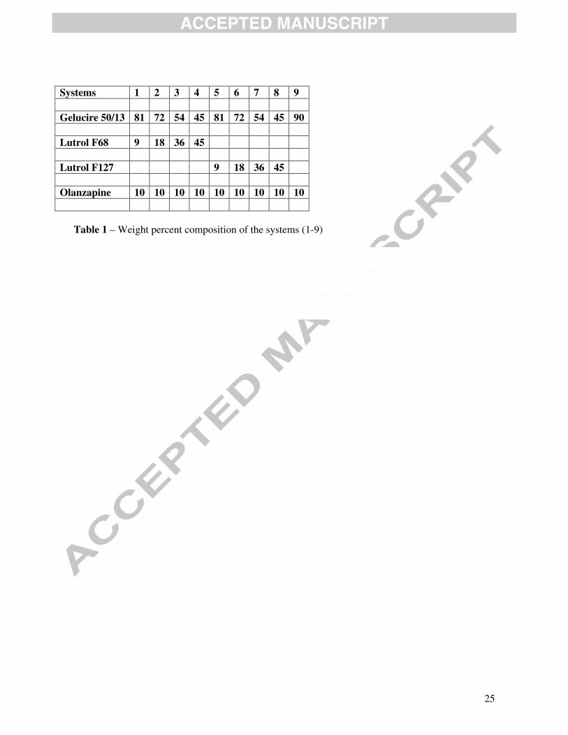
Systems 1 2 3 4 5 6 7 8 9 Gelucire 50/13 81 72 54 45 81 72 54 45 90 Lutrol F68 9 18 36 45 Lutrol F127 9 18 36 45 Olanzapine 10 10 10 10 10 10 10 10 10
Table 1 – Weight percent composition of the systems (1-9)

26
System (% Lutrol F127)
Diameter (Feret) (µm)
Heterogeneity
Aspect ratio
Roundness
Fractal dimension of the contour
5 (9%) 290 (303) 0.64 1.15 1.10 1.08 6 (18%) 290 (297) 0.41 1.18 1.20 1.09 7 (36%) 285 (301) 0.62 1.19 1.24 1.07 8 (45%) 283 (286) 0.50 1.13 1.10 1.07 9 (0%) 260 (275) 0.62 1.14 1.09 1.09
Table 2 – Parameters of the image analysis of systems containing Lutrol F127 at increasing concentration (systems 5-8); system 9 was shown for comparison.
27
Graphical abstract
E
28
Highlights Olanzapine, Gelucire 50/13, Lutrol F68 and F127 were formulated as solid dispersions Microspheres were obtained by ultrasound assisted spray congealing This technique produces defects on the microsphere surface Defects are related to phase de-mixing and non-uniform surface composition Ultrasound treatment of solid dispersions accelerates aging and achieves stability