Embed Size (px)
Citation preview

PERPUSTAKAAN UMP
ill III III 11111111 II Ill III I 11111 0000104513
AP
Initial Assessment of Facial Nerve Paralysis based on Motion Analysis using Optical Flow Method
by
WAN SYAHIRAH BINTI W.SAMSUDIN
(1140610618)
A thesis submitted in fulfillment of the requirements for the degree of Master of Science in Mechatronic Engineering
School of Mechatronic Engineering UN! VERSITI MALAYSIA PERLIS
2015

TABLE OF CONTENTS
PAGE
THESIS DECLARATION ii
ACKNOWLEDGEMENT iii
TABLE OF CONTENTS iv
LIST OF TABLES vii
LIST OF FIGURES x
LIST OF ABBREVIATION xi
ABSTRAK xii
ABSTRACT xiii
CHAPTER 1 INTRODUCTION
1.1 Introduction 1
1.2 Problem Statement 4
1.3 Objectives of Research 5
1.4 Scope of Research 6
1.5 Thesis Outline 7
CHAPTER 2 LITERATURE REVIEW
2.1 Introduction 8
2.2 Facial Nerve 8
2.3 Facial Paralysis 9
2.3.1 Incidence and Prevalence 10
2.3.2 Causes 11
2.3.3 Symptoms 12
2.3.4 Effects 13
2.4 Facial Nerve Assessment Methods 14
2.4.1 Subjective assessment methods 14
2.4.2 Objective assessment methods 19
2.5 Previous Kanade-Lucas-Tomasi (KLT) Research 27
iv

2.6 Summary 29
CHAPTER 3 METHODOLOGY
3.1 Introduction 34
3.2 Ethical Statement 34
3.3 Equipment Installation and Experimental Setup 35
3.4 Landmarks Chosen and Facial Exercise Development Based On 36
Muscle Movement and House-Brackmann (HB) Score
3.5 Data Acquisition 40
3.5.1 Database 40
3.5.2 Subjects 41
3.5.3 Protocols of Experiment 42
3.5.4 Extracting Frames from Video Data 43
3.6 Facial Tracking System 44
3.6.1 Kanade-Lucas-Tomasi (KLT) Tracking Algorithm 45
3.6.2 Facial Tracking System using Optical Flow Method 48
3.6.3 Detect and track the landmarks chosen on facial video data 50
3.7 Compute motion changes using Kanade-Lucas-Tomasi (KLT) method 52
3.8 Data Analysis 55
3.9 Summary 59
CHAPTER 4 RESULTS AND DISCUSSION
4.1 Introduction 60
4.2 Experiment Overview 61
4.2.1 Results Evaluation 61
4.3 Experimental Results 62
4.3.1 Experiment 1: Distance Measurement of Three Exercises in 62
Four Conditions
4.3.2 Experiment 2: Area Measurement of Three Exercises in Four 82
Conditions
4.4 Summary 99

CHAPTER 5 CONCLUSIONS
5.1 Conclusion
101
5.2 Contribution of Research
102
5.3 Research Limitations
103
5.4 Suggestions for Future Work
103
REFERENCES
105
APPENDIX
111
LIST OF PUBLICATIONS
113
vi
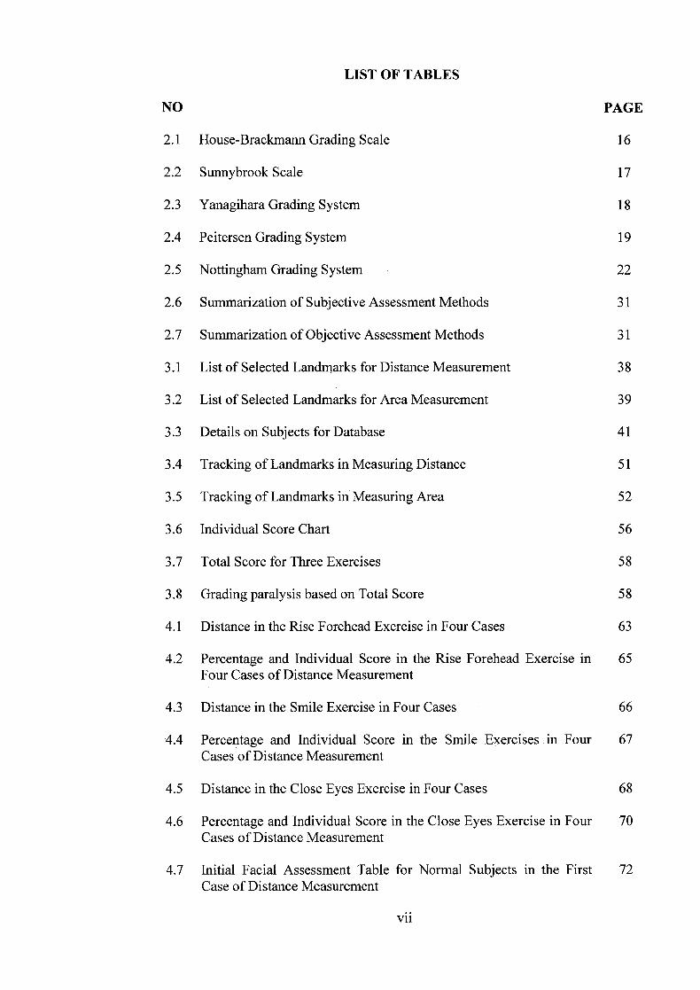
LIST OF TABLES
NO PAGE
2.1 House-Brackmann Grading Scale 16
2.2 Sunnybrook Scale 17
2.3 Yanagihara Grading System 18
2.4 Peitersen Grading System 19
2.5 Nottingham Grading System 22
2.6 Summarization of Subjective Assessment Methods 31
2.7 Summarization of Objective Assessment Methods 31
3.1 List of Selected Landmarks for Distance Measurement 38
3.2 List of Selected Landmarks for Area Measurement 39
3.3 Details on Subjects for Database 41
3.4 Tracking of Landmarks in Measuring Distance 51
3.5 Tracking of Landmarks in Measuring Area 52
3.6 Individual Score Chart 56
3.7 Total Score for Three Exercises 58
3.8 Grading paralysis based on Total Score 58
4.1 Distance in the Rise Forehead Exercise in Four Cases 63
4.2 Percentage and Individual Score in the Rise Forehead Exercise in 65 Four Cases of Distance Measurement
4.3 Distance in the Smile Exercise in Four Cases 66
4.4 Percentage and Individual Score in the Smile Exercises in Four 67 Cases of Distance Measurement
4.5 Distance in the Close Eyes Exercise in Four Cases 68
4.6 Percentage and Individual Score in the Close Eyes Exercise in Four 70 Cases of Distance Measurement
4.7 Initial Facial Assessment Table for Normal Subjects in the First 72 Case of Distance Measurement
vii

4.8 Initial Facial Assessment Table for Patient Subjects in the First Case 73 of Distance Measurement
4.9 Initial Facial Assessment Table for Normal Subjects in the Second 74 Case of Distance Measurement
4.10 Initial Facial Assessment Table for Patient Subjects in the Second 75 Case of Distance Measurement
4.11 Initial Facial Assessment Table for Normal Subjects in the Third 76 Case of Distance Measurement
4.12 Initial Facial Assessment Table for Patient Subjects in the Third 77 Case of Distance Measurement
4.13 Initial Facial Assessment Table for Normal Subjects in the Fourth 78 Condition of Distance Measurement
4.14 Initial Facial Assessment Table for Patient Subjects in the Fourth 79 Case of Distance Measurement
4.15 Accuracy Table for Four Cases of Distance Measurement 80
4.16 Area in the Rise Forehead Exercise in Four Cases 82
4.17 Percentage and Individual Score in the Rise Forehead Exercise in 84 the Four Cases of Area Measurement
4.18 Area in the Smile Exercise in Four Cases
85
4.19 Percentage and Individual Score in the Smile Exercises in Four 86 Cases of Area Measurement
4.20 Area in the Close Eyes Exercise in Four Cases 87
4.21 Percentage and Individual Score in the Close Eyes Exercises in Four 89 Cases of Area Measurement
4.22 Initial Facial Assessment Table for Normal Subjects in the First 90 Caseof Area Measurement
4.23 Initial Facial Assessment Table for Patient Subjects in the First 91 Caseof Area Measurement
4.24 Initial Facial Assessment Table for Normal Subjects in the Second 92 Case of Area Measurement
4.25 Initial Facial Assessment Table for Patient Subjects in the Second 93 Caseof Area Measurement
viii

4.26 Initial Facial Assessment Table for Normal Subjects of Third 94 Caseof Area Measurement
4.27 Initial Facial Assessment Table for Patient Subjects in the Third 95
Caseof Area Measurement
4.28 Initial Facial Assessment Table for Normal Subjects in the Fourth 96 Caseof Area Measurement
4.29 Initial Facial Assessment Table for Patient Subjects in the Fourth 97 Case of Area Measurement
4.30 Accuracy Table for Four Cases of Area Measurement 98
ix

LIST OF FIGURES
NO PAGE
1.1 Head and facial muscles 1
2.1 Anatomy of facial nerve 9
2.2 The measurement of Nottingham Grading System 21
3.1 Flowchart of Methodology 35
3.2 Flowchart of Experiment Protocol for Data Acquisition 42
3.2 (A) and (B) are two rectangle features; (C) Three-rectangle features; 35 (D) Four-rectangle features
3.3 Extracted Frames from Video Data of Subject 44
3.4 Image Registration Problem 45
3.6 Illustration of distance measurement in Rise Forehead Exercise 53
3.7 Illustration of area measurement in Rise Forehead Exercise 54
KI

LIST OF ABBREVIATIONS
AFA Automated Facial Analysis
AU Action Units
AVI Audio Video Interleave
FNF Facial Nerve Paralysis
HB House Brackmann
HBGS House Brackmann Grading System
KLT Kanade-Lucas-Tomasi
LBP Local Binary Pattern
MREC Medical Research and Ethics Committee
SRA Maximum Static Response Array
OP Optical Flow
RAD Resistor-Average-Distance
RBF Radial Basis Function
SLFS Stennert-Limberg-Frentrup Scale
xi

Penilaian Awal Kelumpuhan Saraf Muka Berdasarkan Analisis Pergerakan Menggunakan Kaedah Aliran Optik
ABSTRAK
Pesakit yang mengalami lumpuh saraf muka menghadapi masalah fungsi, kosmetik, dan psikologi yang serius dengan keupayaan untuk berkomunikasi secara lisan dan bukan lisan terjejas. Rehabilitasi untuk kelumpuhan muka bermula dengan penilaian klinikal yang teliti dalam menilai tahap lumpuh. Secara umumnya, untuk penilaian ketidakfungsian saraf muka pada muka pesakit dalam aplikasi klinikal setiap han, system pemerhatian yang subjektif oleh pakar klinikal telah digunakan. Penilaian subjektif terhadap fungsi saraf muka mi dicapai dengan membuat kesimpulan melalui pemerhatian kepada pergerakan muka yang tidak dipaksa. Kaedah-kaedah mi biasanya menghasilkan satu nilai dimana hipotesisnya sepadan dengan tahap kelumpuhan muka. Setakat mi, tiada kaedah penilaian secara subjektif yang boleh menghasilkan makiumat kuantitatifmengenai fungsi sarafmuka dengan tepat. Kajian sebelum mi telah mencadangkan pelbagai kaedah bagi memperolehi data objektif untuk mengukur dan menentukan tahap kelumpuhan muka.Walau bagaimanapun, tiada kaedah kuantitatif yang mengikut piawaian bagi penerangan fungsi saraf muka secara objektif. Mengakui kepentingan penilaian muka mi, kajian mi dijalankan untuk membangunkan penilaian awal yang boleh mengkelaskan subjek kepada normal dan pesakit berdasarkan analisis pergerakan muka menggunakan algoritma aliran optik serta mengkategorikan pesakit kepada enam peringkat mengikut sistem House-Brackmann (HB). Sebelum penilaian awal dibangunkan, dua eksperimen telah dijalankan untuk mencari parameter yang terbaik dan juga ukuran terbaik untuk menilai kelumpuhan muka. Jarak dan luas kawasan telah dipilih sebagai dua parameter yang disiasat dalam kajian mi. Beberapa analisis matematik dan statistic telah dijalankan untuk menentukan ukuran yang terbaik daripada parameter-parameter ini. Keputusan menunjukkan bahawa jarak adalah parameter yang terbaik dan nilai pada bingkai awal senaman adalah pengukuran yang paling penting dalam mengesan perubahan pergerakan muka. Tesis mi juga membentangkan penilaian awal muka yang mengandungi markah individu untuk setiap senaman yang terlibat dalam kajian mi dan juga menggredkan tahap lumpuh untuk setiap pesakit sepadan dengan sistem HB. Kajian-ini telah mendapat keputusan yang memuaskan yang telah disahkan oleh pakar perubatan dalam Otorinolaringologi. Penilaian awal muka mi memainkan peranan penting dalam permulaan system penilaian akhir untuk kelumpuhan muka dan membangunkan program rehabilitasi yang lebih baik untuk pesakit.
xii
Initial Assessment of Facial Nerve Paralysis Based on Motion Analysis using Optical Flow Method
ABSTRACT
Patients with facial nerve paralysis suffer serious functional, cosmetic and psychological problems with impaired ability to communicate both verbally and non-verbally. Rehabilitation for facial paralysis begins with a thorough clinical evaluation in accessing the degree of paralysis. Generally, for assessment of facial nerve dysfunction on patient's face in daily clinical application, observation-based subjective grading systems by clinicians is been employed. Subjective assessments of facial nerve function are accomplished by inferring nerve function through the observation of voluntary facial movement. These methods usually yield a single value which hypothetically corresponds to the severity of facial paralysis. To date, there are no subjective assessment methods that can reliably produce quantitative information regarding facial nerve function. Previous works have proposed a wide range of methods to obtain objective data to quantify and determine the severity of facial paralysis. However, there is no standardized quantitative method for objective description of facial nerve function. Acknowledging the importance of this facial assessment, this research was conducted to develop an initial assessment methodthat can classify normal and patient subjects based on facial motion analysis using Optical Flow algorithm as well as categorizing the severity of patient into six levels according to House-Brackmann (I-TB) system. Prior to initial assessment method been developed, two experiments were conducted to find the best parameter and bestmeasurement for assessing facial paralysis. Distance and area were selected as two measurement parameters which were investigated in this research. A number of mathematical and statistical analyses were performed to determine the best measurement of these parameters. The results indicate that the distance is the best parameter and the value of initial exercise frame is the most important measurement in tracking the changes of facial movement. This thesis also presents the initial facial assessment method which contains the individual scores for each exercise involved in this research and also grading the paralysis for each patient corresponds to HB system. This research showed satisfactory results which was validated by a medical professional in Otorhinolaryngology. This initial facial assessment method may play a pivotal role in initiation of final assessment system for facial paralysis and develop better rehabilitation program for patients.
xlii
CHAPTER 1
INTRODUCTION
1.1 Introduction
Facial nerve incorporates about 7,000 individual nerve fibers. Each fiber
brings the electrical impulses to a specific facial muscle. All information through the
fibers of this nerve permits us to make any facial expression such as laugh, cry,
smile, and frown. Human express the emotions such as sadness, surprise, happiness,
excitement and confusion through facial expressions. For example, a smile can be
interpreted as the sign of happiness while frowning shows some disagreement and
sadness. Facial nerve paralysis will take place when some of the individual nerve
fibers are disrupted. Besides, the movement of facial muscles starts spasming or
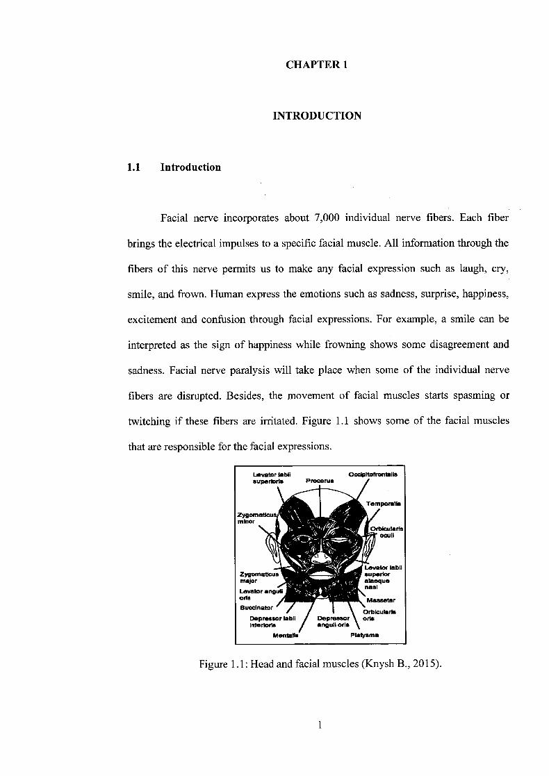
twitching if these fibers are irritated. Figure 1.1 shows some of the facial muscles
that are responsible for the facial expressions.
L.vator labil OcdpitofrOntaIi$ superioris PFOceFU$
Temporalis
ZyQomaticus minor
Orbicuaris oiii
'.1 .
L.evator labil ZyQomaticus superior major alaeque
nasi Levator anguli aria Masseter Buccinator
Depressor labil Depressor aria Inferlorts anguil oris
Mentalls Platysma
Figure 1.1: Head and facial muscles (Knysh B., 2015).
1

Facial nerve paralysis (FNP) is a common problem which involves the
paralysis of any structure innervated by the facial nerve. Literature shows that the
prevalent cause of this paralysis is Bell's palsy, named after Sir Charles Bell (1774-
1842). Bell's palsy is an idiopathic disease category, reporting approximately 50% of
the cases (Brach & VanSwearingen, 1999; Singhi & Jam, 2003).
The other possible causes of facial paralysis are due to birth, trauma,
neurologic syndromes, infection, metabolic, neoplastic, toxic and iatrogenic
(Benecke, 2002). Patients with these paralyses suffer serious functional, cosmetic
and psychological problems with impaired ability to communicate both verbally and
non-verbally. Numbness can occur on the affected side of the face although no actual
sensory loss occurs (Singhi, 2003). Besides, they will be unable to close the eye on
the affected side, which can lead to irritation and cornea! ulceration. Because of that,
the eye should be lubricated with artificial tears until facial paralysis ends (Piercy,
2005). However, the most dramatic impact of the paralysis is its psychological effect
where the patients may have low confidence and fear when interacting with others.
A thorough medical history of patients and physical examination are the
earliest steps in making a facial diagnosis. Clinicians examine whether the forehead
is involved in motor defect or not. This is commonly accomplished by assessing how
well a patient can raise his or her eyebrows. The results from this action help in
determining which part the lesion is in whether in the upper motor neuron or lower
motor neuron of facial nerve component. Rehabilitation is suggested by doctors after
patient's treatment, depending on their condition. It is to regain the function of facial
nerves and improve both the strength and flexibility of the nerves. The failure in
rehabilitation procedures may lead to the continued weakness and inability to
function of facial nerves.
2

The availability of facial rehabilitation is limited, and most individuals with
facial movement disorders have been told to await (spontaneous) recovery or told no
effective intervention exists. Consequently, individuals with this paralysis will deal
with physical, psychological, and social disability daily. The rehabilitation for facial
paralysis begins with a thorough clinical evaluation in accessing the degree of
paralysis. It is important to measure the facial disability from onset to the various
stages of recovery and also to detect changes over time or after treatment. In the past
few decades, several internationally accepted systems have been proposed by
different researchers, yet most of the existing systems are subjective. This subjective
evaluation refers to various facial nerve grading systems, of which the most widely
used is the House-Brackmann (HB) system (Dellanoy& Ward, 2010; Dulguerov,
Wang, Perneger, Marchal, & Lehmann, 2003). The facial nerve evaluation will vary
over many clinicians and it will results in inaccurate assessment of paralysis.
There is no standardized system yet for facial nerve evaluation which has
been accepted for world-wide used. Thus, an objective standardized method, which
is easy to perform, low cost and fast, and simple can be a useful clinical tool to detect
the level of paralysis in patients with facial palsy and monitor their improvement or
performance during and after the rehabilitation procedures. In this study, an initial
facial assessment method is proposed to assist clinicians in assessing the facial nerve
function. Comparisons have been made between two measurement parameters to
determine which one is better to be implemented in the assessment system.
3
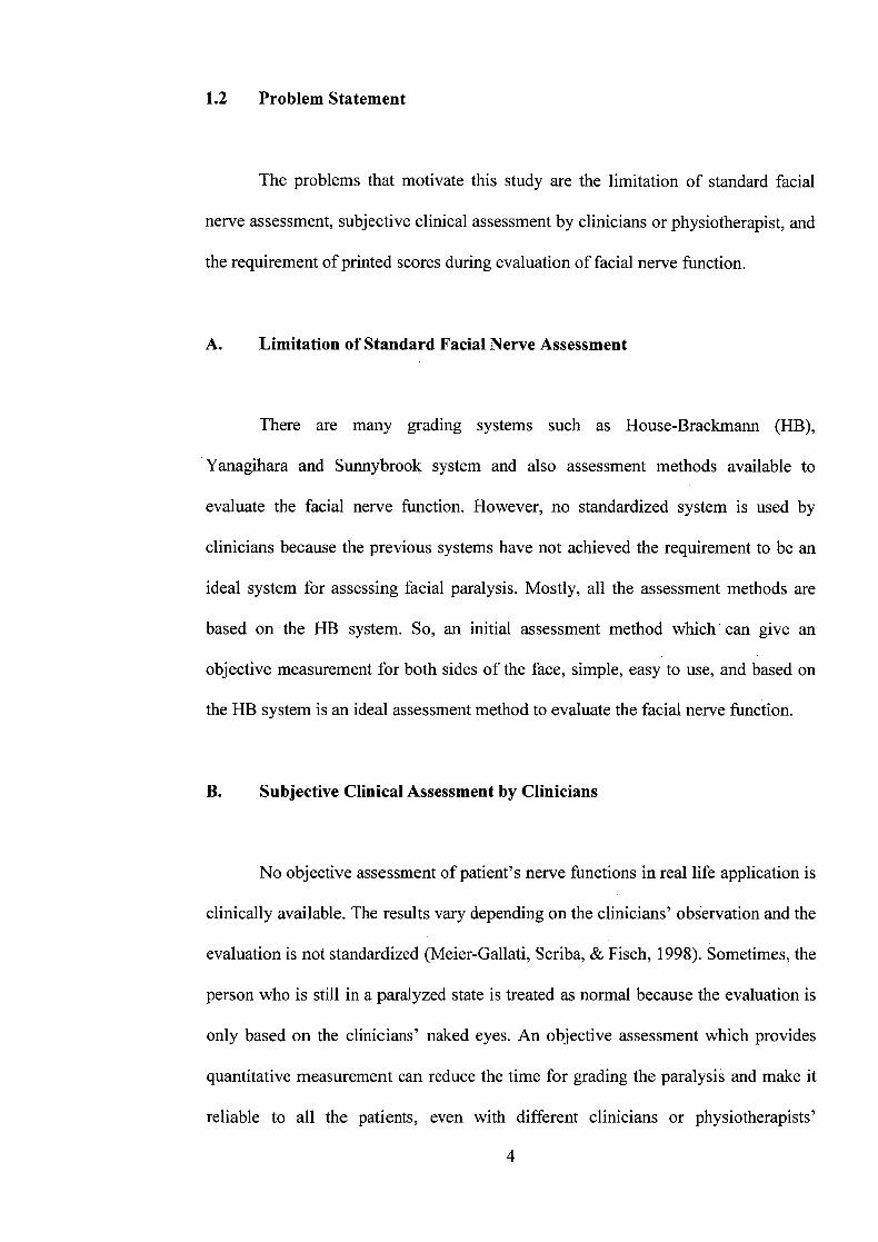
1.2 Problem Statement
The problems that motivate this study are the limitation of standard facial
nerve assessment, subjective clinical assessment by clinicians or physiotherapist, and
the requirement of printed scores during evaluation of facial nerve function.
A. Limitation of Standard Facial Nerve Assessment
There are many grading systems such as House-Brackmann (HB),
Yanagihara and Sunnybrook system and also assessment methods available to
evaluate the facial nerve function. However, no standardized system is used by
clinicians because the previous systems have not achieved the requirement to be an
ideal system for assessing facial paralysis. Mostly, all the assessment methods are
based on the HB system. So, an initial assessment method which can give an
objective measurement for both sides of the face, simple, easy to use, and based on
the HB system is an ideal assessment method to evaluate the facial nerve function.
B. Subjective Clinical Assessment by Clinicians
No objective assessment of patient's nerve functions in real life application is
clinically available. The results vary depending on the clinicians' observation and the
evaluation is not standardized (Meier-Gallati, Scriba, & Fisch, 1998). Sometimes, the
person who is still in a paralyzed state is treated as normal because the evaluation is
only based on the clinicians' naked eyes. An objective assessment which provides
quantitative measurement can reduce the time for grading the paralysis and make it
reliable to all the patients, even with different clinicians or physiotherapists'
4

assessment (Murty, Diver, Kelly, & O'Donoghue, 1994). Even though the patient is
still in the same level of HB scores, the clinicians are able to see how much their
patients have improved in performance of rehabilitation based on the objective
measurement.
C. Requirement of Printed Scores during Evaluation of Facial Nerve
Function
The clinicians and doctors have to refer to the printed scores on sheets of
paper and make their subjective evaluation based on the patients' movements (Isono,
Murata, Tanaka, Kawamoto, & Azuma, 1996; He Soraghan, O'Reilly, & Xing,
2009). Apart from referring to the scores, some of them need to compare the
snapshots taken during the rehabilitation program to evaluate the performance of
patients. The initial objective assessment will assist the clinicians in detecting the
level of paralysis and record the performance of patients. It will help clinicians to
acknowledge the patients' improvement during rehabilitation program.
1.3 Objectives of Research
The main objective of this project is to develop an initial facial assessment for
facial nerve paralysis based on motion analysis using Optical Flow (OF) method. The
sub-objectives of this research include:
1. To explore and identify all possible facial muscles which are involved
in the desired rehabilitation facial exercises.
2. To determine potential landmarks on the face based on muscle
movement and medical professional's recommendation
5

3. To track the landmarks over the time (in the image frames)
successfully by applying optical flow algorithm.
4. To investigate the most suitable measurement parameter based on
motion analysis to be implemented in the initial assessment of facial
nerve function.
5. To validate the performance of the initial facial assessment system,
which is able to differentiate between normal and patient subjects,
detect the level of paralysis with the HB system and compare the
results with a clinician's evaluation.
1.4 Scope of Research
The scope of this research is to develop an initial facial nerve assessment to
evaluate the facial nerve function. This research will analyze two parameters which
are possible to evaluate the facial nerve function, which is distance and area. The
proposed assessment method will be able to differentiate between normal and patient
subjects, determine which side of face that is paralyzed and also able to predict the
level of paralysis based on the HB scoring system. The data for this research are
normal data (healthy subjects) and patient data (Bell's palsy patients from Hospital
Tunku Fauziah (HTF). Bell's palsy is the common cause of facial paralysis which is
reported in HTF. This assessment method will be validated by appropriate medical
professionals from HTR
roll

1.5 Thesis Outline
In Chapter 1, the general idea of this research which is to develop an initial
facial nerve assessment for facial paralysis has been briefly presented. The problem
statement, research's objectives and scope are explained in this chapter. Theoretical
studies and relevant literature are presented in Chapter 2. The physiology study on
facial nerve and facial paralysis are explained thoroughly in this chapter. Previous
works done on facial assessment method for facial paralysis is also discussed in this
chapter. Chapter 3 focuses on the research methodology which is to develop an
initial facial assessment for facial paralysis. The process of data acquisition,
landmark placement, tracking the landmarks on the face, and the scores of the facial
nerve function based on the exercise done by the subjects are discussed in detailed in
this chapter. Results and discussion will be presented in Chapter 4. The results from
the experiment are analyzed and explained in this chapter. Validation of the results
by a medical professional from the Otorhinolaryngology department of HTF is also
presented. Overall conclusion is summarized in Chapter 5. Contributions of the
research are highlighted along with the challenges experienced during the research
period. Finally, suggestions for future work are given at the end of this chapter.
7

CHAPTER 2
LITERATURE REVIEW
2.1 Introduction
This chapter presents the overview of facial nerve and the type of facial nerve
paralysis. The incidence and prevalence, causes, symptoms, and effects to patients
who have facial nerve paralysis are discussed in this chapter. The literature on facial
nerve assessment methods is also discussed.
2.2 Facial Nerve
The facial nerve consists of a motor and also a sensory part, which being
frequently described as the nervus intermedius (pars intermedii of Wrisberg).
Anatomy of the facial nerve is shown in Figure 2.1. The nerve contains 10,000 fibers
out of which 7,000 are myelinated motor fibers (Feghali, Joseph, Fayad Y., &
Murphy M., 2013). Most of the motor fibers travel to the extratemporal portion of the
facial nerve and innervate the muscle of the face, scalp, and auricle, the buccinators
and platysma, the stapedius, the stylohyoideus, and posterior belly of the digastricus
(Kecskes, 2012).
8

Visceral efferent fibers (facial expression muscles, stapedius muscle)
- Visceral motor fibers (lacrimal, salivary glands)
Special sensory fibers (supplies taste to anterior tmo thirds of she tongue)
Greater petrosal rem
Geniculate ganglion Superior
salivatory nucleus
Motor nucleus of facial nerve
internal acoustic
horda meatus tympani nerve
Figure 2.1: Anatomy of facial nerve (Tiemstra & Khatkhate, 2007).
The main function of the facial nerve is to express the voluntary behavior and
spontaneous emotions via innervating 23 facial muscles on each side of the face. The
facial muscles are inserted directly into the skin and the contraction of these muscles
will cause the skin to move. Signals from the complex array of the nerves to the
various muscles instruct the muscles to move in combinations and also as
individually (Kecskes, 2012).
2.3 Facial Paralysis
Facial paralysis is the loss or impairment of motor function of facial muscles
due to damage of the facial nerve (Cranial nerve VII), brainstem nuclei of the facial
nerve, and/or the neuromuscular system innvervated by this nerve. The degree o1
paralysis is from minor weakness to complete paralysis depends on the site and
extent of the lesion (Dunwald, 2010).
Facial paralysis or facial nerve palsy is a serious problem, for both the
afflicted patient and the physician attempting to make a proper diagnosis (Benecke,

2002). Facial paralysis is a disability of communication because as a human, the non-
verbal communication is relied on our facial expression which reveals our innermost
feelings. Those who work with this facial paralysis patients will aware for the need
of rehabilitation in both the physiological and psychosocial aspects of this disability.
Restoring the function and expression to the highest level possible will results in
improved health, self-esteem, self-acceptance, acceptance by others, and quality of
life (Tiemstra & Khatkhate, 2007).
2.3.1 Incidence and Prevalence
The annual prevalence of facial paralysis is approximately between 15 to 40
cases per 100,000 in a general population (Kanerva, Poussa, & Pitkaranta, 2006;
Meier-Gallati, Scriba, & Fisch, 1998; Ross, Fradet, & Nedzelski, 1996) of which
30% of them will developed a facial nerve paresis with sequelae (asymmetry of the
face at rest and during movement, problems with speaking, eating and drinking and
psychosocial problems) varying from very mild to very severe (Beurskens, 2004;
Peitersen, 1994). Among 400 Dutch physiotherapists (response rate 76%) in 1996,
25% of them were involved in the treatment of patients with facial nerve paresis
(Beurskens, 2004).
Both genders are included in this approximation with the peak ages is
between 30 to 50 and 60 to 70 years old (Scriba, Stoeckli, Veraguth, Pollak, & Fisch,
1999). By referring to the US Census Bureau in their International Database,
extrapolation on incidence rate of Bell's palsy has been made. Results show that
China has the highest incidence rate, which is 191,007 cases followed by India and
USA, which have 156,628 and 43,184 respectively.
10
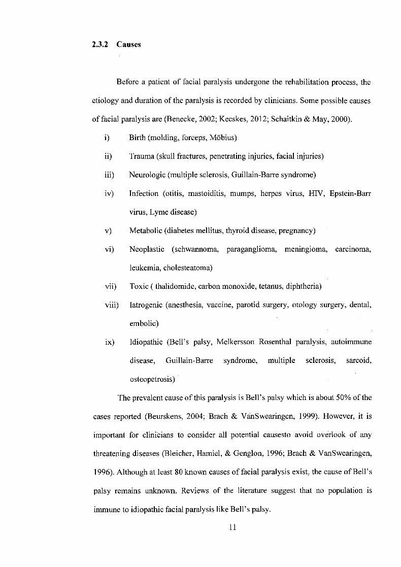
2.3.2 Causes
Before a patient of facial paralysis undergone the rehabilitation process, the
etiology and duration of the paralysis is recorded by clinicians. Some possible causes
of facial paralysis are (Benecke, 2002; Kecskes, 2012; Schaitkin & May, 2000).
i) Birth (molding, forceps, Möbius)
ii) Trauma (skull fractures, penetrating injuries, facial injuries)
Neurologic (multiple sclerosis, Guillain-Barre syndrome)
iv) Infection (otitis, mastoiditis, mumps, herpes virus, HIV, Epstein-Barr
virus, Lyme disease)
V) Metabolic (diabetes mellitus, thyroid disease, pregnancy)
vi) Neoplastic (schwannoma, paraganglioma, meningioma, carcinoma,
leukemia, cholesteatoma)
vii) Toxic (thalidomide, carbon monoxide, tetanus, diphtheria)
viii) latrogenic (anesthesia, vaccine, parotid surgery, otology surgery, dental,
embolic)
ix) Idiopathic (Bell's palsy, Melkersson Rosenthal paralysis, autoimmune
disease, Guillain-Barre syndrome, multiple sclerosis, sarcoid,
osteopetrosis)
The prevalent cause of this paralysis is Bell's palsy which is about 50% of the
cases reported (Beurskens, 2004; Brach & VánSwearingen, 1999). However, it is
important for clinicians to consider all potential causesto avoid overlook of any
threatening diseases (Bleicher, Hamiel, & Genglon, 1996; Brach & VanSwearingen,
1996). Although at least 80 known causes of facial paralysis exist, the cause of Bell's
palsy remains unknown. Reviews of the literature suggest that no population is
immune to idiopathic facial paralysis like Bell's palsy.
11

2.3.3 Symptoms
Issues resulting from facial paralysis include those associated with: (a)
somatic function, (b) social function, (c) psychological wellbeing, and (d)
physiological function (Devriese, 1998). Physical symptoms of facial paralysis in the
upper face include the inability to close the eyes and sagging lower eyelid. Tears may
run from the affected eye, or alternatively, tearing may be reduced. Failure of the
eyelids to completely close, combined with the loss of the blink reflex may cause
irritation, exposure keratopathy, corneal ulcerations, and blindness (Tate &
Tollefson, 2006).
The sagging of the mouth corner is the most notable characteristic in the
lower half of the face. The lack of control over lip closure can result difficulties in
eating, drinking, speech (especially with sounds that require the lips such as /pI and
Ibi) and control of drooling. Also, the nostril on the affected side may collapse,
causing nasal obstruction (Schaitkin & May, 2000). Additionally, patients with facial
paralysis associated with inflammation, as in the case with Bell's palsy and herpes
zoster oticus, may have pain in the mastoid process, neck, ear, or the face (Schaitkin
& May, 2000). Patients will also develop synkinesis which is one sequelae of facial
paralysis (Nakamura, Toda, Sakamaki, Kashima, & Takeda, 2003). Common
examples of synkinesis include eye closure with volitional contraction of mouth
muscles and involuntary movement of the mouth during eye closure. These abnormal
movements in muscle contractions have massive cosmetic and functional
implications.
12

2.3.4 Effects
One of the most socially devastating effects of facial paralysis is the inability
to produce facial expression of emotion. Patients commonly report personal and
work-related problems as well as limited social integration and interpersonal
communication. These problems stem from facial disfigurement and difficulties in
eating, drinking and communicating effectively in social settings. Also, people with
facial paralysis are, often introvert and may become isolated (Goldberg, DeLorie,
Zuker, & Mantkelow, 2003).
The face also is a crucial component of beauty, sexual attractiveness, and
sexual interest (Ekman, 1986). Therefore, patients with facial paralysis experience
pronounce psychological distress. Oftentimes, they must cope with feelings of
shame, decreased self-esteem, anxiety, depression, guilt, anger, and/or fear
(Devriese, 1998; Ekman, 1986).
Nutritional impairment also may be apparent when facial paralysis exists.
Routine and seemingly easy tasks such as eating can be quite challenging for patients
with facial paralysis, who often demonstrate swallowing difficulties that occur in
both oral and pharyngeal phases of deglutition. More specifically, a survey
conducted by Sjogreen, Andersson-Norinder, & Jacobson (2001) revealed that this
group has difficulties in the oral phase including getting food off spoon with lips,
food and liquid leak out of the corners of the mouth and takes a long time to swallow
bites of food. Choking on food and coughing when receiving liquid are some of the
problems in the pharyngeal phase of the swallow.
Verbal communication also may be impaired in this population of patients.
Bilabial incompetence produces characteristics speech error patterns including
substitution, distortions, and omissions of bilabial sounds (/p/, /b/, /m/) and the
13