Embed Size (px)
Citation preview

بسم اهللا الرحمن الرحيم بسم اهللا الرحمن الرحيم

DEXA DEXA … … LATERAL LATERAL LUMBAR LUMBAR
IS IT IMPORTANT IS IT IMPORTANT … … ? ? By
S. S. Tantawy Tantawy, M.D. , M.D.
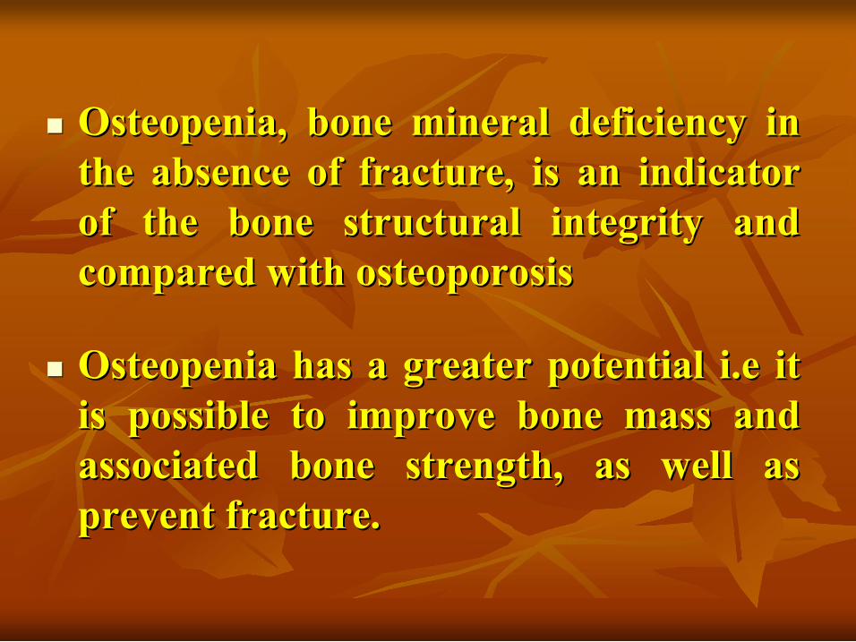
n n Osteopenia Osteopenia, bone mineral deficiency in , bone mineral deficiency in the absence of fracture, is an indicator the absence of fracture, is an indicator of the bone structural integrity and of the bone structural integrity and compared with compared with osteoporosis osteoporosis
n n Osteopenia Osteopenia has a greater potential has a greater potential i.e i.e it it is possible to improve bone mass and is possible to improve bone mass and associated bone strength, as well as associated bone strength, as well as prevent fracture. prevent fracture.

Three degrees of vertebral Three degrees of vertebral deformation are recognized: deformation are recognized:
2 2 Anterior wedge deformity. Anterior wedge deformity.
1 1 End plate deformity. End plate deformity.
3 3 Compression deformity. Compression deformity.

n n A vertebral deformation score is A vertebral deformation score is quantified objectively to study the quantified objectively to study the condition and/or response to treatment condition and/or response to treatment (at least 14 vertebrae)... (at least 14 vertebrae)... T4 through T4 through L4 L4 should be should be roentgenographed roentgenographed. .
n n The anterior, middle and posterior The anterior, middle and posterior heights of the vertebrae are measured heights of the vertebrae are measured and fracture can be then classified as and fracture can be then classified as follow: follow:

n n Moderately deformed (Grade II) 25% to Moderately deformed (Grade II) 25% to 40% reduction in the height and/or a 40% reduction in the height and/or a reduction in the area of 20% to 40%. reduction in the area of 20% to 40%.
n n Mildly depressed (Grade I): 20% to Mildly depressed (Grade I): 20% to 25% decrease in anterior, middle and/or 25% decrease in anterior, middle and/or posterior height and/or reduction in posterior height and/or reduction in 10% to 20% of the area. 10% to 20% of the area.
n n Severely deformed (Grade III) 40% Severely deformed (Grade III) 40% reduction in any height and area. reduction in any height and area.
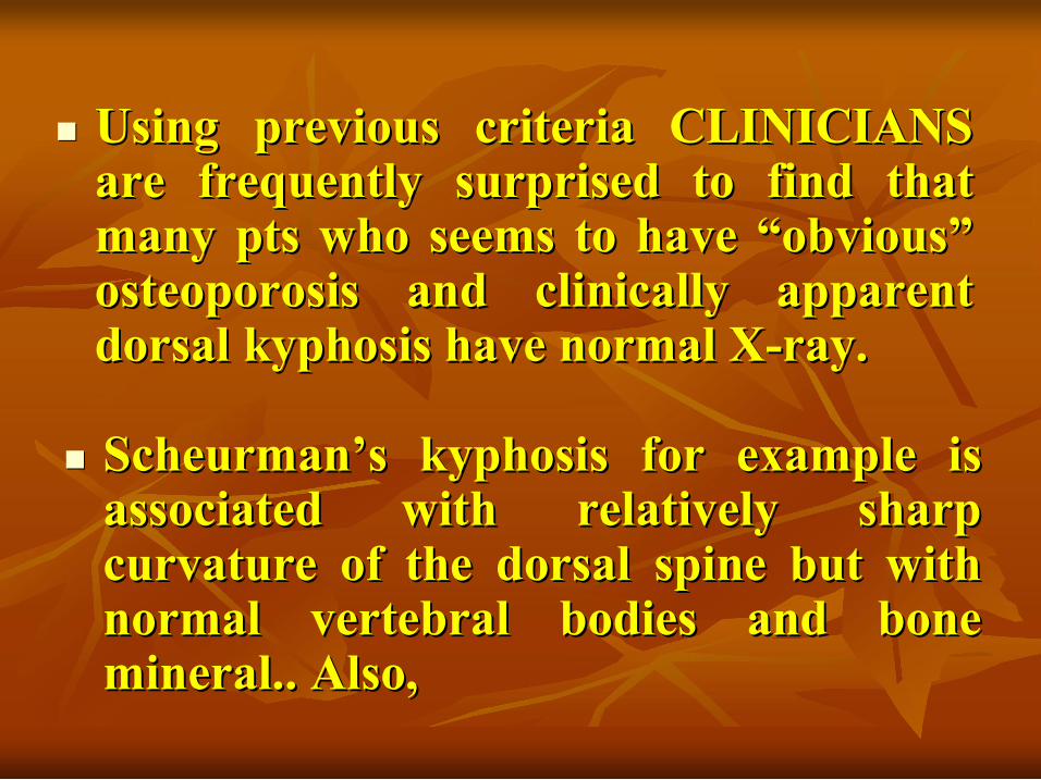
n n Scheurman Scheurman’ ’s s kyphosis kyphosis for example is for example is associated with relatively sharp associated with relatively sharp curvature of the dorsal spine but with curvature of the dorsal spine but with normal vertebral bodies and bone normal vertebral bodies and bone mineral.. Also, mineral.. Also,
n n Using previous criteria CLINICIANS Using previous criteria CLINICIANS are frequently surprised to find that are frequently surprised to find that many pts who seems to have many pts who seems to have “ “obvious obvious” ” osteoporosis and clinically apparent osteoporosis and clinically apparent dorsal dorsal kyphosis kyphosis have normal X have normal X ray. ray.

n n Supine lateral DEXA exam. Is a Supine lateral DEXA exam. Is a promising technique and is better index promising technique and is better index of vertebral bone strength and the of vertebral bone strength and the potential risk for future fracture than potential risk for future fracture than AP DEXA and lateral X AP DEXA and lateral X ray. ray.
n n There are no currently available There are no currently available practical methods of assessing practical methods of assessing microfractures microfractures and/or disruption of and/or disruption of microarchitecture microarchitecture of the plates in the of the plates in the vertebral bone. vertebral bone.

Difficulty with patient positioning is also a Difficulty with patient positioning is also a potential limiting factor and this potential limiting factor and this disadvantage has been resolved by improved disadvantage has been resolved by improved positioning technology in newer positioning technology in newer densitomter densitomter. .
Roentgenograms serve two useful clinical Roentgenograms serve two useful clinical purposes : purposes : 1 1 Disc degeneration. Disc degeneration. 2 2 Osteoarthritis. Osteoarthritis.
As a As a cause of low back pain and they are cause of low back pain and they are helpful in clarifying why some pts with helpful in clarifying why some pts with known osteoporosis can have relatively known osteoporosis can have relatively normal bone mineral density. normal bone mineral density.

Technique Technique Sites measured Sites measured Advantages Advantages Disadvantages Disadvantages
Dual Dual energy energy X X ray ray absorptiometry absorptiometry (DXA) (DXA)
• • Lumbar spine Lumbar spine
• • Proximal Proximal femur femur
• • Total body Total body
• • Forearm Forearm
• • Calcaneus Calcaneus • • Phalanges Phalanges
• • Diagnostic test of Diagnostic test of • • choice choice • • High accuracy, High accuracy, precision, resolution precision, resolution
• • Measures all areas Measures all areas
• • Short scan time, Short scan time, low radiation dose low radiation dose
• • AP spine measurement AP spine measurement influenced by influenced by degenerative sclerosis, degenerative sclerosis, other artifacts other artifacts
• • Combined Combined trabecular trabecular and cortical measure and cortical measure ment ment
Diagnosis of Osteoporosis: Diagnosis of Osteoporosis: Bone Density Assessment Bone Density Assessment

Diagnosis of Osteoporosis: Diagnosis of Osteoporosis: Bone Density Assessment Bone Density Assessment
(cont (cont’ ’d) d) Technique Technique Sites measured Sites measured Advantages Advantages Disadvantages Disadvantages
Quantitative Quantitative ultrasonography ultrasonography
• • Calcaneus Calcaneus
• • Patella Patella
• • Tibia Tibia
Portable Portable • • Uses no Uses no radiation radiation
• • Effectiveness in Effectiveness in predicting fracture predicting fracture risk is controversial risk is controversial
• •Less precise & Less precise & less accurate less accurate than DXA than DXA
Quantitative Quantitative computed computed tomography (QCT) tomography (QCT)
• • Lumbar Lumbar spine spine
• • Allows Allows assessment assessment of of trabecular trabecular bone alone bone alone
• •Greater radiation Greater radiation exposure than DXA exposure than DXA
• • Less precise & Less precise & less accurate in less accurate in spine than DXA spine than DXA

both the vertebral body and neural both the vertebral body and neural arches (cortical/ arches (cortical/trabecular trabecular) ) bone ratio is bone ratio is (50%:50%) all (50%:50%) all mineral with in the path mineral with in the path of photon beam contributes to BMD. of photon beam contributes to BMD.
Sources of errors in PA DEXA of the Sources of errors in PA DEXA of the lumbar spine is an lumbar spine is an areal areal density of the density of the integral bone which include : integral bone which include :

Factors affecting BMD in AP DEXA of Factors affecting BMD in AP DEXA of lumbar spine: lumbar spine:
n n Calcification in the aorta or abdominal lymph Calcification in the aorta or abdominal lymph nodes. nodes.
n n Degenerative disc. Degenerative disc. n n Large lumbar body Large lumbar body osteophytes osteophytes. . n n Osteo Osteo arthretic arthretic changes of the posterior changes of the posterior apophyseal apophyseal joints with consequent hyperostosis joints with consequent hyperostosis
n n Vertebral wedging Vertebral wedging n n Paget Paget’ ’s s disease. disease. n n Sclerotic metastasis. Sclerotic metastasis. n n Vertebral Vertebral haemangioma haemangioma. . n n Residual Residual myodil myodil in the spinal canal and previous in the spinal canal and previous spinal surgery with metallic fixation. spinal surgery with metallic fixation.

Also difficulty in DEXA PA of the Also difficulty in DEXA PA of the lumbar spine may be due to spinal lumbar spine may be due to spinal scoliosis, scoliosis, kyphosis kyphosis or vertebral or vertebral segmentation all may result in segmentation all may result in inaccurate or overestimated values of inaccurate or overestimated values of BMD BMD (Frank et al., 98) (Frank et al., 98). .

Indication For Bone Mass Indication For Bone Mass Measurement Measurement
n n In estrogen In estrogen deficient women, to diagnose deficient women, to diagnose significantly low bone mass to make significantly low bone mass to make decisions about hormone replacement decisions about hormone replacement therapy. therapy.
n n In patient with vertebral abnormalities or In patient with vertebral abnormalities or radiographic radiographic osteopenia osteopenia, to diagnose spinal , to diagnose spinal osteoporosis to make decision about further osteoporosis to make decision about further diagnostic evaluation and therapy. diagnostic evaluation and therapy.

n n In patient receiving long In patient receiving long term term gluco gluco corticoid therapy, to diagnose low bone corticoid therapy, to diagnose low bone mass to adjust therapy. mass to adjust therapy.
n n In patient with primary asymptomatic In patient with primary asymptomatic hyperparathyroidism to diagnose low hyperparathyroidism to diagnose low bone mass to identify those at risk of bone mass to identify those at risk of severe skeletal disease who may be severe skeletal disease who may be candidate for surgical intervention. candidate for surgical intervention.
Other potential Indications Other potential Indications
n n Universal screening for osteoporosis Universal screening for osteoporosis prophylaxis. prophylaxis.
n n Identifying women who are Identifying women who are “ “fast bone fast bone losers losers” ” for more aggressive therapy. for more aggressive therapy.
n n Monitoring bone mass to assess efficacy Monitoring bone mass to assess efficacy of therapy. of therapy.


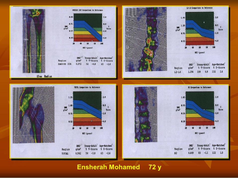
Ensherah Mohamed 72 y

Thoraya Mohamed 54 y

Nahed Abd ElAziz 75 y

Amal Ahmed 40 y

Fatma Abd ElMageed 56 y

Fatma Mohamed 41 y

Kamelya Sadeek 65 y
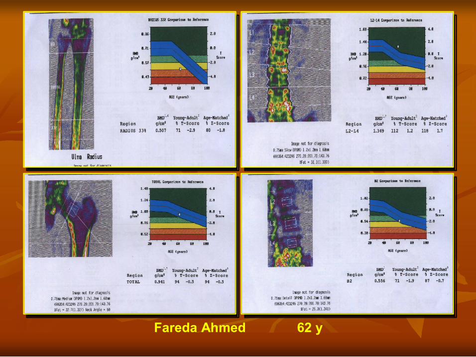
Fareda Ahmed 62 y

Amal Ahmed 50 y
Conclusion Conclusion
*OLD PATIENTS WITH POSSIBLE *OLD PATIENTS WITH POSSIBLE DEGENERATIVE CHANGES TO DEGENERATIVE CHANGES TO OVERCOME POSTERIOR ELEMENTS OVERCOME POSTERIOR ELEMENTS AND LARGE OSTEOPHYTS. AND LARGE OSTEOPHYTS.
LASTLY WE CAN CONLUDE THAT DEXA LASTLY WE CAN CONLUDE THAT DEXA OF THE LATERAL LUMBAR SPINE OF THE LATERAL LUMBAR SPINE
MUST BE DONE IN THE FOLLOWINGS : MUST BE DONE IN THE FOLLOWINGS :

Continue Continue
*IN Pts WITH CALCIFIEC ABD. AORTA *IN Pts WITH CALCIFIEC ABD. AORTA OR CALCIFIED ABDOMINAL OR CALCIFIED ABDOMINAL L.Ns L.Ns. .
*IN Pts SUFFERING FROM PAGET *IN Pts SUFFERING FROM PAGET’ ’S DISEASE S DISEASE LYMPHOMA, SCLEROTIC METASTASIS OR LYMPHOMA, SCLEROTIC METASTASIS OR HAEMANGIOMA. HAEMANGIOMA.
*IF THE ONLY SCREENING METHOD IS *IF THE ONLY SCREENING METHOD IS LUMBAR SPINE ESP. IN OLD PATIENTS LUMBAR SPINE ESP. IN OLD PATIENTS LATERAL DEXA MUST BE DONE AND LATERAL DEXA MUST BE DONE AND FOLLOW FOLLOW UP WILL BE BY THE SAME UP WILL BE BY THE SAME MANNER. MANNER.
*IN PATIENT EXAMINED ROUTINLY BY DEXA FOR *IN PATIENT EXAMINED ROUTINLY BY DEXA FOR THREE SITES; THREE SITES;
n n FOREARM FOREARM n n PA LUMBAR SPINE PA LUMBAR SPINE n n HIP JOINT HIP JOINT
*AND THERE IS DECREASE IN BMD IN BOTH *AND THERE IS DECREASE IN BMD IN BOTH FOREARM AND HIP JOINT WITH NORMAL PA FOREARM AND HIP JOINT WITH NORMAL PA LUMBAR SPINE SO DEXA OF LATERAL LUMBAR LUMBAR SPINE SO DEXA OF LATERAL LUMBAR SPINE MUST BE DONE FOR ACTUAL SPINE MUST BE DONE FOR ACTUAL MEASUREMENT OF BMD OF LUMBAR SPINE AND MEASUREMENT OF BMD OF LUMBAR SPINE AND TO GARD AGAINST FRACTURE RISK TO GARD AGAINST FRACTURE RISK
Continue Continue

Thank you Thank you