Embed Size (px)
Citation preview

commentar y
Kidney International (2010) 78 951
study, acidosis induced with acetazola-mide treatment resulted in the appearance of B2 in the apical membrane of � -inter-calated cells, suggesting that another H + -ATPase or an H + -ATPase that includes both B1 and B2 can be recruited to the plasma membrane when assembly of the holoenzyme is stressed. 13
Intercalated cells are highly dynamic and can rapidly respond to the signals induced by systemic acid / base changes to reduce or increase their rate of vectoral acid secretion. Th e major mechanism uti-lized for adjusting the transport rate in both acute and chronic states is up- or downregulation of the H + -ATPase and anion exchangers (AE1 in � -intercalated and pendrin in � -intercalated cells) inserted into the apical and basolateral membranes. Long-term adaptation uti-lizes this same response in addition to changes in the overall expression level of these transporters and, at some yet-to-be-quantitated rate, the interconversion of � - and � -intercalated cells.
DISCLOSURE The authors declared no competing interests.
ACKNOWLEDGMENTS
This work was supported by the National Institute of Diabetes and Digestive and Kidney Diseases, National Institutes of Health, grant DK59529.
REFERENCES 1 . Schwartz GJ . Plasticity of intercalated cell polarity:
effect of metabolic acidosis . Nephron 2001 ; 87 : 304 – 313 .
2 . Schwartz JH , Li G , Yang Q et al. Role of SNAREs and H + -ATPase in the targeting of proton pump-coated vesicles to collecting duct cell apical membrane . Kidney Int 2007 ; 72 : 1310 – 1315 .
3 . Slotki I , Schwartz JH , Alexander EA . Interrelationship between cell pH and cell calcium in rat inner medullary collecting duct cells . Am J Physiol 1993 ; 265 : C432 – C438 .
4 . Paunescu TG , Ljubojevic M , Russo LM et al. cAMP stimulates apical V-ATPase accumulation, microvillar elongation, and proton extrusion in kidney collecting duct A-intercalated cells . Am J Physiol Renal Physiol 2010 ; 298 : F643 – F654 .
5 . Yang Q , Li G , Singh SK et al. Vacuolar H + -ATPase B1 subunit mutations that cause inherited distal renal tubular acidosis affect proton pump assembly and trafficking in inner medullary collecting duct cells . J Am Soc Nephrol 2006 ; 17 : 1858 – 1866 .
6 . Fejes-Toth G , Chen WR , Rusvai E et al. Differential expression of AE1 in renal HCO3-secreting and -reabsorbing intercalated cells . J Biol Chem 1994 ; 269 : 26717 – 26721 .
7 . Frische S , Kwon TH , Frokiaer J et al. Regulated expression of pendrin in rat kidney in response to chronic NH 4 Cl or NaHCO 3 loading . Am J Physiol Renal Physiol 2003 ; 284 : F584 – F593 .
8 . Al-Awqati Q , Vijayakumar S , Takito J . Terminal differentiation of epithelia from trophectoderm to the intercalated cell: the role of hensin . J Am Soc Nephrol 2003 ; 14 (Suppl 1) : S16 – S21 .
9 . Schwartz GJ , Al-Awqati Q . Role of hensin in mediating the adaptation of the cortical collecting duct to metabolic acidosis . Curr Opin Nephrol Hypertens 2005 ; 14 : 383 – 388 .
10 . Purkerson JM , Tsuruoka S , Suter DZ et al. Adaptation to metabolic acidosis and its recovery are associated with changes in anion exchanger distribution and expression in the cortical collecting duct . Kidney Int 2010 ; 78 : 993–1005 .
11 . Schwartz GJ , Al-Awqati Q . Regulation of transepithelial H + transport by exocytosis and endocytosis . Annu Rev Physiol 1986 ; 48 : 153 – 161 .
12 . Sabolic I , Brown D , Gluck SL , Alper SL . Regulation of AE1 anion exchanger and H( + )-ATPase in rat cortex by acute metabolic acidosis and alkalosis . Kidney Int 1997 ; 51 : 125 – 137 .
13 . Paunescu TG , Da Silva N , Marshansky V et al. Expression of the 56-kDa B2 subunit isoform of the vacuolar H( + )-ATPase in proton-secreting cells of the kidney and epididymis . Am J Physiol Cell Physiol
2004 ; 287 : C149 – C162 .
see original article on page 1016
In 1961, Sarles et al. fi rst described an entity of sclerosing pancreatitis with hypergam-maglobulinemia and hypothesized that this entity was an autoimmune phenomenon. 1 Since then, this disease has been recog-nized as being related to increased IgG4, both as elevated levels of IgG4 in patient serum and in tissue sections with increased IgG4 + plasma cells. In the past few years, the extrapancreatic features of ‘ autoim-mune pancreatitis ’ (AIP) have been recog-nized, 2 thus calling for a new name for the disease. Now, IgG4-related systemic disease has been described in nearly every organ and organ system, fi rst recognized in the pancreas, and then recognized in the liver, gallbladder, other gastrointestinal sites
(sometimes including infl ammatory bowel disease), salivary or lacrimal glands, lung, breast, retroperitoneum, lymph nodes, pituitary gland, prostate, and aorta.
Th e entity of IgG4-related systemic dis-ease, while enjoying increased attention in the literature, has been largely unrecognized in regular clinical practice, especially when it aff ects organs outside of the pancreas. This disease is even more likely to be un diagnosed in the kidney, when renal biopsies of IgG4-related disease are diag-nosed simply as tubulointerstitial nephritis (TIN) without a more specific diagnosis indicating the underlying cause of this dis-ease pattern. As with glomerular diseases, which are, for the most part, specifi c and well defi ned in terms of histologic, ultrastruc-tural, and immunophenotypic features, pathologists and clinicians should strive to identify and describe tubulointerstitial diseases in the same manner, with clinico-pathologic correlation. TIN may be
IgG4-related tubulointerstitial nephritis Lynn D. Cornell 1
Tubulointerstitial nephritis (TIN) is a disease pattern with heterogeneous
causes. Recently a specific subtype of autoimmune TIN, IgG4-related
TIN, has been identified that is part of systemic IgG4-related disease/
autoimmune pancreatitis. On biopsy, this TIN shows an IgG4 + plasma
cell-rich infiltrate, akin to the pancreatic tissue findings in autoimmune
pancreatitis, and may show tubulointerstitial immune complex deposits.
Notably, some cases may be mass-forming. Recognition of this specific
type of TIN can guide appropriate patient therapy.
Kidney International (2010) 78, 951 – 953. doi: 10.1038/ki.2010.342
1 Department of Laboratory Medicine and Pathology,
Mayo Clinic , Rochester , Minnesota , USA
Correspondence: Lynn D. Cornell, Department of
Laboratory Medicine and Pathology, Mayo Clinic,
200 1st Street SW, Rochester, Minnesota 55902,
USA. E-mail: [email protected]
commentar y
952 Kidney International (2010) 78
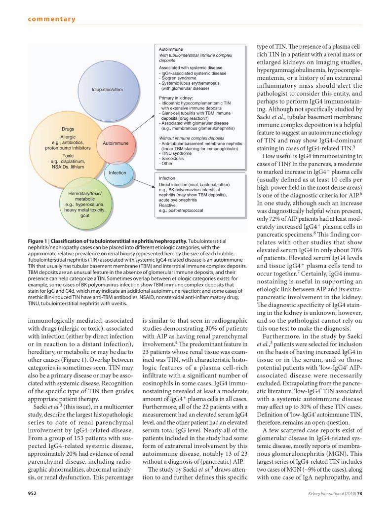
immunologically mediated, associated with drugs (allergic or toxic), associated with infection (either by direct infection or in reaction to a distant infection), hereditary, or metabolic or may be due to other causes ( Figure 1 ). Overlap between categories is sometimes seen. TIN may also be a primary disease or may be asso-ciated with systemic disease. Recognition of the specifi c type of TIN then guides appropriate patient therapy.
Saeki et al. 3 (this issue), in a multicenter study, describe the largest histopathologic series to date of renal parenchymal involvement by IgG4-related disease. From a group of 153 patients with sus-pected IgG4-related systemic disease, approximately 20 % had evidence of renal parenchymal disease, including radio-graphic abnormalities, abnormal urinaly-sis, or renal dysfunction. Th is percentage
is similar to that seen in radiographic studies demonstrating 30 % of patients with AIP as having renal parenchymal involvement. 4 Th e predominant feature in 23 patients whose renal tissue was exam-ined was TIN, with characteristic histo-logic features of a plasma cell-rich infiltrate with a significant number of eosinophils in some cases. IgG4 immu-nostaining revealed at least a moderate amount of IgG4 + plasma cells in all cases. Furthermore, all of the 22 patients with a measurement had an elevated serum IgG4 level, and the other patient had an elevated serum total IgG level. Nearly all of the patients included in the study had some form of extrarenal involvement by this autoimmune disease, notably 13 of 23 without a diagnosis of (pancreatic) AIP.
Th e study by Saeki et al. 3 draws atten-tion to and further defi nes this specifi c
type of TIN. Th e presence of a plasma cell-rich TIN in a patient with a renal mass or enlarged kidneys on imaging studies, hypergammaglobulinemia, hypocomple-mentemia, or a history of an extrarenal inflammatory mass should alert the pathologist to consider this entity, and perhaps to perform IgG4 immunostain-ing. Although not specifi cally studied by Saeki et al. , tubular basement membrane immune complex deposition is a helpful feature to suggest an autoimmune etiology of TIN and may show IgG4-dominant staining in cases of IgG4-related TIN. 5
How useful is IgG4 immunostaining in cases of TIN? In the pancreas, a moderate to marked increase in IgG4 + plasma cells (usually defi ned as at least 10 cells per high-power fi eld in the most dense areas) is one of the diagnostic criteria for AIP. 6 In one study, although such an increase was diagnostically helpful when present, only 72 % of AIP patients had at least mod-erately increased IgG4 + plasma cells in pancreatic specimens. 6 Th is fi nding cor-relates with other studies that show elevated serum IgG4 in only about 70 % of patients. Elevated serum IgG4 levels and tissue IgG4 + plasma cells tend to occur together. 7 Certainly, IgG4 immu-nostaining is useful in supporting an etiologic link between AIP and its extra-pancreatic involvement in the kidney. Th e diagnostic specifi city of IgG4 stain-ing in the kidney is unknown, however, and so the pathologist cannot rely on this one test to make the diagnosis.
Furthermore, in the study by Saeki et al. , 3 patients were selected for inclusion on the basis of having increased IgG4 in tissue or in the serum, and so those potential patients with ‘ low-IgG4 ’ AIP-associated disease were necessarily excluded. Extrapolating from the pancre-atic literature, ‘ low-IgG4 ’ TIN associated with a systemic autoimmune disease may aff ect up to 30 % of these TIN cases. Defi nition of ‘ low-IgG4 ’ autoimmune TIN, therefore, remains an open question.
A few scattered case reports exist of glomerular disease in IgG4-related sys-temic disease, mostly reports of membra-nous glomerulonephritis (MGN). This largest series of IgG4-related TIN includes two cases of MGN ( ~ 9 % of the cases), along with one case of IgA nephropathy, and
Infection
Direct infection (viral, bacterial, other)e.g., BK polyomavirus interstitialnephritis (may show TBM deposits),acute pyelonephritisReactivee.g., post-streptococcal
AutoimmuneWith tubulointerstitial immune complexdeposits
Associated with systemic disease:- IgG4-associated systemic disease- Sjogren syndrome- Systemic lupus erythematosus (with glomerular disease)
Primary in kidney:- Idiopathic hypocomplementemic TIN with extensive immune deposits- Giant-cell tubulitis with TBM immune deposits (drug reaction?)- Associated with glomerular disease (e.g., membranous glomerulonephritis)
Without immune complex deposits- Anti-tubular basement membrane nephritis (linear TBM staining for immunoglobulin)- TINU syndrome- Sarcoidosis- Other
Drugs
Allergice.g., antibiotics,
proton pump inhibitors
Toxice.g., cisplatinum,NSAIDs, lithium
Hereditary/toxic/metabolic
e.g., hyperoxaluria,heavy metal toxicity,
gout
Idiopathic/other
Autoimmune
Infection
Figure 1 | Classification of tubulointerstitial nephritis / nephropathy. Tubulointerstitial nephritis / nephropathy cases can be placed into different etiologic categories, with the approximate relative prevalence on renal biopsy represented here by the size of each bubble. Tubulointerstitial nephritis (TIN) associated with systemic IgG4-related disease is an autoimmune TIN that usually has tubular basement membrane (TBM) and interstitial immune complex deposits. TBM deposits are an unusual feature in the absence of glomerular immune deposits, and their presence can help categorize a TIN. Sometimes overlap between etiologic categories exists: for example, some cases of BK polyomavirus infection show TBM immune complex deposits that stain for IgG and C4d, which may indicate an additional autoimmune reaction; and some cases of methicillin-induced TIN have anti-TBM antibodies. NSAID, nonsteroidal anti-inflammatory drug; TINU, tubulointerstitial nephritis with uveitis.

commentar y
Kidney International (2010) 78 953
three cases of other (undefi ned) glomerular disease. MGN as a potential etiologic link with IgG4-related systemic disease is intriguing because idiopathic MGN is also an IgG4-dominant disease, such that these may have a common pathogenetic link.
IgG4 itself is an unusual antibody and has some unusual physical characteristics. It is the rarest IgG subclass in the circula-tion of normal individuals. Elevated titers of IgG4 are found in conditions of chronic antigen exposure; beekeepers, for exam-ple, show elevated IgG4 directed against bee venom, and patients undergoing allergen immunotherapy develop increased IgG4 titers against the allergen. Compared with IgG1, the IgG4 molecule has weaker interchain disulfi de bridges, so that immunoglobulin half-molecules, composed of one heavy chain and one light chain, dissociate from each other. Once these dissociate, an IgG4 half-mol-ecule may reassociate with another half-molecule with specifi city for a diff erent antigen. 7,8 When IgG4 encounters anti-gen, it can only form small, and presum-ably harmless, immune complexes and may block antigen binding by the more pathogenic IgG1. IgG4 is also unable to fi x complement. In these ways, IgG4 is thought to act as an anti-infl ammatory molecule that can temper the immune response, at least in some circumstances.
While IgG4 is an ‘ anti-inflammatory ’ immunoglobulin, it is perplexing that it is found to be increased in some disease states, most notably IgG4-related systemic disease. Th e mechanism of this disease and its asso-ciation with IgG4 is unclear. IgG4 class switching depends on interleukin-4 and/or interleukin-13 mainly secreted by T-helper 2 cells. Th ese are the same cytokines that promote an IgE response, although IgG4 antibody may be present in the absence of IgE antibody against a particular antigen. Interleukin-10 (IL-10) has an eff ect on IgG4 versus IgE class switching 9 and may be required for IgG4 class-switched B cells to diff erentiate into IgG4-secreting plasma cells. One may speculate that, in IgG4-related systemic disease, an initial insult and process involving production of anti-infl ammatory cytokines, including IL-10 and tumor necrosis factor- α , along with fi brogenic IL-13, drives increased fi brosis, induction of IgG4 class-switched B cells,
and production and massive expansion of IgG4-secreting plasma cells.
TIN is a disease pattern with heteroge-neous causes, both immune and nonim-mune. Th e clinical presentation, laboratory results, and biopsy features are all consid-ered to make a specifi c diagnosis. Th rough the work of Saeki et al. , 3 we now have a clearer view of the clinicopathologic fea-tures of a specifi c type of TIN that is part of systemic IgG4-related disease, which in turn will aid pathologists and clinicians in recognizing and offering appropriate treatment for this disease.
DISCLOSURE The author declared no competing interests.
REFERENCES 1 . Sarles H , Sarles JC , Muratore R et al. Chronic inflam-
matory sclerosis of the pancreas: an autonomous pancreatic disease? Am J Dig Dis 1961 ; 6 : 688 – 698 .
2 . Deshpande V , Chiocca S , Finkelberg D et al. Autoimmune pancreatitis: a systemic immune complex mediated disease . Am J Surg Pathol 2006 ; 30 : 1537 – 1545 .
3 . Saeki T , Nishi S , Imai N et al. Clinicopathological characteristics of patients with IgG4-related tubulo-interstitial nephritis . Kidney Int 2010 ; 78 : 1016–1023 .
4 . Takahashi N , Kawashima A , Fletcher JG et al. Renal involvement in patients with autoimmune pancreatitis: CT and MR imaging findings . Radiology 2007 ; 242 : 791 – 801 .
5 . Cornell LD , Chicano SL , Deshpande V et al. Pseudotumors due to IgG4 immune-complex tubulointerstitial nephritis associated with autoimmune pancreatocentric disease . Am J Surg Pathol 2007 ; 31 : 1586 – 1597 .
6 . Zhang L , Notohara K , Levy MJ et al. IgG4-positive plasma cell infiltration in the diagnosis of auto-immune pancreatitis . Mod Pathol 2007 ; 20 : 23 – 28 .
7 . Aalberse RC , Schuurman J . IgG4 breaking the rules . Immunology 2002 ; 105 : 9 – 19 .
8 . Aalberse RC , Stapel SO , Schuurman J et al. Immunoglobulin G4: an odd antibody . Clin Exp Allergy 2009 ; 39 : 469 – 477 .
9 . Jeannin P , Lecoanet S , Delneste Y et al. IgE versus IgG4 production can be differentially regulated by IL-10 . J Immunol 1998 ; 160 : 3555 – 3561 .
Within the past few years, it has become apparent that fi broblastic growth factor 23 (FGF23), a bone-derived phosphaturic
hormone, plays a central role in the physio-logical regulation of mineral and vitamin D metabolism. FGF23 induces urinary phosphate excretion by supp ress ing the expression of the sodium-phosphate cotransporter. FGF23 also suppresses the synthesis of 1,25-dihydroxyvitamin D [1,25(OH) 2 D] via inhibition of 1 � -hydroxylase and stimulation of 24-hydroxylase. 1 Th ese eff ects are dependent on the presence of Klotho, which converts
Parathyroid resistance to FGF23 in kidney transplant recipients: back to the past or ahead to the future ? Hirotaka Komaba 1 , Masahiro Koizumi 1 and Masafumi Fukagawa 1
Fibroblast growth factor 23 (FGF23) modulates the metabolism of
minerals and vitamin D. In chronic kidney disease (CKD), this process is
disturbed owing to decreased parathyroid expression of FGF23 ’ s
receptor complex Klotho-FGF receptor 1. In this issue, Krajisnik and
colleagues demonstrate that similar alterations occur in parathyroid
glands from kidney transplant recipients in association with a decline in
allograft function. Is it possible that these data can be extrapolated to
general early-stage CKD patients?
Kidney International (2010) 78, 953 – 955. doi: 10.1038/ki.2010.283
1 Division of Nephrology, Endocrinology, and
Metabolism, Tokai University School of Medicine ,
Isehara , Japan
Correspondence: Masafumi Fukagawa, Division
of Nephrology, Endocrinology, and Metabolism,
Tokai University School of Medicine,
143 Shimo-Kasuya, Isehara, 259-1193, Japan.
E-mail: [email protected]
see original article on page 1024