Embed Size (px)
Citation preview

Folia Biologica (Praha) 62, 194-202 (2016)
Original Article
IDH1/2 Mutation and MGMT Promoter Methylation – the Relevant Survival Predictors in Czech Patients with Brain Gliomas(IDH mutation / MGMT methylation / glioma / glioblastoma / astrocytoma / oligodendroglioma)
F. KRAMÁŘ1, M. MINÁRIK2, L. BENEŠOVÁ2, T. HALKOVÁ2, D. NETUKA1, O. BRADÁČ1, V. BENEŠ1
1Department of Neurosurgery and Neurooncology, First Faculty of Medicine, Charles University and Military University Hospital Prague, Czech Republic2Center for Applied Genomics of Solid Tumors (CEGES), Genomac Research Institute, Ltd, Prague, Czech Republic
Abstract. Gliomas are a heterogeneous group of tu-mours varying in prognosis, treatment approach, and overall survival. Recently, novel markers have been identified which are linked to patient prognosis and therapeutic response. Especially the mutation of the enzyme isocitrate dehydrogenase 1 or 2 (IDH1/2) gene and the O6-methylguanine-DNA methyltrans-ferase (MGMT) promoter methylation status seem to be the most important predictors of survival. From 2012 to 2015, 94 Czech patients with primary brain tumours were enrolled into the study. The IDH1/2 mutation was detected by denaturing capillary elec-trophoresis. The methylation status of the MGMT gene and other 46 genes was revealed by MS-MLPA. In all 94 patients, the clinical data were correlated with molecular markers by Kaplan-Meier analyses and Cox regression model. The MGMT promoter methylation status was established and compared to clinical data. In our study eight different probes were used to elucidate the MGMT methylation status; hy-permethylation was proclaimed if four and more probes were positive. This 3 : 5 ratio was tested and
Received January 23, 2016. Accepted June 20, 2016.
This work was supported by grant 14253 from IGA, Ministry of Health, Czech Republic.
Corresponding author: Filip Kramář, Department of Neurosurgery and Neurooncology, First Faculty of Medicine, Charles University and Military University Hospital Prague, Czech Republic. U vojenské nemocnice 1200, 169 02 Prague 6, Czech Republic. E-mail: [email protected]
Abbreviations: DCE – denaturing capillary electrophoresis, GCIMP – gliomaCpG island methylator phenotype, HR – hazard ratio, IDH1/2 – isocitrate dehydrogenase 1 or 2, MGMT – O6methylguanineDNA methyltransferase, MSMLPA – multiplex ligation-dependent probe amplification, KFS – Karnofsky performance status, PNET – primitive neuroectodermal tumour.
confirmed by Kaplan-Meier and Cox analyses. The study confirmed the importance of the IDH1/2 muta-tion and hypermethylation of the MGMT gene promot-er being present in tumour tissue. Both markers are independent positive survival predictors; in the Cox model the IDH hazard ratio was 0.10 and in the case of MGMT methylation it reached 0.32. The methyla-tion analysis of the panel of additional 46 genes did not reveal any other significant epigenetic markers; none of the candidate genes have been confirmed in the Cox regression analyses as an independent prog-nostic factor.
IntroductionGliomas represent a heterogeneous group of primary
brain tumours which differ in prognosis, overall survival and therapeutic response. Unfortunately, the majority of these tumours consist of highgrade tumours, mostly glioblastomas. The knowledge of markers that could predict prognosis or the therapeutic response to oncological treatment is highly needed. In recent years, some progress has been made and new diagnostic and therapeutic advances have been made available for wide utilization. In 2005, a new oncological drug was introduced. Temozolomide improved prognosis and prolonged overall survival in selected glioblastoma patients (Stupp et al., 2005). This selection was based on a molecular marker – promoter of the MGMT gene encoding the demethylation enzyme O6methylguanineDNA methyltransferase (Hegi et al., 2005). In 2008, another molecular marker was revealed – mutation of the isocitrate dehydrogenase type 1 (IDH1) gene (Parsons et al., 2008). Later studies focused on IDH found its important role in pathogenesis of gliomas (Yan et al., 2009).
At present, this mutation is considered as one of the first steps in the glioma development. The mutation itself can change the epigenetic landscape by its toxic

Vol. 62 195
product, 2-hydroxyglutarate (Dang et al., 2009; Noush-mehr et al., 2010). This oncometabolite competitively inhibits the activity of α-ketoglutarate-dependent histone demethylases (Xu et al., 2011; Turcan et al., 2012). DNA methylation preferably appears in CpG islands. The CpG islands are regions of DNA with high frequency of sites where a cytosine nucleotide occurs next to a guanine nucleotide. Cytosines in CpG dinucleotides are often methylated, forming 5methylcytosine, which within a gene can change its expression. In mammals, 70–80 % of CpG cytosines are methylated. 2-hydroxyglutarate indirectly causes DNA hypermethylation by its effect on demethylating enzymes at a large number of DNA loci, indicating the existence of a glioma-CpG island methylator phenotype (G-CIMP).
The presence of IDH mutation in the tumour genome corresponds with favourable prognosis compared to tumours not carrying this mutation. Since its discovery, this fact has been confirmed in many following studies (The Cancer Genome Atlas Research Network, 2015). Apart from MGMT methylation, other epigenetic changes play a role in the pathogenesis of glial tumours, but their importance remains unclear and the results are not as conclusive as in the case of IDH or MGMT. On the other hand, recent development is promising (Sturm et al., 2012; Schiff and Purow, 2013; Shah et al., 2014). How-ever, the overall survival is still limited in many glioma patients. Therefore, novel therapies and appro aches are urgently needed. The aim of our prospective study was to assess the role of selected genetic and epigenetic markers in brain gliomas and confirm or disprove their impact on glioma prognosis and therapeutic response.
Material and MethodsTumour samples were collected during the standard
neurosurgical procedure. All patients (all of them of Czech origin) signed the infor med consent form appro-ved by the hospital ethics committee (Military University Hospital Prague, No. 80-58/39-2012-ÚVN). Each tissue specimen was divided into two parts. One part of a sample was sent for immunohistochemical analysis. The second part of a tissue specimen was immediately frozen and transported at –20 °C to a molecular laboratory for testing. DNA extraction was performed by a commer-cial kit using a standard spin column protocol (JETquick Tissue DNA spin, GENOMED, Loehne, Germany). A typical concentration of extracted DNA was between 10–30 ng/µl. Mutations in IDH genes (IDH1 (R132), IDH2 (R172)) were detected using denaturing capillary electrophoresis (DCE). The method is based on differential melting of wild-type and mutant alleles; a similar approach is used in melting curve analysis. PCR amplicons covering both mutation targets were amplified using fluorescently labelled primers and subsequently resolved at an optimum separating temperature in a standard capillary electrophoretic DNA analyser (ABI PRISM 3100 Genetic analyzer, Applied Biosystems, Foster City, NJ). Full details of the optimization and va-
lidation procedure were described elsewhere (Horbinski et al., 2010).
The methylation status of the MGMT promoter and other 46 genes was examined by methylation-specific multiplex ligation-dependent probe amplification (MS-MLPA) using a combination of kits (ME001, ME002, ME042-CIMP, and ME011 SALSA MLPA kit according to the manufacture’s instructions (MRCHolland, The Netherlands)). MS-MLPA is a semi-quantitative technique based on DNA restriction using a methylation-sensitive restriction enzyme (Hömig-Hölzel and Savola, 2012), and sub sequent detection of products by capillary electrophoresis (ABI PRISM 3100, Applied Biosystems). Clearly, since both DCE and MS-MLPA methods employ fragment analysis in a capillary DNA analyser, the examination of IDH1-2/CIMP status can easily be combined into a common testing protocol. Denaturing capillary electrophoresis was performed to detect IDH1/2 mutations and MSMLPA to identify the methylation status of selected genes according to the study protocol. MSMLPA was primarily focused on identifying the methylation status of the MGMT gene promoter; eight probes were used to detect the precise methylation of the MGMT gene.
The clinical data were collected as well: age at onset of the disease, Karnofsky score (Karnofsky et al., 1948), progression-free survival (PFS), and overall survival (OS).
Statistical analysisStatistical processing of data was performed using
Excel 2013 (Microsoft) and XLSTAT 2014 for Windows (Addinsoft). Charts were constructed in Excel. Asso-ciations between IDH1/2 mutations, methylation of selected genes, and patient survival were tested with univariate and multivariate Cox regression analyses and KaplanMeier survival distribution functions. In MGMT promoter methylation, also the ROC curve analysis was performed to establish the specificity and sensitivity of the used probes.
ResultsFrom 2012 to 2015, 100 Czech patients were enrolled
into the study, all of them Czechs. Two patients were excluded from the study because of histology different from a glial tumour (metastasis of lung carcinoma). Four other patients were not included because of the absence of some statistical data. Only 94 patients were eligible for further statistical analysis. There were 50 men and 44 women included in the study, with the mean age of 52 years and mean Karnofsky score (KPS) 86 points. In all of these patients, a complete set of monitored data was collected. In these 94 patients, the histological analysis according to the WHO classification was performed. Fifty-eight glioblastomas of WHO gr. IV, 13 diffuse astrocytomas gr. II, 12 anaplastic astrocytomas gr. III, two oligodendrogliomas gr. II, four anaplastic
IDH1 Mutation and MGMT Methylation in Brain Gliomas
196 Vol. 62F. Kramář et al.
oligodendrogliomas gr. III, one anaplastic oligoastrocytoma gr. III, one PNET (primitive neuroectodermal tumour) gr. IV, one pilocytic astrocytoma gr. I, one gangliocytoma gr. I and one choroid plexus papilloma gr. I were detected. In all of these samples, detection of IDH1/2 mutation and methyla tion analysis of a panel of 47 genes were performed.
The IDH mutation was found in 37 samples, preferably in grade IIIII gliomas, but also in 10 glioblastomas (secondary glioblastomas), see Table 1. In grade II-III gliomas, the frequency of IDH mutation in our series reached 84 %, in GBMs it was 17 %. Kaplan-Meier analysis of survival according to the presence of IDH1/2 mutation confirmed a highly statistically significant positive effect of IDH mutation on patients’ survival with P value < 0.0001, see Fig. 1. The mean overall survival reached only 20.18 months in the group of wild-type IDH compared to 83.28 months in the group of mutated IDH. A similar result was also found in separate subgroups of grade IIIII gliomas and glioblastomas. In both univariate and multivariate Cox regression model, IDH1/2 mutation remains a significant positive prognostic factor with hazard ratio (HR) 0.10, see Table 2. The role of IDH1/2 is not only limited to OS, but also to PFS. In 70 patients eligible for PFS study, KaplanMeier analysis proved again a significant difference in PFS between patients with mutated and wildtype IDH, see Fig. 2.
The methylation panel consisted of probes detecting the methylation status of 47 genes including MGMT. The complete panel was evaluated in all 94 patients. The MGMT promoter methylation status was detected by eight independent probes (component of ME002, ME011 SALSA kits). None of these eight probes was a statistically significant prognostic factor in the multivariate Cox regression analysis. In the second step, the MGMT
Table 1. Frequency of histological subtypes, grade and IDH1/2 mutation in the series
Histology Grade No IDH1mut IDH2mut
pilocytic astrocytoma I 1 0 0choroid plexus papilloma I 1 0 0gangliocytoma I 1 0 0diffuse astrocytoma II 13 12 0anaplastic astrocytoma III 12 10 0oligodendroglioma II 2 2 0anaplastic oligodendroglioma
III 4 1 1
anaplastic oligoastrocytoma
III 1 1 0
glioblastoma IV 58 9 1PNET IV 1 0 0
Fig. 1. KaplanMeier analysis of survival according to the presence of IDH1/2 mutation (0 – wildtype IDH, 1 – mutated IDH)
Vol. 62 197
promoter methylation group was divided into two subgroups according to the number of methylated loci. Different dividing ratios were tested by the Cox regression model and KaplanMeier analysis. The ratio with the highest statistical significance (log-rank P value = 0.0002) was found: 0–3 methylated probes rated as MGMT promoter non-methylated, 4–8 methylated probes rated as MGMT promoter methylated, see Fig. 3 and Table 2. Based on this ratio, ROC curves were generated defining the specificity and sensitivity for each of the probes, see Fig. 4. Probes can be divided into three groups: the first group is characterized by lower specificity – MGMT3 and MGMT8. Both probes are sensitive (especially MGMT3 probe), but the number of false positive results is high (30 % in MGMT3 and 12 % in MGMT8). Probes in the second group are more specific – MGMT1, 2, 4, 5, and the accuracy of these probes is the highest, more than 84 %; the number of false positive results reaches maxi mally 5,3 % (MGMT4). The MGMT6 and MGMT7 probes are the least sensitive with high number of false negative results (42% in MGMT6 and 23% in MGMT7).
Of the remaining 46 genes, in 20 genes methylation was found in none or only one sample (ATM, BRCA2,
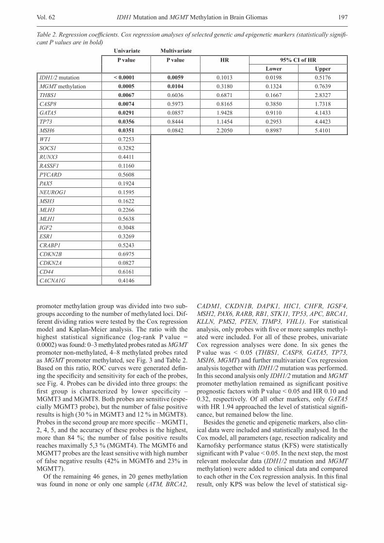
CADM1, CKDN1B, DAPK1, HIC1, CHFR, IGSF4, MSH2, PAX6, RARB, RB1, STK11, TP53, APC, BRCA1, KLLN, PMS2, PTEN, TIMP3, VHL1). For statistical analysis, only probes with five or more samples methylated were included. For all of these probes, univariate Cox regression analyses were done. In six genes the P value was < 0.05 (THBS1, CASP8, GATA5, TP73, MSH6, MGMT) and further multivariate Cox regression analysis together with IDH1/2 mutation was performed. In this second analysis only IDH1/2 mutation and MGMT promoter methylation remained as significant positive prognostic factors with P value < 0.05 and HR 0.10 and 0.32, respectively. Of all other markers, only GATA5 with HR 1.94 approached the level of statistical significance, but remained below the line.
Besides the genetic and epigenetic markers, also clinical data were included and statistically analysed. In the Cox model, all parameters (age, resection radicality and Karnofsky performance status (KFS) were statistically significant with P value < 0.05. In the next step, the most relevant molecular data (IDH1/2 mutation and MGMT methylation) were added to clinical data and compared to each other in the Cox regression analysis. In this final result, only KPS was below the level of statistical sig
Table 2. Regression coefficients. Cox regression analyses of selected genetic and epigenetic markers (statistically signifi-cant P values are in bold) Univariate Multivariate
P value P value HR 95% CI of HRLower Upper
IDH1/2 mutation < 0.0001 0.0059 0.1013 0.0198 0.5176MGMT methylation 0.0005 0.0104 0.3180 0.1324 0.7639THBS1 0.0067 0.6036 0.6871 0.1667 2.8327CASP8 0.0074 0.5973 0.8165 0.3850 1.7318GATA5 0.0291 0.0857 1.9428 0.9110 4.1433TP73 0.0356 0.8444 1.1454 0.2953 4.4423MSH6 0.0351 0.0842 2.2050 0.8987 5.4101WT1 0.7253SOCS1 0.3282RUNX3 0.4411RASSF1 0.1160PYCARD 0.5608PAX5 0.1924NEUROG1 0.1595MSH3 0.1622MLH3 0.2266MLH1 0.5638IGF2 0.3048ESR1 0.3269CRABP1 0.5243CDKN2B 0.6975CDKN2A 0.0827CD44 0.6161CACNA1G 0.4146
IDH1 Mutation and MGMT Methylation in Brain Gliomas

198 Vol. 62
Fig. 2. KaplanMeier analysis of PFS according to IDH1/2 mutation (0 – wildtype IDH, 1 – mutated IDH)
Fig. 3. KaplanMeier survival analysis according to MGMT promoter methylation (0 – unmethylated MGMT, 0–3 probes, 1 – methylated MGMT, 4–8 probes)
F. Kramář et al.

Vol. 62 199
nificance; all other parameters remained relevant survival predictors, see Table 3.
DiscussionGliomas represent serious tumours difficult to treat.
In many cases their prognosis is poor even in case of a very aggressive treatment approach. We present an analysis of our study identifying two independent predictors for patient survival and treatment response. Both IDH1/2 mutation and MGMT promoter methylation are strong parameters confirmed by Kaplan-Meier analysis and Cox regression model. Both of them reduced the
risk of death significantly in our study. We confirmed this fact in accordance with previous studies (Megova et al., 2014; Molenaar et al., 2014; Eckel-Passow et al., 2015). The role of IDH mutation and its positive prognostic effect may not only be limited to prognosis, but there is also evidence that it can positively affect response to chemotherapy (Cairncross et al., 2014). Mu-tated IDH can now be detected by immunohistochemistry (Camelo-Piragua et al., 2010; Takano et al., 2015) and magnetic resonance spectroscopy (Fuente et al., 2015). No drugs currently target mutated IDH, although this remains an area of active research (Li et al., 2015; Suijker et al., 2015). It is obvious that IDH mutation as the first step in tumour development also plays a role in further progression and malignant transformation of low-grade gliomas (Leu et al., 2016).
In contrast to other studies, we have not observed any significant effect of other epigenetic markers (Rankeillor et al., 2014; Rauscher et al., 2014; Lhotska et al., 2015). This can be due to a strong synergistic effect of IDH mutation and MGMT methylation. In univariate Cox analysis, five candidate methylated genes were identified to play a more important role as prognostic markers, but none of them remained statistically significant in multivariate analysis. The same situation was observed in the subgroup of grade IIIII gliomas and subgroup of glioblastomas. Only the GATA5 gene seems to be a factor that can affect prognosis in our series, but more tu
Fig. 4. ROC curve of specificity and sensitivity of each separate MGMT probe
Table 3. Regression coefficients. Multivariate Cox regres-sion analysis of IDH mutation, MGMT methylation, age, radicality of resection and Karnofsky performance status (KFS), (statistically significant P values are in bold) Multivariate
P value HR 95% CI of HRVariable Lower UpperIDH1/2 mutation 0.0007 0.1579 0.0541 0.4610MGMT methylation 0.0171 0.3731 0.1659 0.8390Age 0.0114 1.0334 1.0074 1.0600Resection radicality 0.0012 1.7383 1.2445 2.4281KPS 0.2134 0.9867 0.9660 1.0077
IDH1 Mutation and MGMT Methylation in Brain Gliomas

200 Vol. 62
mour samples should be investigated to verify or exclude this suggestion.
We did not examine the 1p/19q status in our series. 1p/19q co-deletion is known as another positive prognostic predictor, especially in oligodendroglial or mixed (oligoastrocytic) tumours (Cairncross et al., 1998, 2013). Because of a very small number of oligodendroglial tumours investigated in the study (seven patients) we did not examine the 1p/19q status in our series. 1p/19q co deletion is known as another positive prognostic predictor, especially in oligodendroglial or mixed (oligo-astrocytic) tumours (G. Cairncross et al., 2013; J. G. Cairn cross et al., 1998). Because of a very small number of oligodendroglial tumours enrolled in the study (seven patients) we suppose that 1p/19 codeletion cannot significantly affect overall survival in our series, and therefore we did not focus on it and did not examine this marker.
We observed an interesting synergistic effect of MGMT probes. We used eight different probes aiming to different spots within the MGMT promoter and intron 1. None of the probes independently affected prognosis, but positive detection of four and more probes was highly statistically significant in comparison to the minor quantity (three and less probes) in survival analysis. Some of these probes are very sensitive but low in specificity with high rate of false positive results. On the other hand, some probes are specific but sensitivity is low and the number of false negative results can exceed 40 %. Regarding these findings the methylation status must be specified very carefully and correlated with clinical data. Because of the design of the probes (length varying from 179 to 409 bases), one or two short methy-lated segments within the gene probably do not cause gene silencing. On the other hand, the more segments were methylated, the higher was the probability of stopped MGMT gene transcription.
The clinical data included age, KFS and resection radicality. Age is a wellknown factor that affects patient survival. Lower-grade gliomas (gr. II–III) appear in younger people (usually in their thirties, forties) and have better prognosis and overall survival; in our series, the age at surgery in this group was 43 years. Glio blas-toma typically hits the elderly, a history of the disease and overall survival is short; in our series, the mean age of GBM patients was 58 years. In this parameter, our study does not differ from other studies. In general, age slightly increases the risk of death. Karnofsky Performance Status (KPS) did not affect survival in our study. All patients enrolled into the study achieved at least 70 points (av. 86 points) before the surgery with only a very small decrease following the procedure (81 points). That is the reason why KPS could not strike the statistics more noticeably in our series. The second reason might be the fact that only patients with good performance status receive appropriate treatment (Bau chet et al., 2014; Chang-Halpenny et al., 2015). Gross total resection is considered as the next important factor that may significantly affect the overall survival. Espe cially
in comparison with biopsy, the benefit is unambiguous and clear in both low and highgrade gliomas (Senft et al., 2011; Jakola et al., 2014). We confirmed the same finding.
In conclusion, we verified the clinical impact of IDH1/2 mutation and MGMT promoter methylation. These two markers should be diagnosed in glioma patients routinely. Especially in wildtype IDH patients, clinical controls must be performed more frequently, at least every six months in gr. II patients and every three months in gr. III and gr. IV patients. Our data also support active surgical approach; biopsy should be performed only in primarily inoperable cases.
Disclosure of conflict of interestThe authors declare no conflict of interest.
ReferencesBauchet, L., Zouaoui, S., Darlix, A., Menjot de Champfleur,
N., Ferreira, E., Fabbro, M., Kerr, C., Taillandier, L. (2014) Assessment and treatment relevance in elderly glioblastoma patients. Neuro Oncol. 16, 1459-1468.
Cairncross, G., Wang, M., Shaw, E., Jenkins, R., Brachman, D., Buckner, J., Fink, K., Souhami, L., Laperriere, N., Curran, W., Mehta, M. (2013) Phase III trial of chemoradiotherapy for anaplastic oligodendroglioma: long-term results of RTOG 9402. J. Clin. Oncol. 31, 337-343.
Cairncross, J. G., Ueki, K., Zlatescu, M. C., Lisle, D. K., Finkelstein, D. M., Hammond, R. R., Silver, J. S., Stark, P. C., Macdonald, D. R., Ino, Y., Ramsay, D. A., Louis, D. N. (1998) Specific genetic predictors of chemotherapeutic response and survival in patients with anaplastic oligodendrogliomas. J. Natl. Cancer Inst. 90, 1473-1479.
Cairncross, J. G., Wang, M., Jenkins, R. B., Shaw, E. G., Giannini, C., Brachman, D. G., Buckner, J. C., Fink, K. L., Souhami, L., Laperriere, N. J., Huse, J. T., Mehta, M. P., Curran, W. J. (2014) Benefit from procarbazine, lomustine, and vincristine in oligodendroglial tumors is associated with mutation of IDH. J. Clin. Oncol. 32, 783-790.
CameloPiragua, S., Jansen, M., Ganguly, A., Kim, J. C., Louis, D. N., Nutt, C. L. (2010) Mutant IDH1-specific immunohistochemistry distinguishes diffuse astrocytoma from astrocytosis. Acta Neuropathol. 119, 509–511.
Chang-Halpenny, C. N., Yeh, J., Lien, W. W. (2015) Elderly patients with glioblastoma multiforme treated with concurrent temozolomide and standard versus abbreviatedcourse radiotherapy. Perm. J. 19, 15-20.
Dang, L., White, D. W., Gross, S., Bennett, B. D., Bittinger, M. A., Driggers, E. M., Fantin, V. R., Jang, H. G., Jin, S., Keenan, M. C., Marks, K. M., Prins, R. M., Ward, P. S., Yen, K. E., Liau, L. M., Rabinowitz, J. D., Cantley, L. C., Thompson, C. B., Vander Heiden, M. G., Su, S. M. (2009) Cancer-associated IDH1 mutations produce 2-hydroxyglutarate. Nature 462, 739.
EckelPassow, J. E., Lachance, D. H., Molinaro, A. M., Walsh, K. M., Decker, P. A., Sicotte, H., Pekmezci, M., Rice, T., Kosel, M. L., Smirnov, I. V., Sarkar, G., Caron, A. A., Kollmeyer, T. M., Praska, C. E., Chada, A. R., Halder, C., Hansen, H. M., McCoy, L. S., Bracci, P. M., Marshall, R.,
F. Kramář et al.

Vol. 62 201
Zheng, S., Reis, G. F., Pico, A. R., O’Neill, B. P., Buckner, J. C., Giannini, C., Huse, J. T., Perry, A., Tihan, T., Berger, M. S., Chang, S. M., Prados, M. D., Wiemels, J., Wiencke, J. K., Wrensch, M. R., Jenkins, R. B. (2015) Glioma groups based on 1p/19q, IDH, and TERT promoter mutations in tumors. N. Engl. J. Med. 372, 2499-2508.
Fuente, M. I. de la, Young, R. J., Rubel, J., Rosenblum, M., Tisnado, J., Briggs, S., ArevaloPerez, J., Cross, J. R., Campos, C., Straley, K., Zhu, D., Dong, C., Thomas, A., Omuro, A. A., Nolan, C. P., Pentsova, E., Kaley, T. J., Oh, J. H., Noeske, R., Maher, E., Choi, C., Gutin, P. H., Holodny, A. I., Yen, K., DeAngelis, L. M., Mellinghoff, I. K., Thakur, S. B. (2015) Integration of 2-hydroxyglutarate-proton magnetic resonance spectroscopy into clinical practice for disease monitoring in isocitrate dehydrogenasemutant glioma. Neuro Oncol. 18, 283-290.
Hegi, M. E., Diserens, A.C. Gorlia, T., Hamou, M.F., de Tribolet, N., Weller, M., Kross, J. M., Hainfellner, J. A., Mason, W., Mariani, L., Bromberg, J. E. C., Hau, P., Mirimanoff, R. O., Cairncross, J. G., Janzer, R. C., Stupp, R. (2005) MGMT gene silencing and benefit from temozolomide in glioblastoma. N. Engl. J. Med. 352, 997-1003.
Hömig-Hölzel, C., Savola, S. (2012) Multiplex ligation-dependent probe amplification (MLPA) in tumor diagnostics and prognostics. Diagn. Mol. Pathol. 21, 189-206.
Horbinski, C., Kelly, L., Nikiforov, Y. E., Durso, M. B., Nikiforova, M. N. (2010) Detection of IDH1 and IDH2 mutations by fluorescence melting curve analysis as a diagnostic tool for brain biopsies. J. Mol. Diagn. 12, 487-492.
Jakola, A. S., Unsgård, G., Myrmel, K. S., Kloster, R., Torp, S. H., Sagberg, L. M., Lindal, S., Solheim, O. (2014) Surgical strategies in lowgrade gliomas and implications for longterm quality of life. J. Clin. Neurosci. 21, 1304-1309.
Karnofsky, D. A., Abelmann, W. H., Craver, L. F., Burchenal, J. H. (1948) The use of the nitrogen mustards in the palliative treatment of carcinoma. With particular reference to bronchogenic carcinoma. Cancer 1, 634-656.
Leu, S., von Felten, S., Frank, S., Boulay, J.L., Mariani, L. (2016) IDH mutation is associated with higher risk of malignant transformation in lowgrade glioma. J. Neurooncol. 127, 363-372.
Lhotska, H., Zemanova, Z., Cechova, H., Ransdorfova, S., Lizcova, L., Kramar, F., Krejcik, Z., Svobodova, K., Bystricka, D., Hrabal, P., Dohnalova, A., Michalova, K. (2015) Genetic and epigenetic characterization of low-grade gliomas reveals frequent methylation of the MLH3 gene. Genes Chromosomes Cancer 54, 655-667.
Li, L., Paz, A. C., Wilky, B. A., Johnson, B., Galoian, K., Rosenberg, A., Hu, G., Tinoco, G., Bodamer, O., Trent, J. C. (2015) Treatment with a small molecule mutant IDH1 inhibitor suppresses tumorigenic activity and decreases production of the oncometabolite 2-hydroxyglutarate in human chondrosarcoma cells. PLoS One, 10, e0133813.
Megova, M., Drabek, J., Koudelakova, V., Trojanec, R., Kalita, O., Hajduch, M. (2014) Isocitrate dehydrogenase 1 and 2 mutations in gliomas. J. Neurosci. Res. 92, 1611-1620.
Molenaar, R. J., Verbaan, D., Lamba, S., Zanon, C., Jeuken, J. W. M., BootsSprenger, S. H. E., Wesseling, P., Hulsebos, T. J. M., Troost, D., van Tilborg, A. A., Leenstra, S., Vandertop, W. P., Bardelli, A., van Noorden, C. J. F., Bleeker,
F. E. (2014) The combination of IDH1 mutations and MGMT methylation status predicts survival in glioblastoma better than either IDH1 or MGMT alone. Neuro Oncol. 16, 1263-1273.
Noushmehr, H., Weisenberger, D. J., Diefes, K., Phillips, H. S., Pujara, K., Berman, B. P., Pan, F., Pelloski, C. E., Sulman, E. P., Bhat, K. P., Verhaak, R. G. W., Hoadley, K. A., Hayes, D. N., Perou, C. M., Schmidt, H. K., Ding, L., Wilson, R. K., Van Den Berg, D., Shen, H., Bengtsson, H., Neuvial, P., Cope, L. M., Buckley, J., Herman, J. G., Baylin, S. B., Laird, P. W., Aldape, K., Cancer Genome Atlas Research Network (2010) Identification of a CpG island methylator phenotype that defines a distinct subgroup of glioma. Cancer Cell 17, 510-522.
Parsons, D. W., Jones, S., Zhang, X., Lin, J. C.H., Leary, R. J., Angenendt, P., Mankoo, P., Carter, H., Siu, I.M., Gallia, G. L., Olivi, A., McLendon, R., Rasheed, B. A., Keir, S., Nikolskaya, T., Nikolsky, Y., Busam, D. A., Tekleab, H., Diaz, L. A., Hartigan, J., Smith, D. R., Strausberg, R. L., Marie, S. K. N., Shinjo, S. M. O., Yan, H., Riggins, G. J., Bigner, D. D., Karchin, R., Papadopoulos, N., Parmigiani, G., Vogelstein, B., Velculescu, V. E., Kinzler, K. W. (2008) An integrated genomic analysis of human glioblastoma multiforme. Science 321, 1807-1812.
Rankeillor, K. L., Cairns, D. A., Loughrey, C., Short, S. C., Chumas, P., Ismail, A., Chakrabarty, A., Lawler, S. E., Roberts, P. (2014) Methylation-specific multiplex ligation-dependent probe amplification identifies promoter methylation events associated with survival in glioblastoma. J. Neurooncol. 117, 243-251.
Rauscher, J., Beschorner, R., Gierke, M., Bisdas, S., Braun, C., Ebner, F. H., Schittenhelm, J. (2014) WT1 expression increases with malignancy and indicates unfavourable outcome in astrocytoma. J. Clin. Pathol. 67 556-561.
Schiff, D., Purow, B. (2013) Neuro-oncology: five new things. Neurol. Clin. Pract. 3, 326-333.
Senft, C., Bink, A., Franz, K., Vatter, H., Gasser, T., Seifert, V. (2011) Intraoperative MRI guidance and extent of resection in glioma surgery: a randomised, controlled trial. The Lancet Oncol. 12, 997-1003.
Shah, M. A., Denton, E. L., Arrowsmith, C. H., Lupien, M., Schapira, M. (2014) A global assessment of cancer genomic alterations in epigenetic mechanisms. Epigenetics Chro-matin 7, 29.
Stupp, R., Mason, W. P., van den Bent, M. J., Weller, M., Fisher, B., Taphoorn, M. J. B., Belanger, K., Brandes, A. A., Marosi, C., Bogdahn, U., Curschmann, J., Janzer, R. C., Ludwin, S. K., Gorlia, T., Allgeier, A., Lacombe, D., Cairncross, J. G., Eisenhauer, E., Mirimanoff, R. O. (2005) Radiotherapy plus concomitant and adjuvant temozolomide for glioblastoma. N. Engl. J. Med. 352, 987-996.
Sturm, D., Witt, H., Hovestadt, V., KhuongQuang, D.A., Jones, D. T. W., Konermann, C., Pfaff, E., Tönjes, M., Sill, M., Bender, S., Kool, M., Zapatka, M., Becker, N., Zucknick, M., Hielscher, T., Liu, X.Y., Fontebasso, A. M., Ryzhova, M., Albrecht, S., Jacob, K., Wolter, M., Ebinger, M., Schuhmann, M. U., van Meter, T., Frühwald, M. C., Hauch, H., Pekrun, A., Radlwimmer, B., Niehues, T., von Komorowski, G., Dürken, M., Kulozik, A. E., Madden, J., Donson, A., Foreman, N. K., Drissi, R., Fouladi, M., Scheurlen, W., von Deimling, A., Monoranu, C., Roggen
IDH1 Mutation and MGMT Methylation in Brain Gliomas

202 Vol. 62
dorf, W., HeroldMende, C., Unterberg, A., Kramm, C. M., Felsberg, J., Hartmann, C., Wiestler, B., Wick, W., Milde, T., Witt, O., Lindroth, A. M., Schwartzentruber, J., Faury, D., Fleming, A., Zakrzewska, M., Liberski, P. P., Zakrzewski, K., Hauser, P., Garami, M., Klekner, A., Bognar, L., Morrissy, S., Cavalli, F., Taylor, M. D., van Sluis, P., Koster, J., Versteeg, R., Volckmann, R., Mikkelsen, T., Aldape, K., Reifenberger, G., Collins, V. P., Majewski, J., Korshunov, A., Lichter, P., Plass, C., Jabado, N., Pfister, S. M. (2012) Hotspot mutations in H3F3A and IDH1 define distinct epigenetic and biological subgroups of glioblastoma. Cancer Cell 22, 425-437.
Suijker, J., Oosting, J., Koornneef, A., Struys, E. A., Salomons, G. S., Schaap, F. G., Waaijer, C. J. F., WijersKoster, P. M., Briairede Bruijn, I. H., Haazen, L., Riester, S. M., Dudakovic, A., Danen, E., CletonJansen, A.M., van Wijnen, A. J., Bovée, J. V. M. G. (2015) Inhibition of mutant IDH1 decreases D-2-HG levels without affecting tumorigenic properties of chondrosarcoma cell lines. Oncotarget 6, 12505-12519.
Takano, S., Kato, Y., Yamamoto, T., Liu, X., Ishikawa, E., Kaneko, M. K., Ogasawara, S., Matsuda, M., Noguchi, M., Matsumura, A. (2015) Diagnostic advantage of double immunohistochemistry using two mutation-specific anti-IDH antibodies (HMab-1 and MsMab-1) in gliomas. Brain Tu-mor Pathol. 32, 169-175.
IDH1 Mutation and MGMT Methylation in Brain Gliomas
The Cancer Genome Atlas Research Network (2015) Comprehensive, integrative genomic analysis of diffuse lowergrade gliomas. N. Engl. J. Med., 372, 2481–2498.
Turcan, S., Rohle, D., Goenka, A., Walsh, L. A., Fang, F., Yilmaz, E., Campos, C., Fabius, A. W. M., Lu, C., Ward, P. S., Thompson, C. B., Kaufman, A., Guryanova, O., Levine, R., Heguy, A., Viale, A., Morris, L. G. T., Huse, J. T., Mellinghoff, I. K., Chan, T. A. (2012) IDH1 mutation is sufficient to establish the glioma hypermethylator phenotype. Nature 483, 479-483.
Xu, W., Yang, H., Liu, Y., Yang, Y., Wang, P., Kim, S.H., Ito, S., Yang, C., Wang, P., Xiao, M.T., Liu, L., Jiang, W., Liu, J., Zhang, J., Wang, B., Frye, S., Zhang, Y., Xu, Y., Lei, Q., Guan, K.-L., Zhao, S., Xiong, Y. (2011) Oncometabolite 2-hydroxyglutarate is a competitive inhibitor of α-keto glu-tarate-dependent dioxygenases. Cancer Cell 19, 17-30.
Yan, H., Parsons, D. W., Jin, G., McLendon, R., Rasheed, B. A., Yuan, W., Kos, I., BatinicHaberle, I., Jones, S., Riggins, G. J., Friedman, H., Friedman, A., Reardon, D., Herndon, J., Kinzler, K. W., Velculescu, V. E., Vogelstein, B., Bigner, D. D. (2009) IDH1 and IDH2 mutations in gliomas. N. Engl. J. Med. 360, 765-773.