Embed Size (px)
Citation preview
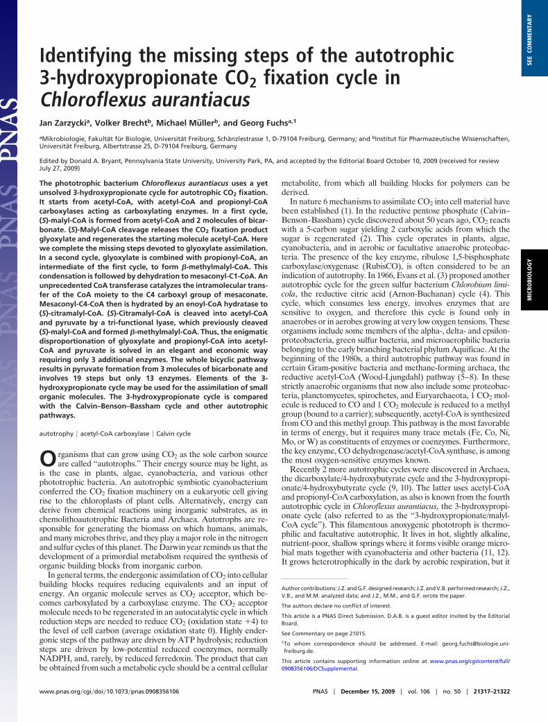
Identifying the missing steps of the autotrophic3-hydroxypropionate CO2 fixation cycle inChloroflexus aurantiacusJan Zarzyckia, Volker Brechtb, Michael Mullerb, and Georg Fuchsa,1
aMikrobiologie, Fakultat fur Biologie, Universitat Freiburg, Schanzlestrasse 1, D-79104 Freiburg, Germany; and bInstitut fur Pharmazeutische Wissenschaften,Universitat Freiburg, Albertstrasse 25, D-79104 Freiburg, Germany
Edited by Donald A. Bryant, Pennsylvania State University, University Park, PA, and accepted by the Editorial Board October 10, 2009 (received for reviewJuly 27, 2009)
The phototrophic bacterium Chloroflexus aurantiacus uses a yetunsolved 3-hydroxypropionate cycle for autotrophic CO2 fixation.It starts from acetyl-CoA, with acetyl-CoA and propionyl-CoAcarboxylases acting as carboxylating enzymes. In a first cycle,(S)-malyl-CoA is formed from acetyl-CoA and 2 molecules of bicar-bonate. (S)-Malyl-CoA cleavage releases the CO2 fixation productglyoxylate and regenerates the starting molecule acetyl-CoA. Herewe complete the missing steps devoted to glyoxylate assimilation.In a second cycle, glyoxylate is combined with propionyl-CoA, anintermediate of the first cycle, to form �-methylmalyl-CoA. Thiscondensation is followed by dehydration to mesaconyl-C1-CoA. Anunprecedented CoA transferase catalyzes the intramolecular trans-fer of the CoA moiety to the C4 carboxyl group of mesaconate.Mesaconyl-C4-CoA then is hydrated by an enoyl-CoA hydratase to(S)-citramalyl-CoA. (S)-Citramalyl-CoA is cleaved into acetyl-CoAand pyruvate by a tri-functional lyase, which previously cleaved(S)-malyl-CoA and formed �-methylmalyl-CoA. Thus, the enigmaticdisproportionation of glyoxylate and propionyl-CoA into acetyl-CoA and pyruvate is solved in an elegant and economic wayrequiring only 3 additional enzymes. The whole bicyclic pathwayresults in pyruvate formation from 3 molecules of bicarbonate andinvolves 19 steps but only 13 enzymes. Elements of the 3-hydroxypropionate cycle may be used for the assimilation of smallorganic molecules. The 3-hydroxypropionate cycle is comparedwith the Calvin–Benson–Bassham cycle and other autotrophicpathways.
autotrophy � acetyl-CoA carboxylase � Calvin cycle
Organisms that can grow using CO2 as the sole carbon sourceare called ‘‘autotrophs.’’ Their energy source may be light, as
is the case in plants, algae, cyanobacteria, and various otherphototrophic bacteria. An autotrophic symbiotic cyanobacteriumconferred the CO2 fixation machinery on a eukaryotic cell givingrise to the chloroplasts of plant cells. Alternatively, energy canderive from chemical reactions using inorganic substrates, as inchemolithoautotrophic Bacteria and Archaea. Autotrophs are re-sponsible for generating the biomass on which humans, animals,and many microbes thrive, and they play a major role in the nitrogenand sulfur cycles of this planet. The Darwin year reminds us that thedevelopment of a primordial metabolism required the synthesis oforganic building blocks from inorganic carbon.
In general terms, the endergonic assimilation of CO2 into cellularbuilding blocks requires reducing equivalents and an input ofenergy. An organic molecule serves as CO2 acceptor, which be-comes carboxylated by a carboxylase enzyme. The CO2 acceptormolecule needs to be regenerated in an autocatalytic cycle in whichreduction steps are needed to reduce CO2 (oxidation state �4) tothe level of cell carbon (average oxidation state 0). Highly ender-gonic steps of the pathway are driven by ATP hydrolysis; reductionsteps are driven by low-potential reduced coenzymes, normallyNADPH, and, rarely, by reduced ferredoxin. The product that canbe obtained from such a metabolic cycle should be a central cellular
metabolite, from which all building blocks for polymers can bederived.
In nature 6 mechanisms to assimilate CO2 into cell material havebeen established (1). In the reductive pentose phosphate (Calvin–Benson–Bassham) cycle discovered about 50 years ago, CO2 reactswith a 5-carbon sugar yielding 2 carboxylic acids from which thesugar is regenerated (2). This cycle operates in plants, algae,cyanobacteria, and in aerobic or facultative anaerobic proteobac-teria. The presence of the key enzyme, ribulose 1,5-bisphosphatecarboxylase/oxygenase (RubisCO), is often considered to be anindication of autotrophy. In 1966, Evans et al. (3) proposed anotherautotrophic cycle for the green sulfur bacterium Chlorobium limi-cola, the reductive citric acid (Arnon-Buchanan) cycle (4). Thiscycle, which consumes less energy, involves enzymes that aresensitive to oxygen, and therefore this cycle is found only inanaerobes or in aerobes growing at very low oxygen tensions. Theseorganisms include some members of the alpha-, delta- and epsilon-proteobacteria, green sulfur bacteria, and microaerophilic bacteriabelonging to the early branching bacterial phylum Aquificae. At thebeginning of the 1980s, a third autotrophic pathway was found incertain Gram-positive bacteria and methane-forming archaea, thereductive acetyl-CoA (Wood-Ljungdahl) pathway (5–8). In thesestrictly anaerobic organisms that now also include some proteobac-teria, planctomycetes, spirochetes, and Euryarchaeota, 1 CO2 mol-ecule is reduced to CO and 1 CO2 molecule is reduced to a methylgroup (bound to a carrier); subsequently, acetyl-CoA is synthesizedfrom CO and this methyl group. This pathway is the most favorablein terms of energy, but it requires many trace metals (Fe, Co, Ni,Mo, or W) as constituents of enzymes or coenzymes. Furthermore,the key enzyme, CO dehydrogenase/acetyl-CoA synthase, is amongthe most oxygen-sensitive enzymes known.
Recently 2 more autotrophic cycles were discovered in Archaea,the dicarboxylate/4-hydroxybutyrate cycle and the 3-hydroxypropi-onate/4-hydroxybutyrate cycle (9, 10). The latter uses acetyl-CoAand propionyl-CoA carboxylation, as also is known from the fourthautotrophic cycle in Chloroflexus aurantiacus, the 3-hydroxypropi-onate cycle (also referred to as the ‘‘3-hydroxypropionate/malyl-CoA cycle’’). This filamentous anoxygenic phototroph is thermo-philic and facultative autotrophic. It lives in hot, slightly alkaline,nutrient-poor, shallow springs where it forms visible orange micro-bial mats together with cyanobacteria and other bacteria (11, 12).It grows heterotrophically in the dark by aerobic respiration, but it
Author contributions: J.Z. and G.F. designed research; J.Z. and V.B. performed research; J.Z.,V.B., and M.M. analyzed data; and J.Z., M.M., and G.F. wrote the paper.
The authors declare no conflict of interest.
This article is a PNAS Direct Submission. D.A.B. is a guest editor invited by the EditorialBoard.
See Commentary on page 21015.
1To whom correspondence should be addressed. E-mail: [email protected].
This article contains supporting information online at www.pnas.org/cgi/content/full/0908356106/DCSupplemental.
www.pnas.org�cgi�doi�10.1073�pnas.0908356106 PNAS � December 15, 2009 � vol. 106 � no. 50 � 21317–21322
MIC
ROBI
OLO
GY
SEE
COM
MEN
TARY

has the capability of fixing inorganic carbon in the light. C.aurantiacus is of particular interest in the study of the evolution ofphotosynthesis and of its autotrophic carbon-fixation cycle, whichhas not been completely elucidated (13–23).
In brief, in a first cycle, 1 acetyl-CoA molecule and 2 bicarbonatemolecules are converted to (S)-malyl-CoA (Fig. 1). Bicarbonatefixation proceeds via acetyl-CoA and propionyl-CoA carboxylation.(S)-Malyl-CoA then is cleaved to glyoxylate and acetyl-CoA, thusclosing the first cycle. A second cycle has been postulated by whichglyoxylate and propionyl-CoA are converted to acetyl-CoA andpyruvate, thus regenerating acetyl-CoA and producing pyruvate asuniversal precursor for biosynthesis (Fig. 1). This second cycle startswith the condensation of glyoxylate and propionyl-CoA to a C5-dicarboxylic acid CoA thioester. As can be seen in Fig. 1, in thecourse of the conversion of this intermediate to citramalyl-CoA,the CoA moiety somehow must be transferred from the ‘‘right’’ tothe ‘‘left’’ carboxyl group of the C5-dicarboxylic acid. The lastproven step of the glyoxylate assimilation cycle is the formation ofmesaconyl-C1-CoA (2-methylfumaryl-CoA) (reaction 11 in Fig. 1).
Here, we demonstrate missing enzymes and intermediates, com-pleting this autotrophic CO2 fixation cycle. This cycle involves anintramolecular CoA transferase that catalyzes the unprecedentedtransfer of the CoA moiety from the C1-carboxyl group to theC4-carboxyl group of the dicarboxylic acid. A common enoyl-CoAhydratase forms (3S)-citramalyl-CoA, the intermediate to becleaved to pyruvate and acetyl-CoA. The advantages of this cyclecompared with the ubiquitous Calvin–Benson–Bassham cycle arediscussed. Under oligotrophic aquatic conditions, elements of the3-hydroxypropionate cycle may be used by other bacteria for theassimilation of small organic molecules.
ResultsConversion of Mesaconyl-C1-CoA to an Unknown CoA Thioester by CellExtracts. We used 3 recombinant enzymes in an assay to synthesizelabeled mesaconyl-C1-CoA from labeled propionyl-CoA and
glyoxylate to be used as substrate for the next missing enzyme of thecycle (Fig. 2, see Fig. 1 for labeling). Extracts of autotrophically andheterotrophically grown cells transformed mesaconyl-C1-CoA (2-methylfumaryl-CoA) at a rate of 2 �mol min�1 (U) mg�1 proteinand 0.6 U mg�1 protein (55 °C), respectively. The product elutedunder different HPLC conditions after mesaconyl-C1-CoA. Ap-proximately 94% of labeled propionyl-CoA was converted tomesaconyl-C1-CoA and the observed product. The equilibriumconcentrations of mesaconyl-C1-CoA and its product were nearlyidentical, indicating a freely reversible reaction with an equilibriumconstant near 1.
Surprisingly, the same product was formed when cell extract wasomitted but a different preparation of the recombinant trifunctional(S)-malyl-CoA/�-methylmalyl-CoA/(S)-citramalyl-CoA (MMC)lyase from C. aurantiacus was used. This enzyme preparation wasderived from the expression of a DNA fragment of C. aurantiacusin Escherichia coli that contained both the gene coding for theMMC lyase and the upstream neighbor gene coding for a putativeCoA transferase. The transferase protein had been ignored becauseit was expressed only in trace quantities.
Identification of the Unknown CoA Thioester as Mesaconyl-C4-CoA.The product formed from mesaconyl-C1-CoA was a CoA thioesteras well, based on its spectral properties (Fig. 2A), migration inHPLC (Fig. 2B), and alkali sensitivity (Fig. 2C). Electrosprayionization mass spectrometry (ESI-MS) determined a mass of 878.0Da (negative ion mode) and 879.8 Da (positive ion mode), corre-sponding to the mass of mesaconyl-C1-CoA or an isomer. However,after alkaline hydrolysis of the 2 labeled CoA esters, onlymesaconate (methylfumarate) was obtained (Fig. 2C); no otherisomer, such as itaconate (methylenesuccinate) or citraconate(methylmaleate), was formed. Interestingly, the same productmixture was obtained when mesaconyl-CoA was synthesized chem-ically (Fig. 2B). One would expect the chemical synthesis to yield
Fig. 1. The complete 3-hydroxypropionate cycle, as studied in C. aurantiacus. [1] Acetyl-CoA carboxylase, [2] malonyl-CoA reductase, [3] propionyl-CoAsynthase, [4] propionyl-CoA carboxylase, [5] methylmalonyl-CoA epimerase, [6] methylmalonyl-CoA mutase, [7] succinyl-CoA:(S)-malate-CoA transferase, [8]succinate dehydrogenase, [9] fumarate hydratase, [10 a, b, c] (S)-malyl-CoA/�-methylmalyl-CoA/(S)-citramalyl-CoA (MMC) lyase, [11] mesaconyl-C1-CoAhydratase (�-methylmalyl-CoA dehydratase), [12] mesaconyl-CoA C1-C4 CoA transferase, [13] mesaconyl-C4-CoA hydratase. Carbon-labeling patterns during theinterconversion of propionyl-CoA plus glyoxylate to pyruvate plus acetyl-CoA via C5 compounds are shown. 14C carbon atoms derived from [1-14C]propionyl-CoAare marked by Œ, and 13C carbon atoms derived from [1,2,3-13C]propionyl-CoA are marked by ■. Note that the cleavage of citramalyl-CoA requires that the CoAmoiety be shifted finally from the ‘‘right’’ carboxyl group of �-methylmalyl-CoA to the ‘‘left’’ carboxyl group of citramalyl-CoA. This shifting is accomplished byan intramolecular CoA transfer (reaction 12). Otherwise, citramalyl-CoA cleavage into pyruvate and acetyl-CoA would not be feasible.
21318 � www.pnas.org�cgi�doi�10.1073�pnas.0908356106 Zarzycki et al.
similar amounts of mesaconyl-C1-CoA and mesaconyl-C4-CoA.Therefore, the product may represent mesaconyl-C4-CoA (3-methylfumaryl-CoA); indeed, this possibility was verified by NMRspectroscopy (see SI Text).
Identification of the CoA Transferase Forming Mesaconyl-C4-CoA andCharacterization of the Enzyme. The enzyme transforming mesaco-nyl-C1-CoA to mesaconyl-C4-CoA obviously was a type of CoAtransferase. A search for putative CoA transferase genes in thegenome led to an ORF (Caur�0175), which clustered with MMClyase (Caur�0174) (as mentioned above) and other genes requiredfor the conversion of propionyl-CoA plus glyoxylate to pyruvateplus acetyl-CoA (Fig. 3). The gene was cloned, expressed in E. colias an N-terminal His10-tagged protein (47 kDa), and the recombi-nant protein was purified. It catalyzed the expected reversibletransformation of mesaconyl-C1-CoA to mesaconyl-C4-CoA (re-action 12 in Fig. 1) at high rates (Vmax of 520 U mg�1 at 55 °C,turnover number 840 s�1) with an apparent Km for mesaconyl-C1-CoA of 0.24 mM. No CoA transferase activity was observed withitaconate, mesaconate, (R)-/(S)-malate, and (R)-/(S)-citramalate asCoA acceptors when acetyl-CoA or succinyl-CoA as CoA wereused as CoA donors. The enzyme exhibited a broad pH optimumat pH20 °C of 7.5–7.8 (extrapolated pH55 °C of 7.1–7.4), with half
maximal activity at pH20 °C 6.6. It did not require free mesaconateas a CoA acceptor, because the enzyme did not form [14C]mesaco-nyl-CoA from [14C]mesaconate plus unlabeled mesaconyl-C1-CoA.Correspondingly, it did not form [14C]mesaconate from[14C]mesaconyl-C1-CoA plus unlabeled mesaconate.
The UV-visible spectrum of the purified recombinant enzymeshowed an absorption band only at 280 nm. Gel filtration indicateda molecular mass of about 73 � 4 kDa, suggesting a homodimericstructure. The high rate of amino acid sequence similarities/identities (ca. 42%/25%) (Table S2 and Fig. S1) with enzymesbelonging to class III CoA transferases (24) suggested that anaspartate residue in the active site forms an acid anhydride with theformerly CoA-activated acid. Although this anhydride intermediateshould react with borohydride and hydroxylamine, resulting inenzyme inactivation, these enzymes exhibit very different sensitiv-ities to these compounds (25–30). The CoA transferase was testedfor inactivation in the presence of its substrate; the enzyme was onlypartially inactivated by hydroxylamine (residual activity about60%), and sodium borohydride had no effect at all. This lack ofeffect may be caused by a closure of the active site upon substratebinding. Such a conformational change has been observed for thecrotonobetainyl-CoA:carnitine CoA transferase (CaiB) from E.coli (31). This change in the conformation of the enzyme mayprevent the inactivator molecules from entering the active site andis consistent with the observation that no mesaconate is releasedduring catalysis.
Further Transformation of Mesaconyl-C4-CoA Involving an Enoyl-CoAHydratase. A coupled photometric assay was developed to measurethe further transformation of mesaconyl-C4-CoA to acetyl-CoAand pyruvate in cell extracts at 45 °C. The specific activities (ex-trapolated to 55 °C) in extracts of autotrophically and heterotroph-ically grown cells were 1.2 and 0.5 U mg�1, respectively. Thisreaction sequence needed no cofactor other than Mg2�. Citramalyl-CoA was observed as an intermediate (as in Fig. 4), indicating thatmesaconyl-C4-CoA was hydrated to citramalyl-CoA, followed bycitramalyl-CoA cleavage to acetyl-CoA and pyruvate. The extrap-olation of specific activities to 55 °C was based on the assumptionthat the reaction rate doubles with an increase in temperatureof 10 °C.
The next missing enzyme, mesaconyl-C4-CoA hydratase, waspurified from autotrophically grown cells (Table S3) SDS-PAGErevealed a polypeptide of 30 kDa, and peptide mass fingerprint
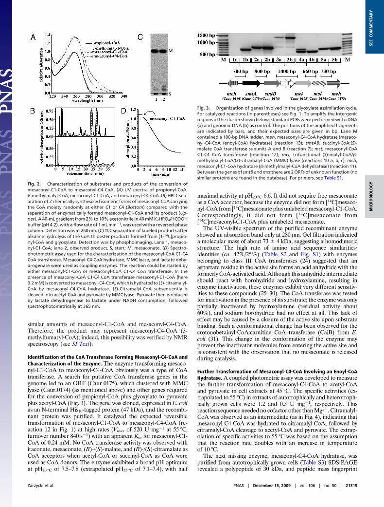
Fig. 2. Characterization of substrates and products of the conversion ofmesaconyl-C1-CoA to mesaconyl-C4-CoA. (A) UV spectra of propionyl-CoA,�-methylmalyl-CoA, mesaconyl-C1-CoA, and mesaconyl-C4-CoA. (B) HPLC sep-aration of 2 chemically synthesized isomeric forms of mesaconyl-CoA carryingthe CoA moiety randomly at either C1 or C4 (Bottom) compared with theseparation of enzymatically formed mesaconyl-C1-CoA and its product (Up-per). A 40-mL gradient from 2% to 10% acetonitrile in 40 mM K2HPO4/HCOOHbuffer (pH 4.2), with a flow rate of 1 mL min�1, was used with a reversed-phasecolumn. Detection was at 260 nm. (C) TLC separation of labeled products afteralkaline hydrolysis of the CoA thioester products formed from [1-14C]propio-nyl-CoA and glyoxylate. Detection was by phosphoimaging. Lane 1, mesaco-nyl-C1-CoA; lane 2, observed product. S, start; M, mesaconate. (D) Spectro-photometric assay used for the characterization of the mesaconyl-CoA C1-C4CoA transferase. Mesaconyl-C4-CoA hydratase, MMC lyase, and lactate dehy-drogenase were used as coupling enzymes. The reaction could be started byeither mesaconyl-C1-CoA or mesaconyl-CoA C1-C4 CoA transferase. In thepresence of mesaconyl-CoA C1-C4 CoA transferase mesaconyl-C1-CoA (here0.2 mM) is converted to mesaconyl-C4-CoA, which is hydrated to (S)-citramalyl-CoA by mesaconyl-C4-CoA hydratase. (S)-Citramalyl-CoA subsequently iscleaved into acetyl-CoA and pyruvate by MMC lyase. Pyruvate then is reducedby lactate dehydrogenase to lactate under NADH consumption, followedspectrophotometrically at 365 nm.
Fig. 3. Organization of genes involved in the glyoxylate assimilation cycle.For catalyzed reactions (in parentheses) see Fig. 1. To amplify the intergenicregions of the cluster shown below, standard PCRs were performed with cDNA(a) and genomic DNA (b) as control. The positions of the amplified fragmentsare indicated by bars, and their expected sizes are given in bp. Lane Mcontained a 100-bp DNA ladder. meh, mesaconyl-C4-CoA hydratase (mesaco-nyl-C4-CoA (enoyl-CoA) hydratase) (reaction 13); smtAB, succinyl-CoA:(S)-malate CoA transferase subunits A and B (reaction 7); mct, mesaconyl-CoAC1-C4 CoA transferase (reaction 12); mcl, trifunctional (S)-malyl-CoA/�-methylmalyl-CoA/(S)-citramalyl-CoA (MMC) lyase (reactions 10 a, b, c); mch,mesaconyl-C1-CoA hydratase (�-methylmalyl-CoA dehydratase) (reaction 11).Between the genes of smtB and mct there are 2 ORFs of unknown function (nosimilar proteins are found in the database). For primers, see Table S1.
Zarzycki et al. PNAS � December 15, 2009 � vol. 106 � no. 50 � 21319
MIC
ROBI
OLO
GY
SEE
COM
MEN
TARY

analysis identified the corresponding ORF (Caur�0180). It waslocated directly adjacent to a cluster of genes coding for severalenzymes of the 3-hydroxypropionate cycle (Fig. 3). The gene wascloned and expressed in E. coli as an N-terminal His10-taggedprotein (33 kDa); the recombinant enzyme was purified, and itsactivity was measured at 45 °C. It catalyzed the expected hydrationof mesaconyl-C4-CoA to (S)-citramalyl-CoA (reaction 13 in Fig. 1).The enzyme exhibited a specific activity of 950 U mg�1 (extrapo-lated to 55 °C, turnover number 1,045 s�1) with an apparent Kmvalue for mesaconyl-C4-CoA of 75 �M and was inhibited bysubstrate concentrations higher than 0.3 mM. Gel filtration indi-cated a molecular mass of about 52 � 5 kDa, suggesting ahomodimeric structure. The amino acid sequence had similarities toa group of uncharacterized conserved proteins (COG3777) with aC-terminal hotdog-fold domain and possibly to an itaconyl-CoAhydratase (AAX86477) from Pseudomonas sp. L1 described byKornberg and associates (32, 33). This group of enzymes apparentlyforms a distinct subclass of the enoyl-CoA hydratase superfamily,based on sequence comparison.
Cleavage of (S)-Citramalyl-CoA into Acetyl-CoA and Pyruvate byRecombinant MMC Lyase and Reconstitution of the Second Cycle fromPurified Enzymes. (S)-Citramalyl-CoA cleavage required anotherenzyme. To assay for this enzyme activity, chemically synthesizedmesaconyl-CoA (both C1- and C4-CoA) was incubated at 45 °Cwith mesaconyl-CoA C1-C4 CoA transferase, and mesaconyl-C4-CoA hydratase to form (S)-citramalyl-CoA. Addition of cell ex-tracts led to a rapid formation of acetyl-CoA and pyruvate. Ex-trapolated to 55 °C, the rate with extracts of autotrophically andheterotrophically grown cells was 2 U mg�1 and 0.6 U mg�1,respectively. Recombinant MMC lyase has been known to catalyzethe cleavage of (S)-citramalyl-CoA into acetyl-CoA and pyruvate(22). We therefore studied the recombinant His10-tagged lyase andfound specific activities for (S)-malyl-CoA cleavage into acetyl-CoAand glyoxylate of 1.1 U mg�1 (reaction 10a in Fig. 1), for propionyl-CoA condensation with glyoxylate to �-methylmalyl-CoA of 10 Umg�1 (reaction 10b), and for (S)-citramalyl-CoA cleavage intoacetyl-CoA and pyruvate of 24 U mg�1 (reaction 10c). (R)-Malyl-
CoA and (R)-citramalyl-CoA were not used. The enzyme catalyzedthe reverse reaction, the condensation of pyruvate and acetyl-CoAto (S)-citramalyl-CoA. However, the equilibrium of this reactionwas strongly in favor of acetyl-CoA and pyruvate (50:1), althoughpyruvate was added in 10-fold excess to acetyl-CoA.
The entire glyoxylate assimilation route (glyoxylate � propionyl-CoA 3 acetyl-CoA � pyruvate) involving 5 enzymatically cata-lyzed reactions could be reconstituted in vitro by using the 4recombinant enzymes MMC lyase, mesaconyl-C1-CoA hydratase,mesaconyl-CoA C1-C4 CoA transferase, and mesaconyl-C4-CoAhydratase (Fig. 4).
Cluster of Genes Involved in the Second Cycle. All genes coding forthe enzymes required for the glyoxylate assimilation route plus the2 genes coding for the subunits of succinyl-CoA:(S)-malate CoAtransferase (Caur�0178, Caur�0179) were clustered in the genome(Fig. 3). A similar gene cluster is present in the related Chloroflexistrains Chloroflexus aggregans, Roseiflexus castenholzii, and Rosei-flexus sp. RS-1 (34). Two additional small ORFs (Caur�0176,Caur�0177), which probably do not have a function in this pathway,were present in C. aurantiacus only. Activity measurements indi-cated that the encoded enzymes were up-regulated to a similarextent under autotrophic growth conditions. Of these 6 genes, 5 areoriented in the same direction. Neighboring genes are co-transcribed, as evidenced by RT-PCR of mRNA (Fig. 3), and mayconstitute an operon. The sixth gene coding for mesaconyl-C4-CoAhydratase (meh) is orientated in the opposite direction. Thus theintergenic region between these clusters may harbor the bindingsites for regulatory proteins.
DiscussionThis work has completed the 3-hydroxypropionate autotrophiccarbon fixation cycle, the fourth autotrophic pathway (Table S4).All characteristic enzymes have been identified and purified. Thecycle results in the net fixation of 3 molecules of bicarbonate into1 molecule of pyruvate, which can be used to produce all cellularbuilding blocks. This pathway is an intertwined bicycle that sche-matically resembles a pretzel (Fig. 1).
In a first cycle starting from acetyl-CoA, 2 bicarbonate moleculesare fixed to form 1 molecule of glyoxylate. In a second cycle,glyoxylate plus propionyl-CoA are disproportionated to acetyl-CoAplus pyruvate without any redox step or additional coenzymeparticipation. Propionyl-CoA is derived from acetyl-CoA and bi-carbonate, as in the first cycle. The reaction sequence glyoxylate �propionyl-CoA 3 acetyl-CoA � pyruvate is associated with astandard free energy change �G°� of �14 kJ mol�1 (35), resultingin an equilibrium constant Kequ [acetyl-CoA] [pyruvate]/[glyoxylate][propionyl-CoA] � 300 and rendering the second cycle almostunidirectional toward pyruvate formation. The reaction sequencerequires only 3 additional enzymes and solves a mechanisticallyintriguing problem, the shift of the CoA moiety, in a most eco-nomical and elegant way. An unparalleled attribute of the completepathway is the crossing of the 2 cycles, because acetyl-CoA con-version to propionyl-CoA is common to both. The stoichiometry,cofactor specificity, energy demand, and other features of the cyclenow can be compared with other CO2 fixation mechanisms.
Comparison with Other Autotrophic Carbon Fixation Pathways. Thestoichiometry of the 3-hydroxypropionate cycle follows theequation: 3 HCO3
� � 5 ATP � 6 NADPH � 1 quinone 3 1pyruvate � 6 NADP � 1 quinoneH2 � 3 ADP � 3 phosphate� 2 AMP � 2 pyrophosphate; this input of energy correspondsto 7 ATP equivalents for pyruvate formation, as required in theCalvin cycle. In terms of the energy demand of an autotrophicpathway, the costs for synthesizing all CO2-fixation–relatedenzymes are important also. Poor catalytic efficiency ofRubisCO, for instance, not only leads to the loss of energyassociated with photorespiration but also pushes the organisms
Fig. 4. HPLC separation of 14C-labeled products formed from [1-14C]propio-nyl-CoA and glyoxylate by different purified recombinant enzymes. (A) Beforeaddition of enzyme. (B) After the addition of MMC lyase. (C) As B, plusmesaconyl-C1-CoA hydratase. (D) As C, plus mesaconyl-CoA C1-C4 CoA trans-ferase. (E) As D, plus mesaconyl-C4-CoA hydratase. (F) As E: formation ofnon-labeled acetyl-CoA and 14C-pyruvate after additional incubation time. Areversed-phase column was developed for 7 min under isocratic conditionswith 100 mM NaH2PO4 (pH 4.0) in 7.5% methanol (vol/vol), followed by a linear10-min gradient from 0% to 60% of 100 mM sodium acetate (pH 4.6) in 90%methanol (vol/vol) at a flow rate of 1 mL min�1. Acetyl-CoA was detected bydiode array detection because of its absorption at 260 nm. Radioactivity wasdetected by solid-state scintillation counting.
21320 � www.pnas.org�cgi�doi�10.1073�pnas.0908356106 Zarzycki et al.

to use special carbon-concentrating mechanisms. Moreover,carboxylases with low catalytic efficiency, such as RubisCO, needto be synthesized in large quantities. In other words, the synthesisof the catalyst itself may devour a huge amount of energy as wellas nitrogen and sulfur sources. Therefore, the expenditures forthe synthesis of all additional enzymes of an autotrophic carbonfixation pathway actually may determine its energy cost. Theother tradeoff is the necessity for low-potential electron donorsfor autotrophic pathways that produce acetyl-CoA or pyruvatedirectly. Balancing that need against the number of uniqueproteins required for integrating the 3-hydroxypropionate cycle(relatively high) or the Calvin cycle (only 2 proteins, but manyof them, are needed) into central metabolism may determinewhether an organism that uses the pathway comes to dominatea given ecosystem. For discussion of other autotrophic pathways,notably those requiring anaerobic conditions, see ref. 36. Acomparison of the respective features of all currently known CO2fixation pathways is provided in Table S4.
Advantages of the 3-Hydroxypropionate Bicycle and Its Elements.Given that organisms with the Calvin cycle have come todominate most ecosystems, they presumably have some advan-tages. In microorganisms the Calvin cycle is found solely inaerobic and facultative aerobic Bacteria, notably in Cyanobacteria.Those bacteria are less energy limited, and therefore the high ATPcosts of the Calvin cycle may be of minor importance. In bacteriathe need for sugar phosphates in biosynthesis of cell wall and ligninprecursors is much less than in plants. The main metabolic fluxesare diverted from acetyl-CoA, pyruvate, oxaloacetate, and 2-oxoglutarate, and their synthesis from 3-phosphoglycerate is partlyconnected with a loss of CO2. Therefore, in bacteria, autotrophicpathways directly yielding acetyl-CoA or pyruvate are more eco-nomical, and this statement holds true for all alternative carbon-fixation pathways, including the 3-hydroxypropionate bicycle. Also,the 2 autotrophic cycles differ with respect to the inorganic carbonspecies used: CO2 in the Calvin cycle versus HCO3
� in the3-hydroxypropionate bicycle. Because the bicarbonate concentra-tion in slightly alkaline water, where Chloroflexus lives, is muchhigher than the concentration of dissolved CO2 (the apparent aciddissociation association constant of HCO3
�/CO2 is 6.3), Chlo-roflexus may profit from using bicarbonate instead of CO2.
Most Chloroflexi probably grow as mixotrophs. Why might theirpathway be better than the Calvin cycle in this regard? A completeor even a rudimentary 3-hydroxypropionate cycle allows co-assimilating trace amounts of organic compounds even under oxicconditions (no enzyme of the cycle is oxygen sensitive), an abilitythat may be advantageous in oligotrophic aquatic habitats. Exam-ples of such substrates are the fermentation products acetate,propionate, and succinate (including their corresponding alcohols),which may be formed by cyanobacteria and possibly by fermentingbacteria associated with the Chloroflexus mats. Numerous othercompounds that are metabolized via acetyl-CoA or propionyl-CoAare included in this substrate spectrum as well. Notably, 3-hydroxypropionate is a very common metabolite that can beassimilated by this mechanism. It is an intermediate in the metab-olism of dimethylsulfoniopropionate (37–40), a ubiquitous osmo-protectant and antioxidant of algae (41, 42); other small sources of3-hydroxypropionate are pyrimidines and �-alanine (43, 44).
However, as discussed later, the limited distribution of thecharacteristic enzymes/genes of the 3-hydroxypropionate cycle sug-gests that these advantages may become effective only in a limitedset of natural niches where Chloroflexi compete successfullyagainst other bacteria and cyanobacteria. Another reason for thelimited occurrence of this cycle may be a late and singular inventionin the Chloroflexi. Interestingly, the autotrophic aerobic Crenar-chaeota use a similar mechanism of converting acetyl-CoA tosuccinyl-CoA via 3-hydroxypropionate (10, 45). However, thiscrenarchaeal autotrophic pathway probably has evolved indepen-
dently, and the involved enzymes show little or no similarity to theChloroflexus enzymes (10).
Occurrence of the 3-Hydroxypropionate Bicycle and Its Elements. Thecompletely sequenced genomes indicate a similar pathway mayoperate in Chloroflexus aggregans, Roseiflexus castenholzii, andRoseiflexus sp. RS-1 (Chloroflexaceae), which contain all postu-lated genes of the cycle. Erythrobacter sp. NAP1 and some pho-totrophic gamma-Proteobacteria (NOR5–3, NOR51-B) harbor theChloroflexus-type genes required for the conversion of acetyl-CoAto succinyl-CoA but lack the other genes of the 3-hydroxypropi-onate bicycle. Likewise, the heterotrophic Congregibacter litoralisand Nitrococcus mobilis and the photolithoautotrophic Chloroher-peton thalassium contain the propionyl-CoA synthase gene only.These bacteria may use a rudimentary cycle for the mixotrophicassimilation of acetate, 3-hydroxypropionate, and/or propionateunder oligotrophic (e.g., marine) conditions. Interestingly, thegamma-proteobacterium strain HTCC2080 possesses the genes fora chimeric 3-hydroxypropionate/4-hydroxybutyrate cycle that evenmay allow autotrophic growth: Genes required for the conversionof acetyl-CoA plus 2 bicarbonate molecules to succinyl-CoA are ofthe Chloroflexus type. In contrast, the regeneration of acetyl-CoAfrom succinyl-CoA seems to correspond to the pathways found inautotrophic Crenarchaeota (9, 10, 46).
Streamlining of the Pathway and Its Genes. The whole cycle requires19 chemical steps, but only 13 enzymes are involved, suggesting thatseveral enzymes are multifunctional (e.g. bifunctional malonyl-CoAreductase, trifunctional propionyl-CoA synthase, and trifunctional(S)-malyl-CoA/�-methylmalyl-CoA/(S)-citramalyl-CoA lyase).Succinyl-CoA:(S)-malate CoA transferase also catalyzes 2 formalsteps, the release of succinate and the activation of (S)-malate. Thestreamlined pathway and the compact genetic organization of mostof its genes indicate an advanced and successful adaptation of theorganism to its natural niche that is not occupied by other autotro-phic bacteria or algae. In 4 members of Chloroflexaceae (see earlierdiscussion) a similar cluster of genes required for the secondglyoxylate assimilation cycle and for the last reactions of the firstglyoxylate formation cycle is present (Fig. 3; for catalyzed reactions,see Fig. 1). Because the encoded enzymes are up-regulated underautotrophic conditions, we expect a common regulator.
An Internal CoA Transferase and an Enoyl-CoA Hydratase. We haveidentified and characterized an enzyme of the CoA transferasefamily (class III) (E. C. 2.8.3.x.) (24) that functions in an intramo-lecular CoA transfer between 2 carboxyl groups (Fig. 1). Itssuggested systematic name is ‘‘mesaconyl-CoA C1-C4 CoA trans-ferase.’’ The amino acid sequence identity and similarity values fordifferent representatives of this family are supplied in Table S2. Ahighly conserved aspartate residue (Asp 169 in the CaiB nomen-clature), which is located in the active site and binds the organic acidin an anhydride bond (47, 48), is conserved in this CoA transferase.Other residues that are important for folding are conserved as well.Regarding the inhibition experiments, the catalytic mechanism ofthis enzyme is enigmatic in view of the presently consideredmechanisms (28). Furthermore, no free carboxylic acid is involved,nor does free mesaconate exchange with mesaconyl-CoA. Addi-tional studies are required to address this tantalizing mechanisticproblem.
Mesaconyl-C4-CoA is hydrated by a rather conventional enoyl-CoA hydratase to (S)-citramalyl-CoA (Fig. 1) that differs stronglyfrom mesaconyl-C1-CoA hydratase (23). Although both enzymesare members of the superfamily of hotdog domain-containingproteins (49), their amino acid sequences are not significantlysimilar. Enoyl-CoA hydratases (E. C. 4.2.1.17) catalyze reversiblereactions of the type (3S)-3-hydroxyacyl-CoA 3 trans-2(or 3)-enoyl-CoA � H2O. Therefore the formation of the (S)-
Zarzycki et al. PNAS � December 15, 2009 � vol. 106 � no. 50 � 21321
MIC
ROBI
OLO
GY
SEE
COM
MEN
TARY

stereoisomer of citramalyl-CoA, as observed experimentally, wasexpected.
Open Questions. The demonstration of all enzyme activities of theCO2 fixation bicycle, their whole-cell regulation, and the organiza-tion of the genes provide final evidence for the operation of theautotrophic CO2 fixation bicycle. The roles of 2 other enzymes,succinyl-CoA:(R)-citramalate CoA transferase and (R)-citramalyl-CoA lyase, which act on the (R)-stereosisomers of (citra)malate (21,24) and which also are up-regulated under autotrophic conditions,are still unclear. Their specific activities are an order of magnitudelower than those of the (S)-citramalate–specific CoA transferaseand lyase. They may be required to recycle (R)-citramalate or(R)-malate formed incidentally by side reactions. The regulationof acetyl-CoA carboxylase, which is the carboxylase of the 3-hydroxypropionate cycle and the initial enzyme in lipid biosynthe-sis, poses another problem that may prevent this cycle from beingintroduced into an organism that uses the Calvin cycle. How fattyacid biosynthesis and acetyl-CoA carboxylase are regulated sepa-rately, depending on the needs of the cell, is not known. Further-
more, it still is unknown if propionyl-CoA carboxylation also iscatalyzed by a bifunctional enzyme, as is the case in the crenar-chaeal 3-hydroxypropionate/4-hydroxybutyrate cycle (10).
Materials and MethodsC. aurantiacus strain OK-70-fl (DSMZ 636) was grown anaerobically and pho-totrophically either under autotrophic conditions with H2 and CO2 (80:20, vol/vol)or under heterotrophic conditions with casamino acids and yeast extract asdescribed elsewhere (20). The genome sequence of the strain J-10-fl was used forprimerdesign.Virtuallynodifferences inthegenesofthe2strainswereobserved.The exact procedures of cloning and heterologous expression of genes from C.aurantiacus in E. coli, enzyme purification, enzyme measurements, syntheses,identification of compounds, and all other methods are described in the SIMaterials and Methods.
ACKNOWLEDGMENTS. G.F. acknowledges the great impact of Professor AchimTrebst on his interest in autotrophy, which traces back to his stay in the Lehrstuhlfuer Biochemie der Pflanzen at Bochum University. This work was supported byDeutsche Forschungsgemeinschaft. The authors thank Nasser Gad’on and ChristaEbenau-Jehle (Freiburg) for invaluable expert technical assistance. Thanks alsoare owed to Ansgar Schlichting, Silke Friedmann, and Birgit E. Alber (Freiburg),who performed preliminary experiments with the CoA transferase.
1. Thauer RK (2007) A fifth pathway of carbon fixation. Science 318:1732–1733.2. Calvin M, Bassham JA (1962) The Photosynthesis of Carbon Compounds (W. A.
Benjamin, Inc., New York).3. Evans MC, Buchanan BB, Arnon DI (1966) A new ferredoxin-dependent carbon reduc-
tion cycle in a photosynthetic bacterium. Proc Natl Acad Sci USA 55:928–934.4. Buchanan BB, Arnon DI (1990) A reverse KREBS cycle in photosynthesis: Consensus at
last. Photosynth Res 24:47–53.5. Utter MF, Wood HG (1951) Mechanisms of fixation of carbon dioxide by heterotrophs
and autotrophs. Adv Enzymol Relat Areas Mol Biol 12:41–151.6. Ljungdahl L, Irion E, Wood HG (1965) Total synthesis of acetate from CO2. I. Co-
methylcobyric acid and CO-(methyl)-5-methoxybenzimidazolylcobamide as interme-diates with Clostridium thermoaceticum. Biochemistry 4:2771–2780.
7. Ljungdahl L, Wood HG (1965) Incorporation of C14 from carbon dioxide into sugarphosphates, carboxylic acids, and amino acids by Clostridium thermoaceticum. J Bac-teriol 89:1055–1064.
8. Kerby R, Zeikus JG (1983) Growth of Clostridium thermoaceticum on H2/CO2 or CO asenergy source. Curr Microbiol 8:27–30.
9. Huber H, et al. (2008) A dicarboxylate/4-hydroxybutyrate autotrophic carbon assimi-lation cycle in the hyperthermophilic Archaeum Ignicoccus hospitalis. Proc Natl AcadSci USA 105:7851–7856.
10. Berg IA, Kockelkorn D, Buckel W, Fuchs G (2007) A 3-hydroxypropionate/4-hydroxybutyrate autotrophic carbon dioxide assimilation pathway in Archaea. Science318:1782–1786.
11. Pierson BK, Castenholz RW (1974) A phototrophic gliding filamentous bacterium of hotsprings, Chloroflexus aurantiacus, gen and sp nov. Arch Microbiol 100:5–24.
12. Castenholz RW, Pierson BK (1995) in Anoxygenic Photosynthetic Bacteria, eds. Blan-kenship RE, Madigan MT, Bauer CE (Kluwer Academic Publishers, Dordrecht, Ger-many), pp. 87–103.
13. Holo H, Grace D (1989) Chloroflexus aurantiacus secretes 3-hydroxypropionate, apossible intermediate in the assimilation of CO2 and acetate. Arch Microbiol 151:252–256.
14. Strauss G, Eisenreich W, Bacher A, Fuchs G (1992) 13C-NMR study of autotrophic CO2fixation pathways in the sulfur-reducing Archaebacterium Thermoproteus neutrophi-lus and in the phototrophic Eubacterium Chloroflexus aurantiacus. Eur J Biochem205:853–866.
15. Eisenreich W, Strauss G, Werz U, Fuchs G, Bacher A (1993) Retrobiosynthetic analysis ofcarbon fixation in the phototrophic Eubacterium Chloroflexus aurantiacus. Eur J Bio-chem 215:619–632.
16. Strauss G, Fuchs G (1993) Enzymes of a novel autotrophic CO2 fixation pathway in thephototrophic bacterium Chloroflexus aurantiacus, the 3-hydroxypropionate cycle. EurJ Biochem 215:633–643.
17. Herter S, et al. (2001) Autotrophic CO2 fixation by Chloroflexus aurantiacus: Study ofglyoxylate formation and assimilation via the 3-hydroxypropionate cycle. J Bacteriol183:4305–4316.
18. Herter S, Busch A, Fuchs G (2002) L-malyl-coenzyme A lyase/beta-methylmalyl-coenzyme A lyase from Chloroflexus aurantiacus, a bifunctional enzyme involved inautotrophic CO2 fixation. J Bacteriol 184:5999–6006.
19. Herter S, Fuchs G, Bacher A, Eisenreich W (2002) A bicyclic autotrophic CO2 fixationpathway in Chloroflexus aurantiacus. J Biol Chem 277:20277–20283.
20. Hugler M, Menendez C, Schagger H, Fuchs G (2002) Malonyl-coenzyme A reductasefrom Chloroflexus aurantiacus, a key enzyme of the 3-hydroxypropionate cycle forautotrophic CO2 fixation. J Bacteriol 184:2404–2410.
21. Alber BE, Fuchs G (2002) Propionyl-coenzyme A synthase from Chloroflexus aurantia-cus, a key enzyme of the 3-hydroxypropionate cycle for autotrophic CO2 fixation. J BiolChem 277:12137–12143.
22. Friedmann S, Alber BE, Fuchs G (2007) Properties of R-citramalyl-coenzyme A lyase andits role in the autotrophic 3-hydroxypropionate cycle of Chloroflexus aurantiacus. JBacteriol 189:2906–2914.
23. Zarzycki J, et al. (2008) Mesaconyl-coenzyme A hydratase, a new enzyme of two centralcarbon metabolic pathways in bacteria. J Bacteriol 190:1366–1374.
24. Heider J (2001) A new family of CoA-transferases. FEBS Lett 509:345–349.25. Friedmann S, Alber BE, Fuchs G (2006) Properties of succinyl-coenzyme A:D-citramalate
coenzyme A transferase and its role in the autotrophic 3-hydroxypropionate cycle ofChloroflexus aurantiacus. J Bacteriol 188:6460–6468.
26. Friedmann S, Steindorf A, Alber BE, Fuchs G (2006) Properties of succinyl-coenzymeA:L-malate coenzyme A transferase and its role in the autotrophic 3-hydroxypropi-onate cycle of Chloroflexus aurantiacus. J Bacteriol 188:2646–2655.
27. Leutwein C, Heider J (2001) Succinyl-CoA:(R)-benzylsuccinate CoA-transferase: Anenzyme of the anaerobic toluene catabolic pathway in denitrifying bacteria. J Bacteriol183:4288–4295.
28. Berthold CL, Toyota CG, Richards NG, Lindqvist Y (2008) Reinvestigation of the catalyticmechanism of formyl-CoA transferase, a class III CoA-transferase. J Biol Chem283:6519–6529.
29. Dickert S, Pierik AJ, Linder D, Buckel W (2000) The involvement of coenzyme A estersin the dehydration of (R)-phenyllactate to (E)-cinnamate by Clostridium sporogenes.Eur J Biochem 267:3874–3884.
30. Kim J, Darley D, Selmer T, Buckel W (2006) Characterization of (R)-2-hydroxyisocap-roate dehydrogenase and a family III coenzyme A transferase involved in reduction ofL-leucine to isocaproate by Clostridium difficile. Appl Environ Microbiol 72:6062–6069.
31. Rangarajan ES, Li Y, Iannuzzi P, Cygler M, Matte A (2005) Crystal structure of Esche-richia coli crotonobetainyl-CoA: Carnitine CoA-transferase (CaiB) and its complexeswith CoA and carnitinyl-CoA. Biochemistry 44:5728–5738.
32. Cooper RA, Itiaba K, Kornberg HL (1965) The utilization of aconate and itaconate byMicrococcus sp. Biochem J 94:25–31.
33. Cooper RA, Kornberg HL (1964) The utilization of itaconate by Pseudomonas sp.Biochem J 91:82–91.
34. Klatt CG, Bryant DA, Ward DM (2007) Comparative genomics provides evidence for the3-hydroxypropionate autotrophic pathway in filamentous anoxygenic phototrophicbacteria and in hot spring microbial mats. Environmental Microbiology 9:2067–2078.
35. Thauer RK, Jungermann K, Decker K (1977) Energy conservation in chemotrophicanaerobic bacteria. Bacteriol Rev 41:100–180.
36. Fuchs G (1989) in Biology of Autotrophic Bacteria, eds. Schlegel HG, Bowien B (ScienceTech Publishers, Madison, WI), pp. 365–382.
37. Ansede JH, Pellechia PJ, Yoch DC (1999) Metabolism of acrylate to beta-hydroxypro-pionate and its role in dimethylsulfoniopropionate lyase induction by a salt marshsediment bacterium, Alcaligenes faecalis M3A. Appl Environ Microbiol 65:5075–5081.
38. Ansede JH, Pellechia PJ, Yoch DC (2001) Nuclear magnetic resonance analysis of[1-13C]dimethylsulfoniopropionate (DMSP) and [1-13C]acrylate metabolism by a DMSPlyase-producing marine isolate of the alpha-subclass of Proteobacteria. Appl EnvironMicrobiol 67:3134–3139.
39. Todd JD, et al. (2007) Structural and regulatory genes required to make the gasdimethyl sulfide in bacteria. Science 315:666–669.
40. Yoch DC (2002) Dimethylsulfoniopropionate: Its sources, role in the marine food web,and biological degradation to dimethylsulfide. Appl Environ Microbiol 68:5804–5815.
41. Johnston AW, et al. (2008) Molecular diversity of bacterial production of the climate-changing gas, dimethyl sulphide, a molecule that impinges on local and global sym-bioses. J Exp Bot 59:1059–1067.
42. Sunda W, Kieber DJ, Kiene RP, Huntsman S (2002) An antioxidant function for DMSPand DMS in marine algae. Nature 418:317–320.
43. Andersen G, et al. (2008) A second pathway to degrade pyrimidine nucleic acidprecursors in eukaryotes. J Mol Biol 380:656–666.
44. Loh KD, et al. (2006) A previously undescribed pathway for pyrimidine catabolism. ProcNatl Acad Sci USA 103:5114–5119.
45. Ishii M, et al. (1997) Autotrophic carbon dioxide fixation in Acidianus brierleyi. ArchMicrobiol 166:368–371.
46. Ramos-Vera WH, Berg IA, Fuchs G (2009) Autotrophic carbon dioxide assimilation inThermoproteales revisited. J Bacteriol 191:4286–4297.
47. Jonsson S, Ricagno S, Lindqvist Y, Richards NG (2004) Kinetic and mechanistic charac-terization of the formyl-CoA transferase from Oxalobacter formigenes. J Biol Chem279:36003–36012.
48. Stenmark P, Gurmu D, Nordlund P (2004) Crystal structure of CaiB, a type-III CoAtransferase in carnitine metabolism. Biochemistry 43:13996–14003.
49. Dillon SC, Bateman (2004) A The hotdog fold: Wrapping up a superfamily of thioes-terases and dehydratases. BMC Bioinformatics 5:109.
21322 � www.pnas.org�cgi�doi�10.1073�pnas.0908356106 Zarzycki et al.