Embed Size (px)
Citation preview

IDENTIFYING COMPONENTS OF THE CHALONE SIGNAL
TRANSDUCTION PATHWAY IN DICTYOSTELIUM DISCOIDEUM
An Undergraduate Research Scholars Thesis
by
BETHANY SUMP
Submitted to the Undergraduate Research Scholars program
Texas A&M University
in partial fulfillment of the requirements for the designation as an
UNDERGRADUATE RESEARCH SCHOLAR
Approved by
Research Advisor: Dr. Richard Gomer
May 2016
Major: Biochemistry and Genetics

1
TABLE OF CONTENTS
Page
ABSTRACT .................................................................................................................................. 1
SECTIONS
I INTRODUCTION ................................................................................................ 3
II METHODS ........................................................................................................... 7
Plasmid .................................................................................................................. 7
Digestion of plasmid ............................................................................................. 7
Restriction-Enzyme-Mediated-Insertional (REMI) mutagenesis ......................... 7
Screening against polyphosphate .......................................................................... 8
Cloning out REMI mutants ................................................................................... 9
Identifying the location of the REMI insert .......................................................... 9
III RESULTS ........................................................................................................... 11
REMI mutants show a nonresponse to inorganic polyphosphate ....................... 11
Plasmid insert is located by inverse PCR ........................................................... 13
Unconfirmed results on interrupted gene ............................................................ 15
IV CONCLUSION .................................................................................................. 16
REFERENCES ........................................................................................................................... 17
ACKNOWLEDGEMENTS ........................................................................................................ 19

2
ABSTRACT
Identifying Components of the Signal Transduction Pathway in Dictyostelium discoideum
Bethany Sump
Department of Biochemistry and Biophysics
Texas A&M University
Research Advisor: Dr. Richard Gomer
Department of Biology
In cancer patients, primary tumors can inhibit the proliferation of distant metastasized tumor
cells. The subsequent removal of the primary tumor causes the metastasized tumor cells to regain
the ability to proliferate. Secreted factors that inhibit the proliferation of the cells that secrete the
factor are called chalones, and while they were originally discovered as a way for tissue size to
be regulated or as a method to control population densities of specific types of cells, chalones
could also be used to control tumor cell proliferation and thus are important to this field of study.
However, their mechanisms for limiting proliferation are still poorly understood. A new possible
chalone, inorganic polyphosphate, has been identified in the model organism Dictyostelium
discoideum, a haploid, eukaryotic social amoeba that has already been used to identify two other
factors that act as chalones. To identify the components of this new chalone’s signal transduction
pathway, procedures such as restriction-enzyme-mediated insertional (REMI) mutagenesis,
inverse PCR, and using homologous recombination to create null mutations are being
implemented.

3
SECTION I
INTRODUCTION
An important observation in our understanding of cancer is that for many tumors, the primary
tumor has the ability to keep metastasized tumor cells in a near-dormant state1. The removal of
the primary tumor appears to have the side effect of allowing the proliferation of the previously
dormant tumor cells1, 2. This leads to the important questions of what is allowing these primary
tumors to hold the metastasized tumor cells in this dormant state, and whether this same method
could be used to stop or slow the growth of tumors as a treatment option 2.
It is a widely accepted idea that some cells have a secreted factor that act as a growth inhibitor as
cells reach high densities, inducing a stationary phase3, 4. This negative feedback loop with the
secreted factors, named chalones, then completely stops proliferation of cells once a certain
threshold has been reached, such as attainment of a certain tissue size 5. Examples of this
negative feedback loop have already been found. A prime case would be the research that has
been done on the function of myostatin as a chalone controlling the proliferation of myoblasts
and ultimately the number of myocytes, or muscle cells, in mice6. Although these chalones are
clearly important, the identity and the mechanisms of actions of these chalones are not known for
most organisms.
The model organism Dictyostelium discoideum is a unicellular, eukaryotic slime mold that is
ideal for further research into chalones5, 7, 8. There have been two secreted factors previously
found that appear to act as chalones in Dictyostelium discoideum, AprA and CfaD 7, 8. AprA has

4
been found to slow proliferation in both wild type and aprA¯ cells without destroying the cells 8.
CfaD has also been found to slow proliferation in Dictyostelium discoideum cells without having
any measureable effect on the growth of the cells 5. Current conclusions lead to the idea cells
have different receptors for both AprA and CfaD, and that both of these receptors must be
activated in order to slow proliferation.5, 7 This would clearly be an advantage for the
Dictyostelium cells to improve their success in an environment that is reaching starvation
conditions. However, since AprA and CfaD only slow proliferation, and at high density
proliferation is still stopped in AprA¯ and CfaD¯ cells, it stands to reason that there must be
another chalone that has the ability to stop proliferation entirely in the Dictyostelium cells that is
completely independent of those already known.
A current hypothesis for the identity of such a chalone in Dictyostelium discoideum is the
molecule polyphosphate (polyP). PolyP is found in plants, animals, fungi, and bacteria, and is
composed of long, linear chains of five to hundreds of phosphates linked with the high energy
phosphoanhydride bonds similarly found in ATP 9, 10. Researchers have already found a number
of roles that polyP plays in the cell by creating knockouts of polyP kinase (PPK), the enzyme
which synthesizes polyP, in Escherichia coli 11 and many other bacteria. Some of the currently
known roles of polyP in E. coli are its role as an energy source, and its role in assisting in the
phosphorylation of alcohols 9. It has been shown that polyP has a very important role to play in
the survival of the cell when in stationary phase, as PPK knockout bacteria lose the ability to
adapt to starvation conditions 11, 12. While the PPK enzymes, PPK1 and PPK2, are most known
for being bacterial, both enzymes have been found to have homologs in a few eukaryotes
including PPK1 being found in multiple photosynthetic eukaryotes and a few non-photosynthetic
5
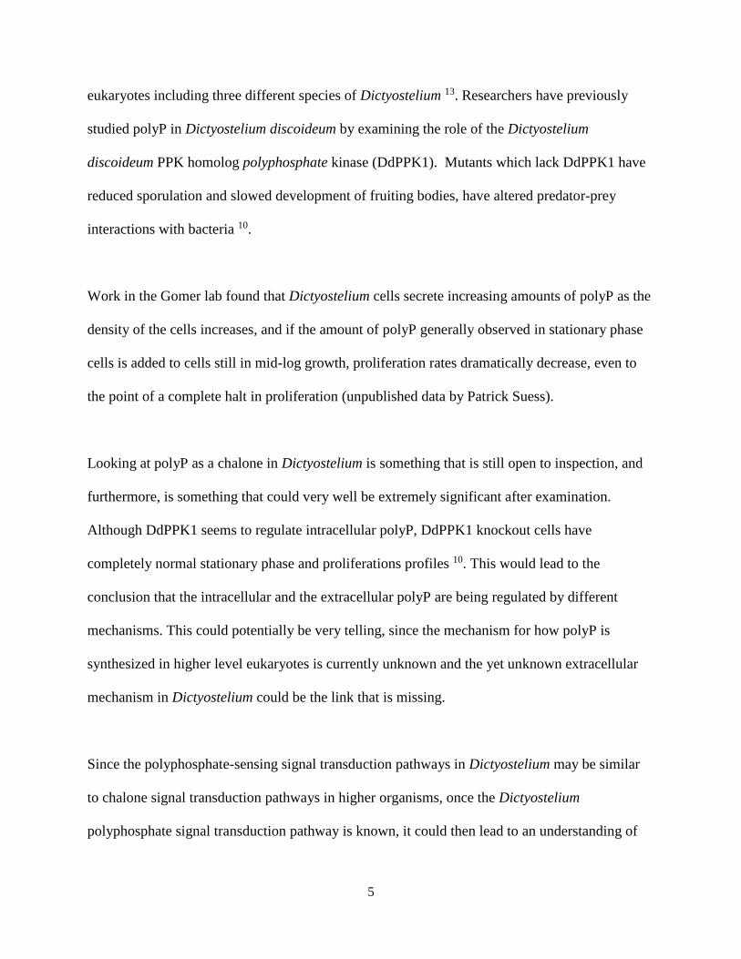
eukaryotes including three different species of Dictyostelium 13. Researchers have previously
studied polyP in Dictyostelium discoideum by examining the role of the Dictyostelium
discoideum PPK homolog polyphosphate kinase (DdPPK1). Mutants which lack DdPPK1 have
reduced sporulation and slowed development of fruiting bodies, have altered predator-prey
interactions with bacteria 10.
Work in the Gomer lab found that Dictyostelium cells secrete increasing amounts of polyP as the
density of the cells increases, and if the amount of polyP generally observed in stationary phase
cells is added to cells still in mid-log growth, proliferation rates dramatically decrease, even to
the point of a complete halt in proliferation (unpublished data by Patrick Suess).
Looking at polyP as a chalone in Dictyostelium is something that is still open to inspection, and
furthermore, is something that could very well be extremely significant after examination.
Although DdPPK1 seems to regulate intracellular polyP, DdPPK1 knockout cells have
completely normal stationary phase and proliferations profiles 10. This would lead to the
conclusion that the intracellular and the extracellular polyP are being regulated by different
mechanisms. This could potentially be very telling, since the mechanism for how polyP is
synthesized in higher level eukaryotes is currently unknown and the yet unknown extracellular
mechanism in Dictyostelium could be the link that is missing.
Since the polyphosphate-sensing signal transduction pathways in Dictyostelium may be similar
to chalone signal transduction pathways in higher organisms, once the Dictyostelium
polyphosphate signal transduction pathway is known, it could then lead to an understanding of

6
the ways that proliferation in higher eukaryotes, such as a patient with a tumor, could be
controlled.

7
SECTION II
METHODS
Plasmid
The plasmid used in the electroporation transformation was pBSR1, obtained from the
Dictyostelium stock center. pBSR1 has a blasticidin S resistance cassette derived from the bsr
gene of Bacillus cereus 14.
Digestion of plasmid
The plasmid pBSR1 was digested using 10X Cutsmart® Buffer and the restriction enzyme
BamHI-Hf (New England BioLabs, Ipswich, MA). This digestion was incubated in a 35°C
incubator for approximately 24 hours. The digested plasmid was purified using a Zymoclean®
Gel DNA Recovery kit (Zymo Research, Irvine, CA). A small sample of the purified pBSR1 was
then tested on a 1% analytical agarose gel for confirmation of proper digestion and purification.
Restriction-Enzyme-Mediated Insertional (REMI) mutagenesis
Dictyostelium discoideum AX-2 wild type cells 15 were grown in shaking culture to mid-log (2-4
x106 cells/mL) in HL-5 medium (Formedium, United Kingdom) supplemented with
streptomycin. For each REMI reaction, 6 x10⁶ cells were collected by centrifugation at 400 x g
for 3 minutes, and washed twice with at least half the original culture volume of H-50
transformation buffer. The recipe for H-50 is 20 mM Hepes, 50 mM KCl, 10 mM NaCl, 1 mM
Mg2SO4, 5 mM NaHCO3, 1 mM Na2HPO4, adjusted to pH 7.0. The cells were then resuspended
into 178 µL ice-cold H-50 buffer per reaction. Each 200 µL REMI reaction consisted of 178 µL

8
of resuspended cells, 20 µL of digested and purified pBSR1 plasmid, and 2 µL of the enzyme
DpnII (New England BioLabs). A negative control was obtained using H-50 buffer instead of the
plasmid and enzyme in these reactions. These cells were then placed in 0.2 mm gap cuvettes and
immediately were electroporated usiong a BioRad Gene Pulser set to 0.85 kV and 1 µF with the
time constants ranging from 0.08-0.2. The cuvettes were then immediately placed in ice for 5
minutes, after which 100 µL aliquots were added dropwise onto 3.5 inch x 0.625 inch plastic
petri dishes that contain 10 mL of HL-5 media supplemented with streptomycin. After
approximately 24 hours at room temperature, 5 µL of 10 mg/mL blasticidin was added to select
for transformed cells. Colonies visible with an inverted microscope appeared after 4-6 days at
room temperature.
Screening against polyphosphate
The transformant plates were then transferred over to shaking culture by first removing the old
media from the plates and then washing the cells off of the surface of the plate with 10 mL fresh
HL5 media and promptly transferred to a flask where 7 µL of 10 mg/mL blasticidin was added to
select for the cells that were successfully transformed with the pBSR1 plasmid, which would
confer a resistance to the drug. These new cultures were then grown to the mid-log range. The
cultures were then diluted to 1.0 x 10⁶ cells/mL and subjected to a treatment of 125 µM
polyphosphate in HL-5, and were left to grow at room temperature on a rotary shaker. Cell
densities were counted approximately every 24 hours. After 48 hours, the cells were collected by
centrifugation and resuspended to 0.5 x 10⁶ cells/mL in HL-5 media supplemented with
streptomycin, but with no polyphosphate, and left to grow for 48 hours, and cell densities were
counted every 24 hours. This process of 48 hours with polyP and 48 hours without polyP is

9
considered one cycle, and the REMIs were subjected to 5-6 cycles to determine how they reacted
to polyP as compared a similarly-treated AX-2 wild type culture.
Cloning out REMI mutants
After the completion of the screening, approximately 1 x 10⁶ cells of the mutants were collected
by centrifugation for 3 minutes at 1,500 rpm. These cells were washed in 3-5 mL of fresh HL5
media with streptomycin and resuspended into 1 mL of drug-free HL5 media. For each mutant
obtained, three plates with E. coli bacterial lawns were obtained and labeled A-C. Different
amounts of the resuspended mutant cells were spread onto each plate; the different amounts
being 100 µL, 300 µL, and 500 µL for plates A, B, and C respectively. It is helpful to add an
extra 200 µL of drug-free HL5 media to plate A to assist in the ease of spreading. After 2-3 days,
colonies appeared and a single colony was used to make three separate streaks (labeled with 1-3
and their plate letter, i.e. B-1, B-2, B-3) onto the edges of a new plate with a bacterial lawn. After
3-4 days these clone colonies were transferred to shaking culture, allowed to grow into mid-log
range, and frozen down until further study. The clones were subsequently tested with the same
protocol as described before of 48 hours with polyphosphate and 48 hours without
polyphosphate to insure that the cloned mutants show the same phenotypic response of
proliferation in the presence of polyphosphate as the original mutant.
Identifying the location of the REMI insert
The location of the REMI insert in the genome of cells that were resistant to polyphosphate was
found using a process called Inverse PCR. First, the genomic DNA was extracted from the
cloned cultures that did not show a proliferation change when in the presence of polyP as

10
previously described. 16 The genomic DNA was then digested with the endonuclease Alu I,
purified with a ZymocleanTM Gel DNA Recovery Kit, and ligated into circular DNA with T4
Ligase. 1 µL of the ligated DNA was used to set up a 15 µL PCR reaction for the mutants and
the AX-2 control sample. These samples were run on a 1% agarose gel to insure that the mutant
samples show bands and the control AX-2 samples do not. Once this was verified, three different
tubes of the same 15 µL PCR reaction were prepared with the same conditions (Forward Primer
– CGTCGATATGGTGCACTCTC; Reverse primer – TGTCGTTAGAACGCGGCTAC;
Thermocycler on the following settings: 94°C for 30 seconds, followed by 32 cycles of 94°C for
30 seconds, 56.3°C for 30 seconds, and 65°C for 90 seconds; this is followed by a 5 minutes of
68°C and the DNA was stored at 4°C until use). The PCR product DNA from the three different
tubes was purified with a ZymocleanTM Gel DNA Recovery Kit and sent off for sequencing
(Lonestar Labs, Houston TX).

11
SECTION III
RESULTS
REMI mutants show a non-response to inorganic polyphosphate
It has previously been found that inorganic polyphosphate acts as a chalone in Dictyostelium
discoideum, by inhibiting proliferation. This has led to interest in the components of the signal
transduction pathway that begins with inorganic polyphosphate as the signal and the stopping of
proliferation in the late G2 phase of the cell cycle as the result. To study this, the process known
as restriction enzyme-mediated insertional (REMI) mutagenesis, was used to create mutants that
could be screened according to their proliferation abilities. When treated with 125 µM
polyphosphate, a number of mutants created by REMI mutagenesis show a continuation of
normal proliferation as opposed to the AX-2 control cells which show almost a complete halt of
proliferation (Figure 1A). This would indicate that the pBSR1 plasmid that was inserted into the
genome to create these mutations was inserted in such a place as to interfere with the signal
transduction pathway between the chalone and the halting of proliferation. It can likewise be
seen that when in a control environment (HL-5 media with no polyphosphate), the REMI
mutants show a slightly higher rate of proliferation than the control AX-2 cells (Figure 1B). This
finding further backs the idea that the REMI mutants have a disrupted signal transduction
pathway, because it shows that they do not respond to even the small amounts of polyphosphate
that would be present in a cell culture even without the experimental conditions adding excess
amounts.

12
Figure 1: REMI mutant 13 shows the phenotype of continued proliferation in the presence of inorganic
polyphosphate. (A) REMI 13 mutants and AX-2 control cells were subjected to 48 hours in HL5 media
supplemented with 125 µM polyphosphate. The control cells show halted proliferation while the REMI mutants
continue to rapidly proliferate. (B) REMI 13 mutants and AX-2 control cells over a 48 hour period in HL5 media.
The mutant cells and the control cells proliferate at approximately the same rate, with the REMI mutants
proliferating slightly faster.
Along with the mutant, REMI 13, seven other REMI mutants showed a proliferation phenotype
similar to that of REMI 13 (Figure 2). Those seven others are called REMI 5, 17A, 17B, 18A,
19A, 20A, and 20B. Because all of these have similar results, we can conclude that they also
have disrupted signal transduction pathways due to the insertion of the pBSR1 plasmid by the
same reasoning as above. These eight mutants can all eventually be tested to discover the
location of their plasmid insertions and thus, their mutations, but for the purposes of this

13
experiment only the mutant with the most significant result (REMI 13) has been used for further
study.
Figure 2: More REMI mutants show the phenotype of continued proliferation even in the presence of
polyphosphate. (A) REMI mutants 17A, 17B, 18A, 19A, 20A, and 20B show greater proliferation than the control
AX-2 cells over repeated 48 hour periods when grown in HL-5 media supplemented with 125 µM polyP. (B) REMI
mutants show a slightly higher rate of proliferation over repeated 48 hour periods than the control AX-2 cells in
normal conditions (HL-5 media with no polyP).
Plasmid insert is located by inverse PCR
The plasmid that was inserted into the genome to cause the mutations in the REMI cultures
theoretically could have been inserted into numerous places in the genome. To find the location
of this insert, we did inverse PCR. After digesting the mutant genomes and ligating the linear
pieces into circles, primers were designed from the known plasmid sequence to amplify the

14
entire piece of circular DNA that included both the REMI vector DNA and the genomic DNA
from Dictyostelium that had been interrupted. This was confirmed on a 1% agarose gel (Figure
3), where it is evident that the AX-2 wildtype DNA has no plasmid insertion, whereas the mutant
genomes do.
Figure 3: Gel electrophoresis image of inverse PCR results after digestion and ligation of control (AX-2) and
mutant (REMI 13-B1) genomes. The AX-2 lane has no bands which proves the lack of plasmid insert, whereas the
three lanes containing REMI 13-B1 all have bands around 1,700 bp.
The bands in the REMI 13-B1 lanes appear at approximately 1,700 base pairs (Figure 3). Since
the sequence of the pBSR1 plasmid is known, we can anticipate where the DpnII cut site is in
relation to the primers designed and thus know that out of the 1,700 base pairs in this small,
circular piece of DNA, 655 of them are from the plasmid. This leaves over 1,000 base pairs of
the DNA belonging to the original Dictyostelium genome and showing us where the insert was
located.

15
Unconfirmed results on interrupted gene
After sending off DNA for sequencing, results indicated that the most likely gene that was
interrupted by the plasmid was a gene called Mediator 20 (Med 20). Mediator 20 codes for
mediator complex subunit protein. Mediators, first discovered in 1990 by Roger Kornberg 17, are
known to be used in support of transcriptional activation, and is known to be heavily associated
with RNA Polymerase II.18 However, these results have not yet been confirmed, as proper
confirmation requires complete knockout of the Med 20 gene by homologous recombination, and
this has yet to be completed.

16
SECTION IV
CONCLUSION
Evidence obtained leads to the conclusion that the mediator protein, Med 20, is a component in
the chalone signal transduction pathway. This conclusion, however, is still uncertain as the
intended phenotype of a non-response to the chalone inorganic polyphosphate, was only obtained
through the interruption of the gene that codes for Med 20, not the complete removal. Further
testing is necessary to prove its role in this pathway.

17
REFERENCES
1. Guba, M. et al. A primary tumor promotes dormancy of solitary tumor cells before
inhibiting angiogenesis. Cancer Res 61, 5575-9 (2001).
2. Gomer, R.H., Jang, W. & Brazill, D. Cell density sensing and size determination. Dev
Growth Differ 53, 482-94 (2011).
3. Soll, D.R., Yarger, J. & Mirick, M. Stationary phase and the cell cycle of Dictyostelium
discoideum in liquid nutrient medium. J Cell Sci 20, 513-23 (1976).
4. Yarger, J., Stults, K. & Soll, D.R. Observations on the growth of Dictyostelium
discoideum in axenic medium: evidence for an extracellular growth inhibitor synthesized by
stationary phase cells. J Cell Sci 14, 681-90 (1974).
5. Choe, J.M., Bakthavatsalam, D., Phillips, J.E. & Gomer, R.H. Dictyostelium cells bind a
secreted autocrine factor that represses cell proliferation. BMC Biochem 10, 4 (2009).
6. Thomas, M. et al. Myostatin, a negative regulator of muscle growth, functions by
inhibiting myoblast proliferation. J Biol Chem 275, 40235-43 (2000).
7. Bakthavatsalam, D., Choe, J.M., Hanson, N.E. & Gomer, R.H. A Dictyostelium chalone
uses G proteins to regulate proliferation. BMC Biol 7, 44 (2009).
8. Brock, D.A. & Gomer, R.H. A secreted factor represses cell proliferation in
Dictyostelium. Development 132, 4553-62 (2005).
9. Rao, N.N., Gomez-Garcia, M.R. & Kornberg, A. Inorganic polyphosphate: essential for
growth and survival. Annu Rev Biochem 78, 605-47 (2009).
10. Zhang, H., Gomez-Garcia, M.R., Brown, M.R. & Kornberg, A. Inorganic polyphosphate
in Dictyostelium discoideum: influence on development, sporulation, and predation. Proc
Natl Acad Sci U S A 102, 2731-5 (2005).
11. Rashid, M.H. et al. Polyphosphate kinase is essential for biofilm development, quorum
sensing, and virulence of Pseudomonas aeruginosa. Proc Natl Acad Sci U S A 97, 9636-41
(2000).
12. Rao, N.N. & Kornberg, A. Inorganic polyphosphate supports resistance and survival of
stationary-phase Escherichia coli. J Bacteriol 178, 1394-400 (1996).
13. Whitehead, M.P., Hooley, P. & MR, W.B. Horizontal transfer of bacterial polyphosphate
kinases to eukaryotes: implications for the ice age and land colonisation. BMC Res Notes 6,
221 (2013).

18
14. Sutoh, K. A transformation vector for dictyostelium discoideum with a new selectable
marker bsr. Plasmid 30, 150-4 (1993).
15. Watts, D.J. & Ashworth, J.M. Growth of myxameobae of the cellular slime mould
Dictyostelium discoideum in axenic culture. Biochem J 119, 171-4 (1970).
16. Pilcher, K.E., Fey, P., Gaudet, P., Kowal, A.S. & Chisholm, R.L. A reliable general
purpose method for extracting genomic DNA from Dictyostelium cells. Nat Protoc 2, 1325-8
(2007).
17. Kelleher, R.J., 3rd, Flanagan, P.M. & Kornberg, R.D. A novel mediator between
activator proteins and the RNA polymerase II transcription apparatus. Cell 61, 1209-15
(1990).
18. Flanagan, P.M., Kelleher, R.J., 3rd, Sayre, M.H., Tschochner, H. & Kornberg, R.D. A
mediator required for activation of RNA polymerase II transcription in vitro. Nature 350,
436-8 (1991).

19
ACKNOWLEDGEMENTS
I would like to give a special thanks to Dr. Richard Gomer and my fellow laboratory members,
most especially Patrick Suess and Dr. Gomer himself as they both played a large role in directing
this project. I would also like to thank the Undergraduate Research Scholars Program at Texas
A&M University for the amazing opportunity this has provided me.