Embed Size (px)
Citation preview
Identification of a New Lipoprotein Export Signal in Gram-NegativeBacteria
Frédéric Lauber, Guy Richard Cornelis, Francesco Renzi
Département de Biologie, Unité de Recherche en Biologie des Microorganismes (URBM), Université de Namur, Namur, Belgium
ABSTRACT Bacteria of the phylum Bacteroidetes, including commensal organisms and opportunistic pathogens, harbor abun-dant surface-exposed multiprotein membrane complexes (Sus-like systems) involved in carbohydrate acquisition. These com-plexes have been mostly linked to commensalism, and in some instances, they have also been shown to play a role in pathogene-sis. Sus-like systems are mainly composed of lipoproteins anchored to the outer membrane and facing the external milieu. Thislipoprotein localization is uncommon in most studied Gram-negative bacteria, while it is widespread in Bacteroidetes. Little isknown about how these complexes assemble and particularly about how lipoproteins reach the bacterial surface. Here, by bioin-formatic analyses, we identify a lipoprotein export signal (LES) at the N termini of surface-exposed lipoproteins of the humanpathogen Capnocytophaga canimorsus corresponding to K-(D/E)2 or Q-A-(D/E)2. We show that, when introduced in sialidaseSiaC, an intracellular lipoprotein, this signal is sufficient to target the protein to the cell surface. Mutational analysis of the LESin this reporter system showed that the amino acid composition, position of the signal sequence, and global charge are criticalfor lipoprotein surface transport. These findings were further confirmed by the analysis of the LES of mucinase MucG, a natu-rally surface-exposed C. canimorsus lipoprotein. Furthermore, we identify a LES in Bacteroides fragilis and Flavobacteriumjohnsoniae surface lipoproteins that allow C. canimorsus surface protein exposure, thus suggesting that Bacteroidetes share anew bacterial lipoprotein export pathway that flips lipoproteins across the outer membrane.
IMPORTANCE Bacteria of the phylum Bacteroidetes are important human commensals and pathogens. Understanding their bi-ology is therefore a key question for human health. A main feature of these bacteria is the presence of abundant lipoproteins attheir surface that play a role in nutrient acquisition. To date, the underlying mechanism of lipoprotein transport is unknown.We show for the first time that Bacteroidetes surface lipoproteins share an N-terminal signal that drives surface localization. Thelocalization and overall negative charge of the lipoprotein export signal (LES) are crucial for its role. Overall, our findings pro-vide the first evidence that Bacteroidetes are endowed with a new bacterial lipoprotein export pathway that flips lipoproteinsacross the outer membrane.
Received 8 July 2016 Accepted 27 September 2016 Published 25 October 2016
Citation Lauber F, Cornelis GR, Renzi F. 2016. Identification of a new lipoprotein export signal in Gram-negative bacteria. mBio 7(5):e01232-16. doi:10.1128/mBio.01232-16.
Invited Editor Thomas J. Silhavy, Princeton University Editor Scott J. Hultgren, Washington University School of Medicine
Copyright © 2016 Lauber et al. This is an open-access article distributed under the terms of the Creative Commons Attribution 4.0 International license.
Address correspondence to Francesco Renzi, [email protected].
Among Gram-negative bacteria, the phylum Bacteroidetes iscomposed of a large diversity of organisms widely distributed
in the environment. Some are saprophytes such as Flavobacteria,found in soil (1) and aquatic environments (2), while others arecommensal organisms of animals. Among the commensal organ-isms, Bacteroides spp. are common members of the intestinal florawhere they play a major role in gut homeostasis (3–7), while Cap-nocytophaga and Porphyromonas spp. are part of the oral flora (8,9). Bacteroides fragilis, a commensal of the human intestine, andCapnocytophaga canimorsus, a common member of the dog oralflora can cause severe systemic human infections (10–15), whilePorphyromonas gingivalis causes severe periodontal diseases (8).The wide distribution of these organisms reflects their high adapt-ability, partially due to their vast array of glycosylhydrolases allow-ing them to degrade nearly all types of carbohydrates they canencounter (7, 16–19). Interestingly, these enzymes are oftensurface-exposed lipoproteins and are part of multiprotein outermembrane (OM) complexes devoted to nutrient acquisition.These complexes, facing the outside environment (20, 21), are
encoded in genetic regions named polysaccharide utilization loci(PUL) (19) that represent a hallmark of this phylum.
To date, most studies have focused on identifying and charac-terizing the functions of these Bacteroidetes surface complexes (5,7, 16–18, 22, 23), but little is known about how they assemble (24)and particularly about how lipoproteins reach the bacterial sur-face. In Gram-negative Proteobacteria, lipoprotein synthesis andtransport have been well studied in model organisms such as Esch-erichia coli (25). Lipoproteins are first synthesized as a precursor inthe cytoplasm before their translocation to the periplasm via theSec (26, 27) or Tat (28–30) machinery. This recognition is medi-ated by the N-terminally located signal peptide II (31), which con-tains a conserved cysteine residue critical for the subsequent stepsof maturation (32, 33). After crossing the inner membrane (IM),lipoprotein precursors remain anchored to the periplasmic side ofthe IM where they are then processed by three enzymes, renderinga final triacylated lipoprotein (34–37). Lipoproteins destined to beinserted into the OM are transported through the aqueous envi-ronment of the periplasm via the dedicated Lol (localization of
RESEARCH ARTICLE
crossmark
September/October 2016 Volume 7 Issue 5 e01232-16 ® mbio.asm.org 1
m
bio.asm.org
on April 6, 2018 - P
ublished by m
bio.asm.org
Dow
nloaded from
m
bio.asm.org
on April 6, 2018 - P
ublished by m
bio.asm.org
Dow
nloaded from
m
bio.asm.org
on April 6, 2018 - P
ublished by m
bio.asm.org
Dow
nloaded from
m
bio.asm.org
on April 6, 2018 - P
ublished by m
bio.asm.org
Dow
nloaded from

lipoproteins) transport machinery, composed of five proteins,LolA, -B, -C, -D, and -E (25, 38). In Proteobacteria, most OMlipoproteins are inserted in the inner leaflet of the OM and thusface the periplasm. The surface localization of OM lipoproteins inBacteroidetes thus implies the existence of a yet unknown dedi-cated recognition and transport mechanism.
The present study deals with the reference strain C. canimorsus5 (39), which encodes 13 PUL. Three of them were recently shownto play critical roles in the biology and pathogenesis of this bacte-rium (40–42). We address the question of how lipoproteins aretargeted to the bacterial surface. We identify a signal sequence(lipoprotein export signal [LES]) present at the N termini ofsurface-exposed lipoproteins, and we show that this signal is suf-ficient to target an intracellular lipoprotein to the cell surface. Weextend our findings to other Bacteroidetes species, namely, Flavo-bacterium johnsoniae and Bacteroides fragilis, identifying their spe-cific LESs, thus showing that they share a new bacterial lipoproteinexport pathway that flips lipoproteins across the outer membrane.
RESULTSIn silico identification of a putative lipoprotein export signal. Inorder to see whether a specific amino acid motif would be respon-sible for the targeting of lipoproteins to the bacterial surface, weexamined in detail the sequences of the lipoproteins detected atthe surface of Capnocytophaga canimorsus strain 5 (17). Whenaligning the mature lipoproteins, a lysine (K), followed by eitheran aspartate (D) or a glutamate (E) residue, appeared to be con-served in close proximity to the N-terminal cysteine at position�1 (see Fig. S1 in the supplemental material). This was refined bya second alignment considering only the 15 N-terminal residues ofthe mature lipoprotein and excluding the invariant first cysteine(Fig. 1A). The resulting consensus motif corresponded to Q-K-D-D-E, located between positions �2 and �6 (Fig. 1B) showingconservation of 16, 72, 48, 44, and 23%, respectively (Fig. 1C). Inorder to determine whether this motif is specific to the surface-exposed lipoproteins, the same analysis was performed on OMlipoproteins facing the periplasm (17). No highly conserved resi-
FIG 1 Alignment of C. canimorsus surface-exposed lipoproteins reveals the presence of an N-terminal conserved motif. (A) MAFFT alignment of the first 15N-terminal amino acids of mature surface-exposed lipoproteins. The first invariant cysteine residue of each sequence was removed before the alignment wasperformed. Highly conserved residues are indicated according to the Clustal color code (R and K in red; D and E in magenta; P in yellow; G in orange; Q, N, S,and T in green, C in pink; A, I, L, M, F, W, and V in blue; H and Y in cyan) (63). MucG (Ccan_17430) is indicated by an asterisk. The derived consensus sequenceis shown below the sequence alignment. (B) Generated WebLogo of the consensus sequence determined in panel A. Positions relative to the �1 cysteine areindicated below the WebLogo. Charged residues are indicated in color. The color code is the same as that used in panel A. (C) Amino acid frequency for eachposition of the consensus sequence, expressed as a percentage. The three most represented amino acids for each position are shown.
Lauber et al.
2 ® mbio.asm.org September/October 2016 Volume 7 Issue 5 e01232-16
m
bio.asm.org
on April 6, 2018 - P
ublished by m
bio.asm.org
Dow
nloaded from

dues were identified in this set of proteins (see Fig. S2 in the sup-plemental material), suggesting that the QKDDE consensus motifcould indeed be a bona fide lipoprotein export signal (LES).
The LES leads to surface localization of the periplasmic lipo-protein sialidase. To verify this hypothesis, we introduced theQKDDE motif in the sequence of the C. canimorsus sialidase(SiaC), an OM lipoprotein that faces the periplasm (42, 43). SiaCharboring the LES (SiaC�2QKDDE�6) (Fig. 2A and B) was detectedat the bacterial surface by immunolabeling, followed by flow cy-tometry and microscopy (Fig. 2D and E). In contrast, wild-type(wt) SiaC and the soluble SiaCC17G variant were not detected. Thisindicated that the addition of the consensus motif to an OM
periplasmic lipoprotein is sufficient to drive its transport to thebacterial surface and hence that this consensus motif repre-sents a LES.
Determination of the minimal consensus motif allowing sur-face localization of sialidase. We next determined the minimalsequence required to constitute a functional LES. We first replacedthe least conserved amino acids of the LES, namely, the �2 Q and�6 E, by alanine residues, generating constructs SiaC�2AKDDE�6
and SiaC�2AKDDA�6 (Fig. 2A). After monitoring protein expres-sion (Fig. 2B), immunolabeling showed that both constructs lo-calized to the bacterial surface (Fig. 2D and E), although to a lowerextent than SiaC�2QKDDE�6 did, thus indicating that the KDD
FIG 2 The LES allows SiaC surface exposure. (A) Wild-type (wt) SiaC and consensus sequence mutant constructs. Amino acids derived from the consensussequence (green boldface) and point mutations (gray boldface) are indicated. The SiaC constructs are referred to by the boldface numbers shown in panel A inpanels B to E. (B) Detection of SiaC by Western blot analysis of total cell extracts of strains expressing the SiaC constructs shown in panel A. Expression of MucGwas monitored as a loading control. (C) Detection of SiaC by Western blot analysis of total lysates (TL) and outer membrane (OM) fractions of bacteriaexpressing different SiaC constructs. Expression of MucG was monitored as a loading control. (D) Quantification of SiaC surface exposure by flow cytometry oflive cells labeled with anti-SiaC serum. The fluorescence intensity of stained cells only is shown (NR, not relevant). The averages from at least three independentexperiments are shown. Error bars represent 1 standard deviation from the mean. Values that are significantly different (P � 0.001) from the value for referenceconstruct 3 are indicated (***). Values that are not significantly different (n.s) from the value for reference construct 3 are indicated. The percentage of stainedcells and standard deviation (SD) are indicated below the bar graph. Values below the detection limit (�2.5%) are shown on a gray background. Values for strainswith a statistically significant lower stained population are shown in red (P � 0.001 compared to reference construct 3). (E) Immunofluorescence microscopyimages of bacteria labeled with anti-SiaC serum. Bar, 5 �m.
Lipoprotein Export in Bacteroidetes
September/October 2016 Volume 7 Issue 5 e01232-16 ® mbio.asm.org 3
m
bio.asm.org
on April 6, 2018 - P
ublished by m
bio.asm.org
Dow
nloaded from

motif is sufficient to target lipoproteins to the cell surface. We thentested whether glutamate was able to functionally replace aspar-tate (SiaC�2AKEEA�6) (Fig. 2A), since both residues were enrichedin the alignment (Fig. 1C). Replacing the two aspartate residueswith two glutamate residues did not prevent surface localizationbut led to a clear reduction of fluorescence (Fig. 2D and E), in linewith the lower conservation of glutamate at positions �4 and �5(Fig. 1C), showing that in C. canimorsus surface lipoproteins, as-partate is preferred over glutamate.
We then generated two SiaC constructs harboring only eitherKD or KE (SiaC�2AKDAA�6 and SiaC�2AKEAA�6) (Fig. 2A), butthese two pairs of residues alone turned out to be very weak LESssince only 29.8% � 4.7% (SiaC�2AKDAA�6) and 16.3% � 2.5%(SiaC�2AKEAA�6) of the cells displayed the proteins at their surface(Fig. 2D). In addition, the fluorescence intensity was weak, 28.2and 29.4%, respectively, of the intensity observed for theSiaC�2AKDDA�6 reference (Fig. 2D). In order to verify that theseconstructs were not impaired in their transport to the OM, wemonitored their presence in isolated outer membrane fractions.Both mutant proteins were found to be anchored to the OM al-though at lower levels than the wt protein, in particular for theconstruct SiaC�2AKDAA�6, suggesting that these mutations couldalso impact to a minor extent OM localization of SiaC (Fig. 2C).Overall, these data supported our hypothesis that K-(D/E)2 rep-resents the minimal LES. These findings also suggested that afunctional LES might require an overall negative charge, sup-ported by the fact that KDD is allowing efficient transport of SiaCto the surface, while only KD is not (Fig. 2D).
We then investigated the importance of the highly conservedlysine residue at position �3 of the LES (Fig. 2A). Unexpectedly,substitution of K alone (SiaC�2QADDE�6) had no impact on thedisplay of SiaC at the bacterial surface (Fig. 2D and E). However,removal of both K and Q (SiaC�2AADDA�6) led to a decrease offluorescence intensity of more than 60% compared to that ofSiaC�2AKDDA�6. Since the glutamine residue itself was not foundto be critical (SiaC�2AKDDA�6 [Fig. 2D]), we conclude that eitherthe �2 Q or the �3 K is required to form a functional LES. Takentogether, these data indicate that the minimal export motif allow-ing surface localization of SiaC is composed of only two negativelycharged amino acids preceded by a positively charged or polarresidue. On the basis of the consensus, we thus defined the mini-mal C. canimorsus LES as being K-(D/E)2 or Q-A-(D/E)2.
Positional effect of the minimal LES on sialidase surface lo-calization. We next addressed the question of the importance ofthe position of the LES. The initial alignment showed that K isconserved mainly at position �3 (72%), to a lower extent at po-sition �2 (13%), and is completely absent from position �4(Fig. 1C). In contrast, D and E were conserved at positions �4,�5, and �6 (48, 44, and 11% for D and 20, 13, and 23% for E,respectively) and completely absent from position �3 (Fig. 1C).This suggested that not only the composition of the export signalcould be crucial but also its position relative to the �1 cysteine.Therefore, we generated constructs in which the KDD motif wasseparated from the �1 cysteine by zero, two, three, or four alanineresidues (Fig. 3A) and compared their surface localization to theconstruct in which the KDD motif is separated from the �1 cys-teine by only one alanine residue (SiaC�2AKDDA�6). Although thefour proteins were expressed (Fig. 3B), none of them were ex-ported as efficiently as the one where only one alanine separatedthe KDD motif from the �1 cysteine (SiaC�2AKDDA�6) (Fig. 3C
and D). All proteins were anchored to the OM, thus again indicat-ing that only the last step of transport to the surface was affected bythese mutations (Fig. 3E). The position of the K-(D/E)2 signalrelative to the �1 cysteine is thus critical for the C. canimorsusLES, and the optimal sequence is C-X-K-(D/E)2-X.
Characterization of the LES of the surface-exposed lipopro-tein MucG. Looking at the LESs of different C. canimorsus surfacelipoproteins (Fig. 1), it appeared that some were quite divergentfrom the consensus. Among these is the LES of mucinase MucG(41) (Ccan_17430), KKEVEEE (Fig 1A; see Fig. S3A in the supple-mental material). We first confirmed that MucG is indeed asurface-exposed lipoprotein (see Fig. S3 in the supplemental ma-terial), and then we tested whether this poorly conserved LESwould drive the export of sialidase to the surface of C. canimorsus.We introduced the MucG LES, KKEVEEE, or part of this se-quence, into SiaC, giving SiaC�2KKEVE�6, SiaC�2KKEVEE�7, andSiaC�2KKEVEEE�8 (see Fig. S4A and S4B in the supplemental ma-terial) and monitored their surface localization (see Fig. S4C andS4D in the supplemental material). SiaC�2KKEVE�6 was onlypoorly transported to the cell surface, while SiaC�2KKEVEE�7 andSiaC�2KKEVEEE�8 showed clear surface localization. Although theoverall protein amount of SiaC�2KKEVE�6 was reduced, the pro-tein appeared to be anchored to the OM (see Fig. S4E in the sup-plemental material). As the only difference between these con-structs was the number of negatively charged amino acids in theLES, this strongly supported our initial findings that the LES re-quires an overall negative charge to drive transport of lipoproteinsto the bacterial surface (Fig. 2).
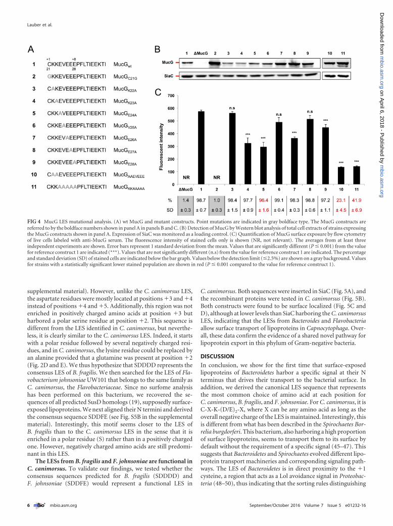
We next wanted to study the MucG LES in its native back-ground. To this aim, we systematically replaced residues 22 to 28of the MucG LES by alanines (Fig. 4A). After verifying that allmutant proteins were expressed (Fig. 4B), we monitored the sur-face exposure of the MucG variants by flow cytometry (Fig. 4C).Replacing K22, V25, and E27 with alanine did not significantlyalter surface exposure of MucG, while mutation of K23, E24, E26,or E28 resulted in a 25 to 50% decrease of surface exposure. Noneof these single mutations completely abolished surface localiza-tion, suggesting that the MucG motif is redundant, presumablydue to the presence of two lysines and four glutamates. The mu-tation of one of those residues could therefore be compensated forby the presence of another one in close proximity, and indeed, allprotein variants we generated harbor an overall negativelycharged functional LES.
Because of this, we generated two additional constructs by mu-tating simultaneously either all negatively or all positively chargedresidues in the MucG LES (Fig. 4A). After having confirmed theircorrect expression (Fig. 4B), we analyzed their surface localizationby flow cytometry (Fig. 4C). As expected, replacing the two lysineresidues (MucGAAEVEEE) led to MucG surface exposure in only23.1% � 4.5% of the cells (Fig. 4C). Furthermore, the fluores-cence intensity in this subset of cells was markedly decreased com-pared to the wt strain (23.8%), indicating that the efficiency oftransport was also strongly affected in this subpopulation. This isin good agreement with our previous findings showing the impor-tance of the K/Q residues for surface export (Fig. 2).
Similarly, MucGKKAAAAA was surface localized in only 41.9% �6.9% of the cells (Fig. 4C), and the fluorescence intensity in thissubpopulation was lower than the fluorescence intensity of the wtstrain (24.5%). This is in agreement with our findings in SiaC that
Lauber et al.
4 ® mbio.asm.org September/October 2016 Volume 7 Issue 5 e01232-16
m
bio.asm.org
on April 6, 2018 - P
ublished by m
bio.asm.org
Dow
nloaded from

an overall negatively charged LES is critical for efficient surfacelocalization.
By combining the data obtained from single and multiple ala-nine substitutions, the minimal LES for optimal MucG surfaceexposure appears to be X-K-(D/E)3 downstream from the �1 cys-teine, hence resembling the LES deduced from previous experi-ments [X-K-(D/E)2-X] (Fig. 2 and 3; see Fig. S4 in the supplemen-tal material).
The LES is conserved in the Bacteroidetes phylum. To deter-mine whether the LES identified in C. canimorsus would be con-served in other Bacteroidetes, we took advantage of the recentlypublished B. fragilis NCTC 9343 surfome study (44) and per-formed an in silico analysis on the N termini of the identifiedsurface lipoproteins (see Fig. S5A in the supplemental material).We found an enrichment in negatively charged amino acids inclose proximity to the �1 cysteine (SDDDD) (see Fig. S5A in the
FIG 3 The position of the minimal LES is crucial for its function. (A) wt SiaC and consensus sequence mutant constructs. Amino acids derived from theconsensus sequence (green boldface) and point mutations (gray boldface) are indicated. The SiaC constructs are referred to by the boldface numbers shown inpanel A in panels B to E. (B) Detection of SiaC by Western blot analysis of total cell extracts of strains expressing the SiaC constructs shown in panel A. MucGexpression was monitored as a loading control. (C) Quantification of SiaC surface exposure by flow cytometry of live cells labeled with anti-SiaC serum. Thefluorescence intensity of stained cells only is shown (NR, not relevant). The averages from at least three independent experiments are shown. Error bars represent1 standard deviation from the mean. Values that are significantly different (P � 0.001) from the value for reference construct 3 are indicated (***). The percentageand standard deviation (SD) of stained cells are indicated below the bar graph. Values below the detection limit (�2.5%) are shown on gray background. Valuesfor strains with a statistically significant lower stained population are shown in red (P � 0.001 compared to the value for the reference construct 3). (D)Immunofluorescence microscopy images of bacteria stained with anti-SiaC serum. Bar, 5 �m. (E) Western blot analysis of total lysates (TL) and outer membrane(OM) fractions of bacteria expressing different SiaC constructs. MucG expression was monitored as a loading control.
Lipoprotein Export in Bacteroidetes
September/October 2016 Volume 7 Issue 5 e01232-16 ® mbio.asm.org 5
m
bio.asm.org
on April 6, 2018 - P
ublished by m
bio.asm.org
Dow
nloaded from
supplemental material). However, unlike the C. canimorsus LES,the aspartate residues were mostly located at positions �3 and �4instead of positions �4 and �5. Additionally, this region was notenriched in positively charged amino acids at position �3 butharbored a polar serine residue at position �2. This sequence isdifferent from the LES identified in C. canimorsus, but neverthe-less, it is clearly similar to the C. canimorsus LES. Indeed, it startswith a polar residue followed by several negatively charged resi-dues, and in C. canimorsus, the lysine residue could be replaced byan alanine provided that a glutamine was present at position �2(Fig. 2D and E). We thus hypothesize that SDDDD represents theconsensus LES of B. fragilis. We then searched for the LES of Fla-vobacterium johnsoniae UW101 that belongs to the same family asC. canimorsus, the Flavobacteriaceae. Since no surfome analysishas been performed on this bacterium, we recovered the se-quences of all predicted SusD homologs (19), supposedly surface-exposed lipoproteins. We next aligned their N termini and derivedthe consensus sequence SDDFE (see Fig. S5B in the supplementalmaterial). Interestingly, this motif seems closer to the LES ofB. fragilis than to the C. canimorsus LES in the sense that it isenriched in a polar residue (S) rather than in a positively chargedone. However, negatively charged amino acids are still predomi-nant in this LES.
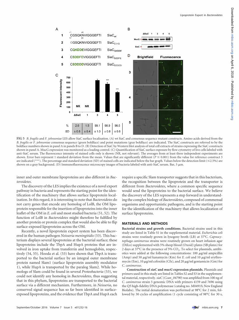
The LESs from B. fragilis and F. johnsoniae are functional inC. canimorsus. To validate our findings, we tested whether theconsensus sequences predicted for B. fragilis (SDDDD) andF. johnsoniae (SDDFE) would represent a functional LES in
C. canimorsus. Both sequences were inserted in SiaC (Fig. 5A), andthe recombinant proteins were tested in C. canimorsus (Fig. 5B).Both constructs were found to be surface localized (Fig. 5C andD), although at lower levels than SiaC harboring the C. canimorsusLES, indicating that the LESs from Bacteroides and Flavobacteriaallow surface transport of lipoproteins in Capnocytophaga. Over-all, these data confirm the evidence of a shared novel pathway forlipoprotein export in this phylum of Gram-negative bacteria.
DISCUSSION
In conclusion, we show for the first time that surface-exposedlipoproteins of Bacteroidetes harbor a specific signal at their Nterminus that drives their transport to the bacterial surface. Inaddition, we derived the canonical LES sequence that representsthe most common choice of amino acid at each position forC. canimorsus, B. fragilis, and F. johnsoniae. For C. canimorsus, it isC-X-K-(D/E)2-X, where X can be any amino acid as long as theoverall negative charge of the LES is maintained. Interestingly, thisis different from what has been described in the Spirochaetes Bor-relia burgdorferi. This bacterium, also harboring a high proportionof surface lipoproteins, seems to transport them to its surface bydefault without the requirement of a specific signal (45–47). Thissuggests that Bacteroidetes and Spirochaetes evolved different lipo-protein transport machineries and corresponding signaling path-ways. The LES of Bacteroidetes is in direct proximity to the �1cysteine, a region that acts as a Lol avoidance signal in Proteobac-teria (48–50), thus indicating that the sorting rules distinguishing
FIG 4 MucG LES mutational analysis. (A) wt MucG and mutant constructs. Point mutations are indicated in gray boldface type. The MucG constructs arereferred to by the boldface numbers shown in panel A in panels B and C. (B) Detection of MucG by Western blot analysis of total cell extracts of strains expressingthe MucG constructs shown in panel A. Expression of SiaC was monitored as a loading control. (C) Quantification of MucG surface exposure by flow cytometryof live cells labeled with anti-MucG serum. The fluorescence intensity of stained cells only is shown (NR, not relevant). The averages from at least threeindependent experiments are shown. Error bars represent 1 standard deviation from the mean. Values that are significantly different (P � 0.001) from the valuefor reference construct 1 are indicated (***). Values that are not significantly different (n.s) from the value for reference construct 1 are indicated. The percentageand standard deviation (SD) of stained cells are indicated below the bar graph. Values below the detection limit (�2.5%) are shown on a gray background. Valuesfor strains with a statistically significant lower stained population are shown in red (P � 0.001 compared to the value for reference construct 1).
Lauber et al.
6 ® mbio.asm.org September/October 2016 Volume 7 Issue 5 e01232-16
m
bio.asm.org
on April 6, 2018 - P
ublished by m
bio.asm.org
Dow
nloaded from
inner and outer membrane lipoproteins are also different in Bac-teroidetes.
The discovery of the LES implies the existence of a novel exportpathway in bacteria and represents the starting point for the iden-tification of the machinery that allows surface lipoprotein local-ization. In this regard, it is interesting to note that Bacteroidetes donot carry genes that encode any homolog of LolB, the OM lipo-protein responsible for the insertion of lipoproteins into the innerleaflet of the OM in E. coli and most studied bacteria (51, 52). Thefunction of LolB in Bacteroidetes might therefore be fulfilled byanother protein or protein complex that would also be able to flipsurface-exposed lipoproteins across the OM.
Recently, a novel lipoprotein export system has been discov-ered in the human pathogen Neisseria meningitidis (53). This bac-terium displays several lipoproteins at the bacterial surface; theselipoproteins include the TbpA and HupA proteins that are in-volved in iron uptake from transferrin and hemoglobin, respec-tively (54, 55). Hooda et al. (53) have shown that TbpA is trans-ported to the bacterial surface by an integral outer membraneprotein named Slam1 (surface lipoprotein assembly modulator1), while HupA is transported by the paralog Slam2. While ho-mologs of Slam could be found in several Proteobacteria (53), wecould not identify any homolog in Bacteroidetes, thus suggestingthat in this phylum, lipoproteins are transported to the bacterialsurface via a different mechanism. Furthermore, in Neisseria, noconserved signal sequence has so far been identified in surface-exposed lipoproteins, and the evidence that TbpA and HupA each
require a specific Slam transporter suggests that in this bacterium,the recognition between the lipoprotein and the transporter isdifferent from Bacteroidetes, where a common specific sequencewould send the lipoproteins to the bacterial surface. We believethe discovery of the LES represents a step forward in understand-ing the complex biology of Bacteroidetes, composed of commensalorganisms and opportunistic pathogens, and is the starting pointfor the identification of the machinery that allows localization ofsurface lipoproteins.
MATERIALS AND METHODSBacterial strains and growth conditions. Bacterial strains used in thisstudy are listed in Table S1 in the supplemental material. Escherichia colistrains were routinely grown in lysogeny broth (LB) at 37°C. Capnocy-tophaga canimorsus strains were routinely grown on heart infusion agar(Difco) supplemented with 5% sheep blood (Oxoid) plates (SB plates) for2 days at 37°C in the presence of 5% CO2. To select for plasmids, antibi-otics were added at the following concentrations: 100 �g/ml ampicillin(Amp) and 50 �g/ml kanamycin (Km) for E. coli and 10 �g/ml erythro-mycin (Em), 10 �g/ml cefoxitin (Cfx), and 20 �g/ml gentamicin (Gm) forC. canimorsus.
Construction of siaC and mucG expression plasmids. Plasmids andprimers used in this study are listed in Tables S2 and S3 in the supplemen-tal material, respectively. siaC (Ccan_04790) was amplified from 100 ng ofC. canimorsus strain 5 genomic DNA with primers 4159 and 7696 usingthe Q5 high-fidelity DNA polymerase (catalog no. M0491S; New EnglandBiolabs). The initial denaturation was performed at 98°C for 2 min, fol-lowed by 30 cycles of amplification (1 cycle consisting of 98°C for 30 s,
FIG 5 B. fragilis and F. johnsoniae LES allow SiaC surface localization. (A) wt SiaC and consensus sequence mutant constructs. Amino acids derived from theB. fragilis or F. johnsoniae consensus sequence (green boldface) and point mutations (gray boldface) are indicated. The SiaC constructs are referred to by theboldface numbers shown in panel A in panels B to D. (B) Detection of SiaC by Western blot analysis of total cell extracts of strains expressing the SiaC constructsshown in panel A. MucG expression was monitored as a loading control. (C) Quantification of SiaC surface exposure by flow cytometry of live cells labeled withanti-SiaC serum. The fluorescence intensity of stained cells only is shown (NR, not relevant). The averages from at least three independent experiments areshown. Error bars represent 1 standard deviation from the mean. Values that are significantly different (P � 0.001) from the value for reference construct 3are indicated (***). The percentage and standard deviation (SD) of stained cells are indicated below the bar graph. Values below the detection limit (�2.5%) areshown on a gray background. (D) Immunofluorescence microscopy images of bacteria labeled with anti-SiaC serum. Bar, 5 �m.
Lipoprotein Export in Bacteroidetes
September/October 2016 Volume 7 Issue 5 e01232-16 ® mbio.asm.org 7
m
bio.asm.org
on April 6, 2018 - P
ublished by m
bio.asm.org
Dow
nloaded from
52°C for 30 s, and 72°C for 2 min) and finally 10 min at 72°C. Afterpurification, the fragment was digested using NcoI and XhoI restrictionenzymes and cloned into plasmid pMM47.A, yielding plasmid pFL117.mucG (Ccan_17430) was cloned in the same way except that primers 7182and 7625 were used for amplification and the fragment was cloned intoplasmid pPM5, yielding plasmid pFL43.
Site-specific point mutations were introduced by amplifying sepa-rately the N- and C-terminal part of each gene using forward and reverseprimers harboring the desired mutations in their sequence in combina-tion with primers 4159 and 7696 for siaC and primers 7182 and 7625 formucG. Both PCR fragments were purified and then mixed in equalamounts for PCR using the PrimeStar HS DNA polymerase (catalog no.R010A; Takara). The initial denaturation step was performed at 98°C for2 min, followed by 30 cycles of amplification (98°C for 10 s, 60°C for 5 s,and 72°C for 3 min 30 s) and finally 10 min at 72°C. Final PCR productswere then cleaned, digested using NcoI and XhoI restriction enzymes, andcloned into plasmids pMM47.A and pPM5 for siaC and mucG, respec-tively. The incorporation of the desired point mutations in all inserts wasconfirmed by sequencing. Plasmids expressing siaC and mucG variantswere transferred to C. canimorsus strain 5 siaC and mucG deletion strains,respectively, by electroporation (39).
Immunofluorescence labeling for flow cytometry (fluorescence-activated cell sorting [FACS]) and microscopy (indirect immunofluo-rescence [IF]) analysis. Bacteria grown for 2 days on SB plates were col-lected, washed once with phosphate-buffered saline (PBS), andresuspended in 1 ml PBS to an optical density at 600 nm (OD600) of 0.1.Five microliters of bacterial suspension (approximately 3 � 105 bacteria)were used to inoculate 2.5 ml of Dulbecco modified Eagle medium(DMEM) (catalog no. 41965-039; Gibco) containing 10% heat-inactivated human serum (HIHS) in 12-well plates (catalog no. 665 180;Greiner Bio-One). Bacteria were harvested after 23 h of growth at 37°C inthe presence of 5% CO2, washed twice with PBS, and resuspended in 1 mlPBS. The optical density at 600 nm of bacterial suspensions was measured,and approximately 3 � 107 bacteria were collected for each strain. Bacteriawere resuspended in 200 �l PBS containing 1% bovine serum albumin(BSA) (wt/vol) and incubated for 30 min at room temperature. Bacteriawere then centrifuged, resuspended in 200 �l of a primary antibody dilu-tion (rabbit anti-SiaC or rabbit anti-MucG antiserum), and incubated for30 min at room temperature. Following centrifugation, bacterial cellswere washed three times before being resuspended in 200 �l of a second-ary antibody dilution (donkey anti-rabbit coupled to Alexa Fluor 488[catalog no. A-21206; Invitrogen]) and incubated for 30 min at roomtemperature in the dark. Following centrifugation, bacteria were washedthree times, resuspended in 200 �l of 4% paraformaldehyde (PFA) (wt/vol) and incubated for 15 min at room temperature in the dark. Finally,bacteria were centrifuged, washed once, and resuspended in 700 �l ofPBS. For flow cytometry analysis, samples were directly analyzed with aBD FACSVerse (BD Biosciences), and data were processed with BD FAC-Suite (BD Biosciences). For microscopy analysis, labeled bacteria wereadded to the top of poly-L-lysine-coated coverslips and allowed to adherefor 30 min at room temperature. After removal of the bacterial suspen-sion, the coverslips were washed three times, mounted upside down onglass slides, and allowed to dry overnight at room temperature in the dark.All microscopy images were captured with an Axioscop (Zeiss) micro-scope with an Orca-Flash 4.0 camera (Hamamatsu) and Zen 2012 soft-ware (Zeiss). For a control, samples were prepared in parallel as describedabove except that rabbit preimmunization serum was used for labeling.
In vivo radiolabeling with [3H]palmitate, immunoprecipitation,and fluorography. Bacteria were grown overnight as described above forimmunofluorescence labeling, except that bacteria were grown in 5 mlmedium in six-well plates (catalog no. 657 160; Greiner Bio-One). After18 h of incubation, [9,10-3H]palmitic acid (32 Ci/mmol) (catalog no.NET043; PerkinElmer Life Sciences) was added to a final concentration of50 �Ci/ml, and incubation was continued for 6 h. Bacteria were thencollected by centrifugation and washed two times with 1 ml PBS, and the
pellets were stored at �20°C until further use. Pellets were resuspended in300 �l PBS containing 1% Triton X-100 (catalog no. 28817.295; VWR)and vortexed 10 s to lyse bacteria. Lysates were centrifuged 2 min at14,000 � g, and the supernatant was transferred to a new tube. MucGproteins were immunoprecipitated by the addition of 15 �l MucG anti-serum for 90 min at room temperature with constant agitation. In parallel,20 �l of protein A agarose slurry (catalog no. P3476; Sigma-Aldrich) waswashed two times with 500 �l of wash buffer (0.1% Triton X-100 in PBS),saturated with 500 �l of 0.2% BSA (wt/vol) for 30 min, and washed againtwo times with wash buffer. The protein A agarose slurry was then addedto the cell lysate, and incubation was continued for 30 min at room tem-perature with constant agitation. Samples were then centrifuged at14,000 � g for 2 min, and the supernatant was discarded. Pellets werewashed five times with 500 �l of wash buffer. Bound proteins were elutedby the addition of 50 �l SDS-PAGE buffer and heating for 10 min at 95°C.Samples were centrifuged again, and supernatants were carefully sepa-rated from the agarose beads and loaded on 10% SDS-polyacrylamidegels. After gel electrophoresis, the gels were fixed in a solution of isopro-panol, water, and acetic acid (25:65:10) overnight and subsequentlysoaked for 30 min in Amplify (NAMP100; Amersham) solution. Gels werevacuum dried and exposed to SuperRX autoradiography film (Fuji) for 13to 21 days until the desired signal strength was reached.
Human salivary mucin degradation. Fresh human saliva was col-lected from healthy volunteers and filter sterilized using 0.22-�m filters(Millipore). Bacteria grown for 2 days on SB plates were collected andwashed once with PBS, and the solution was adjusted to an OD600 of 1.One hundred microliters of bacterial suspension (approximately 5 � 107
bacteria) were then mixed with 100 �l of human saliva and incubated for240 min at 37°C. For a negative control, 100 �l of saliva was incubatedwith 100 �l PBS. Samples were then centrifuged for 5 min at 13,000 � g,and the supernatants were carefully collected and loaded on 10% SDS-polyacrylamide gels. Mucin degradation was monitored by lectin stainingwith peanut agglutinin (PNA) (digoxigenin [DIG] glycan differentiationkit [catalog no. 11210238001; Roche]) according to the manufacturer’sinstructions. Mucin degradation was estimated by loss or reduction ofPNA staining compared to the negative control.
Outer membrane protein purification. Outer membrane proteinswere isolated as described in references 44 and 56 with several modifica-tions. All steps were carried out on ice unless stated otherwise. All sucroseconcentrations are expressed as percentages (wt/vol) in 10 mM HEPES(pH 7.4). Bacteria collected from two plates were washed two times with30 ml of 10 mM HEPES (pH 7.4) before being resuspended in 4.5 ml of10% sucrose. Bacterial cells were then disrupted by two passages througha French press at 35,000 lb/in2. The lysate was collected and centrifugedfor 10 min at 16,500 � g to remove insoluble material. The crude cellextract was then layered on top of a sucrose step gradient composed of1.33 ml of 70% sucrose and 6 ml of 37% sucrose and centrifuged at100,000 � g (28,000 rpm) for 70 min at 4°C in an SW41 Ti rotor. Theyellow material above the 37% sucrose solution and at the 10%/37% in-terface, corresponding to soluble and enriched inner membrane proteins,was collected and diluted to 7 ml with 10 mM HEPES (pH 7.4). Thehigh-density band at the 37%/70% interface, corresponding to enrichedouter membrane proteins, was collected and diluted to 7 ml with 10 mMHEPES (pH 7.4). Membranes from both fractions were then centrifugedat 320,000 � g (68,000 rpm) for 90 min at 4°C in a 70.1 Ti rotor. Thesupernatant of the yellow material fraction, corresponding to soluble pro-teins, was transferred to a fresh tube and stored at �20°C. The pellet of thesame tube, corresponding to a mixture of inner and outer membranefractions, was resuspended in 1 ml of 40% sucrose and stored at �20°C.The supernatant of the outer membrane protein band was discarded, andthe pellet was resuspended in 7 ml of 10 mM HEPES (pH 7.4) containing1% Sarkosyl (catalog no. L5777; Sigma-Aldrich) and incubated at roomtemperature for 30 min with constant agitation. The outer membranefraction was then centrifuged at 320,000 � g for 60 min at 4°C in a 70.1 Tirotor, resuspended in 7 ml of 100 mM Na2CO3 (pH 11), and incubated at
Lauber et al.
8 ® mbio.asm.org September/October 2016 Volume 7 Issue 5 e01232-16
m
bio.asm.org
on April 6, 2018 - P
ublished by m
bio.asm.org
Dow
nloaded from

4°C for 20 min with constant agitation. The outer membrane fraction wasthen centrifuged, washed with 7 ml unbuffered 40 mM Tris, and centri-fuged again. Finally, the purified outer membrane was resuspended in 200to 400 �l unbuffered 40 mM Tris and stored at �20°C. Protein concen-tration of all fractions was assessed using the Bio-Rad protein assay dyereagent (catalog no. 5000006; Bio-Rad) according to the manufacturer’sinstructions. Samples (1 to 2 �g) of total protein from whole-cell lysatesand outer membrane fractions were loaded onto 12% SDS-polyacrylamide gels. After gel electrophoresis, proteins were transferredonto nitrocellulose membranes and analyzed by Western blotting.
Multiple-sequence alignment of lipoproteins. The sequences of 41lipoproteins previously identified as being part of the surface proteome ofC. canimorsus strain 5 (17) were retrieved from the UniProt database (57)(release 2015_12). Additionally, two C. canimorsus 5 proteins (UniProtaccession no. F9YSD4 and F9YTT3) detected at the bacterial surface butpredicted to harbor a signal peptidase I (SPI) signal were reanalyzed withthe PATRIC database (58) and found to possess an SPII signal and thusconsidered lipoproteins, resulting in a final list of 43 surface-exposed pre-dicted lipoproteins (see Table S4 in the supplemental material). The SPIIcleavage site of each protein was then predicted using the LipoP software(59) (1.0 server, default settings), showing that all proteins possess oneclear SPII cleavage site. Accordingly, protein sequences were trimmed totheir predicted mature form. Lists corresponding to either full-lengthprotein sequences or 15 amino acids downstream of the �1 cysteine weregenerated. Data sets were then submitted to multiple-sequence alignmentusing the MAFFT online tool (60) (version 7.268, default settings), andthe output was analyzed using the Jalview software (61) (version 2.9.0b2).The final consensus sequence logo was drawn using WebLogo (62) (ver-sion 2.8.2, default settings). The sequences of the 17 C. canimorsus outermembrane lipoproteins presumably facing the periplasm (17) were pro-cessed in the same way (Table S5). The sequences of the 22 previouslyidentified proteinase K-sensitive Bacteroides fragilis NCTC 9343 surface-exposed lipoproteins (44) were processed in the same way (Table S5).Forty-two Flavobacterium johnsoniae UW101 predicted SusD-like lipo-proteins were identified in the PUL database (PULDB) of the CAZY da-tabase (19), and the corresponding sequences were extracted from theUniProt database and processed as described above (Table S5).
Statistical analysis. All data are presented as means � standard devi-ations (SD). Statistical analyses were done by one-way analysis of variance(ANOVA), followed by Bonferroni test using the GraphPad Prism version5.00 for Windows (GraphPad Software, La Jolla, CA, USA). A P value of�0.05 was considered statistically significant.
SUPPLEMENTAL MATERIALSupplemental material for this article may be found at http://mbio.asm.org/lookup/suppl/doi:10.1128/mBio.01232-16/-/DCSupplemental.
Figure S1, TIF file, 2.6 MB.Figure S2, TIF file, 1.1 MB.Figure S3, TIF file, 2.4 MB.Figure S4, TIF file, 2.9 MB.Figure S5, TIF file, 2.7 MB.Table S1, DOCX file, 0.1 MB.Table S2, DOCX file, 0.1 MB.Table S3, DOCX file, 0.1 MB.Table S4, DOCX file, 0.1 MB.Table S5, DOCX file, 0.1 MB.
ACKNOWLEDGMENTS
We thank P. Manfredi, K. Hack, E. Hess, and E. Lawarée for stimulatingdiscussions and M. Dol, J. Coppine, M. Jadot, and I. Hamer for technicalassistance.
This work was financed by advanced grant 293605-CAPCAN from theEuropean Research Council to Guy R. Cornelis. Francesco Renzi is a post-doctoral fellow “chargé de recherche” of the Belgian National Fund forResearch (FNRS).
The funders had no role in study design, data collection and interpre-tation, or the decision to submit the work for publication.
We declare that we have no competing financial interests.
FUNDING INFORMATIONThis work, including the efforts of Guy R. Cornelis, was funded by EC |European Research Council (ERC) (293605-CAPCAN). This work, in-cluding the efforts of Francesco Renzi, was funded by Fonds De La Re-cherche Scientifique - FNRS (F.R.S. - FNRS) (CR n 16591818).
REFERENCES1. Lauber CL, Hamady M, Knight R, Fierer N. 2009. Pyrosequencing-based
assessment of soil pH as a predictor of soil bacterial community structureat the continental scale. Appl Environ Microbiol 75:5111–5120. http://dx.doi.org/10.1128/AEM.00335-09.
2. Kirchman DL. 2002. The ecology of Cytophaga-Flavobacteria in aquaticenvironments. FEMS Microbiol Ecol 39:91–100. http://dx.doi.org/10.1016/S0168-6496(01)00206-9.
3. Bjursell MK, Martens EC, Gordon JI. 2006. Functional genomic andmetabolic studies of the adaptations of a prominent adult human gutsymbiont, Bacteroides thetaiotaomicron, to the suckling period. J BiolChem 281:36269 –36279. http://dx.doi.org/10.1074/jbc.M606509200.
4. Eckburg PB, Bik EM, Bernstein CN, Purdom E, Dethlefsen L, SargentM, Gill SR, Nelson KE, Relman DA. 2005. Diversity of the humanintestinal microbial flora. Science 308:1635–1638. http://dx.doi.org/10.1126/science.1110591.
5. Koropatkin NM, Cameron EA, Martens EC. 2012. How glycan metab-olism shapes the human gut microbiota. Nat Rev Microbiol 10:323–335.http://dx.doi.org/10.1038/nrmicro2746.
6. Sonnenburg JL, Xu J, Leip DD, Chen CH, Westover BP, WeatherfordJ, Buhler JD, Gordon JI. 2005. Glycan foraging in vivo by an intestine-adapted bacterial symbiont. Science 307:1955–1959. http://dx.doi.org/10.1126/science.1109051.
7. Xu J, Bjursell MK, Himrod J, Deng S, Carmichael LK, Chiang HC,Hooper LV, Gordon JI. 2003. A genomic view of the human-Bacteroidesthetaiotaomicron symbiosis. Science 299:2074 –2076. http://dx.doi.org/10.1126/science.1080029.
8. Mysak J, Podzimek S, Sommerova P, Lyuya-Mi Y, Bartova J, JanatovaT, Prochazkova J, Duskova J. 2014. Porphyromonas gingivalis: majorperiodontopathic pathogen overview. J Immunol Res 2014:476068.http://dx.doi.org/10.1155/2014/476068.
9. Socransky SS, Holt SC, Leadbetter ER, Tanner AC, Savitt E, HammondBF. 1979. Capnocytophaga: new genus of Gram-negative gliding bacteria.III. Physiological characterization. Arch Microbiol 122:29 –33. http://dx.doi.org/10.1007/BF00408042.
10. Brook I. 2010. The role of anaerobic bacteria in bacteremia. Anaerobe16:183–189. http://dx.doi.org/10.1016/j.anaerobe.2009.12.001.
11. Butler T. 2015. Capnocytophaga canimorsus: an emerging cause of sepsis,meningitis, and post-splenectomy infection after dog bites. Eur J ClinMicrobiol Infect Dis 34:1271–1280. http://dx.doi.org/10.1007/s10096-015-2360-7.
12. Gaastra W, Lipman LJ. 2010. Capnocytophaga canimorsus. Vet Micro-biol 140:339 –346. http://dx.doi.org/10.1016/j.vetmic.2009.01.040.
13. Sears CL. 2009. Enterotoxigenic Bacteroides fragilis: a rogue among sym-biotes. Clin Microbiol Rev 22:349 –369. http://dx.doi.org/10.1128/CMR.00053-08.
14. Sears CL, Geis AL, Housseau F. 2014. Bacteroides fragilis subverts mu-cosal biology: from symbiont to colon carcinogenesis. J Clin Invest 124:4166 – 4172. http://dx.doi.org/10.1172/JCI72334.
15. Wexler HM. 2007. Bacteroides: the good, the bad, and the nitty-gritty. ClinMicrobiol Rev 20:593–621. http://dx.doi.org/10.1128/CMR.00008-07.
16. Bauer M, Kube M, Teeling H, Richter M, Lombardot T, Allers E,Würdemann CA, Quast C, Kuhl H, Knaust F, Woebken D, Bischof K,Mussmann M, Choudhuri JV, Meyer F, Reinhardt R, Amann RI,Glöckner FO. 2006. Whole genome analysis of the marine Bacteroidetes‘Gramella forsetii’ reveals adaptations to degradation of polymeric organicmatter. Environ Microbiol 8:2201–2213. http://dx.doi.org/10.1111/j.1462-2920.2006.01152.x.
17. Manfredi P, Renzi F, Mally M, Sauteur L, Schmaler M, Moes S, Jenö P,Cornelis GR. 2011. The genome and surface proteome of Capnocy-tophaga canimorsus reveal a key role of glycan foraging systems in host
Lipoprotein Export in Bacteroidetes
September/October 2016 Volume 7 Issue 5 e01232-16 ® mbio.asm.org 9
m
bio.asm.org
on April 6, 2018 - P
ublished by m
bio.asm.org
Dow
nloaded from

glycoproteins deglycosylation. Mol Microbiol 81:1050 –1060. http://dx.doi.org/10.1111/j.1365-2958.2011.07750.x.
18. McBride MJ, Xie G, Martens EC, Lapidus A, Henrissat B, Rhodes RG,Goltsman E, Wang W, Xu J, Hunnicutt DW, Staroscik AM, Hoover TR,Cheng YQ, Stein JL. 2009. Novel features of the polysaccharide-digestinggliding bacterium Flavobacterium johnsoniae as revealed by genome se-quence analysis. Appl Environ Microbiol 75:6864 – 6875. http://dx.doi.org/10.1128/AEM.01495-09.
19. Terrapon N, Lombard V, Gilbert HJ, Henrissat B. 2015. Automaticprediction of polysaccharide utilization loci in Bacteroidetes species.Bioinformatics 31:647– 655. http://dx.doi.org/10.1093/bioinformatics/btu716.
20. Reeves AR, D’Elia JN, Frias J, Salyers AA. 1996. A Bacteroidesthetaiotaomicron outer membrane protein that is essential for utilizationof maltooligosaccharides and starch. J Bacteriol 178:823– 830.
21. Reeves AR, Wang GR, Salyers AA. 1997. Characterization of four outermembrane proteins that play a role in utilization of starch by Bacteroidesthetaiotaomicron. J Bacteriol 179:643– 649.
22. Koropatkin NM, Martens EC, Gordon JI, Smith TJ. 2008. Starch catab-olism by a prominent human gut symbiont is directed by the recognitionof amylose helices. Structure 16:1105–1115. http://dx.doi.org/10.1016/j.str.2008.03.017.
23. Martens EC, Koropatkin NM, Smith TJ, Gordon JI. 2009. Complexglycan catabolism by the human gut microbiota: the Bacteroidetes Sus-like paradigm. J Biol Chem 284:24673–24677. http://dx.doi.org/10.1074/jbc.R109.022848.
24. Foley MH, Cockburn DW, Koropatkin NM. 2016. The Sus operon: amodel system for starch uptake by the human gut Bacteroidetes. Cell MolLife Sci 73:2603–2617. http://dx.doi.org/10.1007/s00018-016-2242-x.
25. Okuda S, Tokuda H. 2011. Lipoprotein sorting in bacteria. Annu RevMicrobiol 65:239 –259. http://dx.doi.org/10.1146/annurev-micro-090110-102859.
26. Sugai M, Wu HC. 1992. Export of the outer membrane lipoprotein isdefective in secD, secE, and secF mutants of Escherichia coli. J Bacteriol174:2511–2516.
27. Watanabe T, Hayashi S, Wu HC. 1988. Synthesis and export of the outermembrane lipoprotein in Escherichia coli mutants defective in general-ized protein export. J Bacteriol 170:4001– 4007.
28. Hutchings MI, Palmer T, Harrington DJ, Sutcliffe IC. 2009. Lipoproteinbiogenesis in Gram-positive bacteria: knowing when to hold ’em, know-ing when to fold ’em. Trends Microbiol 17:13–21. http://dx.doi.org/10.1016/j.tim.2008.10.001.
29. Thompson BJ, Widdick DA, Hicks MG, Chandra G, Sutcliffe IC,Palmer T, Hutchings MI. 2010. Investigating lipoprotein biogenesis andfunction in the model Gram-positive bacterium Streptomyces coelicolor.Mol Microbiol 77:943–957. http://dx.doi.org/10.1111/j.1365-2958.2010.07261.x.
30. Widdick DA, Dilks K, Chandra G, Bottrill A, Naldrett M, PohlschröderM, Palmer T. 2006. The twin-arginine translocation pathway is a majorroute of protein export in Streptomyces coelicolor. Proc Natl Acad Sci U SA 103:17927–17932. http://dx.doi.org/10.1073/pnas.0607025103.
31. Inouye S, Wang S, Sekizawa J, Halegoua S, Inouye M. 1977. Amino acidsequence for the peptide extension on the prolipoprotein of the Esche-richia coli outer membrane. Proc Natl Acad Sci U S A 74:1004 –1008.http://dx.doi.org/10.1073/pnas.74.3.1004.
32. Hayashi S, Wu HC. 1990. Lipoproteins in bacteria. J Bioenerg Biomembr22:451– 471. http://dx.doi.org/10.1007/BF00763177.
33. Braun V, Wu HC. 1994. Lipoproteins, structure, function, biosynthesisand model for protein export, p 319 –342. In Ghuysen J-M, Hakenbeck R(ed), Bacterial cell wall, vol 27. Elsevier Science, Amsterdam, The Nether-lands.
34. Dev IK, Ray PH. 1984. Rapid assay and purification of a unique signalpeptidase that processes the prolipoprotein from Escherichia coli B. J BiolChem 259:11114 –11120.
35. Hantke K, Braun V. 1973. Covalent binding of lipid to protein. Diglyc-eride and amide-linked fatty acid at the N-terminal end of the murein-lipoprotein of the Escherichia coli outer membrane. Eur J Biochem 34:284 –296. http://dx.doi.org/10.1111/j.1432-1033.1973.tb02757.x.
36. Hussain M, Ichihara S, Mizushima S. 1982. Mechanism of signal peptidecleavage in the biosynthesis of the major lipoprotein of the Escherichia coliouter membrane. J Biol Chem 257:5177–5182.
37. Sankaran K, Wu HC. 1994. Lipid modification of bacterial prolipopro-
tein. Transfer of diacylglyceryl moiety from phosphatidylglycerol. J BiolChem 269:19701–19706.
38. Bos MP, Robert V, Tommassen J. 2007. Biogenesis of the Gram-negativebacterial outer membrane. Annu Rev Microbiol 61:191–214. http://dx.doi.org/10.1146/annurev.micro.61.080706.093245.
39. Mally M, Cornelis GR. 2008. Genetic tools for studying Capnocytophagacanimorsus. Appl Environ Microbiol 74:6369 – 6377. http://dx.doi.org/10.1128/AEM.01218-08.
40. Manfredi P, Lauber F, Renzi F, Hack K, Hess E, Cornelis GR. 2015. Newiron acquisition system in Bacteroidetes. Infect Immun 83:300 –310.http://dx.doi.org/10.1128/IAI.02042-14.
41. Renzi F, Manfredi P, Dol M, Fu J, Vincent S, Cornelis GR. 2015.Glycan-foraging systems reveal the adaptation of Capnocytophaga cani-morsus to the dog mouth. mBio 6:e02507-14. http://dx.doi.org/10.1128/mBio.02507-14.
42. Renzi F, Manfredi P, Mally M, Moes S, Jenö P, Cornelis GR. 2011. TheN-glycan glycoprotein deglycosylation complex (Gpd) from Capnocy-tophaga canimorsus deglycosylates human IgG. PLoS Pathog 7:e1002118.http://dx.doi.org/10.1371/journal.ppat.1002118.
43. Mally M, Shin H, Paroz C, Landmann R, Cornelis GR. 2008. Capnocy-tophaga canimorsus: a human pathogen feeding at the surface of epithelialcells and phagocytes. PLoS Pathog 4:e1000164. http://dx.doi.org/10.1371/journal.ppat.1000164.
44. Wilson MM, Anderson DE, Bernstein HD. 2015. Analysis of the outermembrane proteome and secretome of Bacteroides fragilis reveals a mul-tiplicity of secretion mechanisms. PLoS One 10:e0117732. http://dx.doi.org/10.1371/journal.pone.0117732.
45. Chen S, Zückert WR. 2011. Probing the Borrelia burgdorferi surfacelipoprotein secretion pathway using a conditionally folding protein do-main. J Bacteriol 193:6724 – 6732. http://dx.doi.org/10.1128/JB.06042-11.
46. Kumru OS, Schulze RJ, Rodnin MV, Ladokhin AS, Zückert WR. 2011.Surface localization determinants of Borrelia OspC/Vsp family lipopro-teins. J Bacteriol 193:2814 –2825. http://dx.doi.org/10.1128/JB.00015-11.
47. Schulze RJ, Zückert WR. 2006. Borrelia burgdorferi lipoproteins aresecreted to the outer surface by default. Mol Microbiol 59:1473–1484.http://dx.doi.org/10.1111/j.1365-2958.2006.05039.x.
48. Seydel A, Gounon P, Pugsley AP. 1999. Testing the “�2 rule” for lipo-protein sorting in the Escherichia coli cell envelope with a new geneticselection. Mol Microbiol 34:810 – 821. http://dx.doi.org/10.1046/j.1365-2958.1999.01647.x.
49. Terada M, Kuroda T, Matsuyama SI, Tokuda H. 2001. Lipoproteinsorting signals evaluated as the LolA-dependent release of lipoproteinsfrom the cytoplasmic membrane of Escherichia coli. J Biol Chem 276:47690 – 47694. http://dx.doi.org/10.1074/jbc.M109307200.
50. Yamaguchi K, Yu F, Inouye M. 1988. A single amino acid determinant ofthe membrane localization of lipoproteins in E. coli. Cell 53:423– 432.http://dx.doi.org/10.1016/0092-8674(88)90162-6.
51. Matsuyama S, Yokota N, Tokuda H. 1997. A novel outer membranelipoprotein, LolB (HemM), involved in the LolA (p20)-dependent local-ization of lipoproteins to the outer membrane of Escherichia coli. EMBOJ 16:6947– 6955. http://dx.doi.org/10.1093/emboj/16.23.6947.
52. Tanaka K, Matsuyama SI, Tokuda H. 2001. Deletion of lolB, encoding anouter membrane lipoprotein, is lethal for Escherichia coli and causes ac-cumulation of lipoprotein localization intermediates in the periplasm. JBacteriol 183:6538 – 6542. http://dx.doi.org/10.1128/JB.183.22.6538-6542.2001.
53. Hooda Y, Lai CC, Judd A, Buckwalter CM, Shin HE, Gray-Owen SD,Moraes TF. 2016. Slam is an outer membrane protein that is required forthe surface display of lipidated virulence factors in Neisseria. Nat Micro-biol 1:16009. http://dx.doi.org/10.1038/nmicrobiol.2016.9.
54. Morgenthau A, Pogoutse A, Adamiak P, Moraes TF, Schryvers AB.2013. Bacterial receptors for host transferrin and lactoferrin: molecularmechanisms and role in host-microbe interactions. Future Microbiol8:1575–1585. http://dx.doi.org/10.2217/fmb.13.125.
55. Lewis LA, Dyer DW. 1995. Identification of an iron-regulated outermembrane protein of Neisseria meningitidis involved in the utilization ofhemoglobin complexed to haptoglobin. J Bacteriol 177:1299 –1306.
56. Kotarski SF, Salyers AA. 1984. Isolation and characterization of outermembranes of Bacteroides thetaiotaomicron grown on different carbohy-drates. J Bacteriol 158:102–109.
57. UniProt Consortium. 2015. UniProt: a hub for protein information. Nu-cleic Acids Res 43:D204 –D212. http://dx.doi.org/10.1093/nar/gku989.
Lauber et al.
10 ® mbio.asm.org September/October 2016 Volume 7 Issue 5 e01232-16
m
bio.asm.org
on April 6, 2018 - P
ublished by m
bio.asm.org
Dow
nloaded from
58. Wattam AR, Abraham D, Dalay O, Disz TL, Driscoll T, Gabbard JL,Gillespie JJ, Gough R, Hix D, Kenyon R, Machi D, Mao C, NordbergEK, Olson R, Overbeek R, Pusch GD, Shukla M, Schulman J, StevensRL, Sullivan DE, Vonstein V, Warren A, Will R, Wilson MJ, Yoo HS,Zhang C, Zhang Y, Sobral BW. 2014. PATRIC, the bacterial bioinfor-matics database and analysis resource. Nucleic Acids Res 42:D581–D591.http://dx.doi.org/10.1093/nar/gkt1099.
59. Juncker AS, Willenbrock H, Von Heijne G, Brunak S, Nielsen H, KroghA. 2003. Prediction of lipoprotein signal peptides in Gram-negative bac-teria. Protein Sci 12:1652–1662. http://dx.doi.org/10.1110/ps.0303703.
60. Katoh K, Misawa K, Kuma K, Miyata T. 2002. MAFFT: a novelmethod for rapid multiple sequence alignment based on fast Fourier
transform. Nucleic Acids Res 30:3059 –3066. http://dx.doi.org/10.1093/nar/gkf436.
61. Waterhouse AM, Procter JB, Martin DM, Clamp M, Barton GJ. 2009.Jalview Version 2�a multiple sequence alignment editor and analysisworkbench. Bioinformatics 25:1189 –1191. http://dx.doi.org/10.1093/bioinformatics/btp033.
62. Crooks GE, Hon G, Chandonia JM, Brenner SE. 2004. WebLogo: asequence logo generator. Genome Res 14:1188 –1190. http://dx.doi.org/10.1101/gr.849004.
63. Thompson JD, Gibson TJ, Higgins DG. 2002. Multiple sequence align-ment using ClustalW and ClustalX. Curr Protoc Bioinformatics Chapter2:Unit 2.3. http://dx.doi.org/10.1002/0471250953.bi0203s00.
Lipoprotein Export in Bacteroidetes
September/October 2016 Volume 7 Issue 5 e01232-16 ® mbio.asm.org 11
m
bio.asm.org
on April 6, 2018 - P
ublished by m
bio.asm.org
Dow
nloaded from
Erratum for Lauber et al., Identification of a New Lipoprotein ExportSignal in Gram-Negative Bacteria
Frédéric Lauber, Guy Richard Cornelis, Francesco Renzi
Département de Biologie, Unité de Recherche en Biologie des Microorganismes (URBM), Université de Namur, Namur, Belgium
Volume 7, no. 5, doi:10.1128/mBio.01232-16, 2016. In the Results section (PDF page 5), in Fig. 3, panel D, the microscopy images ofstrain referenced as #5 were duplicates of the strain referenced as #4. The revised Fig. 3 (below) shows the correct microscopy
images.
Published 13 December 2016
Citation Lauber F, Cornelis GR, Renzi F. 2016. Erratum for Lauber et al., Identification of anew lipoprotein export signal in gram-negative bacteria. mBio 7(6):e02007-16. doi:10.1128/mBio.02007-16.
Copyright © 2016 Lauber et al. This is an open-access article distributed under theterms of the Creative Commons Attribution 4.0 International license.
Address correspondence to Francesco Renzi, [email protected].
ERRATUM
crossmark
November/December 2016 Volume 7 Issue 6 e02007-16 ® mbio.asm.org 1
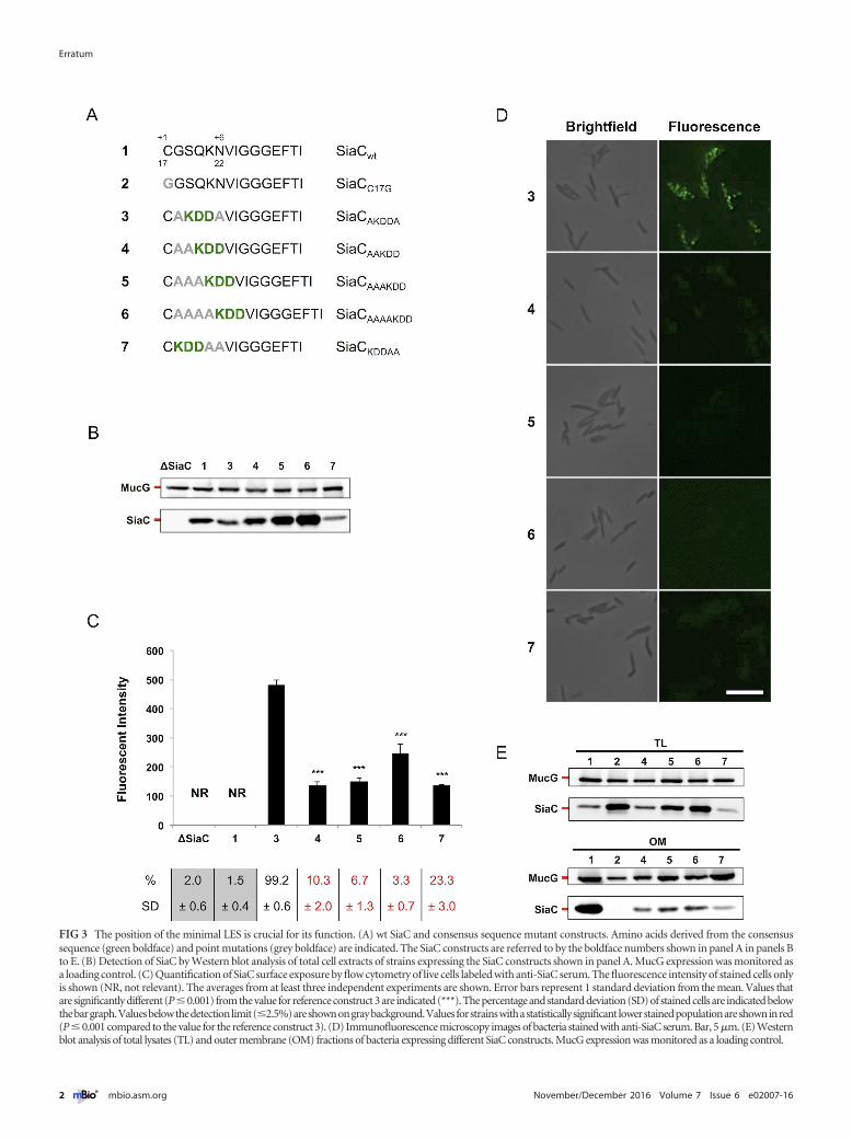
FIG 3 The position of the minimal LES is crucial for its function. (A) wt SiaC and consensus sequence mutant constructs. Amino acids derived from the consensussequence (green boldface) and point mutations (grey boldface) are indicated. The SiaC constructs are referred to by the boldface numbers shown in panel A in panels Bto E. (B) Detection of SiaC by Western blot analysis of total cell extracts of strains expressing the SiaC constructs shown in panel A. MucG expression was monitored asa loading control. (C) Quantification of SiaC surface exposure by flow cytometry of live cells labeled with anti-SiaC serum. The fluorescence intensity of stained cells onlyis shown (NR, not relevant). The averages from at least three independent experiments are shown. Error bars represent 1 standard deviation from the mean. Values thatare significantly different (P�0.001) from the value for reference construct 3 are indicated (***). The percentage and standard deviation (SD) of stained cells are indicated belowthebargraph.Valuesbelowthedetectionlimit(�2.5%)areshownongraybackground.Valuesforstrainswithastatisticallysignificant lowerstainedpopulationareshowninred(P � 0.001 compared to the value for the reference construct 3). (D) Immunofluorescence microscopy images of bacteria stained with anti-SiaC serum. Bar, 5 �m. (E) Westernblot analysis of total lysates (TL) and outer membrane (OM) fractions of bacteria expressing different SiaC constructs. MucG expression was monitored as a loading control.
Erratum
2 ® mbio.asm.org November/December 2016 Volume 7 Issue 6 e02007-16