Embed Size (px)
Citation preview

Identification of a Molecular Activator for Insulin Receptorwith Potent Anti-diabetic Effects*□S
Received for publication, April 12, 2011, and in revised form, August 9, 2011 Published, JBC Papers in Press, September 9, 2011, DOI 10.1074/jbc.M111.247387
Kunyan He‡, Chi-Bun Chan‡1, Xia Liu‡, Yonghui Jia§, Hongbo R. Luo§, Stefan A. France¶, Yang Liu�, W. David Wilson�,and Keqiang Ye‡2
From the ‡Department of Pathology and Laboratory Medicine, Emory University School of Medicine, Atlanta, Georgia 30322, the§Department of Pathology and Lab Medicine, Harvard Medical School and Children’s Hospital, Boston, Massachusetts 02115, the¶School of Chemistry and Biochemistry, Georgia Institute of Technology, Atlanta, Georgia 30332-0400, and the �Department ofChemistry, Georgia State University, Atlanta, Georgia 30033
Insulin exerts its actions through the insulin receptor (IR) andplays an essential role in diabetes. The inconvenient daily injec-tion and undesirable side-effects associated with insulin injec-tiondemandnovel drugs for thediseases.To search for bioactiveinsulinmimetics, we developed an in vitro screening assay usingphospho-IR ELISA. After screening the small molecule chemi-cal libraries, we have obtained a compound (5,8-diacetyloxy-2,3-dichloro-1,4-naphthoquinone) that provokes IR activationby directly binding to the receptor kinase domain to trigger itskinase activity at micromolar concentrations. This compoundselectively activates IR but not other receptors and sensitizesinsulin’s action. Moreover, it elevates glucose uptake in adi-pocytes and has oral hypoglycemic effect in wild-type C57BL/6Jmice and db/db and ob/obmice without demonstrable toxicity.Hence, this promising compound mimics the biological func-tions of insulin and is useful for further drug development fordiabetes treatment.
Type 2 diabetes mellitus is a heterogeneous disease thatresults from defective insulin actions and secretions. Variouspharmacological agents are used to improve glucose homeosta-sis via differentmodes of action: sulfonylureas stimulate insulinsecretion, biguanides (e.g.metformin) promote glucose utiliza-tion and reduce hepatic-glucose production, �-glucosidaseinhibitors (e.g. acarbose) slow down carbohydrate absorptionfrom the gut, and thiazolidinediones enhance cellular insulinaction on glucose metabolism (1). Insulin replacement therapyis also needed when insulin production declines in the patientswith poor glycemic control (2). In recent years, treatment strat-egies have focused on the development of novel therapeuticoptions to substitute insulin therapy. Several small compoundslike demethylasterriquinone-B1 and TLK19780 have beenidentified as the functional insulin mimetics; however, theyhave poor bioavailability or low receptor specificity (3–5).
Therefore, the search for new orally active insulin mimeticswith stringent receptor selectivity is highly warranted.In this report, we demonstrate that the naphthoquinone
derivative 5,8-diacetyloxy-2,3-dichloro-1,4-naphthoquinone(DDN)3 is a highly selective IR activator that interacts directlywith the IR tyrosine kinase domain to induce Akt and ERKphosphorylations. It is also an insulin sensitizer that enhancesinsulin’s action to stimulate glucose uptake. Oral administra-tion of this compound robustly decreases blood glucose inwild-type and diabetic ob/ob and db/db mice. Therefore, DDN is abioactive insulin mimetic with hypoglycemic activity.
EXPERIMENTAL PROCEDURES
Animals—Male C57BL/6J mice, male C57BL/KsJ db/dbmice, and female C57BL/KsJ ob/ob mice were obtained fromthe Jackson Laboratory. Adult animals aged 10–12 weeks wereused. Mice were housed in environmentally controlled condi-tions with a 12-h light/dark cycle and had free access to stand-ard rodent pellet food and water. The animal protocols wereapproved by the Institutional Animal Care and Use Committeeof Emory University. Animal care was given in accordance withinstitutional guidelines.Cells and Reagents—CHO-IR cells (a gift from Dr. Nicholas
Webster, University of California at San Diego) were main-tained in Ham’s F-12 plus 10% fetal bovine serum (FBS), 100units of penicillin-streptomycin, and 200 ng/ml of G-418. NIH3T3-L1 cells were maintained in DMEM with 10% calf bovineserum (CBS) and 100 units of penicillin-streptomycin.HEK293, and mouse embryonic fibroblast (MEF) cells weremaintained inDMEMwith 10% FBS and 100 units of penicillin-streptomycin. All cells were maintained at 37 °C with 5% CO2atmosphere in a humidified incubator. For experiments, cellswere treated in the appropriate serum-free media containingcompounds dissolved in DMSO. Control cells received equiva-lent amounts of DMSO, and the final concentration of DMSOwas always kept below 0.1%. DDN was from InterBioscreen.Insulin, IGF-1, and glutathione S-transferase (GST)-horserad-ish peroxidase were from Sigma. [2-3H]Deoxyglucose was pur-
* This work was supported, in whole or in part, by National Institutes of HealthGrant CA127119 from NCI (to K. Y.).
□S The on-line version of this article (available at http://www.jbc.org) containssupplemental Figs. S1 and S2 and Table S1.
1 To whom correspondence may be addressed: Dept. of Pathology and Lab-oratory medicine, Emory University School of Medicine, Atlanta, GA 30322.Tel.: 404-712-2814; Fax: 404-712-2979; E-mail: [email protected].
2 To whom correspondence may be addressed: Dept. of Pathology and Lab-oratory medicine, Emory University School of Medicine, Atlanta, GA 30322.Tel.: 404-712-2814; Fax: 404-712-2979; E-mail: [email protected].
3 The abbreviations used are: DDN, 5,8-diacetyloxy-2,3-dichloro-1,4-naph-thoquinone; IRKT, intracellular kinase domain of IR; MEF, mouse embryonicfibroblast; IGF-1R, insulin-like growth factor-1 receptor; CSN, 2,3-bismeth-ylsulfanyl-1,4-naphthoquinone; 2NH2-NQ, 2-amino-1,4-naphthoquinone;ATP�S, adenosine 5�-O-(thiotriphosphate); IRS-1, insulin receptor sub-strate 1.
THE JOURNAL OF BIOLOGICAL CHEMISTRY VOL. 286, NO. 43, pp. 37379 –37388, October 28, 2011© 2011 by The American Society for Biochemistry and Molecular Biology, Inc. Printed in the U.S.A.
OCTOBER 28, 2011 • VOLUME 286 • NUMBER 43 JOURNAL OF BIOLOGICAL CHEMISTRY 37379
by guest on January 14, 2019http://w
ww
.jbc.org/D
ownloaded from
by guest on January 14, 2019
http://ww
w.jbc.org/
Dow
nloaded from
by guest on January 14, 2019http://w
ww
.jbc.org/D
ownloaded from

chased from PerkinElmer. Anti-IR � subunit and anti-IRS-1were from Santa Cruz Biotechnology. Anti-phospho-Akt 473,anti-phospho-Erk, and anti-phospho-IR-1146 were from CellSignaling. Anti-phosphotyrosine (pY20) and anti-phospho-IR-1158/1162/1163 were from BD Bioscience and Invitrogen,respectively.GST- intracellular kinase domain of IR (IRTK) andHis-IR were from Invitrogen and R&D, respectively. All otherreagents not mentioned were purchased from Sigma.Cell-based Screening—CHO-IR cells were seeded in a 96-well
plate with 15,000 cells per well in 100 �l of 0.1% FBS medium.Cells were incubated overnight, followed by 15-min treatmentwith 10 �M compounds in DMSO (10 mM stock concentrationfrom the Spectrum Collection Library) at 37 °C. Control wellsreceived vehicle or 100 nM insulin. Cells were lysed, and thephosphorylated receptors were captured with pY20 that wasimmobilized on ELISA plates. Bound receptors were washedthen detected using an anti-IR � antibody, followed by a horse-radish peroxidase-conjugated secondary antibody and colori-metric detection with 3,3�,5,5�-tetramethylbenzidine.IR Activation by Insulin Mimetic Compounds—Cells were
growth in 0.1% FBS medium overnight and then stimulatedwith different concentrations of drugs or insulin (100 nM), andthe whole cell extracts were analyzed by immunoblotting. Aftera 12-h fasting, mice were orally administrated with 20 mg/kg(C57BL/6J) or 5 mg/kg (ob/ob and db/db) DDN in 0.5% meth-ylcellulose and sacrificed at 3, 4, and 5 h following drug admin-istration. Liver and muscle tissues were dissected quickly andfrozen on dry ice. Tissue lysates were prepared and analyzed.IR in Vitro Kinase Assay—Recombinant GST-IRTK was
incubate with 50 mM Tris-HCl (pH 7.4), 10 mM MgCl2, and 50�M ATP with different concentrations of DDN for 15 min at25 °C. Histone H2B (0.35 �g/ml) and [�-32P]ATP (0.25 �Ci/�l)were added, and the samples were further incubated for 10min.The sample was separated on a SDS-polyacrylamide gel andautoradiographed.Partial Proteolysis Assay—200 ng of GST-IRTK was sub-
jected to limited trypsin digestion in the presence or absence of50 �M DDN with 50 mM Tris-HCl (pH 8.2) and 20 mM CaCl2.The sample was separated on a SDS-polyacrylamide gel andstained by silver.UV-absorption Spectra—UV-visible absorption measure-
ment is a simple method that is applicable for exploring struc-tural change and for knowing complex formation (6, 7). In thepresent study, UV-visible absorption for protein binding wasobtained using the Cary 300 UV-visible spectrophotometer(Varian Inc., Palo Alto, CA) equipped with 1.0-cm quartz cells.The spectra were scanned from 200 to 800 nm in Tris buffer(0.01 M Tris, 0.05 M NaCl, 10 mM MgCl2, pH 7.8). During thespectrophotometric titrations, protein (GST or GST-IRTK)was added to a cell containing a constant amount of compound.The absorbance at 602 nm was recorded (data were read threetimes, and mean value was obtain by averaging); the obtaineddata were processed with KaleidaGraph (Synergy Software).Repeated measurements were done for all the samples, and nosignificant differences were observed. (The reproducibilityerror was �5%.) Binding results from the UV titration experi-ments were fit with a one-site interaction model: r � K*Cfree/(1�K*Cfree), where r represents themoles of bound compound
permole of protein,K is themacroscopic binding constant, andCfree is the free compound concentration in equilibrium withthe complex.Insulin Competition Assay—The FITC-insulin (100 nM) was
incubatedwithCHOorCHO-IR cells in the presence of variousconcentrations of DDN in binding buffer (Ham’s F-12, 0.5%BSA, 20mMHEPES (pH 8.0), and 0.1%NaN3) overnight at 4 °C.Cells were washed three times and acquired on a FACScan, andthe mean channel fluorescence of bell-shaped histograms wasanalyzed using the program FlowJ.[2-3H]Deoxyglucose Transport—3T3-L1 preadipocytes and
MEF cells were grown in standard media. After 2 days, 3T3-L1cells were switched to differentiation media with 1 �M dexam-ethasone, 10 �g/ml insulin, and 0.5 mM 3-methyl-1-isobutylx-anthine for 2 days, grown in post-differentiation medium con-taining 10 �g/ml insulin for 5 days, and then placed in standardmedia. NIH 3T3-L1 adipocytes were reseeded into 12-wellplates 10–12 days after differentiation. Cells in 12-well plateswere rinsed three times with PBS at 23 °C and preincubatedwith 500 �l of KRP-HEPES buffer (131.2 mM NaCl, 4.71 mM
KCl, 2.47 mM CaCl2, 1.24 mM MgSO4, 2.48 mM NaH2PO4, 10mM HEPES, and 0.5% BSA (pH 7.45)) containing insulin for 15min or DDN for 30 min at 37 °C. The transport reaction wasinitiated by adding 2-deoxyglucose (final concentration, 100�M) with [2-3H]deoxyglucose (0.2 �Ci/well). After 10-minincubation, cells were washed three times with ice-cold PBSand solubilized in 1% SDS, 1 M NaOH. After neutralization,radioactivity was measured by scintillation counting.BloodGlucose Level Test—After a 12-h fasting, C57BL/6J and
db/db mice were bled from the tail vein for a baseline (time 0)measurement with a glucometer (Accu-Chek, Roche Diagnos-tics). Afterward, animals were orally administered 20 mg/kg(C57BL/6J) or 5 mg/kg (db/db) DDN in 0.5% methylcellulosethrough a feeding needle. Blood glucose levels were measuredat the indicated time points.Intraperitoneal Glucose Tolerance Test—After a 12-h fasting,
ob/obmicewere bled from the tail vein for a baseline (time,�60min) measurement. Then animals were orally administered 5mg/kg DDN in 0.5% methylcellulose through a feeding needle.One hour after the drug administration, glucose was adminis-trated intraperitoneally (2 g/kg). Blood glucose levels weremeasured 0, 30, 60, 90, 120, and 180min after administration ofglucose. The blood was collected in tubes and kept at 4 °C over-night, and plasma was prepared by centrifugation (2000� g, 20min). Plasma insulin was measured by ELISA with a rat insulinkit (Crystal Chem). Insulin was quantified using the insulinstandard curve.Statistic Analysis—Results were expressed as mean � S.E.
from at least three independent experiments and were consid-ered significant when p was �0.05. Statistic analysis was per-formed by using PRISM (GraphPad).
RESULTS
DDN and CSN Activate IR and Insulin-mediated SignalingCascades—To search for small compounds that can rapidlyactivate IR, we have developed a cell-based ELISA assay usingan IR stably transfectedCHO-IR cell line.We seeded the cells ina 96-well plate and treated the cells with 10 �M (15 min) com-
DDN Is an Insulin Mimetic Compound
37380 JOURNAL OF BIOLOGICAL CHEMISTRY VOLUME 286 • NUMBER 43 • OCTOBER 28, 2011
by guest on January 14, 2019http://w
ww
.jbc.org/D
ownloaded from

pounds from several chemical libraries containing �4500 nat-ural products and synthetic chemicals. The cell lysates weresubjected to the sandwich ELISA using immobilized anti-phos-photyrosine antibody and anti-IR antibody. The positive hitswere subsequently analyzed onCHO-IGF-1R cells, and only thecompounds that failed to activate IGF-1R were chosen for fur-ther study. Among the positive hits, we identified six 1,4-naph-thoquinone compounds that can selectively activate IR but notIGF-1R (Fig. 1A). ELISA results revealed that all these com-pounds could significantly induce IR phosphorylation (Fig. 1B).Surprisingly, immunoblotting analysis demonstrated that only2,3-bismethylsulfanyl-1,4-naphthoquinone (CSN) and DDNstrongly stimulated tyrosine phosphorylation of the IR � sub-unit in Tyr-1146 (Fig. 2A, first panel) and Tyr-1158/1162/1163(Fig. 2A, second panel). Total IR tyrosine phosphorylation wasfurther confirmed by immunoprecipitation using anti-phos-photyrosine (pY20) antibody (Fig. 2A, third panel). Presumably,the discrepant IR phosphorylation levels between ELISA andimmunoblotting assay were caused by nonspecific signal and alow detection limit of the ELISA. Consistent with IR activation,
the tyrosine phosphorylation of IRS-1 of CHO-IR cells was alsoincreasedwhen stimulated by bothDDNandCSN (Fig. 2A, fifthpanel). A titration assay showed both compounds activated IRin a dose-dependent manner. At 5 �M, both compounds evi-dently stimulated IR phosphorylation and downstreamAkt andERK activation in a dose-dependent manner (Fig. 2B). Thetime-course assay demonstrated that CSN activated IR inCHO-IR cells at 5–15 min, peaked at 60 min, and the signaldecreased at 2 h (Fig. 2C,middle panel). In contrast, significantIR phosphorylation started 5 min after DDN stimulation, andthe signals escalated with the time course (Fig. 2C, right panel).Although insulin activated IR signaling at 0.05 �M, these com-pounds stimulated the IR signal cascades at 5 �M (Fig. 2D).Neither IGF-1R nor EGFR was activated by any of these com-pounds even at 10 �M, underscoring that these molecules pos-sess selectivity toward different receptors (Fig. 2E). Hence, bothDDN and CSN potently and specifically stimulate IR and itsdownstream signaling cascades in intact cells.DDN Displays Hypoglycemic Effect in Mice—To explore
whether DDN and CSN can mimic insulin in provoking IR and
FIGURE 1. Identification of novel insulin mimetics. A, chemical structures of six compounds of 1,4-naphthoquinone derivatives. B, effects 1,4-naphthoqui-none derivatives on IR phosphorylation in CHO-IR cells. CHO-IR cells were treated with different 1,4-naphthoquinone derivatives (5 �M) or insulin (100 nM) for15 min. The amount of phosphorylated IR receptors was quantified by sandwich ELISA using immobilized anti-phosphotyrosine antibodies (pY20) anti-IRantibody. The activities of tested compounds were expressed as a percentage of the vehicle (DMSO) (n � 3).
DDN Is an Insulin Mimetic Compound
OCTOBER 28, 2011 • VOLUME 286 • NUMBER 43 JOURNAL OF BIOLOGICAL CHEMISTRY 37381
by guest on January 14, 2019http://w
ww
.jbc.org/D
ownloaded from

its downstream IRS-1 activation in vivo, we intraperitoneallyadministrated various doses of DDN and CSN into C57BL/6Jmice. However, CSNwas lethal to themice even at low doses (1mg/kg) (data not shown). Hence, we only focused on DDN forthe subsequent experiments.We tested ifDDNpossesses hypo-glycemic functions. After oral injection, DDN significantlyreduced blood glucose inC57BL/6mice as comparedwith vehi-cle control. The kinetics of DDN in lowering blood glucose isdifferent from that of insulin. Thirty minutes after intraperito-neal injection of insulin, the blood glucose concentration wasreduced; however, a significant decrease of blood glucose couldonly be detected after 3 h of DDN administration. On the otherhand, injection of the inactive analog 2-amino-1,4-naphthoqui-none (2NH2-NQ) at the same dosage had no effect (see Fig. 3A
for the percentage change and supplemental Fig. S1 for theabsolute blood glucose concentrations). To monitor the IR sig-naling cascades in insulin-sensitive tissues, we injected 20mg/kg DDN into C57BL/6J mice via oral gavage, sacrificed theanimals at different time points, and monitored IR tyrosinephosphorylation by immunoblotting. In liver andmuscle, DDNelicited demonstrable IR activation after 3–4 h (Fig. 3B, firstpanel). Immunoprecipitation using pY20 antibody also con-firmed IRS-1 tyrosine phosphorylation in both tissues afterstimulated by DDN (Fig. 3B, third panel); in contrast, DDN didnot stimulate IGF-IR activation in either muscle or liver (Fig.3B, fifth panel), which concurred with our in vitro observation(Fig. 2E). Neither IR nor IRS-1 phosphorylations wereincreased after 2NH2-NQ injection (Fig. 3B). To test whether
FIGURE 2. DDN and CSN activate IR and its downstream signaling. A, DDN and CSN activate IR and its downstream signaling in CHO-IR cells. CHO-IR cellswere treated with insulin (100 nM) or various 1,4-naphthoquinone derivatives (5 �M) for 15 min. The cell lysates were analyzed by immunoblotting withanti-phospho-IR antibodies, or immunoprecipitated by pY20 and analyzed using anti-IR and anti-IRS-1, respectively. B, DDN and CSN induce IR phosphoryla-tion in a dose-dependent manner. CHO-IR cells were treated with the small compounds at different concentrations for 15 min, and the IR phosphorylation wasmonitored by immunoprecipitation and immunoblotting. C, DDN and CSN induce IR phosphorylation in a time-dependent manner. CHO-IR cells were treatedwith DDN (5 �M), CSN (5 �M), and insulin (10 nM) for various times, and the IR phosphorylation was monitored by immunoprecipitation and immunoblotting.D, DDN and CSN induce IR phosphorylation and its downstream signaling in a dose-dependent manner. CHO-IR cells were treated with various concentrationsof DDN or CSN for 30 min, or insulin for 15 min, and the IR phosphorylation and its downstream signaling were monitored by immunoprecipitation andimmunoblotting. E, DDN and CSN cannot induce IGF-1R or EGFR phosphorylation. CHO-IGF-1R or HEK293 cells were treated with EGF (100 nM), IGF-1 (100 nM),DDN (10 �M), or CSN (10 �M) for 15 min. The cell lysates were analyzed by immunoblotting with anti-pIGF-1R and anti-pEGFR.
DDN Is an Insulin Mimetic Compound
37382 JOURNAL OF BIOLOGICAL CHEMISTRY VOLUME 286 • NUMBER 43 • OCTOBER 28, 2011
by guest on January 14, 2019http://w
ww
.jbc.org/D
ownloaded from

DDN has an acute effect as insulin, DDN or 2NH2-NQ wasinjected into the animal through the vena cava. The liver wasthen dissected and homogenized after 5 min. Immunoprecipi-tation using pY20 antibody demonstrated that DDN, but not2NH2-NQ, quickly activated IR (Fig. 3C, first panel) and Aktand ERK (Fig. 3C, third and fifth panels, respectively).Similarly to the observation in C57/BL6 mice, DDN induced
IR signaling in the well establishedmicemodels of non-insulin-dependent diabetes mellitus. Oral injection of DDN enhanced
IR and ERK phosphorylations in db/db (Fig. 4A) and ob/ob (Fig.4B) mice. One hour after oral administration of DDN, lowerblood glucose was found in db/dbmice as compared with vehi-cle control (Fig. 4C). In contrast to the findings in C57/BL6mice, the time needed for DDN to lower the blood glucose indb/dbmice was comparable to that of insulin injection, but theeffect of DDN could be sustained till the end of the experiment,whereas the effect of insulin vanished 2 h after injection. Oraladministration of DDN to ob/ob mice also led to significant
FIGURE 3. DDN activates insulin signaling pathways in vivo and possesses hypoglycemic activity. A, DDN reduces blood glucose in C57BL/6J mice.C57BL/6J mice were starved for 12 h and then orally administrated with vehicle (0.5% methylcellulose), DDN (20 mg/kg), or the inactive analog 2NH2-NQ (20mg/kg). Human insulin (1 unit/kg) was injected intraperitoneally as a positive control. Blood glucose was monitored before and after dosing at various timepoints as indicated (*, p � 0.05; **, p � 0.01; ***, p � 0.001, n � 10 –13, Two-way analysis of variance versus vehicle). B, DDN provokes IR phosphorylation andits downstream signaling in C57BL/6J mice. Three-month-old C57BL/6J mice were starved for 12 h and then orally administrated with vehicle (0.5% methyl-cellulose), DDN (20 mg/kg), or 2NH2-NQ (20 mg/kg). Cell lysates were prepared from liver and muscle tissues, which were collected at 3, 4, and 5 h after drugadministration and analyzed by immunoprecipitation and immunoblotting. C, DDN quickly activates IR phosphorylation and its downstream signaling.Three-month-old C57BL/6J mice were starved for 12 h and then injected with vehicle, 5 mg/kg DDN, or 5 mg/kg 2NH2-NQ through the vena cava. After 5 min,the liver was collected and analyzed by immunoblotting using specific antibodies.
DDN Is an Insulin Mimetic Compound
OCTOBER 28, 2011 • VOLUME 286 • NUMBER 43 JOURNAL OF BIOLOGICAL CHEMISTRY 37383
by guest on January 14, 2019http://w
ww
.jbc.org/D
ownloaded from

improvement in glucose tolerance, which was comparable tothe effect of insulin injection (Fig. 4D). Nonetheless, single oraldoses of DDN had no significant effect on plasma insulin levelsin ob/ob mice (data not shown), suggesting the hypoglycemicfunction of DDN did not act through the change of insulinsecretion.Treatment ofwild-typeC57BL/6Jmice (n� 5)with daily oral
administration of DDN (5 mg/kg) for 16 days did not affect thefood intake (supplemental Fig. S1) or blood biochemistry (sup-plemental Table S1). Pathological examination also failed todetect any demonstrable toxicity in brain, heart, liver, kidney,and muscle (data not shown), indicating that the compoundmight not possess any intolerable toxicity. Hence, DDNmimicsinsulin and effectively lowers blood glucose in wild-type andinsulin-resistant db/db and ob/obmice.DDN Induces Cellular Glucose Uptake—The observation
that DDN effectively reduced circulating glucose in animalssuggested that it might enhance glucose absorption. Therefore,we examined the effect of DDN on glucose uptake in adi-pocytes, a well characterized cellularmodel for insulin-inducedglucose homeostasis. Differentiated 3T3-L1 adipocytes weretreated with various concentrations of DDN, and the activationof Akt was monitored. As expected, DDN elicited a dose-de-pendent IR and Akt activation (Fig. 5A). Moreover, DDN esca-lated [3H]deoxyglucose uptake in a dose-dependent manner(Fig. 5B), which concurred with the Akt phosphorylationpattern.
It is noteworthy that DDN sensitized the insulin activity. Indifferentiated 3T3-L1 cells, 5 nM insulin was able to trigger IRphosphorylation (Fig. 5C, first panel, lane 2). However, DDN atthe same concentration did not induce any IR activation (Fig.5C, first panel, lane 3). When the two insulin and DDN wereadded together, IR phosphorylation was higher than thoseobserved when insulin or DDN was administrated separately,suggesting DDN can potentiate insulin activity. Akt phospho-rylation tightly correlated with the IR activity after insulin orDDN stimulation (Fig. 5C, third panel). In addition, DDN ele-vated insulin activity in triggering glucose uptake in differenti-ated 3T3-L1 cells. Consistent with the IR activation pattern,stimulation with 5 nM insulin, but not DDN, significantlyincreased the [3H]deoxyglucose uptake in 3T3-L1 cells. Wheninsulin and DDN were administrated together, the magnitudeof glucose uptake was further increased (Fig. 5D).To confirm that the DDN-induced biological effect was IR-
dependent, we knocked down IR in the differentiated adi-pocytes using siRNA and monitored the insulin signaling.Western blot analysis showed that IR expression was signifi-cantly decreased in cells transfectedwith siRNA against IR (Fig.5E, first panel). Both insulin- and DDN-triggered ERK phos-phorylation were reduced when IR was depleted, underscoringthat IR is the major molecular target of DDN to trigger ERKactivation (Fig. 5E, secondpanel).Wealso treatedphosphatidyl-inositol 3-kinase p85 subunit-ablated (p85��/�) MEF withDDN and determined its glucose uptake activity. It has been
FIGURE 4. DDN displays hypoglycemic function in diabetic mouse models. A, DDN activates insulin signaling in db/db mice. Three-month-old mice werestarved for 12 h and then orally administrated with vehicle (0.5% methylcellulose) or DDN (5 mg/kg). Cell lysates were prepared from liver and muscle tissues,which were collected at 1 and 2 h after drug administration and analyzed by immunoprecipitation and immunoblotting. B, DDN activates insulin signaling inob/ob mice. Three-month-old mice were starved for 12 h and then orally administrated with vehicle (0.5% methylcellulose) or DDN (5 mg/kg). Cell lysates wereprepared from liver and muscle tissues, which were collected at 1 and 2 h after drug administration and analyzed by immunoprecipitation and immunoblot-ting. C, hypoglycemic function of DDN in db/db mice. The animals were starved for 12 h and then orally injected with vehicle (0.5% methylcellulose) or DDN (5mg/kg). Human insulin (1 unit/kg) was injected intraperitoneally as a positive control. Blood glucose was monitored before and after drug administration at 1-hintervals (*, p � 0.05; **, p � 0.01, n � 5; two-way analysis of variance versus vehicle). D, DDN improves the glucose tolerance in ob/ob mice. The animals werestarved for 12 h and then orally dosed with vehicle (0.5% methylcellulose) or DDN (5 mg/kg). Human insulin (1 unit/kg) was injected intraperitoneally as apositive control. A bonus of glucose (2 gm/kg) was injected intraperitoneally 1 h later. Blood glucose was measured at 30-min intervals (*, p � 0.05; **, p � 0.01,n � 5; two-way analysis of variance versus vehicle control).
DDN Is an Insulin Mimetic Compound
37384 JOURNAL OF BIOLOGICAL CHEMISTRY VOLUME 286 • NUMBER 43 • OCTOBER 28, 2011
by guest on January 14, 2019http://w
ww
.jbc.org/D
ownloaded from

reported that MEFs with p85 ablation are defective in insulin-stimulated glucose uptake (8). Both insulin and DDN triggeredrobust [3H]deoxyglucose uptake in wild-typeMEF cells but notp85��/� cells (Fig. 5F), suggesting that an intact IR/phosphati-
dylinositol 3-kinase/Akt cascade is necessary for DDN to exertits hypoglycemic function.DDN Binds to the Kinase Domain of Insulin Receptor—The
synergistic function of DDN on insulin-induced IR activation
FIGURE 5. DDN enhances cellular glucose uptake. A, DDN provokes IR signaling in differentiated 3T3-L1 adipocytes. The 3T3-L1 cells were stimulated withinsulin (50 nM) for 15 min or DDN (0.1, 0.25, and 0.5 �M) for 30 min. Insulin signaling in the cell lysates was tested by immunoblotting. B, DDN stimulates glucoseuptake. Differentiated 3T3-L1 adipocytes were stimulated with insulin (50 nM) for 15 min or DDN (0.1, 0.25, and 0.5 �M) for 30 min. [2-3H]Deoxyglucose was thenadded, and the cells were further incubated for 10 min. [2-3H]Deoxyglucose uptake by adipocytes was measured by scintillation counting (*, p � 0.05; **, p �0.01 versus control, Student’s t test, n � 3). C, DDN synergizes insulin signaling. Differentiated 3T3-L1 adipocytes were stimulated with 5 nM insulin, 5 nM DDN,or a combination of the two drugs. Cell lysates were then prepared for Western blot using specific antibodies as indicated. D, DDN synergizes insulin activity inpromoting glucose uptake. Differentiated 3T3-L1 adipocytes were stimulated with 5 nM insulin, 5 nM DDN, or a combination of the two drugs. [2-3H]Deoxyg-lucose was then added, and the cells were further incubated for 10 min. [2-3H]Deoxyglucose uptake by adipocytes was measured by scintillation counting (*,p � 0.05; **, p � 0.01, Student’s t test, n � 3). E, IR is necessary for DDN to induce ERK phosphorylation. Differentiated 3T3-L1 adipocytes were transfected withcontrol siRNA or siRNA against IR. The cells were then stimulated with insulin (100 nM) for 15 min or DDN (5 �M) for 30 min. Cell lysates were analyzed by Westernblot. F, the glucose uptake induced by DDN in p85� knock-out MEF cells. Wild-type and p85��/� MEF cells were treated with 100 nM insulin for 15 min or 5 �M
DDN for 30 min. The uptake of [2-3H]deoxyglucose was monitored by liquid scintillation counting (*, p � 0.05; **, p � 0.01 versus control, Student’s t test, n �3).
DDN Is an Insulin Mimetic Compound
OCTOBER 28, 2011 • VOLUME 286 • NUMBER 43 JOURNAL OF BIOLOGICAL CHEMISTRY 37385
by guest on January 14, 2019http://w
ww
.jbc.org/D
ownloaded from

suggests that insulin andDDNmight share differential IR bind-ing sites. We therefore performed insulin competition assayusing flow cytometry (9). CHO-IR cells were incubated with100 nM FITC-insulin in the presence of DDN at different con-centrations. The binding of FITC-insulin to the cell surface IRincreased the fluorescent signal of CHO-IR cells, leading to aright shift of the peak. No significant change of the shifted peakposition was observed in the presence of DDN (Fig. 6A, toppanel). However, a left shift of the fluorescent peak wasdetected, when 5 �M unlabeled insulin was added, suggesting asuccessful competition between unlabeled insulin and FITC-insulin for the ligand binding (Fig. 6A,middle panel). As a neg-ative control, FITC-insulin did not bind to the IR-deficientparental CHO cells (Fig. 6A, bottom panel). These results sug-gest that DDN and insulin might possess different ligand bind-ing sites on IR.Several small molecules bind to IRTK and provoke its activa-
tion (3–5). Thus, DDN might also interact with the IRTK toactivate IR. To test this possibility, we performed an UV-ab-sorption spectra analysis using recombinant IRTK. Although
the GST-tagged IRTK (GST-IRTK, amino acids 1011–1382)had no absorption from 200 to 800 nm, addition of DDN, butnot its structural related analog, naptho[2,3-d]thiazole-4,9-di-one-2-[(4-chlorophenyl)amino] enhanced the peak absorptionat 560 and 602 nm (Fig. 6B), suggesting a specific interactionoccurs between DDN and IRTK. A titration assay revealed thatthe binding constant Kd of DDN to GST-IRTK was �3.27 �1.06 �M. We also performed the partial proteolysis analysis toconfirm theDDN-IRTK interaction as previously described (4).We first incubated recombinant GST-tagged IRTK with eitherDMSO or DDN, followed by limited trypsin digestions. InDMSO treatment, GST-IRTKwas digested into several smallerfragments of various molecular weights. However, DDN pre-treatment protected IRTK against trypsin digestion with moreintact IRTK observed (Fig. 6C). As a positive control, the pres-ence of ATP�S also altered the proteolysis pattern. Whereas aband of �37 kDa presented strongly in the ATP�S-incubatedIRTK, a band of �30 kDa was found in the control samples butattenuated in the ATP�S-treated sample.We further examinedif DDN alters the kinase activity of IRTK through direct inter-
FIGURE 6. DDN interacts and activates the insulin receptor. A, DDN does not complete with insulin for IR binding site. FITC-insulin was incubated withCHO-IR cells in the presence of various concentrations of DDN (top panel) or unlabeled insulin (middle panel). Parental CHO cells were used as a negative control(bottom panel). The fluorescence-labeled cells were analyzed on flow cytometry. B, DDN binds to IRTK. UV-visible spectra were scanned from 200 to 800 nm forIRTK fragment in the presence of DDN or its analog. When DDN binds to IRTK, it elicits the absorption alteration at 560 and 602 nm. C, DDN protects IRTK fromproteolysis. GST-IRTK was subjected to limited trypsin digestion in the presence of DDN (50 �M), ATP�S (5 mM), or both. The reaction mixture was resolved bySDS-PAGE, followed by silver staining. D, DDN increases IRTK activity in vitro. Recombinant GST-IRTK was incubated with different concentrations of DDN or itsinactive analog 2NH2-NQ, and the kinase activity of IRTK was examined by monitoring the phosphorylation of histone H2B using autoradiography.
DDN Is an Insulin Mimetic Compound
37386 JOURNAL OF BIOLOGICAL CHEMISTRY VOLUME 286 • NUMBER 43 • OCTOBER 28, 2011
by guest on January 14, 2019http://w
ww
.jbc.org/D
ownloaded from
action by performing an in vitro histone phosphorylation assay(4). As expected, DDN, but not the inactive analog 2NH2-NQ,activated the recombinant IRTK to phosphorylate histone H2B(Fig. 6D). Therefore, DDN activates IR by increasing the kinaseactivity of IR through direct interaction.
DISCUSSION
In this report, we have identified the 1,4-naphthoquinonederivative DDN as a new small molecular IR activator in vitroand in vivo. DDN has a simple chemical structure with promi-nent effect in selectively provoking IR activation and loweringblood glucose in animals. Thus, it represents a novel prototypefor further chemical modification to generate a powerful ther-apeutic drug for diabetes.Zhang et al. have reported that DAQ B1 is an orally active IR
ligand with anti-diabetic activity (4). Webster and his col-leagues have conducted an extensive structure-activity rela-tionship study on DAQ B1 derivatives and successfully identi-fied the mono-indolyl-dihydroxybenzoquinones ZL-196 andLD-17, which activate IR signaling and effectively lower bloodglucose in db/dbmice (10). However, these compounds cannotselectively induce IR, and they also provoke other receptor tyro-sine kinases, including IGF-1R, NGFR, and EGFR (10, 11). Incontrast, DDN specifically activates IR and its downstream cas-cades. A few classes of non-peptidyl IR activators have also beenreported with different modes of actions. For example,TLK19780 is an insulin sensitizer, which potentiates insulin-triggered IR phosphorylation (3, 12). However, it is inactivewhen administrated alone. We also found that DDN potenti-ates the action of insulin in promoting IR activation and up-reg-ulating glucose uptake. This additive effect might be a result ofdifferential usage of IR ligand binding sites for insulin andDDN. Indeed, we have shown that DDN does not completewith insulin for IR binding but binds to the IR kinase domaindirectly. Hence, IR could simultaneously interact with bothinsulin on its extracellular domain andDDNon its intracellularkinase domain.When administrated orally, DDNhas hypoglycemic function
in both normal and diabetic mice models, and the glucose-low-ering effect could be observed after 1-h administration. It ispossible that themetabolites of DDN, in addition toDDN itself,might also possess the hypoglycemic activity in vivo. However,our cell-based in vitro studies showed that DDNdirectly boundIR and activated it within 5–15min, suggesting DDN per se hasa significant role in provoking IR activation. Moreover, IR acti-vation in liver and muscle could be observed in 5 min, whenDDN was injected into the bloodstream directly through thevena cava. It is interesting to note that DDN takes shorter timeto reduce the blood glucose level in db/dbmice than in normalC57BL/6 mice. One of the possible explanations is that thedb/db mice have more adipose tissue than the normal mice,which may provide more insulin-responsive “storage sites” forthe blood glucose. Thus, the db/db mice have more “targets”uponwhichDDNcan exert its hypoglycemic function. Anotherpossibility is that db/dbmice have higher intestinal permeabil-ity (13). Because DDN is given via oral administration in thecurrent studies, the higher absorption in the intestine of db/dbmice thus promotes more DDN into the bloodstream, leading
to a better efficacy in lowering blood glucose. It is noteworthythat a similar observation is made in the study using anotherquinone-based insulin mimetic Compound 2h. Compound 2his more potent in lowing blood glucose in db/dbmice than thelean control, suggesting that it may be a common characteristicfor quinone-based insulin mimetics (11).CSN is also an effective IR activator in vitro. However, it is
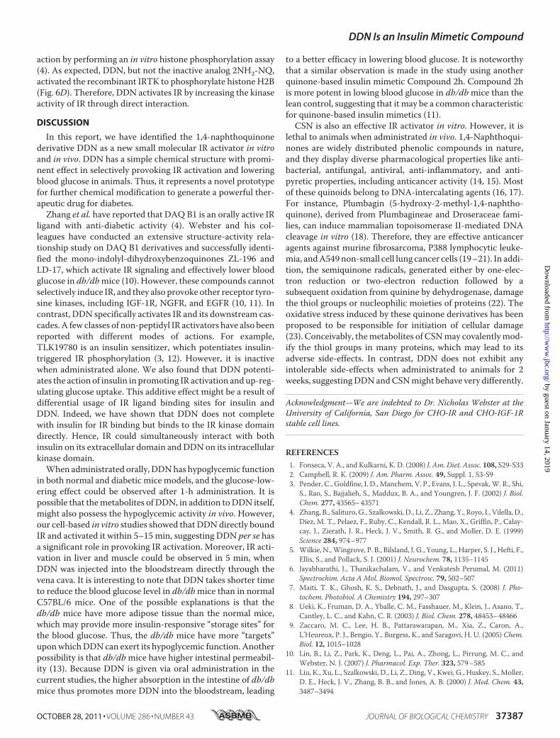
lethal to animals when administrated in vivo. 1,4-Naphthoqui-nones are widely distributed phenolic compounds in nature,and they display diverse pharmacological properties like anti-bacterial, antifungal, antiviral, anti-inflammatory, and anti-pyretic properties, including anticancer activity (14, 15). Mostof these quinoids belong to DNA-intercalating agents (16, 17).For instance, Plumbagin (5-hydroxy-2-methyl-1,4-naphtho-quinone), derived from Plumbagineae and Droseraceae fami-lies, can induce mammalian topoisomerase II-mediated DNAcleavage in vitro (18). Therefore, they are effective anticanceragents against murine fibrosarcoma, P388 lymphocytic leuke-mia, andA549non-small cell lung cancer cells (19–21). In addi-tion, the semiquinone radicals, generated either by one-elec-tron reduction or two-electron reduction followed by asubsequent oxidation from quinine by dehydrogenase, damagethe thiol groups or nucleophilic moieties of proteins (22). Theoxidative stress induced by these quinone derivatives has beenproposed to be responsible for initiation of cellular damage(23). Conceivably, themetabolites of CSNmay covalentlymod-ify the thiol groups in many proteins, which may lead to itsadverse side-effects. In contrast, DDN does not exhibit anyintolerable side-effects when administrated to animals for 2weeks, suggestingDDNandCSNmight behave very differently.
Acknowledgment—We are indebted to Dr. Nicholas Webster at theUniversity of California, San Diego for CHO-IR and CHO-IGF-1Rstable cell lines.
REFERENCES1. Fonseca, V. A., and Kulkarni, K. D. (2008) J. Am. Diet. Assoc. 108, S29-S332. Campbell, R. K. (2009) J. Am. Pharm. Assoc. 49, Suppl. 1, S3-S93. Pender, C., Goldfine, I. D., Manchem, V. P., Evans, J. L., Spevak,W. R., Shi,
S., Rao, S., Bajjalieh, S., Maddux, B. A., and Youngren, J. F. (2002) J. Biol.Chem. 277, 43565–43571
4. Zhang, B., Salituro, G., Szalkowski, D., Li, Z., Zhang, Y., Royo, I., Vilella, D.,Díez, M. T., Pelaez, F., Ruby, C., Kendall, R. L., Mao, X., Griffin, P., Calay-cay, J., Zierath, J. R., Heck, J. V., Smith, R. G., and Moller, D. E. (1999)Science 284, 974–977
5. Wilkie, N.,Wingrove, P. B., Bilsland, J. G., Young, L., Harper, S. J., Hefti, F.,Ellis, S., and Pollack, S. J. (2001) J. Neurochem. 78, 1135–1145
6. Jayabharathi, J., Thanikachalam, V., and Venkatesh Perumal, M. (2011)Spectrochim. Acta A Mol. Biomol. Spectrosc. 79, 502–507
7. Maiti, T. K., Ghosh, K. S., Debnath, J., and Dasgupta, S. (2008) J. Pho-tochem. Photobiol. A Chemistry 194, 297–307
8. Ueki, K., Fruman, D. A., Yballe, C. M., Fasshauer, M., Klein, J., Asano, T.,Cantley, L. C., and Kahn, C. R. (2003) J. Biol. Chem. 278, 48453–48466
9. Zaccaro, M. C., Lee, H. B., Pattarawarapan, M., Xia, Z., Caron, A.,L’Heureux, P. J., Bengio, Y., Burgess, K., and Saragovi, H. U. (2005) Chem.Biol. 12, 1015–1028
10. Lin, B., Li, Z., Park, K., Deng, L., Pai, A., Zhong, L., Pirrung, M. C., andWebster, N. J. (2007) J. Pharmacol. Exp. Ther. 323, 579–585
11. Liu, K., Xu, L., Szalkowski, D., Li, Z., Ding, V., Kwei, G., Huskey, S., Moller,D. E., Heck, J. V., Zhang, B. B., and Jones, A. B. (2000) J. Med. Chem. 43,3487–3494
DDN Is an Insulin Mimetic Compound
OCTOBER 28, 2011 • VOLUME 286 • NUMBER 43 JOURNAL OF BIOLOGICAL CHEMISTRY 37387
by guest on January 14, 2019http://w
ww
.jbc.org/D
ownloaded from

12. Manchem, V. P., Goldfine, I. D., Kohanski, R. A., Cristobal, C. P., Lum,R. T., Schow, S. R., Shi, S., Spevak, W. R., Laborde, E., Toavs, D. K., Villar,H. O., Wick, M. M., and Kozlowski, M. R. (2001) Diabetes 50, 824–830
13. Brun, P., Castagliuolo, I., Di Leo, V., Buda, A., Pinzani, M., Palù, G., andMartines, D. (2007) Am. J. Physiol. Gastrointest. Liver Physiol. 292,G518-G525
14. Kim, B. H., Yoo, J., Park, S. H., Jung, J. K., Cho, H., and Chung, Y. (2006)Arch. Pharm. Res. 29, 123–130
15. Babula, P., Adam, V., Havel, L., and Kizek, R. (2007)Ceska. Slov. Farm. 56,114–120
16. Yamashita, N., Maruyama, M., Yamazaki, K., Hamazaki, T., and Yano, S.(1991) Clin. Immunol. Immunopathol. 59, 335–345
17. Prasad, V. S., Devi, P. U., Rao, B. S., and Kamath, R. (1996) Indian J. Exp.Biol. 34, 857–858
18. Fujii, N., Yamashita, Y., Arima, Y., Nagashima, M., and Nakano, H. (1992)Antimicrob. Agents Chemother. 36, 2589–2594
19. Krishnaswamy, M., and Purushothaman, K. K. (1980) Indian J. Exp. Biol.18, 876–877
20. Singh, U. V., and Udupa, N. (1997) Indian J. Physiol. Pharmacol. 41,171–175
21. Hsu, Y. L., Cho, C. Y., Kuo, P. L., Huang, Y. T., and Lin, C. C. (2006)J. Pharmacol. Exp. Ther. 318, 484–494
22. Babich, H., Stern, A., and Munday, R. (1993) Toxicol. Lett. 69, 69–7523. Smith, M. T. (1985) J. Toxicol. Environ. Health 16, 665–672
DDN Is an Insulin Mimetic Compound
37388 JOURNAL OF BIOLOGICAL CHEMISTRY VOLUME 286 • NUMBER 43 • OCTOBER 28, 2011
by guest on January 14, 2019http://w
ww
.jbc.org/D
ownloaded from

Yang Liu, W. David Wilson and Keqiang YeKunyan He, Chi-Bun Chan, Xia Liu, Yonghui Jia, Hongbo R. Luo, Stefan A. France,
Anti-diabetic EffectsIdentification of a Molecular Activator for Insulin Receptor with Potent
doi: 10.1074/jbc.M111.247387 originally published online September 9, 20112011, 286:37379-37388.J. Biol. Chem.
10.1074/jbc.M111.247387Access the most updated version of this article at doi:
Alerts:
When a correction for this article is posted•
When this article is cited•
to choose from all of JBC's e-mail alertsClick here
Supplemental material:
http://www.jbc.org/content/suppl/2011/09/09/M111.247387.DC1
http://www.jbc.org/content/286/43/37379.full.html#ref-list-1
This article cites 23 references, 7 of which can be accessed free at
by guest on January 14, 2019http://w
ww
.jbc.org/D
ownloaded from
VOLUME 286 (2011) PAGES 37379 –37388DOI 10.1074/jbc.A111.247387
Identification of a molecular activator for insulinreceptor with potent anti-diabetic effects.Kunyan He, Chi-Bun Chan, Xia Liu, Yonghui Jia, Hongbo R. Luo,Stefan A. France, Yang Liu, W. David Wilson, and Keqiang Ye
PAGE 37383:
In the article cited above, the fourth panel of Fig. 3B on the right sidecontained duplicated bands of the loading control for anti-IRS-1. Inaddition, on the right panels of Fig. 3C, the labels for the positive controlfor insulin and 2-NH2-NQwere switched. Thesemistakes occurred dueto an error in labeling of the scanned images during final assembly of thefigures. The original data for these panels are shown below as a correc-tion. This error does not alter the scientific conclusions of the paper.
THE JOURNAL OF BIOLOGICAL CHEMISTRY VOL. 287, NO. 16, p. 13050, April 13, 2012© 2012 by The American Society for Biochemistry and Molecular Biology, Inc. Published in the U.S.A.
13050 JOURNAL OF BIOLOGICAL CHEMISTRY VOLUME 287 • NUMBER 16 • APRIL 13, 2012
ADDITIONS AND CORRECTIONS
Authors are urged to introduce these corrections into any reprints they distribute. Secondary (abstract) services are urged to carry notice ofthese corrections as prominently as they carried the original abstracts.


![Synthesis and Evaluation of Potent KCNQ2/3-Specific ...molpharm.aspetjournals.org/content/molpharm/89/6/667.full.pdf[Activator] n/([Activator] 1 EC 50 n. DV 1/2 is the change in V](https://img.dokumen.tips/doc/110x75/5ed57336c0b3156ac4174bc8/synthesis-and-evaluation-of-potent-kcnq23-specific-activator-nactivator.jpg)