Embed Size (px)
Citation preview
Proc. Natl. Acad. Sci. USAVol. 88, pp. 8203-8207, September 1991Medical Sciences
Identification and molecular cloning of a soluble humangranulocyte-macrophage colony-stimulating factor receptor
(hematopoiesis/secreted receptors/cytokine receptor/PCR)
MARIBETH A. RAINES*t, LIDE LIu*, SHIRLEY G. QUAN*, VICTOR JOE*, JOHN F. DIPERSIOt,AND DAVID W. GOLDE**Division of Hematology-Oncology, University of California School of Medicine, Los Angeles, CA 90024-1678; and tUniversity of Rochester Medical Center,Rochester, NY 14642
Communicated by Paul A. Marks, June 17, 1991
ABSTRACT Granulocyte-macrophage colony-stimulat-ing factor (GM-CSF) plays an important role in hematopoiesisand host defense via interaction with specific cell-surfacereceptors in target tissues. We identified a truncated, solubleform of the low-affinity GM-CSF receptor (GMR) in chorio-carcinoma cells. Low-affinity GMR cDNAs encoding both themembrane-bound and soluble receptors were obtained by PCRusing primers corresponding to the published sequence. Clonesencoding the soluble receptor were identical in sequence to themembrane-bound form but contained a 97-nucleotide internaldeletion. The amino acid sequence of this deleted cDNApredicts a protein that lacks the 84 C-terminal amino acids ofthe membrane-bound receptor, including the transmembraneand cytoplasmic domains, and contains 16 different aminoacids at its C terminus. Expression of the soluble GMR cDNAin murine *-AM cells as well as GM-CSF-dependent myeloid32Dc13 cells produced a secreted protein that retained itscapacity to bind GM-CSF in solution. RNase protection anal-ysis indicates that this variant cDNA is derived from a naturallyoccurring mRNA. Soluble receptors have been identified forseveral other hematopoietin receptors and may be a generalfeature of this class. The striking similarity between the solubleform of the GMR and other hematopoietin receptors suggeststhat soluble binding proteins may play an important role inregulating the broad spectrum of biological responses mediatedby these cytokines.
Granulocyte-macrophage colony-stimulating factor (GM-CSF) is one of a family of hematopoietic growth factors thatstimulates the proliferation and differentiation of myeloidprogenitor cells and enhances the function of mature myeloideffector cells (1). In addition to its action on cells of hema-topoietic origin, GM-CSF has been shown to interact withseveral types of nonhematopoietic cells (2-5). The biologicaleffects of GM-CSF are initiated by binding to specific cell-surface receptors (2, 6-8). High- and low-affinity forms oftheGM-CSF receptor (GMR) have been described on bothhematopoietic and nonhematopoietic cells and recent datahave provided insight into the structural nature of these twoforms. A cDNA clone encoding the low-affinity GMR wasisolated (9) and shown to transduce a proliferative signalwhen transfected into a GM-CSF-dependent murine cell line(10). More recently, Hayashida et al. (11) have identified acell-surface protein that has no intrinsic GM-CSF bindingactivity but can reconstitute a high-affinity form of the GMRif coexpressed with the low-affinity receptor. Thus, the GMRappears to be similar to the interleukin 2 (IL-2) and IL-6receptors in that it is a multisubunit complex in which
additional components contribute to high-affinity binding andsignaling capacity.While receptors are classically expressed as membrane-
bound proteins, there are many cytokine receptors that areexpressed as soluble binding proteins. In this report, wepresent the cloning of a human cDNA encoding a solubleform of the low-affinity GMR (sGMR).§ Our results indicatethat this is a naturally occurring variant of the low-affinityGMR that may play an important role in regulating GM-CSF-mediated responses.
MATERIALS AND METHODSCell Lines and Reagents. BeWo and JAR choriocarcinoma
cell lines were obtained from the American Type CultureCollection. H/-AM cells were kindly provided by R. Mulligan(Massachusetts Institute of Technology) (12). The GM-CSF-dependent murine cell line 32D clone 3 (32Dc13) was a giftfrom J. Greenberger (University of Massachusetts) (13). Allcells were grown in Iscove's medium supplemented with 10%calf serum and 2 mM glutamine.
Unlabeled recombinant GM-CSF purified from Chinesehamster ovary (CHO) cells was kindly provided by S. Clark(Genetics Institute, Cambridge, MA). 125I-labeled GM-CSF(125I-GM-CSF) was purchased from New England Nuclear/DuPont and was used for all GM-CSF binding experiments.RNA Extraction and PCR Cloning. Total cellular RNA and
polyadenylylated mRNA were isolated as described (14) andused to synthesize oligo(dT)-primed cDNA (15). Upon com-pletion, cDNA reactions were diluted 1:5 with distilled waterand used as a template for PCR. Two sets of overlappingprimers were used for amplification: primers A, 5'-TTCTCTCTGACCAGCA (positions 131-147) and 5'-ACATGGTTCCTGAGTC (positions 660-676), which definea 530-base-pair (bp) 5' fragment of the GMR; primers B,5'-CGTGAATACTAGTCAAAGAGG (positions 439-459)and 5'-TTACTGGATCCTGGGATTACAGGCGTGA (posi-tions 1494-1511), which define 1072 bp of 3' sequences. PCRamplification was performed under the conditions recom-mended by the supplier (Perkin-Elmer) and used 40 cycleswith 1 min at 940C, 2 min at 60'C, and 3 min at 720C. AmplifiedDNA from the 530-bp fragment (primers A) was cloned intothe Sma I site of Bluescript KS+ vector (Stratagene). The1072-bp amplified DNA fragments (primers B) were cloned asKpn I/BamHI fragments using an internal Kpn I site (position501) and a BamHI site present in the PCR primer. Clones
Abbreviations: G-CSF, granulocyte colony-stimulating factor; GM-CSF, granulocyte-macrophage colony-stimulating factor; GMR,GM-CSF receptor; IL, interleukin; nt, nucleotide(s); sGMR, solubleGM-CSF receptor.tTo whom reprint requests should be addressed.§The sequence reported in this paper has been deposited in theGenBank data base (accession no. M73832).
8203
The publication costs of this article were defrayed in part by page chargepayment. This article must therefore be hereby marked "advertisement"in accordance with 18 U.S.C. §1734 solely to indicate this fact.
8204 Medical Sciences: Raines et al.
containingGMR sequences were verified by double-strandedDNA sequencing using T7 DNA polymerase (Pharmacia).
Expression of Recombinant GMR cDNAs. Clones compris-ing the entire coding region of the membrane-bound (GMR)and soluble (sGMR) receptors were constructed by ligatingthe different 3' Kpn I/BamHI fragments (primer B, PCRclones) with the same 5' fragment (370-bp BamHI/Kpn Ifragment from the primer A PCR clone). These fragmentswere cloned into Bluescript KS+ and the retroviral vectorLXSN (16).
Retroviral cDNA clones were transfected into the 4-AMpackaging cell line using calcium phosphate and stable trans-fectants selected by growth in G418 as described (14). Cell-free supernatants from stable transfectant clones were usedto infect 32Dc13 cells. G418-resistant 32Dc13 clones repre-sent pools taken from 105 infected cells. *-AM and 32Dc13clones were assayed for GMR expression by RNA PCR aswell as by binding to 1251I-GM-CSF.
Radioligands and Binding Assays. Binding of I251-GM-CSFto cells was performed as described (6) with incubation timesof 30 min at 230C. Binding of concentrated medium wassimilar to that described for solubilized membranes (2).Specific binding was determined as the difference betweentotal bound counts and those blocked by competition withexcess unlabeled GM-CSF.RNase Protection Analysis. A PCR clone encoding the 3'
1010-bp Kpn I/BamHI fragment (from primer B PCR) of themembrane-bound GMR was digested with Ase I (position960) and used as a template for in vitro transcription. Syn-thesis of the radioactively labeled antisense T7 RNA probeand RNase protection analysis were performed as described*by the supplier (Promega).
RESULTSIsolation and Characterization of Human GMR cDNAs.
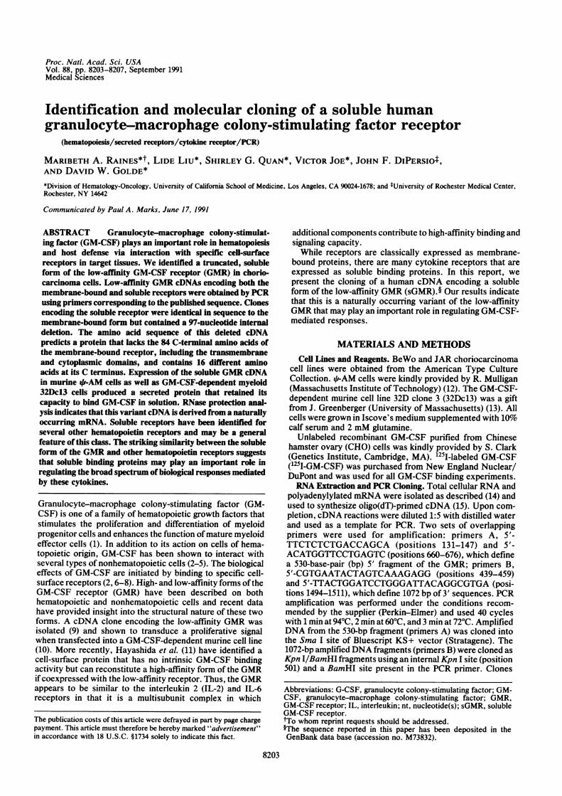
Poly(A)+ RNA from the BeWo choriocarcinoma cell line wasused to isolate cDNA clones encoding the low-affinity humanGMR. Two sets of overlapping primers corresponding to thepublished sequence were used to amplify GMR-specificcDNA sequences with the RNA-directed PCR technique(Fig. LA). A number of PCR-amplified clones were obtainedthat spanned either the 5' or 3' end of the GMR codingsequence. The nucleotide sequence of the cDNA clones
A
Proc. Natl. Acad. Sci. USA 88 (1991)
isolated was identical to that reported by Gearing et al. (9).Several clones spanning the 3' end of the GMR codingsequence (6 of 26 analyzed) contained a 97-nucleotide (nt)deletion (nt 1096-1192), which removed the entire transmem-brane domain of the GMR (Fig. 1B). This deletion occurredwithin a repeated pentanucleotide sequence, GGTTC, mak-ing it difficult to precisely identify its boundaries. This variantcDNA (sGMR) predicts a protein product that encodes atruncated GMR, where 84 C-terminal amino acids of theGMR are replaced by 16 different amino acids (Fig. 1C).Furthermore, this protein should be secreted from the cellsince it lacks a transmembrane domain but retains its signalsequence, suggesting that it encodes a soluble form of theGMR.
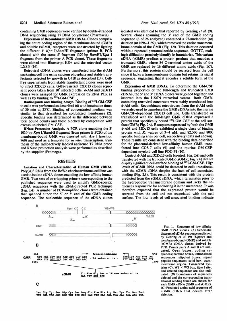
Expression of GMR cDNAs. To determine the GM-CSFbinding properties of the full-length and truncated GMRcDNAs, the 5' and 3' cDNA clones were joined together andinserted into the LXSN retroviral vector. Both GMR-containing retroviral constructs were stably transfected into4p-AM cells. Recombinant retroviruses from the *-AM cellswere also used to transduce the GMR cDNAs into the murineGM-CSF-dependent 32Dc13 cell line. Cells transfected ortransduced with the full-length GMR cDNA expressed aprotein that specifically bound 125I-GM-CSF at the cell sur-face (GMR; Fig. 2A). Receptors expressed by both the GMR4,-AM and 32Dc13 cells exhibited a single class of bindingprotein with Kd values of 3-4 nM, and 82,500 and 8000specific binding sites per cell, respectively (data not shown).These results are consistent with the binding data describedfor the placental-derived low-affinity human GMR trans-fected into COS-7 cells (9) and the murine GM-CSF-dependent myeloid cell line FDC-P1 (10).
Control 4,-AM and 32Dc13 cells (control; Fig. 2A) and cellstransfected with the truncated GMR (sGMR; Fig. 2A) did notdisplay significant cell-surface binding of 125I-GM-CSF. Highlevels of sGMR RNA could be detected in cells transfectedwith the sGMR cDNA despite the lack of cell-associatedbinding (Fig. 2A). This result is consistent with the proteinpredicted from the sGMR cDNA, which terminates prior tothe hydrophobic transmembrane domain and lacks the se-quences responsible for anchoring it in the membrane. It wastherefore expected that the expressed protein would besecreted from the cell and not associated with the cellsurface. The low levels of cell-associated binding indicate
KpnICC CC WSxWS
A D i 0.1 kbA .* AZM
4-'~~~~~~~~-1V//
GMR
sGMR F--r -
B
314 trensmembrene 350GMR Glu Phe Gly Set Asp Asp Gly Lys Arg Phe Leu
GAA TTT T GAC GAC GGG AAC 24 amino acids - TTT AAA fGGTT CTT1089 1199
sGMR Glu Phe Gly Ser 16 new amino acids
GAA TTTC
Leu Gly Tyr Ser Gly Cys Ser Arg Gln Pho His Arg Ser Lys Thr Asn EndTTA-GGA TAC AGC GGC TGT TCC CGC CAG TTC CAC AGA TCA AAG ACA AAC TGA
FIG. 1. Structure of low-affinityGMR cDNA clones. (A) Schematicdiagram ofcDNA sequence publishedby Gearing et al. (9) (Upper) andmembrane-bound (GMR) and soluble(sGMR) cDNA clones derived byPCR. Primer pairs A and B are indi-cated. Open boxes, coding se-quences; hatched boxes, untranslatedsequences; stippled boxes, signalpeptide sequences; solid box, trans-membrane region. Conserved cys-teine (C), WS x WS box, Kpn I site,and deleted sequences are also indi-cated. (B) Boundaries of sequencesdeleted and the corresponding trans-lational reading frame are shown foreach GMR cDNA (GMR and sGMR).(C) Predicted amino acid sequence ofsGMR cDNA that occurs afterdeletion.
13-0-

Proc. Natl. Acad. Sci. USA 88 (1991) 8205
B
Iz4 'r - _ - im _- -I T- _ - 4- & 4.fi-
2 3 , 3 5 2 2 E 3 2 o 3 2 I
0 0 0 0 0 0 0 0 0
9-AM 32DcI3 (p-AM 32DcI3
FIG. 2. GM-CSF binding characteris-tics of recombinant GMR cDNAs ex-pressed by stable transfectants. (A) Spe-cific binding of 2 nM 1251-GM-CSF to 2.5x i05 murine *-AM and GM-CSF-dependent 32Dc13 control cells and celllines expressing either the membrane-bound (GMR-1 and GMR-2) or solubleform (sGMR-1 and sGMR-2) of the GMRcDNA. Specific binding is defined as thedifference between total bound countsand those displaced by a 50-fold excess ofunlabeled GM-CSF. For each cell line orconditioned medium analyzed, n = 6. (B)Specific binding of 1 nM 125I-GM-CSF to25 ul of 5-fold concentrated conditionedmedium from control L-AM and GM-CSF-dependent 32Dc13 cells and fromcells expressing the respective GMRcDNAs.
that the protein is effioiently secreted into the medium andthat the 16 unique C-terminal amino acids are not sufficientto stably anchor the protein in the membrane.To conclusively demonstrate the secretion of the recom-
binant protein by the sGMR-transfected cells, we analyzedthe medium of transfected and untransfected cells for theirability to specifically bind 1251-GM-CSF in solution (Fig. 2B).Only conditioned medium from cells transfected with thesGMR cDNA contained significant levels of 125I-GM-CSFbinding activity. In these samples, 50-70%o of the total boundcounts were displaced with a 50-fold excess of unlabeledGM-CSF. The level of specificity observed in this assay issimilar to the specificity observed in the cell-associatedbinding experiments described above (data not shown). De-tection of a soluble GM-CSF binding protein from cellsexpressing sGMR was reproducible by using several differentpreparations of conditioned medium and was observed inboth the Di-AM transfectants and in the transduced 32Dc13cells. Although the sGMR appears to be a low-affinity GMRwith a dissociation constant similar to the membrane-boundform, we have not yet been able to achieve saturable bindingwith concentrated conditioned medium and therefore cannotassign a definite dissociation constant to this protein. None-theless, these results demonstrate that the sGMR cDNAencodes a soluble form ofthe GMR that is efficiently secretedand that retains its ability to bind GM-CSF. In contrast to thesGMR-producing cells few, if any, GMRs were detected inmedium from untransfected cells (control) or from cellstransfected with the full-length GMR cDNA (GMR; Fig. 2B),suggesting that the membrane-bound receptor is not shedwhen expressed in this context.
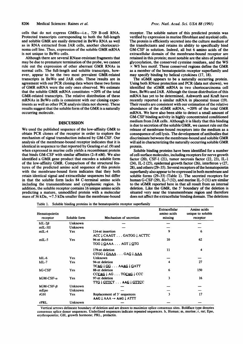
Expression of sGMR mRNA. RNase protection analysiswas used to demonstrate that the sGMR cDNA was not anartefact ofPCR cloning and was present as a bona fide mRNAspecies in cells. A 602-nt antisense RNA probe was synthe-sized from one of the 3' PCR clones encoding the membrane-bound GMR. This RNA probe contained 551-nt of GMR-specific sequences and contained the 97 nt that are deleted inthe sGMR cDNA (Fig. 3B). Thus, the full-length GMRmRNA should protect a 551-nt RNase-resistant fragment,while the soluble GMR mRNA should yield two resistantproducts, one -317 nt and the other 136 nt. Total RNA fromGMR- and sGMR-transfected qD-AM cells (GMR-*2- andsGMR-qi2) were used as positive controls and exclusivelyexpressed the full-length or deleted mRNAs, respectively.The predominant transcripts protected by the transfected cellmRNAs yield the predicted 551-nt fragment in the GMR-4f-AM RNA and the 317- and 136-nt fragment in sGMR-iFi-AMmRNA. The protected transcripts corresponding to the sol-uble GMR mRNA produced broader bands since there is a5-bp repeat at the deletion boundary and thus protected
fragments are produced that differ in size by several nucle-otides.
Additional RNase-resistant products are also apparent andare presumably due to the protection of prematurely termi-nated transcripts contaminating the RNA probe. No RNase-resistant transcripts were observed in the RNA taken from
A SaMt 0Om< l 0
(~ <CMCCa
726-*
553-_500-
417-_
311 -
249-
200-..i3
.1151 -_140-
118-
..
is
Al
t.
B960_
551 nt
a136nt1j., a 317nt
602
551
-317
-136
1511 17 602 nt-i_ RNA probe
_-+ GMR-i sGMR
FIG. 3. RNase protection analysis. (A) Total RNA from BeWo,JAR, 729-6 cells, or L-AM cells transfected with the membrane-bound (lane GMR-4i) or soluble (lane sGMR-/i) GMR was hybridizedto a 602-nt T7 RNA transcript. Samples were treated with RNase T2,precipitated, and analyzed on a 4% denaturing polyacrylamide gel.Sizes (nt) of protected fragments are indicated and were determinedrelative to the mobility of 32P-labeled Hinfi-digested 4X174 DNA(lane M). Undigested probe (lane probe) and digested probe (lanetRNA) are also shown. (B) Schematic diagram ofT7 RNA transcripttranscribed from PCR cDNA clone (primers B) containing mem-brane-bound GMR sequences (solid box) in Bluescript KS+. Pre-dicted sizes of protected products corresponding to the membrane-bound (GMR) or soluble (sGMR) GMR mRNA are indicated.
0._
._
D E
00:L
en
UNCM
Medical Sciences: Raines et al.
8206 Medical Sciences: Raines et al.
cells that do not express GMRs -i.e., 729 B-cell RNA.Protected transcripts corresponding to both the full-lengthand soluble GMR are easily detected in BeWo RNA as wellas in RNA extracted from JAR cells, another choriocarci-noma cell line. Thus, expression of the soluble GMR mRNAis not unique to BeWo cells.Although there are several RNase-resistant fragments that
may be due to premature termination ofthe probe, we cannotrule out the expression of other aberrant GMR RNAs innormal cells. The full-length and soluble transcripts, how-ever, appear to be the two most prevalent GMR-relatedtranscripts in BeWo and JAR cells. These results are inagreement with our PCR cloning data where these two formsof GMR mRNA were the only ones observed. We estimatethat the soluble GMR mRNA constitutes -20% of the totalGMR-related transcripts. The relative distribution of thesemRNAs in BeWo cells is consistent with our cloning exper-iments as well as other PCR analysis (data not shown). Theseresults suggest that the soluble form ofthe GMR is a naturallyoccurring molecule.
DISCUSSIONWe used the published sequence of the low-affinity GMR toobtain PCR clones of the receptor in order to explore themechanism of signal transduction induced by GM-CSF. Ouranalysis of the membrane-bound receptor indicates that it isidentical in sequence to that reported by Gearing et al. (9) andwhen expressed in murine cells yields a recombinant proteinthat binds GM-CSF with similar affinities (2-8 nM). We alsoidentified a GMR gene product that encodes a soluble formof the low-affinity GMR. Comparison of the structural fea-tures of the predicted amino acid sequence of this proteinwith the membrane-bound form indicates that they bothretain identical signal and extracellular sequences but differin that the soluble form lacks 84 C-terminal amino acidsincluding the transmembrane and cytoplasmic region. Inaddition, the soluble receptor contains 16 unique amino acidspredicting a mature, unmodified protein with a molecularmass of 36 kDa, -7.5 kDa smaller than the membrane-bound
receptor. The soluble nature of this predicted protein wasverified by expression in murine fibroblast and myeloid cells.The protein is efficiently secreted into the culture medium ofthe transfectants and retains its ability to specifically bindGM-CSF in solution. Indeed, all but 6 amino acids of theextracellular domain of the membrane-bound receptor areretained in this protein; most notable are the sites ofpotentialglycosylation, the conserved cysteine residues, and the WSx WS box motif. These conserved regions define the GMRas a member of the hematopoietin receptor superfamily andmay specify binding by helical cytokines (17, 18).The sGMR appears to be a naturally occurring protein.
Using both RNase protection and PCR (data not shown), weidentified the sGMR mRNA in two choriocarcinoma celllines, BeWo and JAR. Although the tissue distribution of thismRNA has yet to be determined, Ashworth and Kraft haverecently reported a similar mRNA in placental tissue (19).Their results are consistent with our estimation ofthe relativeabundance of the sGMR mRNA, -20% of the total GMRmRNA. We have also been able to detect a small amount ofGM-CSF binding activity in highly concentrated conditionedmedium from JAR cells. Although it is likely that this bindingis due to secretion ofthe soluble GMR, we cannot rule out therelease of membrane-bound receptors into the medium as aconsequence of cell lysis. The development ofantibodies thatdiscriminate between the membrane-bound and soluble GMRwill aid in characterizing the naturally occurring soluble GMRprotein.
Soluble binding proteins have been identified for a numberofcell-surface molecules, including receptors for nerve growthfactor (20), CSF-1 (21), tumor necrosis factor (22, 23), IL-1(24), IL-2 (25), epidermal growth factor (26), interferon y (27,28), and others (29-35). Several receptors ofthe hematopoietinsuperfamily also appear to be expressed in both membrane andsoluble forms (29-33) (Table 1). The secreted receptors forhuman G-CSF (29), IL-7 (32), and murine IL-5 (31) are similarto the sGMR reported here in that all result from an internaldeletion. Like the GMR, the 5' boundary of the deletion issituated very near the transmembrane region and thereforedoes not affect the extracellular binding domain. The deletions
Table 1. Soluble binding proteins in the hematopoietin receptor superfamilyExtracellular Amino acids
Hematopoietin amino acids unique to solublereceptor Soluble form Mechanism of secretion missing receptor
hIL-2,f UnknownmIL-311 UnknownmIL-4 Yes 114-nt insertion 9 6
ACC CAAGT ... GATGG I ACTTCmIL-5 Yes 94-nt deletion 10 62
TGG l AAA ... AGT | GTG
179-nt deletion 11 4GTGGIA . ... GAQJAAA
hIL-6 Yes UnknownhIL-7 Yes 94-nt deletion 4 27
TCMA IG . .. AAAG l ATThG-CSF Yes 88-nt deletion 6 150
CCCA I AG . .. TGC ICCChGM-CSF-a Yes 97-nt deletion 3 16
TTQ I DTTCT . .. AA5G I GTCChGM-CSF-/3 UnknownmEpo UnknownrGH Yes Replacement of 3' sequences 3 17
AAG I AAA -* AAG I ATTrPRL Unknown
Vertical arrows delineate boundary of deletion and are drawn to maximize splice consensus sites. Boldface type denotesconsensus splice donor sequences. Underlined sequences indicate repeated sequences. h, Human; m, murine; r, rat; Epo,erythropoietin; GH, growth hormone; PRL, prolactin.
Proc. Natl. Acad. Sci. USA 88 (1991)

Proc. Natl. Acad. Sci. USA 88 (1991) 8207
encompass most, if not all, of the transmembrane region andcause a shift in the translational reading frame such that alltransmembrane and cytoplasmic C-terminal amino acids areremoved. In each case, several amino acids unique to thesoluble form are added. As would be predicted from theextracellular sequences expressed by these proteins, the sol-uble forms of IL-5, IL-7, and GMR are secreted and retaintheir ligand-binding capacity.Although soluble receptors for growth hormone, IL-6, and
IL-4 have been detected in vivo (27, 28, 34), their mechanismof secretion is unknown. The striking parallels in the dele-tions described above suggest that the receptors for IL-5,IL-7, G-CSF, and GM-CSF use alternative mRNA splicing asa mechanism to generate their soluble counterparts. Exam-ination of the sequences situated directly 5' to the deletionboundaries (Table 1) reveals sequences reminiscent of con-
sensus splice donor sites ((C/A)AG). The extent of thedeletion (-95 nt) and the sequences encompassed (the entiretransmembrane region) suggests that the deleted sequencesconstitute a single exon. Such a notion is consistent with theintron-exon organization of the transmembrane region ofother membrane-bound molecules. Removal of the trans-membrane domain by alternative splicing has been describedin other systems and is best exemplified in IgM antibodysecretion (35). Although alternative splicing seems to be themost likely mechanism, we cannot rule out the possibilitythat these deleted mRNAs arose from a gene locus distinctfrom that encoding the membrane-bound receptor.The prevalence of soluble forms of hematopoietin recep-
tors suggests that they are of biological importance. SolubleGM-CSF receptors may modulate GM-CSF-mediated re-sponses by competing with the membrane-bound receptor forGM-CSF binding. It may act as a direct inhibitor of GMRfunction or in a feedback mechanism to turn off GM-CSF-stimulated responses. It is not known whether the subunitof the GMR can influence these processes. Association ofsoluble receptors with other membrane-bound componentshas been documented for soluble IL-6 and epidermal growthfactor receptors (36, 37). For example, the soluble IL-6receptor can associate with gpl3O to transduce a signal (36).The sGMR may be of clinical significance since it could playa role in regulating the differentiation and function of hem-atopoietic cells. Soluble GM-CSF receptors could also act todampen inflammatory responses and therefore have thera-peutic potential.
We thank Scott Friedenberg, Caroline Tomongin, Rex Hsei, andJoe Garcia for their expert technical assistance and Dorothy Parkerand Elizabeth Koers for manuscript preparation. This work wassupported by U.S. Public Health Service Grants CA30388, CA32737,and HL42107. M.A.R. is a postdoctoral trainee supported by De-partment of Health and Human Services Public Health ServiceNational Institutional Research Award T32 CA09056.
1. Gasson, J. C. (1991) Blood 77, 1131-1145.2. DiPersio, J. F., Hedvat, C., Ford, C. F., Golde, D. W., Gas-
son, J. C. (1991) J. Biol. Chem. 266, 279-286.3. Baldwin, G. C., Gasson, J. C., Kaufman, S. E., Quan, S. G.,
Williams, R. E., Avalos, B. R., Gazdar, A. F., Golde, D. W.,DiPersio, J. F. (1989) Blood 73, 1033-1037.
4. Bussolino, F., Wang, J. M., Defilippi, P., Turrini, F., Sanavio,F., Edgell, C.-J. S., Aglietta, M., Arese, P. & Mantovani, A.(1989) Nature (London) 337, 463-471.
5. Dedhar, S., Tojo, A., Kitamura, T., Urabe, A., Miyazono, K.& Takaku, F. (1988) Proc. Nail. Acad. Sci. USA 85, 9253-9257.
6. Gasson, J. C., Kaufman, S. E., Weisbart, R. H., Tomonaga,M. & Golde, D. W. (1986) Proc. Nail. Acad. Sci. USA 83,669-673.
7. Park, L. S., Friend, D., Gillis, S. & Urdal, D. L. (1986) J. Exp.Med. 164, 251-262.
8. Chiba, S., Tojo, A., Kitamura, T., Urabe, A., Miyazono, K. &Takaku, F. (1990) Leukemia 4, 29-36.
9. Gearing, D. P., King, J. A., Gough, N. M. & Nicola, N. A.(1989) EMBO J. 8, 3667-3676.
10. Metcalf, D., Nicola, N. A., Gearing, D. P. & Gough, N. M.(1990) Proc. Nail. Acad. Sci. USA 87, 4670-4674.
11. Hayashida, K., Kitamura, T., Gorman, D. M., Arai, K.-i.,Yokota, T. & Miyajima, A. (1990) Proc. Nail. Acad. Sci. USA87, %55-9659.
12. Cone, R. D. & Mulligan, R. C. (1984) Proc. Nail. Acad. Sci.USA 81, 6349-6353.
13. Greenberger, J. S., Sakakeeny, M. A., Humphries, R. K.,Eaves, C. J. & Eckner, R. J. (1983) Proc. Natl. Acad. Sci. USA80, 2931-2935.
14. Raines, M. A., Maihle, N. J., Moscovici, C., Moscovici, M. G.& Kung, H.-J. (1988) J. Virol. 62, 2444-2452.
15. Gubler, U. & Hoffman, B. J. (1983) Gene 25, 263-269.16. Miller, A. D. & Rosman, G. J. (1989) -BioTechniques 7, 980-
990.17. Bazan, J. F. (1990) Proc. Nail. Acad. Sci. USA 87, 6934-6938.18. Bazan, J. F. (1990) Immunol. Today 11, 350-354.19. Ashworth, A. & Kraft, A. (1990) Nucleic Acids Res. 18, 7178.20. DiStefano, P. S. & Johnson, E. M., Jr. (1988) Proc. Nail.
Acad. Sci. USA 85, 270-274.21. Downing, J. R., Roussel, M. F. & Sherr, C. J. (1989) Mol. Cell.
Biol. 9, 2890-28%.22. Gray, P. W., Barrett, K., Chantry, D., Turner, M. & Feld-
mann, M. (1990) Proc. Nail. Acad. Sci. USA 87, 7380-7384.23. Kohno, T., Brewer, M. T., Baker, S. L., Schwartz, P. E.,
King, M. W., Hale, K. K., Squires, C. H., Thompson, R. C. &Vannice, J. L. (1990) Proc. Nail. Acad. Sci. USA 87, 8331-8335.
24. Symons, J. A. & Duff, G. W. (1990) FEBS Lett. 272, 133-136.25. Robb, R. J. & Kutny, R. M. (1987) J. Immunol. 139, 855-862.26. Maihle, N. J., Flickinger, T. W., Raines, M. A., Sanders,
M. L. & Kung, H. J. (1991) Proc. Nail. Acad. Sci. USA 88,1825-1829.
27. Novick, D., Engelmann, H., Wallach, D. & Rubinstein, M.(1989) J. Exp. Med. 170, 1409-1414.
28. Novick, D., Engelmann, H., Wallach, D., Leitner, O., Revel,M. & Rubinstein, M. (1990) J. Chromatogr. 510, 331-337.
29. Fukunaga, R., Seto, Y., Mizushima, S. & Nagata, S. (1990)Proc. Nail. Acad. Sci. USA 87, 8702-8706.
30. Mosley, B., Beckmann, M. P., March, C. J., Idzerda, R. L.,Gimpel, S. D., VandenBos, T., Friend, D., Alpert, A., Ander-son, D., Jackson, J., Wignall, J. M., Smith, C., Gallis, B.,Sims, J. E., Urdal, D., Widmer, M. B., Cosman, D. & Park,L. S. (1989) Cell 59, 335-348.
31. Takaki, S., Tominaga, A., Hitoshi, Y., Mita, S., Sonoda, E.,Yamaguchi, N. & Takatsu, K. (1990) EMBO J. 9, 4367-4374.
32. Goodwin, R. G., Friend, D., Ziegler, S. F., Jerzy, R.+ Falk,B. A., Gimpel, S., Cosman, D., Dower, S. K., March, C. J.,Namen, A. E. & Park, L. S. (1990) Cell 60, 941-951.
33. Baumbach, W. R., Homer, D. L. & Logan, J. S. (1989) GenesDev. 3, 1199-1205.
34. Femandez-Botran, R. & Vitetta, E. S. (1990) Proc. Nail. Acad.Sci. USA 87, 4202-4206.
35. Early, P., Rogers, J., Davis, M. (1980) Cell 20, 313-319.36. Hibi, M., Murakami, M., Saito, M., Hirano, T., Taga, T. &
Kishimoto, T. (1990) Cell 63, 1149-1157.37. Basu, A., Raghunath, M., Bishayee, S. & Das, M. (1989) Mol.
Cell. Biol. 9, 671-677.
Medical Sciences: Raines et al.


![NUEVO LA PODERoso 83 8207' TRES 2 S] a Eta I e BP 381, N ...geneticaglobal.com.ar/assets/images/toros/53_Hermano.pdf · nuevo la poderoso 83 8207' tres 2 s] a eta i e bp 381, n' la](https://img.dokumen.tips/doc/110x75/5f084aec7e708231d4214a52/nuevo-la-poderoso-83-8207-tres-2-s-a-eta-i-e-bp-381-n-nuevo-la-poderoso-83.jpg)