Embed Size (px)
Citation preview

266Publicación en línea, mayo 2021
DOI: https://doi.org/10.18781/R.MEX.FIT.2103-4
Armenta-López SE, Valenzuela-Solano C and Hernández-Martínez R. 2021. Identification and molecular analysis of races of Fusarium oxysporum f. sp. lycopersici isolated from tomato in Baja California, Mexico. Mexican Journal of Phytopathology 39(2): 266-288.
Primera publicación DOI: 29 de Abril, 2021.First DOI publication: April 29, 2021.
Resumen. Baja California es uno de los princi-pales productores de jitomate (Solanum lycopersi-cum) en México. La marchitez vascular, causada por Fusarium oxysporum f. sp. lycopersici (Fol), es una de las principales enfermedades que afectan al cultivo del jitomate en el estado de Baja California; sin embargo, se desconoce la identidad de las espe-cies de Fusarium, la ocurrencia de Fol y las razas
Identification and molecular analysis of races of Fusarium oxysporum f. sp. lycopersici isolated from tomato
in Baja California, Mexico
Identificación y análisis molecular de razas de Fusarium oxysporum f. sp. lycopersici aisladas de jitomate en Baja California, México
Sara Elodia Armenta-López, Junta Local de Sanidad Vegetal del Valle del Fuerte, Lázaro Cárdenas 315 Pte. Col. Centro, Los Mochis, Sinaloa; Cesar Valenzuela-Solano, Sitio Experimental Costa de Ensenada- INIFAP. Calle del Puerto Núm. 375-23 Fracc. Playa Eda. Ensenada, B. C. 22880; Rufina Hernández-Martínez*, Centro de Investigación Científica y de Educación Superior de Ensenada (CICESE). Carretera Ensenada-Tijuana 3918, Zona Playitas, 22860 Ensenada, B.C. Autora de Correspondencia: [email protected]
Recibido: 04 de Marzo, 2021. Aceptado: 23 de Abril, 2021.
Abstract. In Baja California, Fusarium oxysporum f. sp. lycopersici (Fol) is the causal agent of vascular wilt of tomato; however, the races present in the state remain unknown. The objective of this work was to isolate and identify Fusarium spp. and to validate a multiplex-PCR system to identify strains Fol and at the race level. Sixty symptomatic plants were collected, of which 45 isolates were obtained. When analyzed microscopically, it was confirmed that 44 corresponded to Fusarium spp. The use of the sp13 and sp23 oligonucleotide pairs that amplified the pgx4 and pg1 polygalacturonase genes, respectively, indicated the presence of Fol races 1 and 3 in the area. Sequence analysis of the elongation factor of the 1α translation (TEF1-α) of representative strains served to confirm its identity and the presence of Fol and F. solani in tomato plants with symptoms of vascular wilt. Finally, a RAPD analysis showed that the oligonucleotide OPA-11, generates a specific banding pattern in

Publicación en línea, mayo 2021 267
Fully BilingualRevista Mexicana de FITOPATOLOGÍA
Mexican Journal of Phytopathology
presentes. El objetivo del presente trabajo fue aislar e identificar aislados de Fusarium de plantas enfer-mas de jitomate y validar métodos de diagnóstico para identificar Fol a nivel de raza. Se colectaron 60 plantas sintomáticas, de las que se obtuvieron 45 aislados. Al ser analizadas microscópicamente se confirmó que 44 pertenecieron al género Fusa-rium. El uso de los pares de oligonucleótidos sp13 y sp23, que amplifican los genes de poligalacturo-nasas pgx4 y pg1, indicaron la presencia de Fol ra-zas 1 y 3 en la zona. El análisis de secuencias del factor de elongación de la traducción 1α (TEF1-α) de cepas representativas confirmó la identifica-ción de la presencia de Fol y de Fusarium solani en plantas de jitomate con síntomas de marchitez vascular. Finalmente, un análisis RAPD, identificó que el oligonucleótido OPA-11 genera un patrón de bandeo especifico en aislados de la raza 1 por lo que podría emplearse como una forma rápida para la identificación de cepas de esta raza.
Palabras clave: polimorfismos de ADN, xilema, enfermedad vascular, hongos
El jitomate (Solanum lycopersicum) es un cul-tivo de gran importancia económica en muchos países. México ocupa el segundo lugar con una producción de 2,923,163 t, en una superficie de 47,151 ha (SIAP, 2019). Fusarium oxysporum f. sp. lycopersici (Fol) es el agente causal de la mar-chitez vascular del jitomate. Actualmente, se cono-cen tres razas fisiológicas de Fol que difieren en su grado de virulencia (Biju et al., 2017; Chang et al., 2018). Para su detección se han utilizado distintas técnicas, como métodos moleculares (Chang et al., 2018; Murugan et al., 2020) y variedades diferen-ciales de jitomate con distintos genes de resistencia a cada una de las razas (Cai et al., 2003; Pirayesh et al., 2018), y la comparación de secuencias
Fol race 1, so it could be used as a quick way to identify them.
Key words: DNA Polymorphisms, xylem, vascular disease, fungi
The tomato (Solanum lycopersicum) is a crop of great economic importance in many countries. Mexico is its second largest producer, with 2,923,163 t on a surface of 47,151 ha (SIAP, 2019). Fusarium oxysporum f. sp. lycopersici (Fol) is the causal agent of vascular wilting in tomato. Three physiological races of Fol are currently known, which vary in their degree of virulence (Biju et al., 2017; Chang et al., 2018). Different techniques have been used to detect them, such as molecular methods (Chang et al., 2018; Murugan et al., 2020), along with different varieties of tomatoes with different genes for resistance to each of the races (Cai et al., 2003; Pirayesh et al., 2018) and the comparison of nucleotide sequences of the polygalacturonase genes (Kawabe et al., 2005; Murugan et al., 2020). These enzymes degrade polygaracturonic acid and are found in all of the organs of the plant, as well as in the cell walls of the tomato fruit, and they act as pectin depolymerizers. They therefore help invade and colonize host tissues (Martel et al., 1998). Hirano and Arie (2006) compared the partial nucleotide sequences of an endopolygalacturonase gene (pg1) and an exopolygalacturonase gene (pgx4) of the special forms of (ff. spp.) lycopersici and radicis-lycopersici (which causes the rotting of the tomato crown), and designed the pairs of oligonucleotides (sp13 y sp23), based on Single Nucleotide Polymorphisms (SNPs), which, combined, help distinguish the Fol races.
In Mexico, races 1, 2 and 3 of Fol have been reported in the state of Sinaloa (Valenzuela-Ureta et al., 1996; Carrillo et al., 2003; Ascencio-Álvarez

Publicación en línea, mayo 2021 268
Fully BilingualRevista Mexicana de FITOPATOLOGÍAMexican Journal of Phytopathology
nucleotídicas de los genes de poligalacturonasas (Kawabe et al., 2005; Murugan et al., 2020). Estas enzimas son degradadoras de ácido poligalacturó-nico, presente en todos los órganos de la planta y de la pared celular del fruto del jitomate, y actúan como depolimerizadoras de pectina, por lo que fa-cilitan la invasión y colonización de tejido del hos-pedero (Martel et al., 1998). Hirano y Arie (2006) compararon las secuencias nucleotídicas parciales de un gen de endopoligalacturonasa (pg1) y otro de una exopoligalacturonasa (pgx4) de las formas es-peciales (ff. spp.) lycopersici y radicis-lycopersici (causante de la pudrición de la corona del jitomate) con las que diseñaron los pares de oligonucleóti-dos (sp13 y sp23), basados en polimorfismos de un solo nucleótido (SNPs, Single Nucleotid Polymor-phims), que en conjunto permiten diferenciar las razas de Fol.
En México, se ha reportado la presencia de las razas 1, 2 y 3 de Fol en el estado de Sinaloa (Va-lenzuela-Ureta et al., 1996; Carrillo et al., 2003; Ascencio-Álvarez et al., 2008), las razas 2 y 3 en Baja California Sur (Holguín-Peña, 2005) y las razas 2 y 3 en San Luis Potosí (Hernández et al., 2014). Adicionalmente, se ha reportado la presen-cia de Fusarium circinatum y Fusarium andiyazi ocasionando marchitez en jitomate en los estados de Morelos, Puebla y Tlaxcala (Isaac et al., 2018). En Baja California, se conoce de la presencia de Fusarium en jitomate, pero no se han identificado las especies. La correcta identificación de las va-riantes patogénicas de Fusarium presentes en una determinada zona de cultivo, es indispensable para establecer la utilización de cultivares. El objetivo de este trabajo fue aislar y caracterizar especies de Fusarium asociadas a plantas de jitomate con síntomas de marchitez vascular en Baja California, México y al mismo tiempo validar un método mo-lecular para diferenciarlos.
et al., 2008), races 2 and 3 in Baja California Sur (Holguín-Peña, 2005), and races 2 and 3 in San Luis Potosí (Hernández et al., 2014). Additionally, the presence of Fusarium circinatum and Fusarium andiyazi have been reported to cause wilting in tomatoes in the states of Morelos, Puebla and Tlaxcala (Isaac et al., 2018). In Baja California, the presence of Fusarium is known in tomato, although the species have not yet been identified. The correct identification of the pathogenic variants of Fusarium found in a particular cultivation area is crucial to establish the use of cultivars. The aim of this investigation was to isolate and characterize Fusarium species related to tomato plants with vascular wilt symptoms in Baja California, Mexico, as well as to validate a molecular method to differentiate them.
MATERIALS AND METHODS
Collection of samples, isolation and morphologic characterization of strains
Tomato plants with symptoms of vascular wilt were gathered from four locations: two from the town of Maneadero and two from the town of San Quintín, in the municipal area of Ensenada, Baja California. In total, 60 tomato plants were collected, with different levels of vascular wilt symptoms. Five samples of the Cherry variety were taken from San Quintín Este, from an open-air plantation. Ten plants established in bags were taken from a greenhouse in San Quintín Norte, five of which were Cherry Zebra and five, Strawberry tomatoes. From Maneadero Sur, three greenhouses with plants grown in the soil were sampled; two Cherry tomato plants were taken from the first one, along with three Heirloom tomato plants. From the
Publicación en línea, mayo 2021 269
Fully BilingualRevista Mexicana de FITOPATOLOGÍA
Mexican Journal of Phytopathology
MATERIALES Y MÉTODOS
Colecta de muestras, aislamiento y caracteriza-ción morfológica de cepas
Se colectaron plantas de jitomate con sínto-mas de marchitez vascular en cuatro sitios, dos del poblado de Maneadero y dos del poblado de San Quintín del municipio de Ensenada, Baja Califor-nia. En total, se colectaron 60 plantas de jitomate con diferentes niveles de síntomas de marchitez vascular. De San Quintín Este, se obtuvieron cinco muestras de la variedad tipo Cherry de un cultivo establecido a cielo abierto. De San Quintín Norte, se obtuvieron diez plantas de cultivo en invernade-ro establecidos en bolsas, cinco tipo Cherry Zebra y cinco tipo Fresa. De Maneadero Sur, se muestrea-ron tres invernaderos con cultivos establecidos en suelo, en el primero se tomaron dos plantas del tipo Cherry y tres del tipo Heirloom; en el segundo, cin-co muestras del tipo Heirloom, y en el tercero cinco muestras, una de la variedad G5, tres de la variedad RG-871 y una de la variedad TC. Finalmente, en Maneadero Norte se tomaron muestras de 30 plan-tas tipo Cherry establecidos a cielo abierto.
Las muestras previamente etiquetadas, fueron depositadas en bolsas de polietileno e inmediata-mente trasladadas al laboratorio de Fitopatología del Centro de Investigación Científica y de Edu-cación Superior de Ensenada (CICESE) para su procesamiento. Las plantas se lavaron con agua co-rriente, se secaron con toallas de papel y en el tallo, a aproximadamente 10 cm de altura de la corona se realizaron tres cortes transversales y se obtuvie-ron trozos de tejido sintomático. Se sumergieron en alcohol al 75% y se flamearon rápidamente. Poste-riormente, con la ayuda de una navaja esterilizada al calor, se cortaron rebanadas de alrededor de 0.5 cm y se colocaron sobre medio de cultivo agar papa-dextrosa (PDA), suplementado con cloranfenicol
second one, five Hierloom samples were taken, and in the third one, five G5 samples, three of the RG-871 variety and one TC. Finally, in Maneadero Norte, 20 samples were taken of Cherry tomato plants grown in the open field.
The samples were labelled and placed in polyethylene bags and moved to the Phytopathology Laboratory of the Scientific Research and Higher Education Center of Ensenada (Centro de Investigación Científica y de Educación Superior de Ensenada, CICESE) for processing. The plants were washed with tap water, dried with paper towels and, on the stem, approximately 10 cm above the root crown, three cross sectional cuts were made and pieces of symptomatic tissue were taken. They were submerged in alcohol at 75% and quickly flamed. Later, using a heat-sterilized blade, 0.5 cm pieces were cut and placed in a potato-dextrose-agar (PDA) culture medium, supplementing with chloramphenicol (15 μg mL-1), and the Petri dishes were incubated in the dark at 25 ± 2 °C. To obtain the pure cultures from the fungal growth found, seven days after harvesting, hyphae tips were transferred into a PDA medium and their putative identification was carried out using morphological observation. From those primary plantations, monosporic strains were obtained, which were kept in glycerol at -20 °C until their use.
The fungal isolates were grown in PDA and agar-carnation (CLA) (Fisher et al., 1982), two of the standard media used to identify Fusarium species (Leslie and Summerell, 2008). The Petri dishes were stored at 27 °C in complete darkness, following recommendations for their identification. After 15 days, the morphology and pigmentation of the culture in PDA were analyzed, along with the production of sporodochia, sclerotia, reproductive structures and macroconidia in a CLA medium using an Olympus SZX12 stereoscopic microscope and an Axiovert 200 Zeiss® inverted microscope.

Publicación en línea, mayo 2021 270
Fully BilingualRevista Mexicana de FITOPATOLOGÍAMexican Journal of Phytopathology
(15 μg mL-1) y las cajas se incubaron en la oscuri-dad a 25 ± 2 °C. Para la obtención de cultivos pu-ros del crecimiento fúngico observado, alrededor de siete días después de la siembra, se transfirieron puntas de hifas a medio PDA y su identidad puta-tiva se hizo por observación morfológica. A partir de éstos cultivos primarios se obtuvieron cepas mo-nospóricas, que se mantuvieron en glicerol a -20 °C hasta su utilización.
Los aislamientos fúngicos se cultivaron en PDA y agar-clavel (CLA) (Fisher et al., 1982), dos de los medios estándar utilizados en la identificación de las especies de Fusarium (Leslie y Summerell, 2008). Las cajas se mantuvieron a 27 °C en total obscuridad, de acuerdo a lo recomendado para su identificación. Después de 15 días se analizó la morfología y pigmentación de la colonia en PDA y la producción de esporodoquios, esclerocios, las estructuras de reproducción y las macroconidias en medio CLA usando un microscopio estereoscopio Olympus SZX12 y un microscopio invertido Axio-vert 200 Zeiss®.
Caracterización molecular de razas de F. oxys-porum f. sp lycopersici
Para la extracción de ADN, las cepas se cultiva-ron en papa dextrosa líquido en agitación a 27 °C durante cuatro días. El micelio se recuperó por fil-tración, se congeló y liofilizó en tubos de micro-centrifuga de 2 ml y se pulverizó usando varillas de vidrio de punta aguzada. El ADN genómico se obtuvo empleando el método de CTAB al 3% y al final se ajustó a 50 μg mL-1.
Para la identificación de las razas de Fol se mez-claron dos pares de oligonucleótidos reportados previamente (Hirano y Arie, 2006) para realizar un PCR múltiple. El par sp13-f (5’-GTCAGTCCATT-GGCTCTCTC-3´) y sp13-r (5’-TCCTTGACAC-CATCACAGAG-3’) amplifican un fragmento de
Molecular characterization of races of F. oxysporum f. sp lycopersici
For the extraction of DNA, the strains were grown in a potato dextrose liquid, shaking at 27 °C for four days. The mycelium was recovered by filtration, frozen and lyophilized in 2 ml microcentrifuge tubes, and pulverized using glass rods with pointed tips. The genomic DNA was obtained using the CTAB method at 3% and finally, it was adjusted to 50 μg mL-1.
To identify the races of Fol, two pairs of previously reported oligonucleotides (Hirano and Arie, 2006) were mixed to carry out a multi-plex PCR. The sp13-f (5’-GTCA GTCCATTG-GCTCTCTC-3´) and sp13-r (5’-TCCTTGACAC-CATCACAGAG-3’) pair amplify a 445 pb fragment of the gene of one exopolygalacturonase (pgx4) in races 1 and 3, but not in race 2; the pair of sp23-f (5’-CCTCTTGTCTTTGTCTCACGA-3’) and sp23-r (5’-GCAACAGGTCGTGGGGAAAA-3’) was designed in the site of deletion of two nucleo-tides (nt 259-260) in the gene of an endopolygalac-turonase (pg1) and it amplifies a fragment of 518 pb in races 2 and 3, but not in race 1. In the isolates of F. oxysporum f. sp. radicis-lycopersici, these oligonucleotides did not amplify any fragments (Hirano and Arie, 2006). After testing several con-ditions of amplification by combining the oligo-nucleotides, the reaction mixture used consisted of 1X buffer (Axygen), 0.2 μg mL-1de BSA, 0.2 mM of dNTPs, 2.5 mM of MgCl2, 0.4 µM of each of the four oligonucleotides, 0.1 U μg-1 of Taq polymerase (5 U, Axygen) and 50 ng of DNA in a final volume of 25 µL. Amplification consisted of a 94 °C cycle for 1 min, followed by 45 cycles at 94 °C for 1 min, 61 °C for 1 min, 72 °C for 1 min and a final extension at 72 °C for 10 min, using a BioRad T100 thermocycler. The products were evaluated by electrophoresis in agarose gel at 1% and photo-

Publicación en línea, mayo 2021 271
Fully BilingualRevista Mexicana de FITOPATOLOGÍA
Mexican Journal of Phytopathology
445 pb del gen de una exopoligalacturonasa (pgx4) en las razas 1 y 3 pero no en la raza 2; el par sp23-f (5’-CCTCTTGTCTTTGTCTCACGA-3’) y sp23-r (5’-GCAACAGGTCGTGGGGAAAA-3’) fue di-señado en el sitio de deleción de dos nucleótidos (nt 259-260) en el gen de una endopoligalacturo-nasa (pg1) y amplifica un fragmento de 518 pb en las razas 2 y 3, pero no en la raza 1. En los aisla-dos de F. oxysporum f. sp. radicis-lycopersici estos oligonucleótidos no amplifican ningún fragmento (Hirano y Arie, 2006). Después de probar varias condiciones de amplificación combinado los oligo-nucleótidos, La mezcla de reacción usada consistió en 1X buffer (Axygen), 0.2 μg mL-1de BSA, 0.2 mM de dNTPs, 2.5 mM de MgCl2, 0.4 µM de cada uno de los cuatro oligonucleótidos, 0.1 U μg-1 de Taq polimerasa (5 U, Axygen) y 50 ng de ADN en un volumen final de 25 µL. La amplificación consistió en un ciclo de 94 °C por 1 min, seguido de 45 ci-clos de 94 °C por 1 min, 61 °C por 1 min y 72 °C por 1 min, y una extensión final a 72 °C por 10 min, usando un termociclador BioRad T100. Los productos se evaluaron en electroforesis en gel de agarosa al 1% y se fotografiaron utilizando un fotodocumentador Quantity One de BioRad. El control positivo fue ADN extraído de Fol raza 1 y Fol raza 2, proporcionadas amablemente por el Dr. Raymundo García del Centro de Investigación en Alimentos y Desarrollo (CIAD), unidad Culiacán.
Una vez analizados los productos de PCR, y para confirmar que el fragmento amplificado co-rrespondía a la secuencia esperada, se selecciona-ron al azar cinco aislados de las razas determina-das. Los productos amplificados seleccionados se cortaron del gel y se purificaron utilizando el kit DNA clean y concentrator (Zymo Research) si-guiendo las instrucciones del fabricante y los frag-mentos generados fueron enviados a secuenciar a Eton Bioscience Inc. (San Diego, California). Una vez obtenidas las secuencias, éstas se compararon
graphed using a BioRad Quantity One photodocu-menter. The positive control was DNA taken from race 1 Fol and race 2 Fol, kindly provided by Dr. Raymundo García of the Food Research and De-velopment Center (Centro de Investigación en Ali-mentos y Desarrollo, CIAD), Culiacán.
After analyzing the PCR products, and to confirm that the amplified fragment matched the expected sequence, five isolates were chosen at random from the determined races. The amplified products were cut out of the gel and purified using the DNA Clean and Concentrator kit (Zymo Research), following the instructions by the manufacturer and the fragments generated were sent for sequencing to Eton Bioscience Inc. (San Diego, California). Once the sequences were obtained, they were compared using the Basic Local Alignment Search Tool (BLAST) algorithm from the National Center for Biotechnology Information (NCBI). Sequences from the fragments of pgx4 and pg1 were recovered and they were all aligned using the program BioEdit v7.2.3 (copyright (c) 1997-2013, Tom Hall).
To confirm that the isolates obtained and analyzed with the multiplex PCR were from F. oxysporum, some of them were selected and a fragment of approximately 700 pb from the TEF1-α region was amplified by PCR, using oligonucleotides EF-1 and EF-2 (O’Donnell et al., 1998), used earlier to distinguish between species of Fusarium (Lievens et al., 2009). The reaction of the PCR was similar to the one used to amplify the polygalacturonase genes, but the concentration was reduced to 1.5 mM of MgCl2. The amplification consisted of an initial denaturalization of 94 °C for 3 min, followed by 40 cycles of 1 min at 94 °C, 1 min at 61.5 °C, 1 min at 72 °C and a final extension of 10 min at 72 °C. The fragments were evaluated by electrophoresis in agarose gel, purified and sequenced as described earlier. For the comparative analysis of the sequences, we accessed the Fusarium-ID (http://

Publicación en línea, mayo 2021 272
Fully BilingualRevista Mexicana de FITOPATOLOGÍAMexican Journal of Phytopathology
utilizando el algoritmo Basic Local Alignment Search Tool (BLAST) del National Center for Bio-technology Information (NCBI). Se recuperaron secuencias de los fragmentos de pgx4 y pg1 y todas se alinearon utilizando el programa BioEdit v7.2.3 (copyright (c) 1997-2013, Tom Hall).
Para confirmar que los aislados obtenidos y analizados con la PCR múltiple pertenecían a F. oxysporum, se seleccionaron algunos de ellos y se amplificó por PCR un fragmento de alrededor de 700 pb de la región del TEF1-α, usando los oligo-nucleótidos EF-1 y EF-2 (O’Donnell et al., 1998). Usados anteriormente para diferenciar entre espe-cies de Fusarium (Lievens et al., 2009). La reac-ción de PCR fue similar a la usada para amplificar los genes de poligalacturonasas, excepto que se disminuyó la concentración a 1.5 mM de MgCl2. La amplificación constó de una desnaturalización inicial de 94 °C por 3 min, seguido de 40 ciclos de 1 min a 94 °C, 1 min a 61.5 °C, 1 min a 72 °C y una extensión final de 10 min a 72 °C. Los fragmentos se evaluaron por electroforesis en gel de agarosa, se purificaron y secuenciaron como se describió de manera previa. Para el análisis comparativo de las secuencias, se accedió al banco de datos Fusarium-ID (http://isolate.fusariumdb.org) y al del NCBI, usando el algoritmo de BLAST. Algunas secuen-cias que mostraron máxima similitud con las ob-tenidas en este trabajo se obtuvieron del banco de datos, y todas se alinearon usando el programa Bio-Edit v7.2.3. Finalmente, se obtuvo el filograma uti-lizando el programa MEGAX (Kumar et al., 2018).
Identificación de razas de Fol con el uso de va-riedades diferenciales
Para confirmar la identificación de las razas de-terminadas por PCR múltiple, se utilizaron cuatro genotipos de jitomate; Bonny Best (sin resistencia), Manapal (resistente a Fol raza 1), Walter (resistente
isolate.fusariumdb.org) and NCBI databanks, using the BLAST algorithm. Some sequences that showed the maximum similarity with those obtained in this work were recovered from the genebank, and they were all aligned using the program BioEdit v7.2.3. Finally, the phylogram was obtained using the program MEGAX (Kumar et al., 2018).
Identification of races of Fol with the use of differential varieties
Four tomato genotypes were used to confirm the identity of the races determined by multiplex PCR: Bonny Best (without resistance), Manapal (resistant to Fol race 1), Walter (resistant to Fol race 2) and I3R3 (resistant to Fol race 3) (Ascencio-Álvarez et al., 2008).
Seeds of each variety were planted in polyethylene trays with 72 pits in a 2:1 mixture of Peat moss-perlite and after 30 days, they were transplanted into 1 L polyethylene pots. Twenty-one days after transplanting, six plants of each variety were inoculated with one out of five of the isolates selected (the same ones used in the sequencing analysis), adding 10 mL of a suspension of 1X10⁻⁷ conidia mL-1 two centimeters from the stem (Baysal et al., 2009). Fol strain races 1 and 2 were used as positive controls and the negative control consisted of 10 mL of water. The plants were kept under greenhouse conditions at a variable temperature of 36±16 °C, distributed in a completely random design. Damages were evaluated on a daily basis until the observation of symptoms in the control strains (approximately 15 days after inoculation), based on the arbitrary damage scale proposed previously (Vakalounakis and Fragkiadakis, 1999), where: 0 = plant without symptoms, 1 = plant with slight wilting or similar to a lack of water, 2 = plant similar to scale 1 + yellow or dry leaves in 50% of the foliage, 3 = plant similar to scale 1 + yellow or

Publicación en línea, mayo 2021 273
Fully BilingualRevista Mexicana de FITOPATOLOGÍA
Mexican Journal of Phytopathology
Fol raza 2) e I3R3 (resistente a Fol raza 3) (Ascen-cio-Álvarez et al., 2008).
Las semillas de cada variedad, se sembraron en charolas de polietileno de 72 cavidades en una mezcla 2:1 de Peat moss-perlita. Después de 30 días, se trasplantaron en macetas de polietileno de 1 L. A los 21 días del trasplante, seis plantas de cada variedad se inocularon con uno de cin-co aislados seleccionados (los mismos usados en el análisis de secuenciación), agregando 10 mL de una suspensión de 1X10⁻⁷ conidios mL-1 a dos centímetros del tallo (Baysal et al., 2009). Como controles positivos se usaron las cepas Fol razas 1 y 2 y como control negativo 10 mL de agua. Las plantas se mantuvieron en condiciones de inverna-dero a temperatura variable 36±16 °C, distribuidas en un diseño completamente al azar. La evaluación de daños se realizó diario hasta la observación de síntomas en las cepas control (aproximadamente 15 días después de la inoculación), basándose en la escala arbitraria de daño propuesta previamente (Vakalounakis y Fragkiadakis, 1999), donde: 0 = planta sin síntomas, 1 = planta con marchitez leve o similar a la falta de agua, 2 = planta similar a gra-do 1 + hojas amarillas o secas, en menos del 50% del follaje, 3 = planta similar a escala 1 + hojas amarillas o secas en un 50% o más del follaje y 4 = plantas completamente marchitas. Cada una de las escalas representa los siguientes porcentajes de daño (0%, 25%, 50%, 75% y 100%). Las plantas que presentaron un porcentaje de marchitez vas-cular del 20% o menos fueron catalogadas como resistentes a las cepas inoculadas.
Evaluación de la variabilidad genética de Fusa-rium spp. por polimorfismos de ADN amplifica-dos al azar
Para determinar la variabilidad inter e intra-específica entre los aislados, se utilizó la técnica
dry leaves in 50% or more of the foliage and 4 = plant completely wilted. Each one of these scales represents the percentages of damage of 0%, 25%, 50%, 75% and 100%, respectively. The plants that displayed a percentage of vascular wilting of 20% or less were considered resistant to the strains inoculated.
Evaluation of the genetic variability of Fusarium spp. by randomly amplified DNA polymorphisms
To determine the inter and intraspecific vari-ability between the isolates, we used the RAPD, or Random Amplified Polymorphic DNA technique (Assigbetse et al., 1994; Jiménez et al., 2001; Luna-Paez et al., 2004). The size and number of fragments generated in strains of Fol race 1 and Fol race 2 were determined by using the universal oligonucleotides OPA-01 (5’-CAGGCCCTTC-3’), OPA-03 (5’-AGTCAGCCAC-3’), OPA-05 (5´-A GGGGTCTTG-3´), OPA-11 (5´-CAATCGCCGT -3´), OPA-15 (5’-TTCCGAACCC-3’), OPA16 (5’-AGCCAGCGAA-3’) and OPA 17 (5’-GACC-GCTTGT-3’) (Operon Technologies Inc). The re-action mixture consisted of 0.75 mM of Taq master Mix (Mercury, Cat# 790005), 25 ng of DNA and 0.1 mM of one of the oligonucleotides in a final volume of 25 μL. The conditions for amplification were 94 °C for 2 min, followed by 35 cycles at 94 °C for one min, 36 °C for 1 min and 72 °C for 1.5 min, with a final extension of 10 min at 72 °C. The amplified fragments were analyzed in 1.5% agarose gels and their size was determined using the pro-gram Quantity one (1D- Analysis Software), with a 100bp molecular weight marker as a reference. Based on the results, the oligonucleotides selected were those which displayed different striping pat-terns to the amplicons obtained for each race, and they were used for the typing of the strains obtained in this study.

Publicación en línea, mayo 2021 274
Fully BilingualRevista Mexicana de FITOPATOLOGÍAMexican Journal of Phytopathology
de amplificación al azar de ADN polimórfico (RAPD) (Assigbetse et al., 1994; Jiménez et al., 2001; Luna-Paez et al., 2004). El tamaño y nú-mero de fragmentos generados en las cepas tipo Fol raza 1 y Fol raza 2, se determinó mediante el uso de los oligonucleótidos universales: OPA-01 (5’-CAGGCCCTTC-3’), OPA-03 (5’-AGTCAGC-CAC-3’), OPA-05 (5´-AGGGGTCTTG-3´), OPA-11 (5´-CAATCGCCGT -3´), OPA-15 (5’-TTCC-GAACCC-3’), OPA16 (5’-AGCCAGCGAA-3’), y OPA 17 (5’-GACCGCTTGT-3’) (Operon Techno-logies Inc). La mezcla de reacción consistió en 0.75 mM de Taq master Mix (Mercury, Cat# 790005), 25 ng de ADN y 0.1 mM de uno de los oligonu-cleótidos en un volumen final de 25 μL. Las con-diciones para la amplificación fueron, 94 °C por 2 min; seguida de 35 ciclos de 94 °C por un min; 36 °C por 1 min y 72 °C por 1.5 min, y una extensión final de 10 min a 72 °C. Los fragmentos amplifica-dos se analizaron en geles de agarosa al 1.5% y su tamaño se determinó con el programa Quantity one (1D- Analysis Software) usando como referencia un marcador de peso molecular de 100 bp. Con base en los resultados, se eligieron los oligonucleó-tidos que mostraron patrones diferentes de bandeo de los amplicones obtenidos de cada raza y se usa-ron para la tipificación de las cepas obtenidas en este estudio.
RESULTADOS
En todas las plantas muestreadas se observó ne-crosis en los haces vasculares. De las 60 muestras colectadas, se obtuvieron 45 aislados, tres de San Quintín Este (RG-2, RG-3 y RG-4), tres de San Quintín Norte (RE-2, RE-3 y RE-4), nueve de Ma-neadero Sur SO-2, SO-4, SO-5, SO-7, SO-8, SO-9, SO-11, SO-13 y SO-15) y 30 de Maneadero Norte (Voip1 a Voip30) (Cuadro 1). Adicionalmente, de
RESULTS
Necrosis was observed in all vascular bundles. Out of the 60 samples gathered, 45 isolates were obtained, three of which were taken from San Quintín Este (RG-2, RG-3 and RG-4), three from San Quintín Norte (RE-2, RE-3 and RE-4), nine from Maneadero Sur SO-2, SO-4, SO-5, SO-7, SO-8, SO-9, SO-11, SO-13 and SO-15) and 30 from Maneadero Norte (Voip1 to Voip30) (Table 1). Additionally, microorganisms different to F. oxysporum, including Botrytis sp., Penicillium sp. and bacteria were isolated from 15 plants. In five symptomatic plants sampled in area II, the wilt observed was considered to be caused by bacteria. All isolates different to Fusarium spp. were discarded.
Morphological characterization of fungal isolates
In PDA medium, the isolates displayed four types of colony morphology, named A, B, C and D in this investigation. The isolates in group A (Voip-1 to Voip-5) presented white cottonlike mycelia, irregularly shaped, with a violet center in the top and bottom (Figure 1A). The isolates from group B (Voip2 to Voip4, Voip7, Voip10, Voip12, Voip13, Voip23, Voip25, Voip26 and Voip28), presented cottonlike mycelia, a filamentous edge with a faint violet color and the presence of yellow or colorless sporodochia (Figure 1B and 1E). The isolates from group C (RG-2, RG-4, RE-2, RE-4, SO-4, SO-5, SO-8, SO-9, SO-11, SO-13, Voip6, Voip8, Voip9, Voip11, Voip14 to Voip22, Voip24 and Voip27) presented abundant cottonlike mycelia with a filamentous shape and a circular edge with a faint violet color at the top and white to violet at the bottom (Figure 1C). Finally, the isolates in group D (RG-3, RE-3, SO-2, SO-7 and SO-15) presented

Publicación en línea, mayo 2021 275
Fully BilingualRevista Mexicana de FITOPATOLOGÍA
Mexican Journal of Phytopathology
15 plantas se aislaron microorganismos diferentes a F. oxysporum, incluidos Botrytis sp., Penicillium sp. y bacterias. En cinco plantas sintomáticas mues-treadas en el San Quintín Norte, se considera que la marchitez observada fue ocasionada por bacterias. Todos los aislados diferentes a Fusarium spp. fue-ron descartados.
Caracterización morfológica de los aislados fún-gicos
En medio PDA, los aislados mostraron cuatro tipos de morfología colonial, aquí denominados A, B, C y D. Los aislados en el grupo A (Voip-1 a Voip-5) presentaron micelio algodonoso, forma irregular con coloración blanca y centro violeta en el anver-so y reverso (Figura 1A). Los aislados del grupo B (Voip2 a Voip4, Voip7, Voip10, Voip12, Voip13, Voip23, Voip25, Voip26 y Voip28), presentaron micelio algodonoso, borde filamentoso con colora-ción violeta tenue y la presencia de esporodoquios
cottonlike mycelia, a filamentous shape and edges, with white to cream color at the top and cream colored at the bottom, and the presence of yellow or colorless sporodochia and structures that seemed similar to sclerotia (Figure 1D and 1F).
In the CLA culture medium, with the exception of isolate SO-7, they all presented typical F. oxysporum structures (Leslie and Summerell, 2008), including macroconidia (Figures 1G and 1K), microconidia (Figure 1G), monophyllids (Figura 1M), conidiogenic structures (Figure 1L-1O) and false heads (Figures 1L-1O).
Molecular identification of races of Fol
The fragments generated by multiplex PCR using the pairs of oligonucleotides sp13 and sp23, indicated the presence of Fol race 1 and Fol race 3 in Baja California. According to this analysis, 36 isolates out of a total of 45 corresponded to Fol race 1, found in the four areas sampled; seven to
Cuadro 1. Identificación de los aislamientos obtenidos de plantas de jitomate de distintos varietales en cuatro áreas muestreadas.
Table 1. Identification of the isolates obtained from tomato plants from different varietals in four sampled areas.
Área ID muestra Variedad Sistema
San Quintín Este RG-2 Cherry Suelo/fertirrigaciónSan Quintín Este RG-3 Cherry Suelo/fertirrigaciónSan Quintín Este RG-4 Cherry Suelo/fertirrigación
San Quintín Norte RE-2 Cherry zebra Bolsa/musgo de turba y vermiculitaSan Quintín Norte RE-3 Cherry zebra Bolsa/musgo de turba y vermiculitaSan Quintín Norte RE-4 Cherry zebra Bolsa/musgo de turba y vermiculita
Maneadero Sur SO-2 Cherry Suelo/fertirrigaciónManeadero Sur SO-4 Cherry Suelo/fertirrigaciónManeadero Sur SO-5 Heirloom Suelo/fertirrigaciónManeadero Sur SO-7 Heirloom Suelo/fertirrigaciónManeadero Sur SO-8 Heirloom Suelo/fertirrigaciónManeadero Sur SO-9 Heirloom Suelo/fertirrigaciónManeadero Sur SO-11 Heirloom Suelo/fertirrigaciónManeadero Sur SO-13 Heirloom Suelo/fertirrigaciónManeadero Sur SO-15 RG-871 Suelo/fertirrigación
Maneadero Norte Voip-1 a Voip-30 Cherry Suelo/fertirrigación

Publicación en línea, mayo 2021 276
Fully BilingualRevista Mexicana de FITOPATOLOGÍAMexican Journal of Phytopathology
Figura 1. Características de aislados de Fusarium oxysporum f. sp lycopersici obtenidos de jitomate. Colonias de grupos representativos A) Voip5, B) Voip23, C) Voip14, D) SO-15. E) esporodoquios, F) esclerocios observados en algunas colonias. Micrografías de macroconidios (G-K) y falsas cabezas (L-P) observados en los aislados de los cuatro grupos morfológicos de Fusarium y en F. solani. G y L) Voip5, H y M) Voip23, I y N) Voip14, J y O I) SO-15 E K y P) SO-7.
Figure 1. Characteristics of Fusarium oxysporum f. sp lycopersici isolates obtained from tomato. Cultures of representative groups A) Voip5, B) Voip23, C) Voip14, D) SO-15. E) Sporodochia, F) sclerotia observed in some cultures. Micro-graphs of macroconidia (G-K) and false heads (L-P) observed in the isolates of the four Fusarium morphological groups and in F. solani. G and L) Voip5, H and M) Voip23, I and N) Voip14, J and O I) SO-15 E K and P) SO-7.

Publicación en línea, mayo 2021 277
Fully BilingualRevista Mexicana de FITOPATOLOGÍA
Mexican Journal of Phytopathology
amarillos o incoloros (Figura 1B y 1E). Los aisla-dos del grupo C (RG-2, RG-4, RE-2, RE-4, SO-4, SO-5, SO-8, SO-9, SO-11, SO-13, Voip6, Voip8, Voip9, Voip11, Voip14 a Voip22, Voip24 y Voip27), presentaron micelio algodonoso abundante con forma filamentosa y borde circular con coloración violeta tenue en el anverso y blanco a ligeramente violeta en el reverso (Figura 1C). Finalmente, los aislados en el grupo D (RG-3, RE-3, SO-2, SO-7 y SO-15) presentaron micelio algodonoso, forma y bordes filamentosos con coloración blanca a crema en el anverso y crema en el reverso y la presencia de esporodoquios amarillos o incoloros y estructu-ras semejantes a esclerocios (Figura 1D y 1F).
En medio de cultivo CLA, a excepción del aisla-do SO-7, todos presentaron estructuras caracterís-ticas de F. oxysporum (Leslie y Summerell, 2008), entre otros: macroconidios (Figuras 1G - 1J), mi-croconidios (Figura 1G), monofiálides (Figura 1M), estructuras conidiógenas (falsas cabezas) (Fi-guras 1L a 1O).
Identificación molecular de razas de Fol
Los fragmentos generados por PCR múltiple usando los pares de oligonucleótidos sp13 y sp23, indicaron la presencia de Fol raza 1 y Fol raza 3 en Baja California. De acuerdo a este análisis, 36 ais-lados de un total de 45, correspondieron a Fol raza 1, presente en las cuatro áreas muestreadas; siete a Fol raza 3, y de los dos restantes (SO-2 y SO-11) no se amplificaron fragmentos con ninguno de los pares de oligonucleótidos utilizados (Figura 2).
Para comparar los resultados, se secuenciaron los fragmentos amplificados con el par de oligo-nucleótidos sp13 de pgx4 de los aislados Voip4, Voip6, Voip8, Voip9 y Voip14, identificados como Fol raza 1 y los aislados RG-3, RE-2, SO-13 y SO-15, identificados como Fol raza 3. Al comparar las secuencias obtenidas de pgx4 con la base de datos de
Fol race 3 and of the two remaining (SO-2 and SO-11) no fragments were amplified with any of the pairs of oligonucleotides used (Figure 2).
In order to compare results, the amplified fragments were sequenced with the pair of oligonucleotides sp13 of pgx4 of isolates Voip4, Voip6, Voip8, Voip9 and Voip14, identified as Fol race 1 and isolates RG-3, RE-2, SO-13 and SO-15, identified as Fol race 3. When comparing the sequences obtained from pgx4 with the NCBI database, all isolates displayed a similarity of 99% with sequence AB256797.1 (Hirano and Arie, 2006), which confirmed that they belonged to Fol race 1. Only in isolate SO-13 (race 3) were nucleotide polymorphisms found in positions 64 and 65 with sequence AB256797.1, with a G replacing A. On the other hand, the comparison of sequences of the fragments amplified with the pair of oligonucleotides sp23 of pg1 of the strains identified as race 3, RG-3, RE-2, SO-13 and SO-15 have a T instead of C in the nucleotide position 342 and do not present nucleotides A and T in sites 54 and 66, which would identify them as belonging to race 3 and they showed a similarity of 99% with sequence AB256794.1, which belongs to an isolate of F. oxysporum f. sp. tulipae and with AB256778.1 of F. oxysporum f. sp. melonis. RE-3, it displayed a G in site 342, differentiating from the rest of the isolates.
Amplification of the elongation factor (TEF1α)
Based on the analysis of the multiplex PCR, the TEF1α of the following isolates were amplified: Voip4, Voip6, Voip8, Voip9, Voip14, SO-7 (Fol race 1), RG-3, RE-2, SO-13, SO-15 (Fol race 3), SO-2 and SO-11 (not identified by the multiplex PCR). In the majority, fragments of around 700pb were amplified, except for SO-11 (around 650 pb) and SO-2, where no amplicons were obtained,

Publicación en línea, mayo 2021 278
Fully BilingualRevista Mexicana de FITOPATOLOGÍAMexican Journal of Phytopathology
NCBI, todos los aislados mostraron 99% de simi-litud con la secuencia AB256797.1 (Hirano y Arie, 2006), lo que confirmó que corresponden a Fol raza 1. Únicamente en el aislado SO-13 (raza 3), se identificó la presencia de polimorfismos nucleo-tídicos en las posiciones 64 y 65 con la secuencia AB256797.1, con una G sustituyendo A. Por otra parte, la comparación de secuencias de los frag-mentos amplificados con el par de oligonucleótidos sp23 de pg1 de las cepas identificadas como raza 3, RG-3, RE-2, SO-13 y SO-15, poseen una T en lu-gar de C en la posición nucleotídica 342 y además no presentan los nucleótidos A y T en los sitios 54 y 66, que las identificarían como raza 3 y mostraron 99% de similitud con la secuencia AB256794.1, perteneciente a un aislado de F. oxysporum f. sp. tulipae y con AB256778.1 de F. oxysporum f. sp.
despite having checked the integrity of the DNA and having attempted the PCR several times. When analyzing the amplified sequences of TEF1-α, we verified that all isolates, excluding SO-7, belong to the species F. oxysporum. Isolate SO-7 was grouped with the sequences of F. solani (KT357542.1 and KT357549.1) (Figure 3).
Identification of races using different tomato cultivars
The pathogenicity tests of the Fol isolates in tomato plants showed the susceptibility of the varieties according to the variety and the isolate of the fungus. In the Bonny Best cultivar, all isolates caused wilting. Voip9 caused the most damage (41%), followed by Voip14, RE-2, SO-13 and Fol
Figura 2. Fragmentos generados por los pares de oligonucleótidos sp13 (que amplifica un fragmento de 445 pb del gen pgx4,
en las razas 1 y 3) y sp23 (que amplifica un fragmento de 518 pb del gen pg1, en las razas 2 y 3) en los aislados de Fusarium oxysporum. Se obtuvo un fragmento de unas 445 pb, en los aislados RG-2, RE-4, SO-4, SO-7, SO-8 y SO-9. Mientras que los aislados RG-3, RG-4, RE-2, RE-3, SO-5, SO 13 y SO15, generaron 2 fragmentos de unas 445 y 518 pb, aprox. Los aislados SO-2 y SO-11, no mostraron fragmentos.
Figure 2. Fragments generated by the pairs of oligonucleotides sp13 (which amplifies a fragment of 445 pb of the gene pgx4 in races 1 and 3) and sp23 (which amplifies a fragment of 518 pb in gene pg1 in races 2 and 3) in Fusarium oxysporum isolates. A fragment of approximately 445 pb was obtained in isolates RG-2, RE-4, SO-4, SO-7, SO-8 and SO-9. Meanwhile, isolates RG-3, RG-4, RE-2, RE-3, SO-5, SO 13 and SO15 generated two fragments of ap-proximately 445 and 518 pb. Isolates SO-2 and SO-11 showed no fragments.

Publicación en línea, mayo 2021 279
Fully BilingualRevista Mexicana de FITOPATOLOGÍA
Mexican Journal of Phytopathology
melonis. RE-3, presentó una G en el sitio 342, dife-renciándose del resto de los aislados.
Amplificación del factor de elongación (TEF1α)
Con base en el análisis de la PCR múltiple, se amplificó el TEF1α de los aislados: Voip4, Voip6, Voip8, Voip9, Voip14, SO-7 (Fol raza 1), RG-3, RE-2, SO-13, SO-15 (Fol raza 3), SO-2 y SO-11 (no identificados por la PCR múltiple). De la ma-yoría, se amplificaron fragmentos de alrededor de 700 pb excepto de SO-11 (alrededor de 650 pb) y en SO-2 en donde no se obtuvo amplicón, pese a que la integridad del ADN fue verificada y se inten-tó la PCR varias veces. Al analizar las secuencias amplificadas del TEF1-α, se verificó que todos los aislados, excepto SO-7 pertenecen a la especie F. oxysporum. El aislado SO-7 se agrupo con las se-cuencias de F. solani (KT357542.1 y KT357549.1) (Figura 3).
Identificación de razas utilizando cultivares di-ferenciales de jitomate
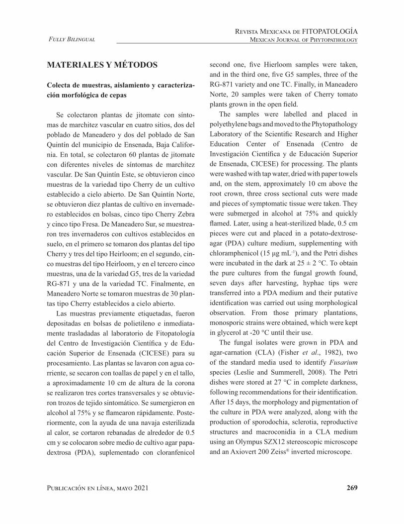
La prueba de patogenicidad de los aislados de Fol en plantas de jitomate mostró una susceptibili-dad de las variedades de jitomate acorde a la varie-dad y aislamiento del hongo. En el cultivar Bonny Best, todos los aislados causaron marchitez. Voip9 fue el que causó mayor daño (41%), seguido de Voip14, RE-2, SO-13 y Fol R2 (38%); Voip-8, RG-E, SO-11 y Fol R1 (32%) y Voip4 y RE-3 (29%). Los que menor daño ocasionaron (25%) fueron los aislados Voip6, SO-15 y SO-7. Las plantas no ino-culadas no mostraron ningún daño (Figura 4a).
En Manapal, el aislado Voip9 fue el que cau-só mayor daño (54%), seguido de SO-15 (41%); Voip14, RG-13, RE-2 y SO-13 y Fol R2 (33%); Fol R1 (29%); RE-3 y SO-7 (25%), Voip4, Voip6, Voip8 y SO-11 y las plantas control (9%) (Figura 4b).
R2 (38%); Voip-8, RG-E, SO-11 and Fol R1 (32%) and Voip4 and RE-3 (29%). The least damaging (25%) were isolates Voip6, SO-15 and SO-7. Plants not inoculated presented no damage (Figure 4a).In Manapal, isolate Voip9 caused the most damage (54%), followed by SO-15 (41%); Voip14, RG-13, RE-2 and SO-13 and Fol R2 (33%); Fol R1 (29%); RE-3 and SO-7 (25%), Voip4, Voip6, Voip8 and SO-11 and the control plants (9%) (Figure 4b).
In Walter, all isolates produced over 10% of vascular wilting, except Fol R2, which caused 9%; Voip8, Voip9 and Fol R1 (45%); Voip14, RE-3, SO-7 (41%); RG-3 y SO-15, (37%); RE-2 and SO-11 (33%); SO-13, Voip4 and Voip6, less than 20% (Figure 4c). In I3R3, isolate Fol R1 produced the least wilting (58%), followed by Voip8, Voip14 and SO-13 (53%); RG-3 (50%); RE-2 (45%), Voip9 (41%), Vop6 and RE-2 (38%); SO-15 (32%), SO-11 (25%), Voip4 (19%), SO-7 (8%) and RE-3 (3%) and the control (8%) (Figure 4d). Table 2 shows a summary of the results obtained in the tests for the identification of races according to comparisons between the presence of fragments generated by the sets of race-specific oligonucleotides, the sequences obtained and the in planta tests.
Genetic variability of isolates of Fol (RAPD)
Oligonucleotides OPA-01, OPA-15, OPA-16 and OPA-17 produced no fragments in Fol races1 and 2, and were therefore discarded for the analysis of variability between races used as a type. The approximate number and sizes of the fragments observed with OPA-03 were eight in Fol race 1, sized 1430, 519, 691, 752, 988, 1380, 1413 and 1926 pb, and four in Fol race 2, sized 519, 717, 1000 y 1430 pb. With the OPA-05 primer, three fragments of approximately 790, 954 and 1430 pb in Fol race 1 were observed, along with four in Fol race 2 sized 636, 790, 954 and 1616 pb. Finally, with primer OPA-11, four fragments were obtained

Publicación en línea, mayo 2021 280
Fully BilingualRevista Mexicana de FITOPATOLOGÍAMexican Journal of Phytopathology
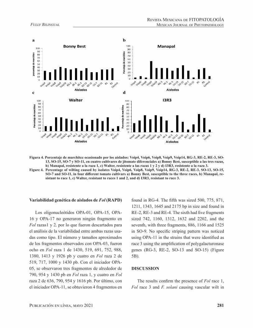
En Walter, todos los aislados produjeron arriba del 10% de marchitez vascular, excepto Fol R2, que ocasiono 9%; Voip8, Voip9 y Fol R1 (45%); Voip14, RE-3, SO-7 (41%); RG-3 y SO-15, (37%); RE-2 y SO-11 (33%); SO-13, Voip4 y Voip6, menos del 20% (Figura 4c). En I3R3, el aislado Fol R1 fue el que mayor marchitez produjo (58%); seguido de Voip8, Voip14 y SO-13 (53%); RG-3 (50%); RE-2 (45%), Voip9 (41%), Vop6 y RE-2 (38%); SO-15 (32%), SO-11 (25%), Voip4 (19%), SO-7 (8%) y RE-3 (3%) y el control (8%) (Figura 4d). El Cua-dro 2, muestra un resumen de los resultados obte-nidos en las pruebas para la identificación de razas de acuerdo a comparaciones entre la presencia de fragmentos generados por los sets de oligonucleóti-dos raza-específicos, las secuencias obtenidas y las pruebas in planta.
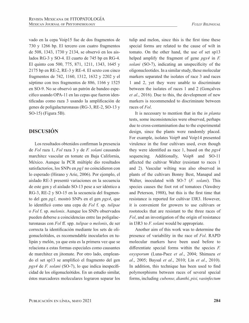
in Fol race 1 with approximately 664, 776, 1273 and 1660 pb, and in Fol race 2, five fragments sized 430, 607, 725, 1284 and 1637 pb (Figure 5A).
In the evaluation of the isolates obtained in this investigation and using oligonucleotide OPA-05, no specific patterns were found (data not shown), therefore its use was discarded. With OPA-11, seven stripe patterns were obtained, the first of which displayed four fragments sized 664, 776, 1273 and 1660 pb, and it was found in 29 of the isolates obtained, identified as Fol race 1 in the previous tests (Voip1-Voip16, Voip18-Voip26, Voip28-Voip30 and SO-11). The second pattern, found in strain Voip15 consisted of two fragments, sized 730 and 1266 bp. The third, with four fragments sized 508, 1343, 1730 and 2134, was found in isolates RG-3 and SO-4. The fourth, sized 745 bp, was
Figura 3. Filograma utilizando el método Neighbor-Joining basado en secuencias de la región TEF1-α con 500 réplicas.
Hubo un total de 591 posiciones en el conjunto de datos final. Los análisis evolutivos se realizaron en MEGA X (Kumar et al., 2018). El árbol está dibujado a escala, con la longitud de las ramas en las mismas unidades que las distancias evolutivas utilizadas para inferir el árbol filogenético. Las distancias evolutivas se calcularon utilizando el método de máxima probabilidad compuesta (Maximum Composite Likelihood).
Figure 3. Phylogram using the Neighbor-Joining method, based on sequences of the TEF1-α region with 500 replicas. There was a total of 591 positions in the final set of data. The evolutionary analyses were carried out in MEGA X (Kumar et al., 2018). The tree is drawn to scale, with the length of the branches in the same units as the evolutionary dis-tances used to infer the phylogenetic tree. Evolutionary distances were calculated using the Maximum Composite Likelihood method.
Publicación en línea, mayo 2021 281
Fully BilingualRevista Mexicana de FITOPATOLOGÍA
Mexican Journal of Phytopathology
Variabilidad genética de aislados de Fol (RAPD)
Los oligonucleótidos OPA-01, OPA-15, OPA-16 y OPA-17 no generaron ningún fragmento en Fol razas1 y 2, por lo que fueron descartados para el análisis de la variabilidad entre ambas razas usa-das como tipo. El número y tamaños aproximados de los fragmentos observados con OPA-03, fueron ocho en Fol raza 1 de 1430, 519, 691, 752, 988, 1380, 1413 y 1926 pb y cuatro en Fol raza 2 de 519, 717, 1000 y 1430 pb. Con el iniciador OPA-05, se observaron tres fragmentos de alrededor de 790, 954 y 1430 pb en Fol raza 1, y cuatro en Fol raza 2 de 636, 790, 954 y 1616 pb. Por último, con el iniciador OPA-11, se obtuvieron 4 fragmentos en
found in RG-4. The fifth was sized 500, 775, 871, 1211, 1343, 1645 and 2175 bp in size and found in RE-2, RE-3 and RE-4. The sixth had five fragments sized 742, 1160, 1312, 1632 and 2202, and the seventh, with three fragments, 886, 1166 and 1525 in SO-9. No specific striping pattern was noticed using OPA-11 in the strains that were identified as race 3 using the amplification of polygalacturonase genes (RG-3, RE-2, SO-13 and SO-15) (Figure 5B).
DISCUSSION
The results confirm the presence of Fol race 1, Fol race 3 and F. solani causing vascular wilt in
Figura 4. Porcentaje de marchitez ocasionado por los aislados: Voip4, Voip6, Voip8, Voip9, Voip14, RG-3, RE-2, RE-3, SO-13, SO-15, SO-7 y SO-11, en cuatro cultivares de jitomate diferenciales a) Bonny Best, susceptible a las tres razas, b) Manapal, resistente a la raza 1, c) Walter, resistente a las razas 1 y 2 y d) I3R3, resistente a la raza 3.
Figure 4. Percentage of wilting caused by isolates Voip4, Voip6, Voip8, Voip9, Voip14, RG-3, RE-2, RE-3, SO-13, SO-15, SO-7 and SO-11, in four different tomato cultivars a) Bonny Best, susceptible to the three races, b) Manapal, re-sistant to race 1, c) Walter, resistant to races 1 and 2, and d) I3R3, resistant to race 3.
Publicación en línea, mayo 2021 282
Fully BilingualRevista Mexicana de FITOPATOLOGÍAMexican Journal of Phytopathology
Fol raza 1 con tamaños aproximados de 664, 776, 1273 y 1660 pb y en Fol raza 2, cinco fragmentos de 430, 607, 725, 1284 y 1637 pb (Figura 5A).
En la evaluación de los aislados obtenidos en esta investigación, usando el oligonucleótido OPA-05, no se encontraron patrones específicos (datos no mostrados), por lo que se descartó su uso. Con OPA-11, se obtuvieron siete patrones de bandas con los siguientes tamaños aproximados. El primer patrón mostró cuatro fragmentos de 664, 776, 1273 y 1660 pb y fue observado en veintinueve aisla-dos obtenidos identificados como Fol raza 1 en las pruebas anteriores (Voip1-Voip16, Voip18-Voip26, Voip28-Voip30 y SO-11). El segundo patrón obser-
Cuadro 2. Identificación de razas de acuerdo a comparaciones entre la presencia de fragmentos generados por los sets de oligonucleótidos raza-específicos, las secuencias obtenidas y las pruebas in planta.
Table 2. Identification of races by comparison between the presence of fragments generated by the sets of race-specific oligonucleotides, the sequences obtained and the in planta tests.
AisladoPresencia de fragmento
Análisis de secuencias PCR-múltiple Cultivares diferenciales Raza
pgx4 pg1 pgx4 pg1 Raza Bonny Best Manapal Walter I3R3
Voip4 • X + 1 S R R S 1Voip6 • X + 1 S R R S 1Voip8 • X + 1 S R S S 1Voip9 • X + 1 S S S S XVoip14 • X + 1 S S S S XRG-3 • • + - 3 S S S S XRE-2 • • + - 3 S S S S XRE-3 • • + - 3 S S S R 3SO-13 • • - - 3 S S R S XSO-15 • • + - 3 S S S S XSO-7* • X ND S S S R F. solaniSO-11& X X ND S R R S 1
*El fragmento de SO-7, no se secuencio debido a que no pertenece a Fusarium oxysporum. / * Fragment SO-7 was not sequenced because it does not belong to Fusarium oxysporum.&SO-11 sólo se usó para inocular plantas, no se obtuvieron fragmentos con ningún par de los oligonucleótidos utilizados en los métodos de identificación de razas. / &SO-11 was only used to inoculate plants; no fragments were obtained with any pair of the oligonucleotides used in the race identification methods.• Presencia de fragmento. / • Presence of fragment.X ausencia de fragmento. / X absence of fragment.+Coincidencia de la secuenciación con la raza según los fragmentos amplificados. / +Coincidence of sequencing with race, depending on the fragments amplified.-Presencia de SNPs en la secuencia amplificada, respecto a Hirano y Arie, 2006. / -Presence of SNPs in the amplified sequence, regarding Hirano and Arie, 2006.
tomato in Baja California, Mexico. Although the multiplex PCR provided satisfactory results, the SNPs in pg1 did not meet expectations (Hirano and Arie, 2006). For example, isolate RE-3 presented variations in the sequence of this gene and isolate SO-13, despite being identical to RG-3, RE-2 and SO-15 in the sequence of fragment of gene pg1, displayed SNPs in gene pgx4, which identified it as a strain of Fol f. sp. tulipae or Fol f. sp. melonis. Although the SNPs observed may be due to coincidences between the polygalacturonases with Fol ff. spp. tulipae or melonis, should the identification with the sets of oligonucleotides be correct, it is recommendable to inoculate them in

Publicación en línea, mayo 2021 283
Fully BilingualRevista Mexicana de FITOPATOLOGÍA
Mexican Journal of Phytopathology
Figura 5. Patrones de bandas observados en el análisis RAPD en cepas de Fusarium oxysporum f. sp. lycopersici (Fol). A. Patrones de bandas observados en las cepas tipo de Fol razas 1 y 2, generados por los oligonucleótidos universales OPA-03, OPA-05 y OPA-11. B. Fragmentos generados por el oligonucleótido OPA-11, que muestran siete patrones de bandas en los aislados de Fusarium obtenidos de jitomate. Los aislados Voip8-Voip24 presentaron el mismo patrón de bandas, al igual que los RE-2, RE-3 y RE-4, los otros cinco presentaron diferente número y tamaño de fragmentos.
Figure 5. Banding patterns observed in the RAPD analysis in strains of Fusarium oxysporum f. sp. lycopersici (Fol). A. Banding pattern observed in Fol-type strains, races 1 and 2, generated by universal nucleotides OPA-03, OPA-05 and OPA-11. B. Fragments generated by oligonucleotide OPA-11, showing seven bands pattern in the isolates of Fusarium obtained from tomato. Isolates Voip8-Voip24 presented the same pattern of bands, like RE-2, RE-3 and RE-4; the other five presented different fragment numbers and sizes.
Publicación en línea, mayo 2021 284
Fully BilingualRevista Mexicana de FITOPATOLOGÍAMexican Journal of Phytopathology
vado en la cepa Voip15 fue de dos fragmentos de 730 y 1266 bp. El tercero con cuatro fragmentos de 508, 1343, 1730 y 2134, se observó en los ais-lados RG-3 y SO-4. El cuarto de 745 bp en RG-4. El quinto con 500, 775, 871, 1211, 1343, 1645 y 2175 bp en RE-2, RE-3 y RE-4. El sexto con cinco fragmentos de 742, 1160, 1312, 1632 y 2202 y el séptimo con tres fragmentos de 886, 1166 y 1525 en SO-9. No se observó un patrón de bandeo espe-cifico usando OPA-11 en las cepas que fueron iden-tificadas como raza 3 usando la amplificación de genes de poligalacturonasas (RG-3, RE-2, SO-13 y SO-15) (Figura 5B).
DISCUSIÓN
Los resultados obtenidos confirman la presencia de Fol raza 1, Fol raza 3 y de F. solani causando marchitez vascular en tomate en Baja California, México. Aunque la PCR múltiple dio resultados satisfactorios, los SNPs en pg1 no coincidieron con lo esperado (Hirano y Arie, 2006). Por ejemplo, el aislado RE-3 presentó variaciones en la secuencia de este gen y el aislado SO-13 pese a ser idéntico a RG-3, RE-2 y SO-15 en la secuencia del fragmen-to del gen pg1, mostró SNPs en el gen pgx4, que lo identificó como una cepa de Fol f. sp. tulipae o Fol f. sp. melonis. Aunque los SNPs observados pueden deberse a coincidencias entre las poligalac-turonasas con Fol ff. spp. tulipae o melonis, de ser correcta la identificación mediante los sets de oli-gonucleótidos, es recomendable inocularlos en tu-lipán y melón, ya que esta es la primera vez que se relaciona a estas formas especiales como causantes de marchitez en jitomate. Por otro lado, emplean-do el set sp13 se amplificó el fragmento del gen pgx4 de F. solani (SO-7), lo que indica inespecífi-cidad de los oligonucleótidos. En un estudio similar, éstos marcadores moleculares lograron separar los
tulip and melon, since this is the first time these special forms are related to the cause of wilt in tomato. On the other hand, the use of set sp13 helped amplify the fragment of gene pgx4 in F. solani (SO-7), indicating an unspecificity of the oligonucleotides. In a similar study, these molecular markers separated the isolates of race 3 and races 1 and 2, yet they were unable to discriminate between the isolates of races 1 and 2 (Gonçalves et al., 2016). Due to this, the development of new markers is recommended to discriminate between races of Fol.
It is necessary to mention that in the in planta tests, some inconsistencies were observed, perhaps due to cross-contamination due to the experimental design, since the plants were randomly placed. For example, isolates Voip9 and Voip14 presented virulence in the four cultivars used, even though they were identified as race 1, based on the pgx4 sequencing. Additionally, Voip8 and SO-11 affected the cultivar Walter (resistant to races 1 and 2). Vascular wilting was also observed in plants of the cultivars Bonny Best, Manapal and Walter, inoculated with SO-7 (F. solani). This species causes the foot rot of tomatoes (Vawdrey and Peterson, 1988), but this is the first time that resistance is reported for cultivar I3R3. However, it is convenient for growers to use cultivars or rootstocks that are resistant to the three races of Fol, and an investigation of the origin of resistance in I3R3 to F. solani would be appropriate.
Another aim of this work was to determine the presence of variability in the race of Fol. RAPD molecular markers have been used before to differentiate special forms within the species F. oxysporum (Luna-Paez et al., 2004; Shimazu et al., 2005; Baysal et al., 2010; Lin et al., 2010). In addition, this technique has been used to find polymorphisms between races of several special forms, including cubense, dianthi, pisi, vasinfectum

Publicación en línea, mayo 2021 285
Fully BilingualRevista Mexicana de FITOPATOLOGÍA
Mexican Journal of Phytopathology
aislados de la raza 3 de las razas 1 y 2, pero no fueron capaces de discriminar entre los aislados de las razas 1 y 2 (Gonçalves et al., 2016). Por ello, se recomienda desarrollar nuevos marcadores para discriminar entre las razas de Fol.
Es preciso mencionar que en las pruebas in planta, se observaron algunas inconsistencias, qui-zá debidas a contaminación cruzada, por el diseño experimental, ya que las plantas fueron ubicadas completamente al azar. Por ejemplo, los aislados Voip9 y Voip14, presentaron virulencia en los cua-tro cultivares utilizados, aunque fueron identifica-dos como raza 1, con base en la secuenciación de pgx4. Además, Voip8 y SO-11, afectaron al cultivar Walter (con resistencia a las razas 1 y 2). También se observó marchitez vascular en las plantas de los cultivares Bonny Best, Manapal y Walter, inocu-ladas con SO-7 (F. solani), ésta especie causa pu-drición del pie del jitomate (Vawdrey y Peterson, 1988), pero esta es la primera vez que se reporta la resistencia del cultivar I3R3. Aunque, es con-veniente que los productores utilicen cultivares o portainjertos resistentes a las tres razas de Fol, sería adecuado investigar el origen de la resistencia en I3R3 a F. solani.
Otro objetivo de este trabajo fue determinar la presencia de variabilidad en las razas de Fol. El uso de marcadores moleculares RAPDs, se ha utilizado con anterioridad para diferenciar formas especia-les dentro de la especie F. oxysporum (Luna-Paez et al., 2004; Shimazu et al., 2005; Baysal et al., 2010; Lin et al., 2010). Además, por medio de esta técnica se ha detectado la presencia de polimorfis-mos entre razas de varias formas especiales, inclui-das, cubense, dianthi, pisi, vasinfectum y ciceris (Bentley et al., 1994; Grajal et al., 1993; Migheli et al., 1998; Assigbetse et al., 1994; Jiménez et al., 2001; Katkar y Mane, 2012). Los oligonucleótidos universales OPA-03, OPA-05 y OPA-11, generaron distintos patrones de bandas en las razas tipo 1 y
and ciceris (Bentley et al., 1994; Grajal et al., 1993; Migheli et al., 1998; Assigbetse et al., 1994; Jiménez et al., 2001; Katkar and Mane, 2012). Universal oligonucleotides OPA-03, OPA-05 and OPA-11 generated different banding patterns in races type 1 and 2. However, only oligonucleotide OPA-11 presented different patterns. The number and size of the fragments generated by Fol race 3 was equal to those found in isolates RE-2 and RE-4 (Figure 5). Although the former displayed a SNP in gene pg1 and affected cultivar I3R3, it most likely belongs to race 3. Isolates Voip1-Voip16, Voip18-Voip26, Voip28-Voip30 and SO-11 displayed stripe patterns similar to those for race 1 and generated only the fragment of gene pgx4, which confirms that it belongs to that race, although in some isolates, such as Voip14 and Voip24, the stripes were the faintest (Figure 5). OPA-11 also produced a unique stripe pattern for F. solani (SO-7), and it can therefore be used to quickly determine the presence of this pathogen in different economically important cultivars.
CONCLUSIONS
Out of 60 tomato plants with vascular wilting, 44 F. oxysporum and one F. solani. isolates were obtained. Although the sets of race-specific oligonucleotides were used in a satisfactory way in a multiplex PCR reaction, they displayed unspecificity. The in planta tests with different cultivars helped confirm the presence of Fol races 1 and 3 in Baja California. For Fol race 3, the results were confirmed with the sequences of the fragments of gene pg1. Race 1 prevailed in sampled plants, infecting the cultivars Herlum, Cherry, Cherry-zebra and Cherry, whereas race 3 was found to be related to the cultivar Herlum. Using the RAPD technique, genetic polymorphisms were found that

Publicación en línea, mayo 2021 286
Fully BilingualRevista Mexicana de FITOPATOLOGÍAMexican Journal of Phytopathology
2. Sin embargo, solamente con el oligonucleótido OPA-11 se observaron diferencias en los patrones. El número y tamaño de los fragmentos generados por Fol raza 3 (RE-3) fue igual al observado en los aislados RE-2 y RE-4 (Figura 5B). El primero, aunque presentó un SNP en el gen pg1 y afectó al cultivar I3R3, lo más probable es que pertenezca a la raza 3. Los aislados Voip1-Voip16, Voip18-Voip26, Voip28-Voip30 y SO-11 mostraron patro-nes de bandas similares a la raza 1 y generaron solo el fragmento del gen pgx4, lo que confirma que pertenecen a esa raza. Aunque en algunos aislados como Voip14 y Voip24 las bandas fueron más te-nues (Figura 5). OPA-11 también generó un patrón de bandas único para F. solani (SO-7), por lo que puede ser utilizado para una determinación rápida de la presencia de este patógeno en diferentes culti-vares económicamente importantes.
CONCLUSIONES
De 60 plantas de jitomate con síntomas de marchitez vascular, se obtuvieron 44 aislados de F. oxysporum y uno de F. solani. Aunque los sets de oligonucleótidos raza-específicos se usaron sa-tisfactoriamente en una reacción de PCR múltiple, mostraron inespecificidad. Las pruebas in planta con cultivares diferenciales, permitieron confirmar la presencia de Fol razas 1 y 3 en Baja California. Para Fol raza 3, los resultados fueron corroborados con las secuencias de los fragmentos del gen pg1. La raza 1 prevaleció en las plantas muestreadas, in-fectando a los cultivares Herlum, Cherry, Cherry-zebra y Cherry, mientras que la raza 3 se encontró asociada al cultivar tipo Herlum. Usando la técnica de RAPD se encontraron polimorfismos genéticos que permiten diferenciar a la Fol raza 1 de Fol raza 2 y Fol raza 3.
help distinguish Fol race 1 from Fol race 2 and Fol race 3.
End of the English version
LITERATURA CITADA
Ascencio-Álvarez A, López-Benítez A, Borrego-Escalante F, Rodríguez-Herrera SA, Flores-Olivas A, Jiménez-Díaz F y Gámez-Vázquez, AJ. 2008. Marchitez vascular del jito-mate: I. Presencia de razas de Fusarium oxysporum f. sp. lycopersici (Sacc.) Snyder y Hansen en Culiacán, Sinaloa, México. Revista Mexicana de Fitopatología 26(2):114-120. https://www.redalyc.org/articulo.oa?id=61226203
Assigbetse KB, Fernández D, Dubois MP and Geiger JP. 1994. Differentiation of Fusarium oxysporum f. sp. vasinfectum races on cotton by Random Amplified Polymorphic DNA (RAPD) analysis. Phytopathology 84: 622-626. https://www.apsnet.org/publications/phytopathology/backissues/Documents/1994Articles/Phyto84n06_622.PDF
Baysal Ö, Siragusa M, Ikten H, Polat I, Gümrükcü E, Yigit F, Carimi JA and da Silva JT. 2009. Fusarium oxysporum f. sp. lycopersici races and their genetic discrimination by molecular markers in West Mediterranean region of Tur-key. Physiological and Molecular Plant Pathology 74(1): 68-75. https://doi.org/doi:10.1016/j.pmpp.2009.09.008
Baysal Ö., Siragusa M, Gumrukcu E, Zengin S, Carimi F, Sa-jeva M and Teixeira da Silva, JA. 2010. Molecular charac-terization of Fusarium oxysporum f. melongenae by ISSR and RAPD markers on eggplant. Biochemical genetics 48(5-6):524–37. https://doi.org/doi:10.1007/s10528-010-9336-1
Bentley S, Pegg KG and Dale JL. 1994. Optimization of RAPD‐PCR fingerprinting to analyse genetic variation within populations of Fusarium oxysporum f. sp. cuben-se. Journal of Phytopathology 142:164-78. https://doi.org/doi:10.1111/j.1439-0434.1994.tb00008.x
Biju VC, Fokkens L, Houterman PM, Rep M and Cornelissen BJ. 2017. Multiple evolutionary trajectories have led to the emergence of races in Fusarium oxysporum f. sp. lyco-persici. Applied and environmental microbiology 83(4). https://doi.org/10.1128/AEM.02548-16
Cai G, Gale LR, Schneider RW, Kistler HC, Davis RM, Elias KS and Miyao EM. 2003. Origin of race 3 of Fusarium oxysporum f. sp. lycopersici at a single site in California. Phytopathology 93(8):1014-1022. https://doi.org/10.1094/PHYTO.2003.93.8.1014
Carrillo Fasio JA, Montoya Rodríguez TJ, García Estrada RS, Cruz Ortega JE, Márquez Zequera I y Sañudo Barajas AJ. 2003. Razas de Fusarium oxysporum f. sp. lycopersici. Snyder y Hansen en jitomate (Lycopersicon esculentum Mill) en el valle de Culiacán, Sinaloa, México. Revista Mexicana de Fitopatología 21:123-127. https://www.re-dalyc.org/articulo.oa?id=61221205.

Publicación en línea, mayo 2021 287
Fully BilingualRevista Mexicana de FITOPATOLOGÍA
Mexican Journal of Phytopathology
Chang YD. Du B, Wang L, Ji P, Xie Y J, Li X F and Wang JM. 2018. A study on the pathogen species and physio-logical races of tomato Fusarium wilt in Shanxi, China. Journal of Integrative Agriculture 17:1380-1390. https://doi.org/10.1016/S2095-3119(18)61983-5
Gonçalves AM, Costa H, Fonseca MEN, Boiteux LS, Lopes CA and Reis A. 2016. Variability and geographical distri-bution of Fusarium oxysporum f. sp. lycopersici physio-logical races and field performance of resistant sources in Brazil. In V International Symposium on Tomato Disea-ses: Perspectives and future directions in tomato protection 1207 (pp. 45-50). https://biblioteca.incaper.es.gov.br/digi-tal/bitstream/123456789/3237/1/goncalves2018.pdf
Grajal MJ, Simon CJ and Muehlbauer FJ. 1993. Use of Ran-dom Amplified Polymorphic DNA (RAPD) to characte-rize race 2 of Fusarium oxysporum f. sp. pisi. Phytopa-thology 83:612-614. https://www.apsnet.org/publications/phytopathology/backissues/Documents/1993Articles/Phyto83n06_612.PDF
Hernández Martínez R, López Benítez A, Borrego Escalante F, Espinoza Velázquez J, Sánchez Aspeytia D, Maldonado Mendoza IE y López Ochoa LA. 2014. Razas de Fusarium oxysporum f. sp. lycopersici en predios jitomateros en San Luis Potosí. Revista Mexicana de Ciencias Agrícolas 5(7):1169-1178. https://cienciasagricolas.inifap.gob.mx/editorial/index.php/agricolas/article/view/838/661
Hirano Y and Arie T. 2006. PCR-based differentiation of Fu-sarium oxysporum ff. sp. lycopersici and radicis-lycoper-sici and races of F. oxysporum f. sp. lycopersici. Journal of General Plant Pathology 72(5):273-283. https://doi.org/10.1007/s10327-006-0287-7
Holguín-Peña RJ. 2005. Fusarium wilt of tomato caused by Fusarium oxysporum f. sp. lycopersici race 3 in Baja Ca-lifornia Sur, México. Plant Disease 89(12):1360-1360. https://doi.org/10.1094/PD-89-1360C
Isaac MR and Leyva-Mir SG, Sahagun-Castellanos J, Camara-Correia K, Tovar-Pedraza JM y Rodriguez-Perez JE. 2018. Occurrence, identification, and pathogenicity of Fusarium spp. associated with tomato wilt in Mexico. Notulae Bo-tanicae Horti Agrobotanici Cluj-Napoca 46(2):484-493. https://doi.org/10.15835/nbha46211095
Jiménez Gasco MM, Pérez E and Jiménez RM. 2001. Identi-fication of pathogenic races 0, 1B/C, 5, and 6 of Fusarium oxysporum f. sp. ciceris with random amplified polymor-phic DNA (RAPD). European Journal of Plant Pathology 107:237-248. https://doi.org/10.1023/A:1011294204630
Katkar M and Mane SS. 2012. Characterization of Indian races of Fusarium oxysporum f. sp ciceris through RAPD mar-kers. International Journal of Agriculture, Environment and Biotechnology 5(4):323-328. https://www.indianjo-urnals.com/ijor.aspx?target=ijor:ijaeb&volume=5&issue=4&article=003
Kawabe M, Kobayashi Y, Okada G, Yamaguchi I, Teraoka T and Arie T. 2005. Three evolutionary lineages of tomato wilt pathogen, Fusarium oxysporum f. sp. lycopersici, ba-sed on sequences of IGS, MAT1, and pg1, are each com-posed of isolates of a single mating type and a single or closely related vegetative compatibility group. Journal of General Plant Pathology 71(4):263-272. https://doi.org/10.1007/s10327-005-0203-6
Kumar S, Stecher G, Li M, Knyaz C and Tamura K. 2018. MEGA X: Molecular Evolutionary Genetics Analysis across computing platforms. Molecular Biology and Evo-lution 35:1547-1549. https://doi.org/10.1093/molbev/msy096
Lievens B, Van Baarlen P, Verreth C, Van Kerckhove S, Rep M and Thomma BP. 2009. Evolutionary relationships between Fusarium oxysporum f. sp. lycopersici and F. oxysporum f. sp. radicis-lycopersici isolates inferred from mating type, elongation factor-1α and exopolygalacturo-nase sequences. Mycological research 113(10):1181-1191. https://www.sciencedirect.com/science/article/abs/pii/S0953756209001452
Leslie JF and Summerell BA. 2008. The Fusarium laboratory manual. John Wiley & Sons.
Lin YH, Chen KS, Chang JY, Wan YL, Hsu CC, Huang JW and Chang PFL. 2010. Development of the molecular me-thods for rapid detection and differentiation of Fusarium oxysporum and F. oxysporum f. sp. niveum in Taiwan. New Biotechnology 27(4):409-18. https://doi.org/10.1016/j.nbt.2010.05.005
Luna-Paez A, Silva-Rojas HV, Marbán-Mendoza N y Va-ladez-Moctezuma E. 2004. Variabilidad genética de Fusarium oxysporum Schlechtend.: Fr. f. sp. ciceris (Padwick) Matuo y K. Sato mediante PCR-RAPD’S en el Bajío de México. Revista Mexicana Fitopatología 22:44-51. http://scielo.sld.cu/scielo.php?pid=S1562-30092009000300004&script=sci_abstract
Martel MB, Létoublon R and Fèvre M. 1998. Purification and characterization of two endopolygalacturonases secreted during the early stages of the saprophytic growth of Sclero-tinia sclerotiorum. FEMS microbiology letters 158(1):133-138. https://doi.org/10.1111/j.1574-6968.1998.tb12812.x
Migheli Q, Briatore E and Garibaldi A. 1998. Use of random amplified polymorphic DNA (RAPD) to identify races 1, 2, 3 and 8 of Fusarium oxysporum f. sp. dianthi in Italy. European Journal of Plant Pathology. 104:49-57. https://doi.org/10.1023/A:1008682328764
Murugan L, Krishnan N, Venkataravanappa V, Saha S, Mishra AK, Sharma BK and Rai AB. 2020. Molecular characteri-zation and race identification of Fusarium oxysporum f. sp. lycopersici infecting tomato in India. 3 Biotech 10(11):1-12. https://doi.org/10.1007/s13205-020-02475-z
O’Donnell K. Kistler HC, Cigelnik E and Ploetz RC. 1998. Multiple evolutionary origins of the fungus causing Pana-ma disease of banana: concordant evidence from nuclear and mitochondrial gene genealogies. Proceedings of the National Academy of Sciences 95(5):2044-2049. https://doi.org/10.1073/pnas.95.5.2044
Pirayesh S, Zamanizade H and Morid B. 2018. Molecular Identification of Physiological Races of Fusarium oxyspo-rum f. sp. lycopersici and radicis lycopersici Causal Agent of Fusarium Wilt of Tomato in Iran. Journal of Agricultural Science and Technology 20(1):193-202. https://jast.moda-res.ac.ir/article-23-4759-en.html
Shimazu J, Yamauchi N, Hibi T, Satou M, Horiuchi S and Shirakawa T. 2005. Development of sequence tagged site markers to identify races of Fusarium oxysporum f. sp. lactucae. Journal of General Plant Pathology 71(3):183–189. https://doi.org/10.1007/s10327-005-0183-6

Publicación en línea, mayo 2021 288
Fully BilingualRevista Mexicana de FITOPATOLOGÍAMexican Journal of Phytopathology
SIAP, 2019. Recuperado 13 de octubre de 2020: http://info-siap.siap.gob.mx:8080/agricola_siap_gobmx/Resumen-Delegacion.do
Valenzuela-Ureta JG, Lawn DA, Heisey RF and Zamudio-Guzman V. 1996. First report of Fusarium wilt race 3, caused by Fusarium oxysporum f. sp. lycopersici, of tomato in Mexico. Plant Disease 80(1):105. https://www.apsnet.org/publications/plantdisease/backissues/Documents/1996Abstracts/PD_80_0105A.htm.
Vakalounakis DJ and Fragkiadakis GA. 1999. Genetic diver-sity of Fusarium oxysporum isolates from cucumber: Di-fferentiation by Pathogenicity, Vegetative Compatibility, and RAPD Fingerprinting. Phytopathology 89(2):161-8. https://doi.org/10.1094/PHYTO.1999.89.2.161
Vawdrey LL and Peterson RA. 1988. Fusarium solani, the cau-se of foot rot of tomatoes in Central Queensland. Australa-sian Plant Pathology, 17(1):24-25. https://doi.org/10.1071/APP9880024