Embed Size (px)
Citation preview

APPLIED AND ENVIRONMENTAL MICROBIOLOGY,0099-2240/98/$04.0010
Sept. 1998, p. 3313–3319 Vol. 64, No. 9
Copyright © 1998, American Society for Microbiology. All Rights Reserved.
Identification and Characterization of Leuconostoc carnosum,Associated with Production and Spoilage of
Vacuum-Packaged, Sliced, Cooked HamK. J. BJORKROTH,1* P. VANDAMME,2 AND H. J. KORKEALA1
Department of Food and Environmental Hygiene, University of Helsinki, Helsinki, Finland,1
and Department of Microbiology, University of Ghent, Ghent, Belgium2
Received 26 February 1998/Accepted 15 June 1998
Leuconostoc carnosum was shown to be the specific spoilage organism in vacuum-packaged, sliced, cookedham showing spoilage during 3 weeks of shelf life. Identification of the specific spoilage organism was done byuse of phenotypic data and ClaI, EcoRI, and HindIII reference strain ribopatterns. One hundred L. carnosumisolates associated with the production and spoilage of the ham were further characterized by pulsed-field gelelectrophoresis (PFGE), together with some meat-associated Leuconostoc species: L. citreum, L. gelidum,L. mesenteroides subsp. dextranicum, and L. mesenteroides subsp. mesenteroides. ApaI and SmaI digests dividedthe industrial L. carnosum strains into 25 different PFGE types, ApaI and SmaI types being consistent. Onlyone specific PFGE type was associated with the spoiled packages. This type also was detected in air andraw-meat mass samples. The spoilage strain did not produce bacteriocins. Only seven isolates belonging tothree different PFGE types produced bacteriocins. Similarity analysis of the industrial L. carnosum strainsrevealed a homogeneous cluster which could be divided into eight subclusters consisting of strains having atmost three-fragment differences. The L. carnosum cluster was clearly distinguished from the other meat-associated leuconostoc clusters, with the exception of the L. carnosum type strain. Ribotyping can be veryhelpful in the identification of L. carnosum, but its discriminatory power is too weak for strain characterization.PFGE provides good discrimination for studies dealing with the properties of homogeneous L. carnosumstrains.
Lactic acid bacteria (LAB) are the major spoilage bacteria invacuum-packaged, cooked meat products (1, 2, 10, 13, 25, 27,31, 38, 44, 47, 56). Lactobacillus and Leuconostoc have beenthe main genera associated with the spoilage of these products,Lactobacillus sake and Lactobacillus curvatus being isolatedcommonly (12, 16, 18, 19, 24, 27, 30, 35, 39, 43–46). Comparedto aerobic spoilage bacteria, spoilage LAB produce their typ-ical sensory changes, such as souring, gas formation, and/orslime formation, later, at the stationary phase (29, 44), anda vacuum-packaged product is usually expected to maintaingood sensory quality for at least 3 to 4 weeks. However, due toan increased level of LAB contamination or particularly activespoilage strains, spoilage may occur during the shelf-life pe-riod, subjecting the producer to recalls (30, 31, 33, 46).
In an LAB contamination study of vacuum-packaged, sliced,cooked ham, 982 LAB isolates from the spoiled product andproduction line were characterized in order to determine theunderlying reasons for fluctuations in product quality (4, 6).Many lots had been showing spoilage changes, i.e., sour odorand taste, before the sell-by date. In that study, ribotyping (21)was used as a tool for contamination analysis. Based on EcoRIand HindIII ribopatterns, two major spoilage LAB types, typesG and A, were detected. Contamination with these spoilageLAB was shown to have occurred postcooking, and a probablesite of air-mediated contamination from the macerated raw-meat mass to the cooked product was revealed. Because type Gshowed the typical EcoRI and HindIII ribopatterns of L. sake(5), no further identification or characterization studies were
warranted. However, the most important specific spoilage or-ganism, type A, was not identified to the species level. Type Ahad been detected as the dominant type in the maceratedraw-meat mass and in the spoiled packages with the strongestchanges in sensory characteristics (6). It had also persisted inthe plant during the 1-year study period, consisting of twoseparate large-scale contamination experiments (4, 6).
In this study, we set out to identify type A LAB to the specieslevel and characterize in more detail the 100 isolates possess-ing the type A EcoRI and HindIII ribopatterns. Since pheno-typic characteristics alone are seldom sufficient for speciesidentification of LAB (15), a reference strain library wascreated by ribotyping and was used with phenotypic data.Pulsed-field gel electrophoresis (PFGE) was applied in orderto provide further strain-level characterization. Production ofbacteriocins was determined for evaluation of the impact ofthis characteristic in a population associated with process con-tamination and product spoilage.
MATERIALS AND METHODS
Bacterial strains. One hundred type A LAB possessing the same EcoRI andHindIII ribopatterns had been isolated during a contamination study of a meatplant (6). All isolates were gram-positive, oval cocci isolated from a maceratedraw-meat mass, air in the macerating room, surfaces and air in the cooking room,worker’s gloves, surfaces of the ham prior to slicing, and vacuum-packaged,sliced, cooked ham cultured on the sell-by date. Isolates originating from differ-ent sources are listed in Table 1.
In order to obtain a library for species identification, the following referencestrains were ribotyped with ClaI, EcoRI, and HindIII: Leuconostoc carnosumNCFB (National Collection of Food Bacteria) 2776T, Leuconostoc citreum (Leu-conostoc amelibiosum) D1 (35), Leuconostoc fallax CCUG (Culture Collection ofUniversity of Gothenburg) 30061T, Leuconostoc gelidum NCFB 2775T, Leu-conostoc lactis CCUG 30064T, Leuconostoc mesenteroides subsp. mesenteroidesDSM (Deutsche Sammlung von Mikroorganismen) 20343T, Leuconostoc mesen-teroides subsp. cremoris CCUG 21965T, Leuconostoc mesenteroides subsp. dex-tranicum DSM 20484T, Leuconostoc pseudomesenteroides DSM 20193T, Weissellahalotolerans ATCC (American Type Culture Collection) 35410T, Weissella viri-
* Corresponding author. Mailing address: Department of Food andEnvironmental Hygiene, Faculty of Veterinary Medicine, University ofHelsinki, P.O. Box 57, FIN-00014 Helsinki, Finland. Phone: 358-50-5976555. Fax: 358-9-70849718. E-mail: [email protected].
3313
on March 24, 2019 by guest
http://aem.asm
.org/D
ownloaded from
descens ATCC 12706T, and Weissella paramesenteroides DSM 20288T. In addi-tion, the previously established (5, 7) ClaI, EcoRI, and HindIII Lactobacillusribotypes were compared with the Leuconostoc and Weissella ribotypes charac-terized in this study.
The meat-associated reference strains L. carnosum NCFB 2776T, L. citreum(L. amelibiosum) D1 (35), L. gelidum NCFB 2775T, L. mesenteroides subsp.dextranicum DSM 20484T, L. mesenteroides subsp. mesenteroides DSM 20343T, L.pseudomesenteroides DSM 20193T, and W. paramesenteroides DSM 20288T werecharacterized by PFGE along with the industrial isolates.
All strains were maintained in MRS broth (Difco, Detroit, Mich.) at 270°Cand cultured with MRS broth or MRS agar (Oxoid, Basingstoke, United King-dom) as previously described (28).
Phenotypic characterization. The anaerobic growth of all industrial isolates onRogosa selective Lactobacillus agar (Orion Diagnostica, Espoo, Finland) wasdetermined, and the scheme of Villiani et al. (55) was used for the presumptiveidentification of Leuconostoc spp. Gas production from glucose was tested withmodified MRS broth in Durham tubes (51). Production of ammonia from argi-nine was observed by the method of Briggs (14), and dextran formation wasstudied with 5% sucrose-containing agar (22). Fermentation of carbohydrateswas determined by use of the API 50 CH Lactobacillus identification system(Biomerieux, Marcy l’Etoile, France) for five randomly selected isolates (I27a,M1f, V8a, M6f, and P31a), which were also tested for the ability to producedifferent lactic acid isomers by an enzymatic method (57) with D- and L-lactatedehydrogenases (Boehringer GmbH, Mannheim, Federal Republic of Germa-ny). The five randomly selected isolates were also tested for growth in MRSbroth at 8, 10, 15, and 37°C.
Bacteriocin determination. The agar spot test method modified by Schillingerand Lucke (48) was used for screening bacteriocin activity. Based on existingliterature, L. mesenteroides subsp. mesenteroides DSM 20343T was selected as theindicator bacterium (3, 26, 40, 54, 58).
In vitro isolation of DNA and ribotyping for species identification. Referencestrains and the five randomly selected industrial isolates, already known topossess similar EcoRI and HindIII ribotypes, were characterized with ClaI,EcoRI, and HindIII (New England BioLabs, Beverly, Mass.). These enzymeswere selected because they characterize LAB well (4–6). DNA was isolated bythe guanidium thiocyanate method of Pitcher et al. (42) as modified by Bjorkrothand Korkeala (4) by combined lysozyme and mutanolysin treatments. Restrictionendonuclease treatment of 3 mg of DNA was done as specified by the manufac-
turer (New England BioLabs). Genomic blotting was done by vacuum blotting(Vacugene; Pharmacia, Uppsala, Sweden), and the ribosomal DNA probe forribotyping was labeled by reverse transcription (avian myeloblastosis virus re-verse transcriptase [Promega, Madison, Wis.]; Dig DNA labeling kit [Boehr-inger]) as previously described by Blumberg et al. (11). Membranes were hybrid-ized at 68°C as described by Bjorkroth and Korkeala (5). Similarity between allribopatterns was determined visually.
In situ DNA isolation and PFGE. Cells were harvested from 2 ml of MRSbroth cultures grown overnight at 30°C. DNA isolation in situ from agaroseblocks was performed as described by Maslow et al. (37) with the modificationsdescribed by Bjorkroth et al. (9). Initially, 11 rare-cutting restriction enzymes,ApaI, AscI, EagI, MluI, NotI, NruI, RsrII, SacII, SmaI, XbaI, and XhoI, weretested for the cleavage of DNA of three strains (NCFB 2776T, M6f, and I27a).ApaI and SmaI, which produced convenient numbers of fragments with discrim-inatory patterns, were chosen for the cleavage of all strains. The samples wereelectrophoresed through a 1.2% (wt/vol) agarose gel (SeaKem Gold; FMC Bio-Products, Rockland, Maine) in 0.53 TBE (45 mM Tris, 4.5 mM boric acid [pH8.3], 1 mM sodium EDTA) at 14°C by use of a Gene Navigator system with thehexagonal electrode (Pharmacia). Interpolation ramping from 0.5 to 15 s for 20 hat 200 V was used for both enzyme digests.
PFGE data management. Photographs of the PFGE banding patterns werescanned with a ScanJet 4c/T scanner (Hewlett-Packard Co., Boise, Idaho). Nu-merical analysis of macrorestriction patterns was performed with a GelComparsystem (version 4.0; Applied Maths, Kortijk, Belgium). The similarity between allpairs was expressed by Dice coefficient correlation, and clustering by the un-weighted pair-group method with arithmetic averages was used for the construc-tion of the dendrogram. Types were considered closely related (53) in thepresence of at most a three-band difference (one genetic event). This relation-ship was indicated in the type nomination by a shared roman numeral.
RESULTS
The 100 isolates did not grow on Rogosa selective Lactoba-cillus agar; all produced gas from glucose but did not produceammonia from arginine. Fifteen isolates (11 different PFGE
TABLE 1. Division of the isolates into different types and certain phenotypic properties
Typea Isolatesb
Restriction enzymeprofile Production ofc:
ApaI SmaI Slime from sucrose Bacteriocin
A I-a M2n, M2h A1 S1 2 2A I-b M3h, M6f, M6h A2 S2 2 2A I-c P31a, M41, M4m A3 S3 2 2A I-d I27e A4 S4 1 2A I-e M5i, M5j A5 S5 2 2A I-f P36b A6 S6 2 2A I-g M6j A7 S7 1 2A I-h V8a–o, V9a–o, V11a–m, V13a–o, I2b, I27a, M5o, M6a A8 S8 2 2A I-i M6o A9 S9 2 2A I-j M2e, M2l, M2o, M3f, M3l A10 S10 2 1A I-k M6g A11 S11 1 2A II-a I1b A12 S12 2 2A II-b I1c, I1f A13 S13 2 2A II-c I26b, I28b A14 S14 1 2A II-d I2a A15 S15 2 2A III-a M2j, M3e, M3m A16 S16 1 2A III-b M1e A17 S17 1 2A III-c M5k A18 S18 1 2A IV M2d A19 S19 2 1A V-a M3o, M6i A20 S20 1 2A V-b M1f A21 S21 1 1A VI M1j A22 S22 1 2A VII-a I27f A23 S23 1 2A VII-b M1i A24 S24 2 2A VIII M1c A25 S25 2 2
a Types sharing the same roman numeral differ by at most three bands in the restriction enzyme profiles.b Sampling was described previously (6). Sources were as follows: M, raw-meat mass; P, surface; I, air; V, spoiled product. Groups of lowercase letters indicate a series
of isolates; e.g., a–o indicates 15 isolates from V8a to V8o.c 1, production; 2, no production.
3314 BJORKROTH ET AL. APPL. ENVIRON. MICROBIOL.
on March 24, 2019 by guest
http://aem.asm
.org/D
ownloaded from
types) produced slime from sucrose, and bacteriocins wereproduced by 7 isolates (Table 1). The five isolates tested pro-duced only D-lactic acid and had similar fermentation patternsfor the utilization of ribose, D-glucose, D-fructose, a-methyl-D-glucoside, N-acetylglucosamine, cellobiose, saccharose, treha-lose, b-gentiobiose, D-turanose, and gluconate. Growth oc-curred at 8, 10, and 15°C but not at 37°C.
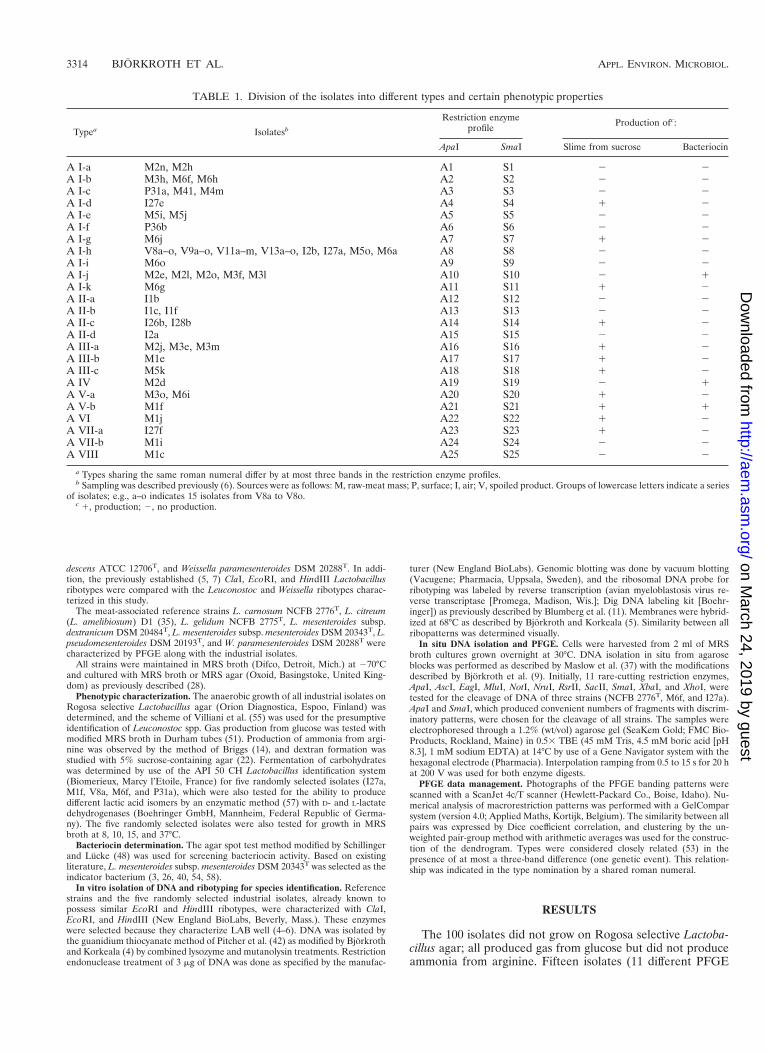
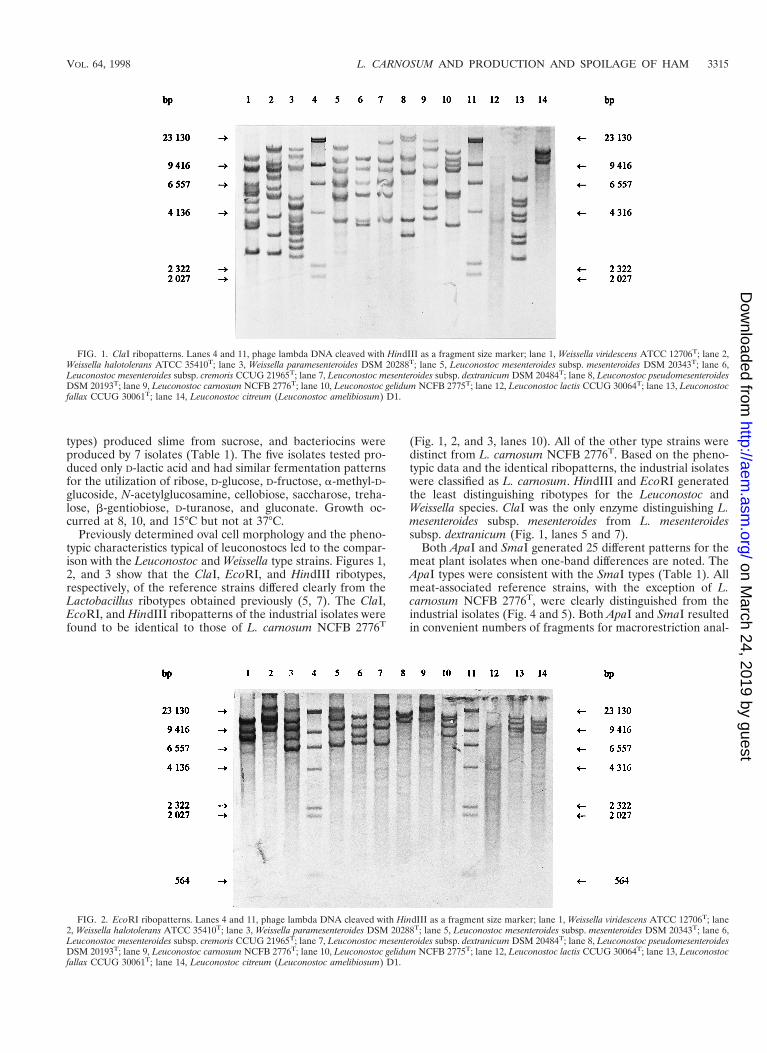
Previously determined oval cell morphology and the pheno-typic characteristics typical of leuconostocs led to the compar-ison with the Leuconostoc and Weissella type strains. Figures 1,2, and 3 show that the ClaI, EcoRI, and HindIII ribotypes,respectively, of the reference strains differed clearly from theLactobacillus ribotypes obtained previously (5, 7). The ClaI,EcoRI, and HindIII ribopatterns of the industrial isolates werefound to be identical to those of L. carnosum NCFB 2776T
(Fig. 1, 2, and 3, lanes 10). All of the other type strains weredistinct from L. carnosum NCFB 2776T. Based on the pheno-typic data and the identical ribopatterns, the industrial isolateswere classified as L. carnosum. HindIII and EcoRI generatedthe least distinguishing ribotypes for the Leuconostoc andWeissella species. ClaI was the only enzyme distinguishing L.mesenteroides subsp. mesenteroides from L. mesenteroidessubsp. dextranicum (Fig. 1, lanes 5 and 7).
Both ApaI and SmaI generated 25 different patterns for themeat plant isolates when one-band differences are noted. TheApaI types were consistent with the SmaI types (Table 1). Allmeat-associated reference strains, with the exception of L.carnosum NCFB 2776T, were clearly distinguished from theindustrial isolates (Fig. 4 and 5). Both ApaI and SmaI resultedin convenient numbers of fragments for macrorestriction anal-
FIG. 1. ClaI ribopatterns. Lanes 4 and 11, phage lambda DNA cleaved with HindIII as a fragment size marker; lane 1, Weissella viridescens ATCC 12706T; lane 2,Weissella halotolerans ATCC 35410T; lane 3, Weissella paramesenteroides DSM 20288T; lane 5, Leuconostoc mesenteroides subsp. mesenteroides DSM 20343T; lane 6,Leuconostoc mesenteroides subsp. cremoris CCUG 21965T; lane 7, Leuconostoc mesenteroides subsp. dextranicum DSM 20484T; lane 8, Leuconostoc pseudomesenteroidesDSM 20193T; lane 9, Leuconostoc carnosum NCFB 2776T; lane 10, Leuconostoc gelidum NCFB 2775T; lane 12, Leuconostoc lactis CCUG 30064T; lane 13, Leuconostocfallax CCUG 30061T; lane 14, Leuconostoc citreum (Leuconostoc amelibiosum) D1.
FIG. 2. EcoRI ribopatterns. Lanes 4 and 11, phage lambda DNA cleaved with HindIII as a fragment size marker; lane 1, Weissella viridescens ATCC 12706T; lane2, Weissella halotolerans ATCC 35410T; lane 3, Weissella paramesenteroides DSM 20288T; lane 5, Leuconostoc mesenteroides subsp. mesenteroides DSM 20343T; lane 6,Leuconostoc mesenteroides subsp. cremoris CCUG 21965T; lane 7, Leuconostoc mesenteroides subsp. dextranicum DSM 20484T; lane 8, Leuconostoc pseudomesenteroidesDSM 20193T; lane 9, Leuconostoc carnosum NCFB 2776T; lane 10, Leuconostoc gelidum NCFB 2775T; lane 12, Leuconostoc lactis CCUG 30064T; lane 13, Leuconostocfallax CCUG 30061T; lane 14, Leuconostoc citreum (Leuconostoc amelibiosum) D1.
VOL. 64, 1998 L. CARNOSUM AND PRODUCTION AND SPOILAGE OF HAM 3315
on March 24, 2019 by guest
http://aem.asm
.org/D
ownloaded from
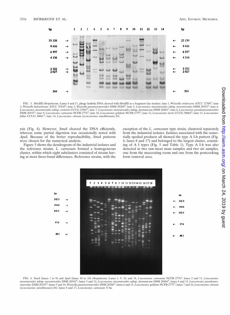
ysis (Fig. 4). However, SmaI cleaved the DNA efficiently,whereas some partial digestion was occasionally noted withApaI. Because of the better reproducibility, SmaI patternswere chosen for the numerical analysis.
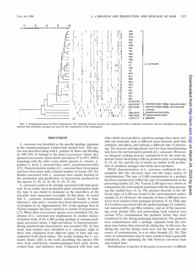
Figure 5 shows the dendrogram of the industrial isolates andthe reference strains. L. carnosum formed a homogeneouscluster, within which eight subclusters consisted of strains hav-ing at most three-band differences. Reference strains, with the
exception of the L. carnosum type strain, clustered separatelyfrom the industrial isolates. Isolates associated with the senso-rially spoiled products all showed the type A I-h pattern (Fig.4, lanes 8 and 17) and belonged to the largest cluster, consist-ing of A I types (Fig. 5 and Table 1). Type A I-h was alsodetected in two raw-meat mass samples and two air samples,one from the macerating room and one from the postcookingform removal area.
FIG. 3. HindIII ribopatterns. Lanes 4 and 11, phage lambda DNA cleaved with HindIII as a fragment size marker; lane 1, Weissella viridescens ATCC 12706T; lane2, Weissella halotolerans ATCC 35410T; lane 3, Weissella paramesenteroides DSM 20288T; lane 5, Leuconostoc mesenteroides subsp. mesenteroides DSM 20343T; lane 6,Leuconostoc mesenteroides subsp. cremoris CCUG 21965T; lane 7, Leuconostoc mesenteroides subsp. dextranicum DSM 20484T; lane 8, Leuconostoc pseudomesenteroidesDSM 20193T; lane 9, Leuconostoc carnosum NCFB 2776T; lane 10, Leuconostoc gelidum NCFB 2775T; lane 12, Leuconostoc lactis CCUG 30064T; lane 13, Leuconostocfallax CCUG 30061T; lane 14, Leuconostoc citreum (Leuconostoc amelibiosum) D1.
FIG. 4. SmaI (lanes 1 to 9) and ApaI (lanes 10 to 18) ribopatterns. Lanes 1, 9, 10, and 18, Leuconostoc carnosum NCFB 2776T; lanes 2 and 11, Leuconostocmesenteroides subsp. mesenteroides DSM 20343T; lanes 3 and 12, Leuconostoc mesenteroides subsp. dextranicum DSM 20484T; lanes 4 and 13, Leuconostoc pseudomes-enteroides DSM 20193T; lanes 5 and 14, Weissella paramesenteroides DSM 20288T; lanes 6 and 15, Leuconostoc gelidum NCFB 2775T; lanes 7 and 16, Leuconostoc citreum(Leuconostoc amelibiosum) D1; lanes 8 and 17, Leuconostoc carnosum V-8a.
3316 BJORKROTH ET AL. APPL. ENVIRON. MICROBIOL.
on March 24, 2019 by guest
http://aem.asm
.org/D
ownloaded from
DISCUSSION
L. carnosum was identified as the specific spoilage organismin the vacuum-packaged, cooked ham studied here. This spe-cies was described along with L. gelidum by Shaw and Hardingin 1989 (50). It belongs to the main Leuconostoc cluster des-ignated Leuconostoc sensu stricto and shares 97 to 99% rRNAhomology with the other sensu stricto species: L. citreum, L.gelidum, L. lactis, L. mesenteroides, and L. pseudomesenteroides(15). Characterization studies of L. carnosum have been sparseand have been done with a limited number of strains (50, 58).Studies associated with L. carnosum have mainly focused onthe production and purification of bacteriocins produced bythis species (3, 20, 23, 26, 40, 41, 49, 52, 54).
L. carnosum seems to be strongly associated with ham prod-ucts. In an earlier meat production plant contamination study(6), type A was found to dominate in the microflora of theraw-pork mass macerated overnight. In this plant, we notedthat L. carnosum contamination occurred mainly in ham,whereas L. sake and L. curvatus have been detected in a varietyof products (4, 6). Approximately 36% of the spoilage flora inVienna sausages has been reported to consist of leuconostocs(17). When these Leuconostoc species were identified (18), theabsence of L. carnosum was emphasized. In another charac-terization study of the LAB causing spoilage in vacuum-pack-aged, processed meats, a high prevalence of bacteriocin-pro-ducing psychrotropic leuconostocs was revealed (58). In thatstudy, nine isolates were identified as L. carnosum; eight ofthese nine originated from different types of ham and oneoriginated from sliced turkey. The strains forming the L. car-nosum cluster (III) in the work describing this species (50)were from cold-stored, vacuum-packaged beef, pork, bacon,cooked ham, and luncheon meat. Compared with ham and
other whole-meat products, emulsion sausages have more vari-able raw materials, such as different meat mixtures, pork skinemulsion, and spices, and undergo a different type of process-ing. The process and ingredients used for ham manufacturingmay favor the survival and/or growth of L. carnosum. However,an adequate cooking process, considered to be the most im-portant factor destroying LAB on products prior to packaging(1, 33, 34, 36), and the use of nitrite are similar in the produc-tion of emulsion sausages and whole-meat products.
PFGE characterization of L. carnosum confirmed the as-sumption that the raw-meat mass was the major source ofcontamination. The type of LAB contamination in a producthas been considered to reflect the type of contamination in theprocessing facility (25, 38). Various LAB types were shown tocontaminate the environment associated with the ham process-ing line studied here (4, 6). The greatest diversity in the dif-ferent types of LAB was found in the environmental surfacesamples (4, 6). However, the majority of these LAB types havenever been isolated from packaged products (4, 6). Only typeA I-h isolates associated with the spoiled packages (V isolates),raw material (M5o and M6a isolates), and air of the macerat-ing room (isolate I2b) and postcooking form removal area(isolate I27a) contaminated the products before they weretransferred to the slicing-packaging department. The productswere contaminated with a spoilage organism from the raw-meat mass before they entered the slicing line. In this case, theslicing line and the slicing room were not the main site andsource of contamination, as is so often thought (25, 38). Thisroute of contamination may be more common than is generallyconsidered, also explaining the link between raw-meat massand cooked ham.
Identification of species of the genus Leuconostoc is difficult
FIG. 5. Dendrogram based on SmaI ribopatterns. The similarity between all pairs was expressed by Dice coefficient correlation, and the unweighted pair-groupmethod with arithmetic averages was used for the construction of the dendrogram.
VOL. 64, 1998 L. CARNOSUM AND PRODUCTION AND SPOILAGE OF HAM 3317
on March 24, 2019 by guest
http://aem.asm
.org/D
ownloaded from
(15, 55), which apparently is the main reason for the sparsepopulation characterizations published. Leuconostoc spp. arephenotypically related to Weissella spp., heterofermentativelactobacilli, and pediococci and form a natural phylogeneticgroup with Weissella confusa, W. halotolerans, Weissellakandleri, Weissella minor, and W. viridescens (15). Due to thevariable results obtained, sugar fermentation patterns are oflittle value in the species identification and could lead to mis-classification (15). For presumptive identification, the schemeproposed by Villiani et al. (55) was found practical. However,in this scheme L. carnosum is supposed to form dextran. Only15 of the 100 isolates tested here (11 of the established 25PFGE types) formed slime from sucrose, lessening the value ofthis characteristic in L. carnosum identification.
It has been stated that reliable differentiation between L.carnosum and L. gelidum is impossible without DNA-DNAhybridization (15). Our results indicate that ribotyping can beused to distinguish L. carnosum from the other phenotypicallyrelated leuconostocs. However, care must be taken when en-zymes are selected for species identification by ribotyping. Us-ing HindIII-based ribopatterns, Villiani et al. (55) could notdistinguish L. mesenteroides subsp. mesenteroides from L. mes-enteroides subsp. dextranicum and L. lactis. We found HindIIIand EcoRI to be the least distinguishing enzymes and ClaI tobe the only enzyme generating a clear one-band shift in thepatterns of these two subspecies (Fig. 1, lanes 5 and 7). ClaImay thus provide better results for the discrimination of L.mesenteroides subspecies. However, the HindIII pattern of theL. lactis type strain was clearly distinguished from the patternsof the L. mesenteroides subspecies (Fig. 3, lanes 5, 7, and 12).Despite its value in species identification and LAB contami-nation studies dealing with a diversity of species, ribotypingcannot be used for strain characterization when such a homo-geneous population, such as the population of L. carnosumisolated from the meat production plant studied here, is as-sessed.
Only one type, A I-h (Fig. 4, lanes 8 and 17), from the largestlineage, was associated with the sensorially spoiled packages;however, even the production environment was not over-whelmingly contaminated by this specific organism. Strains ofthis type may possess characteristics that aid in growth nicheoccupation. Specific spoilage organisms have been consideredto have better competitive ability, enabling them to prevail inthe microflora present (8, 13, 32). Differences in the generationtime, production of bacteriocins, strong ability to produceslime or volatile compounds causing sensorial spoilage, andbetter resistance to different stress factors, such as cold, heat,and disinfectants, are factors considered to be associated withspecific spoilage organisms. For the L. carnosum populationstudied here, the production of bacteriocins was not found tobe a common characteristic, as reported by Yang and Ray (58).The nine L. carnosum isolates studied by Yang and Ray (58) allinhibited L. mesenteroides. The true general impact of bacte-riocin production in the development of spoilage flora is stillnot clear. Studies of bacteriocin production have mainly fo-cused on the use of bacteriocins or cultures producing bacte-riocins as biopreservatives. Biopreservatives are inoculated at ahigh initial concentration or a dense population in a freshlyprepared product. This situation differs clearly from the situ-ation in which some or one species in a contaminating floragradually occupies a niche in a package and, finally, whenreaching the stationary phase, spoils the product.
Molecular typing methods also provide valuable informationfor applied microbiology. They can contribute to knowledge ofdifferent bacterial populations associated with food processingand enable future research to be focused accurately on specific
spoilage organisms and their specific characteristics. Such workwill rely mainly on the reliable species identification and goodstrain characterization of specific spoilage organisms.
REFERENCES
1. Allen, J. R., and E. M. Foster. 1960. Spoilage of vacuum-packed slicedprocessed meats during refrigerated storage. Food Res. 25:19–25.
2. Alm, F., I. Erichsen, and N. Molin. 1961. The effect of vacuum packaging onsome sliced processed meat products as judged by organoleptic and bacte-riological analysis. Food Technol. 15:199–203.
3. Becker, B., W. H. Holzapfel, and A. von Holy. 1994. Effect of pH and thebacteriocin carnocin 54 on growth and cell morphology of two Leuconostocstrains. Lett. Appl. Microbiol. 19:126–128.
4. Bjorkroth, J., and H. Korkeala. 1996. Evaluation of Lactobacillus sake con-tamination in vacuum packaged sliced cooked meat products by ribotyping.J. Food Prot. 59:398–401.
5. Bjorkroth, J., and H. Korkeala. 1996. rRNA gene restriction patterns as acharacterization tool for Lactobacillus sake strains producing ropy slime. Int.J. Food Microbiol. 30:293–302.
6. Bjorkroth, J., and H. Korkeala. 1997. Use of rRNA gene restriction patternsto evaluate lactic acid bacterium contamination of vacuum-packaged slicedcooked whole-meat products in a meat processing plant. Appl. Environ.Microbiol. 63:448–453.
7. Bjorkroth, J., and H. Korkeala. 1997. Characterization of Lactobacillus fruc-tivorans spoilage in ketchup. J. Food Prot. 60:505–509.
8. Bjorkroth, J., and H. Korkeala. 1997. Ropy slime-producing Lactobacillussake strains possess a strong competitive ability against a commercial bio-preservate. Int. J. Food Microbiol. 38:117–123.
9. Bjorkroth, J., J. Ridell, and H. Korkeala. 1996. Characterization of Lacto-bacillus sake strains associated with production of ropy slime by randomlyamplified polymorphic DNA (RAPD) and pulsed-field gel electrophoresis(PFGE) patterns. Int. J. Food Microbiol. 31:59–68.
10. Blickstad, E., and G. Molin. 1983. The microbial flora of smoked pork loinand frankfurter sausage stored in different gas atmospheres at 4°C. J. Appl.Bacteriol. 54:45–56.
11. Blumberg, H. M., J. A. Kielbauch, and I. K. Wachsmuth. 1991. Molecularepidemiology of Yersinia enterocolitica O:3 infections: use of chromosomalDNA restriction fragment length polymorphism of rRNA genes. J. Clin.Microbiol. 29:2368–2374.
12. Borch, E., and G. Molin. 1988. Numerical taxonomy of psychrotrophic lacticacid bacteria from prepacked meat and meat products. Antonie Leeuwen-hoek 54:301–323.
13. Borch, E., M.-L. Kant-Muermans, and Y. Blixt. 1996. Bacterial spoilage ofmeat and cured meat products. Int. J. Food Microbiol. 33:103–120.
14. Briggs, M. 1953. The classification of lactobacilli by means of physiologicaltests. J. Appl. Bacteriol. 54:45–56.
15. Dellaglio, F., L. M. T. Dicks, and S. Torriani. 1995. The genus Leuconostoc,p. 235–278. In B. J. B. Wood and W. Holzapfel (ed.), The genera of lacticacid bacteria. Blackie Academic and Professional, Glasgow, United King-dom.
16. Dykes, G. A., T. J. Britz, and A. von Holy. 1994. Numerical taxonomy andidentification of lactic acid bacteria from spoiled, vacuum-packaged Viennasausages. J. Appl. Bacteriol. 76:246–252.
17. Dykes, G. A., T. E. Cloete, and A. von Holy. 1991. Quantification of micro-biological populations associated with the manufacture of vacuum-packaged,smoked Vienna sausages. Int. J. Food Microbiol. 13:239–248.
18. Dykes, G. A., T. E. Cloete, and A. von Holy. 1994. Identification of Leuconos-toc species associated with the spoilage of vacuum-packaged Vienna sau-sages by DNA-DNA hybridization. Food Microbiol. 11:271–274.
19. Dykes, G. A., T. E. Cloete, and A. von Holy. 1995. Taxonomy of lactic acidbacteria associated with vacuum-packaged processed meat spoilage by mul-tivariate analysis of cellular fatty acids. Int. J. Food Microbiol. 28:89–100.
20. Felix, J. V., M. A. Papathanasopoulos, A. A. Smith, A. von Holy, and J. W.Hastings. 1994. Characterization of leucocin B-Ta11a: a bacteriocin fromLeuconostoc carnosum Ta11a isolated from meat. Curr. Microbiol. 29:207–212.
21. Grimont, F., and P. A. D. Grimont. 1986. Ribosomal ribonucleic acid generestriction as potential taxonomic tools. Ann. Inst. Pasteur/Microbiol. 137B:165–175.
22. Harrigan, W. F., and M. E. McCance. 1976. Laboratory methods in food anddairy microbiology. Academic Press, Inc., New York, N.Y.
23. Hastings, J. W., M. E. Stiles, and A. von Holy. 1994. Bacteriocins of leu-conostocs isolated from meat. A review paper. Int. J. Food Microbiol. 24:75–81.
24. Holzapfel, W. H., and E. S. Gerber. 1986. Predominance of Lactobacilluscurvatus and Lactobacillus sake in the spoilage association of vacuum-pack-aged meat products, p. 26. In Abstracts of the 32nd European Meeting ofMeat Research Workers.
25. Kempton, A. G., and S. R. Bobier. 1970. Bacterial growth in refrigerated,vacuum-packed luncheon meats. Can. J. Microbiol. 16:287–297.
3318 BJORKROTH ET AL. APPL. ENVIRON. MICROBIOL.
on March 24, 2019 by guest
http://aem.asm
.org/D
ownloaded from
26. Keppler, K., R. Geisen, and W. H. Holzapfel. 1994. An a-amylase sensitivebacteriocin of Leuconostoc carnosum. Food Microbiol. 11:39–45.
27. Korkeala, H., and P. Makela. 1989. Characterization of lactic acid bacteriaisolated from vacuum-packed cooked ring sausages. Int. J. Food Microbiol.9:33–43.
28. Korkeala, H., and S. Lindroth. 1987. Differences in microbial growth in thesurface layer and at the center of vacuum-packaged cooked ring sausage. Int.J. Food Microbiol. 4:105–110.
29. Korkeala, H., T. Alanko, P. Makela, and S. Lindroth. 1989. Shelf-life ofvacuum-packed cooked ring sausages at different chill temperatures. Int. J.Food Microbiol. 9:237–247.
30. Korkeala, H., T. Suortti, and P. Makela. 1988. Ropy slime formation invacuum-packed cooked meat products caused by homofermentative lacto-bacilli and a Leuconostoc species. Int. J. Food Microbiol. 7:339–347.
31. Korkeala, H. J., and K. J. Bjorkroth. 1997. Microbiological spoilage andcontamination of vacuum-packaged cooked sausages: a review. J. Food Prot.60:724–731.
32. Leisner, J., G. G. Greer, and M. E. Stiles. 1996. Control of beef spoilage bya sulfide-producing Lactobacillus sake strain with bacteriocinogenic Leu-conostoc gelidum UAL187 during anaerobic storage at 2°C. Appl. Environ.Microbiol. 62:2610–2614.
33. Makela, P., and H. Korkeala. 1987. Lactobacillus contamination of cookedring sausages at sausage processing plants. Int. J. Food Microbiol. 5:323–330.
34. Makela, P., H. Korkeala, and J. Laine. 1990. Raw materials of cooked ringsausages as a source of spoilage lactic acid bacteria. J. Food Prot. 53:965–968.
35. Makela, P., U. Schillinger, H. Korkeala, and W. H. Holzapfel. 1992. Classi-fication of ropy slime producing lactic acid bacteria based on DNA-DNAhomology, and identification of Lactobacillus sake and Leuconostoc amelibio-sum as dominant spoilage organisms in meat products. Int. J. Food Micro-biol. 16:167–172.
36. Makela, P. M., H. J. Korkeala, and J. J. Laine. 1992. Survival of ropy slimeproducing lactic acid bacteria in heat processes used in the meat industry.Meat Sci. 31:463–471.
37. Maslow, J. N., A. M. Slutsky, and R. D. Arbeit. 1993. Application of pulsed-field electrophoresis to molecular epidemiology, p. 563–572. In D. H. Pers-ing, T. F. Smith, F. C. Tenover, and T. J. White (ed.), Diagnostic molecularmicrobiology: principles and application. American Society for Microbiol-ogy, Washington, D.C.
38. Mol, J. H. H., J. E. A. Hietbring, H. W. M. Mollen, and J. van Tinteren. 1971.Observations on the microflora of vacuum packaged sliced cooked meatproducts. J. Appl. Bacteriol. 34:377–397.
39. Morishita, Y., and K. Shiromizu. 1986. Characterization of lactobacilli iso-lated from meat and meat products. Int. J. Food Microbiol. 3:19–29.
40. Papathanosopoulos, M. A., J. W. Hastings, and A. von Holy. 1994. Antibac-terial activity of three Leuconostoc strains isolated from vacuum-packagedprocessed meats. J. Basic Microbiol. 34:173–182.
41. Parente, E., M. Moles, and A. Riccardi. 1996. Leucocin F10, a bacteriocinfrom Leuconostoc carnosum. Int. J. Food Microbiol. 33:231–234.
42. Pitcher, D. G., N. A. Saunders, and R. J. Owen. 1989. Rapid extraction of
bacterial genomic DNA with guanidium thiocyanate. Lett. Appl. Microbiol.8:151–156.
43. Reuter, G. 1970. Laktobazillen und eng verwandte Mikroorganismen inFleisch und Fleischerzeugnissen. 2. Mitteilung: die Charakterisierung derisolierten Laktobazillenstamme. Fleischwirtschaft 50:954–962.
44. Reuter, G. 1970. Untersuchungen zur Mikroflora von verpackten, aufge-schnittenen Bruh- und Kochwursten. Arch. Lebensmittelhyg. 21:257–264.
45. Reuter, G. 1970. Laktobazillen und eng verwandte Mikroorganismen inFleisch und Fleischwaren. 4. Mitteilung: die Okologie von Laktobazillen,Leuconostoc-Species und Pediokokken. Fleischwirtschaft 50:1397–1399.
46. Reuter, G. 1975. Classification problems, ecology and some biochemicalactivities of lactobacilli of meat products, p. 221–229. In J. C. Can, C. V.Cutting, and G. C. Whiting (ed.), Lactic acid bacteria in beverages and food.Academic Press Ltd., London, England.
47. Schillinger, U., and F.-K. Lucke. 1987. Lactic acid bacteria on vacuum-packaged meat and their influence on shelf life. Fleischwirtschaft 67:1244–1249.
48. Schillinger, U., and F.-K. Lucke. 1989. Antibacterial activity of Lactobacillussake isolated from meat. Appl. Environ. Microbiol. 55:1901–1906.
49. Schillinger, U., B. Becker, and W. H. Holzapfel. 1995. Antilisterial activity ofcarnocin 54, a bacteriocin from Leuconostoc carnosum. Food Microbiol.12:31–37.
50. Shaw, B. G., and C. D. Harding. 1989. Leuconostoc gelidum sp. nov. andLeuconostoc carnosum sp. nov. from chill-stored meats. Int. J. Syst. Bacteriol.39:217–223.
51. Smittle, R. B., and M. C. Cirigcliano. 1992. Salad dressings, p. 975–983. In C.Vanderzandt and D. F. Splittstoesser (ed.), Compendium of methods for themicrobiological examination of foods. American Public Health Association,Washington, D.C.
52. Stiles, M. E. 1994. Bacteriocins produced by Leuconostoc species. J. DairySci. 77:2718–2724.
53. Tenover, F. C., R. D. Arbeit, R. V. Goering, P. A. Mickelsen, B. E. Murray,D. H. Persing, and B. Swaminathan. 1995. Interpreting chromosomal DNArestriction patterns produced by pulsed-field gel electrophoresis: criteria forbacterial strain typing. J. Clin. Microbiol. 33:2233–2239.
54. van Laack, R. L. J. M., U. Schillinger, and W. H. Holzapfel. 1992. Charac-terization and partial purification of a bacteriocin produced by Leuconostoccarnosum LA44A. Int. J. Food Microbiol. 16:183–195.
55. Villiani, F., G. Moschetti, G. Blaiotta, and S. Coppola. 1997. Characteriza-tion of Leuconostoc mesenteroides by analysis of soluble whole-cell proteinpattern, DNA fingerprinting and restriction of ribosomal DNA. J. Appl.Microbiol. 82:578–588.
56. von Holy, A., T. E. Cloete, and W. H. Holzapfel. 1991. Quantification andcharacterization of microbial populations associated with spoiled, vacuum-packed Vienna sausages. Food Microbiol. 8:95–104.
57. Von Krush, U., and A. Lompe. 1982. Schnellest zum qualitativen Nachweissvon L (1) and D (2) Milchsaure fur die Bestimmung von Milchsaurebakte-rien. Milchwissenshaft 37:65–68.
58. Yang, R., and B. Ray. 1994. Prevalence and biological control of bacteriocin-producing psychrotrophic leuconostocs associated with spoilage of vacuum-packaged processed meats. J. Food Prot. 57:209–217.
VOL. 64, 1998 L. CARNOSUM AND PRODUCTION AND SPOILAGE OF HAM 3319
on March 24, 2019 by guest
http://aem.asm
.org/D
ownloaded from