Embed Size (px)
Citation preview
Indra Yudhipratama Page 1 of 16
Gas-phase spectroscopy for conformational study of
polypeptides
Indra Yudhipratama
Department of Chemistry, University of Sheffield, Sheffield S3 7HF, UK.
E-mail: [email protected]
Abstract. Gas-phase spectroscopy has been used for several decades for biomolecules
such as polypeptides. The study in gas-phase spectroscopy reveals the intrinsic
properties of the polypeptides that are commonly hidden in its biological environment.
Besides that, gas-phase spectra of polypeptides can reveal the intramolecular
interactions within the peptide sequence. Dispersion and H-bonding interaction are
mainly investigated in the gas-phase spectroscopy; in some cases salt-bridge
interactions can also be observed. In the gas phase, polypeptides exhibit canonical
structures rather than in zwitterion form. Furthermore, gas-phase spectra of
polypeptides are mainly consistent with the computational calculations which lead to
conformational determination. Thus, it can give a good benchmark for force fields in
computational calculation.
Introduction
The secondary structures of proteins, such as α-helices, β-sheets, or β- or γ-turns,
mainly determine the overall 3-dimensional structures of protein and these structures
are mainly stabilised by interactions such as hydrogen bonding (H-bonding) and
dispersion interactions. Besides that, the interaction with its biological environment also
contributes to the overall structure which means the intrinsic properties of the protein
are hidden. Therefore, to have a better understanding about protein structure, proteins or

Indra Yudhipratama Page 2 of 16
polypeptides must be in isolated condition where no solvent molecules are present. One
of the methods that can be used to study peptides in this isolated condition is gas-phase
spectroscopy.
Gas-phase spectroscopy can provide information about intrinsic properties of the
building block of polypeptide which is commonly hidden in its surrounding [1]. Besides
that, from this spectroscopy the strength of interactions within the polypeptide that give
the 3D structure can also be observed. One of the advantages of gas-phase spectroscopy
is it can give the best data for comparison with theoretical calculations [1]. Furthermore,
the theoretical calculation will be commonly used to account for the spectrum of the
polypeptide. Despite the combination of gas-phase spectroscopy and theoretical
calculations, the study in the gas-phase spectroscopy of polypeptides emphasises the
probing of molecular interaction rather than determining the structure.
This technique implies the polypeptide needs to be brought into gas phase to study
the electronic spectrum of the polypeptide. However, there are some problems to
vaporise the polypeptide in the experimental conditions, which are low vapour pressure
of polypeptide and decomposition under extreme heat.
In this review, the use of gas-phase spectroscopy to study the conformations of
polypeptides will be discussed which will give a better understanding about the
interactions within the polypeptide chains.
Experimental Techniques
Vaporisation of polypeptides
Bringing polypeptides to the gas phase is tricky in experimental conditions due to
low vapour pressure and tendency to decompose during heating. However, there are

Indra Yudhipratama Page 3 of 16
many techniques that are commonly used to bring this type of molecule to the gas phase
but these techniques are mainly used in mass spectrometry. One of the methods that can
be used to bring polypeptide into gas phase is laser-desorption jet cooling method.
In this technique, the sample is mixed with graphite powder and applied to a solid
bar such as graphite and then the laser brings the sample, in this case polypeptide, to gas
phase by desorbing from the mixture and cool it down by supersonic expansion with the
noble gase such as He [2] or Ar [3,4] as the carrier gas. Furthermore, the common laser
source in this technique is Nd:YAG laser (1064 nm) [2]. Another method to produce
gas phase polypeptide is by using nanoelectrospray from an acidic solution of volatile
solvents and the molecule is trapped using multipole ion trap. Stearn’s group vaporised
protonated Ala-Tyr and Tyr-Ala dipeptides via nanoelectrospray from 2 x 10⁻⁴
M
solutions of dipeptides in 1:1 methanol:H2O solvent and the ions are trapped using
hexapole ion trap [5]. This method is commonly used to study the isolated protonated
peptides which have overall positive charge [5,6].
Electronic spectroscopy
Types of electronic spectroscopy in the study of peptides are double-resonance
spectroscopy such as resonant 2-photons ionisation (R2PI), IR-UV or UV-UV
spectroscopy. These methods are used to explore different energy level of the molecules
using electromagnetic radiation as shown in figure 1.

Indra Yudhipratama Page 4 of 16
Figure 1 Diagram from [1] showing of how the energy levels can be explored
using electronic spectroscopy techniques.
With the ionisation process in R2PI technique, it has the advantage of mass
selectivity [1]. Meanwhile in UV-UV or IR-UV, one of the pulses excites the molecule
so the ground state is depleted and the other pulse follows the first pulse as a probe with
a time gap between 50-100 ms [7]. Because the first beam causes a depletion of the
ground state, it is called a burn laser pulse, and due to this depletion a signal decrease of
the probe laser pulse which cause a ‘hole’ in the spectrum; hence UV-UV or IR-UV
spectroscopy is known as spectral-hole burning (SHB) [1]. In IR-UV technique, IR
pulses act as the burn laser pulse to excite vibrational transition which gives the
advantage of this technique to be isomer selective and also can be used in structural
assignment.
Another method to probe the conformation of small peptides or amino acids can be
done by examining X-ray photoemission (XPS) and near edge X-ray absorption fine
structure (NEXAFS) spectra as conducted by Feyer’s group in study of amino acids
alanine and threonine [8] and dipeptides Gly-Gly [9]. These methods probe the
electronic transition within the molecule and the peak-shifting could predict the
interaction within the peptides.

Indra Yudhipratama Page 5 of 16
Discussion
The use of laser in gas-phase spectroscopy implies the need of chromophore within
the polypeptides and there are only three amino acids that contain chromophore:
phenylalanine (Phe), tryptophan (Trp), and tyrosine (Tyr) which contains benzene,
indole and phenol respectively as illustrated in figure 2 [1].
Figure 2 Amino acids with chromophore side group.
Tryptophan was used in the first reported study using laser desorption spectroscopy
which investigated the conformation of tryptophan, Trp-Gly, Gly-Trp, and Trp-Gly-Gly
peptide by Cable’s group [2]. This type of amino acids could give more detailed
information about the environment of the chromophore due to their spectra being highly
affected by its surrounding. This high dependency on its environment can be seen in
UV spectrum of amino acids or peptides such as in figure 3. The UV spectrum would
show a vibrational progression which arises from the chromophore and it can reveal its
environment. Then, the interpretation of UV spectrum is mainly extracting the Franck-
Condon activity which is related to the ground states and excited states of the peptides.
A study by Chin’s group in conformational analysis on β-turn motifs in gas-phase small
peptides showed a complex vibrational progression spectrum of Ac-Gly-Phe-NH2
(figure 3c) which implies a distinct structure of the peptides in the ground states relative
to its excited states [4].

Indra Yudhipratama Page 6 of 16
Figure 3 UV spectra from [4] of a) Ac-Phe-NH2, b) Ac-Phe-Gly-NH2, and c)
Ac-Gly-Phe-NH2 in the region of Phe due to first π-π* transition.
A similar vibrational progression between Ac-Phe-NH2 and Ac-Phe-Gly-NH2 (figure 3a
and 3b) shows that the environment of benzene ring is similar in those peptides.
However, the presence of chromophore side group could give a computational
calculation challenge as it is difficult to treat stacking interaction that arises from this
chromophore computationally [1].
Gas-phase spectroscopy can provide information about the interactions that stabilise
the secondary structure such as H-bonding interaction. This interaction could give a
signature band in IR spectroscopy where the NH stretching frequency is intense, broad
and shifted to lower frequency (red-shifted). This signature band can also tell how
strong the interaction is as stronger H-bonding interaction could red-shift the NH
stretching frequency significantly. The early study on gramicidin structure in gas phase
relied heavily on this signature band. The spectra reported by Abo-Riziq’s group (figure
4) show a highly red-shifted NH stretching frequency which is consistent with its
helical structure. Besides that, from these spectra it also shows that there is no

Indra Yudhipratama Page 7 of 16
H-bonding interaction on OH group of tyrosine which give a sharp band at around
3600 cm-1
[10].
Figure 4 The helical structure and spectra of gramicidin taken from [8].
Řeha’s group demonstrated the strength of H-bonding interaction from the IR spectra
of Phe-Gly-Gly peptides where a less broad and intense peak were observed [3]. This
study enabled the IR spectra to be accounted with the calculated spectra of the global
minimum of Phe-Gly-Gly. It was shown the calculated structure of the global minimum
of the peptides has three weak H-bonding interactions due to the distance being more
than 2 Å [3]. It is noteworthy that the structure of global minimum is similar to the
crystal structure of Phe-Gly-Gly in Cambridge Structural Database (CSD) with the only
difference being no intramolecular H-bonding interaction in the crystal structure [3].
Other structural motifs that are also observed in secondary structures of gas-phase
peptides are β- and γ- turns. Usually, the turning structural motif is formed with the aid
of proline (Pro) or glycine (Gly) amino acids. The used of proline to exhibit turning

Indra Yudhipratama Page 8 of 16
structure is due to the pyrrolidine side chain, while steric-free side chain of glycine aids
the formation turning motifs. Besides that, the turning structural motifs are also
stabilised by H-bonding interaction at the backbone.
Study by Chin’s group in the secondary structure of Ac-Phe-NH2 (NAPA),
Ac-Phe-Gly-NH2 (Ac-FG-NH2) and Ac-Gly-Phe-NH2 (Ac-GF-NH2) dipeptides in the
gas phase where NH stretching frequencies were selectively used to account for the
structure [4]. Within this peptides sequences, each amino acid has the local preference
of structural motifs such as Phe prefers βL or γ-turn while Gly prefers γL/D. This study
also used the red-shifted NH stretching frequency to account the strength the H-bonding
interaction. A weakly red-shifted NH stretching frequency in NAPA and Ac-FG-NH2
shows possible C5 H-bonding or NH–π electron of benzene interaction. The density
functional theory (DFT) and optimisation at the B3LYP/6-31+G(d) level shows this
peak arises due to C5 H-bonding interaction which implies the stabilisation of β-turn
motif [4]. On the other hand, C7 H-bonding interaction causes the NH stretching
frequency to be more red-shifted which is observed in the IR spectrum around
3350 cm-1
. This different position of C5 and C7 H-bonding interaction implies C7 is
stronger interaction than C5 which might due to less strained structure of C7 interaction
relative to C5. In Ac-GF-NH2, this intermediate band due to C5 interaction is absent
which means only C7 intramolecular interactions occur in Ac-GF-NH2 and it is
consistent with the calculated spectra of this conformation. This implies Ac-FG-NH2
adopts βL-γ conformation, which is similar to NAPA, while Ac-GF-NH2 adopts γ-γ
conformations [4]. Hence, this study showed that the secondary structure is sequence
dependence. A further study by Alauddin’s group in small peptides sequence containing
either cysteine or serine also shows the local preference of amino acids in turning motifs

Indra Yudhipratama Page 9 of 16
as cysteine prefer C5 interaction with SH atom as H-bonding acceptor while serine
prefer C6 interaction with OH group as H-bonding donor [11].
Figure 5 Intramolecular interactions in small peptides.
Another study in small peptide conformation of β- and γ- turns was conducted by
Compagnon’s group which investigated the backbone conformation. This study used a
dipeptide of α-aminoisobutyric acid (Aib) and proline which is protected using
benzyloxycarbonyl (Z) at N-terminal and –NHMe group at C-terminal [12]. This
sequence of peptide is noteworthy as the benzene chromophore is provided by the
protecting group rather than the amino acids. Another point to note in this sequence is
the use of Aib, which is a rare amino acid with 2 methyl groups on α-carbon, to give a
steric strain in the backbone.
In the peptide sequence of Z-Aib-Pro-NHMe, there are 2 possible H-bonding
interactions that can be accommodated in this structure, which are γ-turn and β-turn as
shown in figure 6.

Indra Yudhipratama Page 10 of 16
Figure 6 Possible intramolecular interaction in Z-Aib-Pro-NHMe
In this study, the carbonyl stretching frequency was used to probe rather than NH
stretching frequency, which is more useful as shown by the interaction in figure 6. From
the IR spectra of this peptide, three peaks were observed in the region of carbonyl
stretching frequency. The computational calculation aids the assignment of these
carbonyls with C=O close to Z-cap [CO(1)] is at higher frequency relative to the other
bands, the intermediate band is assigned to carbonyl linking C=O close to NHMe
[CO(3)], and the red region carbonyl stretching frequency is assigned to C=O linking
Pro with Aib [CO(2)] [12]. The highly red-shifted band of C=O implies that there is H-
bonding interaction with CO(2), and the blue region of carbonyl stretching shows no H-
bonding interaction with CO(1). This implies Z-Aib-Pro-NHMe exhibits γ-turn and
from the computational calculation the structure exhibit helical structure [12]. This
helical structure is aided by Aib which has strong preference to form helical structure.
However, this study revealed that secondary structure of this peptide in the gas phase is
different to the crystal structure; the crystal structure has β-turn motif [12]. Comparing
this study with study by Řeha’s group earlier, this difference between the condensed
and isolated structure is striking but this study underline the importance of investigation
of biomolecules in the isolated condition. In this phase, the biomolecules are
disentangled from its environment to reveal its intrinsic properties.

Indra Yudhipratama Page 11 of 16
Most of the studies of peptides in the gas phase were limited to a small peptide
sequence, mainly a maximum of five amino acids, which is due to increasing the
number amino acids in polypeptide consequently increases the molecular weight. Thus,
it is more difficult to bring the peptides into gas phase. The study in peptide with high
molecular weight was pioneered by a study conducted by Abo-Riziq’s group on the
structure of gramicidin in the gas phase [10]. Gramicidin is a relatively large peptide
with 15 residues with molecular weight between 1845 – 1898 a.m.u. which is larger
than the peptides which had been commonly used. In this study, as discussed earlier, it
was revealed that the gramicidin exhibits helical structure and it was assigned only by
the IR spectroscopy. Further study by Rijs et al. confirmed helical structure of
gramicidin A and C which is consistent with the theoretical calculations and the
experimental spectra [13]. Another study in peptide with high molecular weight was
done by Nagornova’s group on a decapeptide cyclo-VOLFPVOLFP, where “O” is
designated to ornithine, and this decapeptide was probed using cold-ion spectroscopy
which allows vibrational resolution in the UV and IR spectra [14]. The assignment of
this decapeptide was done by assessing the shifting of the NH stretching band and its
computational calculation. Furthermore, molecular dynamic simulation showed a more
compact structure of this decapeptide with H-bonding interaction with π-electron of Phe
chromophore which is absent in the crystallised species [14].
In the gas phase, amino acids and polypeptides are always observed in their
canonical structures, not as zwitterions. This means the study of isolated peptides
always concentrates on H-bonding and π-π dispersion interactions which mainly
stabilise the secondary structure of polypeptides. However, there is another stabilising
interaction that is slightly challenging to study in isolated phase which is salt-bridge
interaction. A study conducted by Rijs’ group showed a zwitterionic structure of the
Indra Yudhipratama Page 12 of 16
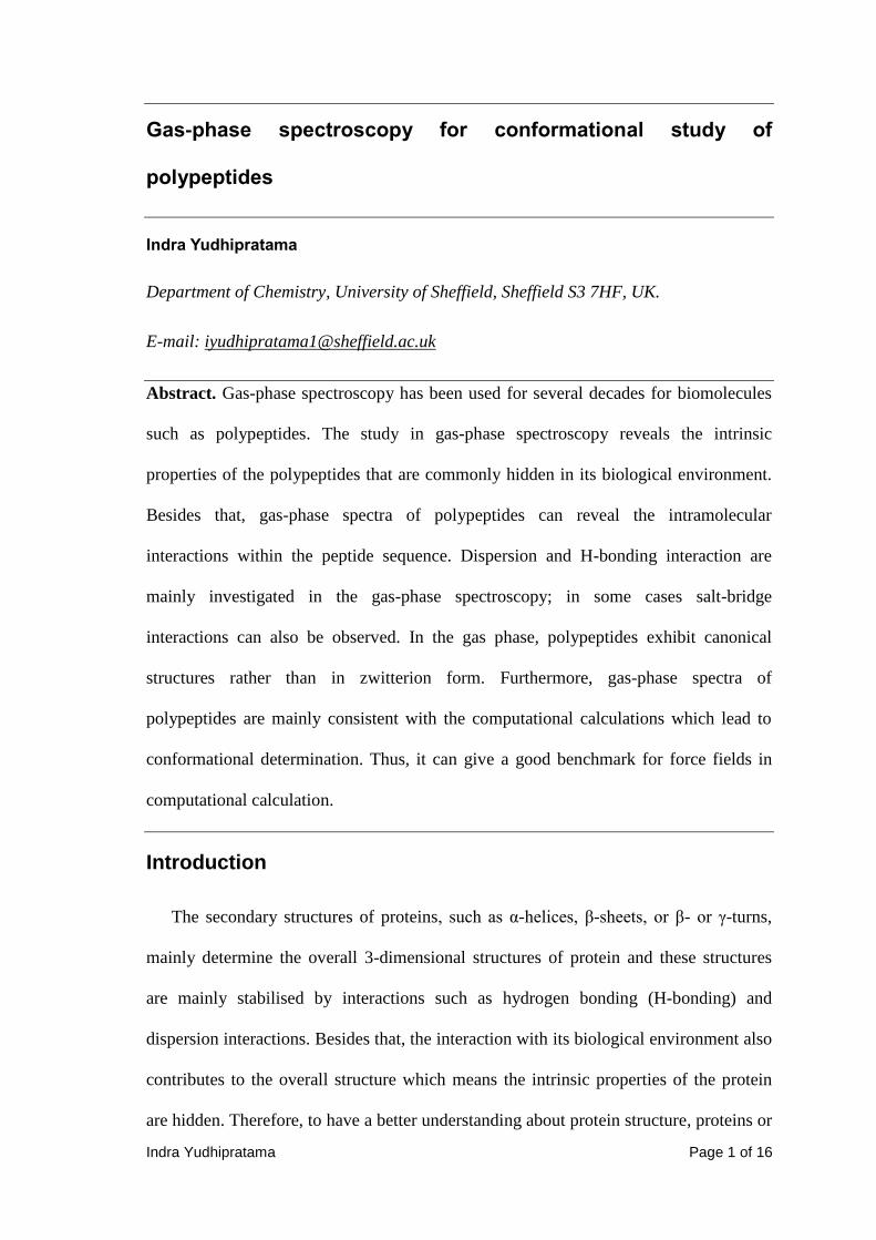
side chain can be formed in the gas phase [15]. It is noteworthy that the overall charge
is still neutral and to exhibit the zwitterionic structure the peptide needs to have the
most acidic and the most basic side groups which are glutamic acid (Glu) and arginine
(Arg). In this study, Ac-Glu-Ala-Phe-Ala-Arg-NHMe (EAFAR) was used and
phenylalanine which serves as chromophore [15]. The way to observe this zwitterionic
structure was done by comparing the IR spectrum of EAFAR with Z-Glu-OH and Z-
Glu-NH2. The signature band of the unprotonated residue of Glu in the region around
1850 cm-1
which is corresponding to carboxylic acid’s C=O stretching frequency was
used in this study [15].
Figure 7 The IR spectra from [15] of a) capped EAFAR b) Z-Glu-OH and
c) Z-Glu-NH2.

Indra Yudhipratama Page 13 of 16
In the spectrum of EAFAR, the absence of a band around 1850 cm-1
indicates the
deprotonation of COOH residue of Glu in the gas phase EAFAR [15]. Besides that, it is
also observed that the stretching frequency of the carbonyl amide is significantly red-
shifted which indicates H-bonding interaction. The conformational search of EAFAR
then conducted by comparing computed and experimental IR spectrum of EAFAR and
it was found the experimental spectrum corresponds to the spectrum of EAFAR global
minimum which shows salt bridge interaction and exhibits helical structure (figure 8).
Figure 8 Diagram from [15] showing calculated low-energy conformations of
zwitterionic EAFAR.
A further study conducted by Jaeqx’s group using Z-Glu-Alan-Arg-NHMe (n = 0, 1,
3) confirmed the salt bridge interaction in gas-phase peptides [7]. On the
conformational search of this peptide, there are 3 possible ways the salt bridge
interaction can be formed as shown in figure 9.
Figure 9 Diagram from [7] showing different types of salt bridge interactions in
conformational search of Z-Glu-Alan-Arg-NHMe.

Indra Yudhipratama Page 14 of 16
Jaeqx’s group used the COO– symmetric carbonyl stretching frequency to observe
this interaction due to its sensitivity to the environment. Another interesting feature
from Z-Glu-Alan-Arg-NHMe peptides is the Arg residue also interacts with benzene
ring of the Z-cap via NH-π electrons interaction; the exact interaction between Arg
residue and π-electrons of benzene is different between each system. This study shows
that all systems exhibit the A-type interactions and this is also observed in the previous
study. Furthermore, Jaeqx’s group also noted that B-type interaction is possible to
observe when Glu and Arg residues are further apart and it might be due less strain on
the backbone as the peptide becomes longer [7].
Conclusions
In the past decades, gas-phase spectroscopy has been used to investigate
biomolecules, such as polypeptides, in its isolated phase. These studies give several
methods to bring biomolecules into gas phase such as laser desorption and electrospray
techniques which are then coupled with laser spectroscopy.
The studies in the gas-phase spectroscopy of polypeptides reveal the intrinsic
properties of the polypeptides such as the intramolecular interactions within the peptide
sequence. Mainly, the secondary structure of peptides in the gas phase is stabilised by
H-bonding and dispersion interactions. Several studies also started to look at the
possibility of salt-bridge interactions in the isolated condition by using Glu and Arg in
the peptides due to Glu and Arg has the most acidic and basic residue respectively. The
gas-phase spectroscopy is mainly used to investigate the intramolecular interactions in
the peptides rather than determination of the secondary structure. This conformational
search of secondary structure is aided by computational calculation which consistently
shows the agreement between theoretical calculation and experimental spectra. Thus,

Indra Yudhipratama Page 15 of 16
the intrinsic structural motifs of the polypeptides can be revealed and commonly the
structure resembles in its crystallised form or in its biological environment.
Furthermore, this shows the advantage of gas-phase spectroscopy that can be used as a
benchmark for force fields in computational calculation.
References
1 M. S. de Vries and P. Hobza, “Gas-Phase Spectroscopy of Biomolecular Building
Blocks”, Annu. Rev. Phys. Chem., 2007, 58, 585-612.
2 J. R. Cable, M. J. Tubergen, and D. H. Levy, “Laser desorption Molecular Beam
Spectroscopy: The Electronic Spectra of Tryptophan Peptides in the Gas Phase”, J. Am.
Chem. Soc., 1987, 109, 6198-6199.
3 D. Řeha, V. Valdés, J. Vondrášek, P. Hobza, A. Abu-Riziq, B. Crews, and M. S. de
Vries, “Structure and IR Spectrum of Phenylalanyl-Glycyl-Glycil Tripeptide in the Gas-
Phase: IR/UV Experiments, Ab Initio Quantum Chemical Calculations, and Molecular
Dynamic Simulations, Chem. Eur. J., 2005, 11, 6803-6817.
4 W. Chin, J. P. Dognon, F. Piuzzi, B. Tardivel, I. Dimicoli, and M. Mons, “Intrinsic
Folding of Small Peptide Chains: Spectroscopic Evidence for the Formation of β-Turns
in the Gas Phase”, J. Am. Chem. Soc., 2004, 127, 707-712.
5 J. A. Stearns, M. Guidi, O. V. Boyarkin, and T. R. Rizzo, “Conformation-specific
infrared and ultraviolet spectroscopy of tyrosine-based protonated dipeptides”, J. Chem.
Phys., 2007, 127, 154322-1 – 154322-7.
6 J. A. Stearns, O. V. Boyarkin, and T. R. Rizzo, “Spectroscopic Signatures of Gas-
Phase Helices: Ac-Phe-(Ala)5-Lys-H+ and Ac-Phe-(Ala)10-Lys-H
+”, J. Am. Chem. Soc.,
2007, 45, 13820-13821.
Indra Yudhipratama Page 16 of 16
7 S. Jaeqx, J. Oomens, and A. Rijs, “Gas-phase salt bridge interactions between
glutamic acid and arginine”, Phys. Chem. Chem. Phys., 2013, 15, 16341-16352.
8 V. Feyer, O. Plekan, R. Richter, M. Careno, K. C. Prince, and V. Carravetta, “Core
Level Study of Alanine and and Threonine”, J. Phys. Chem. A, 2008, 112, 7806-7815.
9 V. Feyer, O. Plekan, R. Richter, M. Careno, K. C. Prince, and V. Carravetta,
“Photoemission and Photoabsorption Spectroscopy of Glycil-Glycine in the Gas Phase”,
J. Phys. Chem. A, 2009, 113, 10726-10733.
10 A. Abo-Riziq, B. O. Crews, M. P. Callahan, L. Grace, and M. S. de Vries,
“Spectroscopy of Isolated Gramicidin Peptides”, Angew. Chem., 2006, 118, 5290-5293.
11 M. Alauddin, H. S. Biswal, E. Gloaguen, and M. Mons, “Intra-residue interactions
in proteins: interplay between serine or cysteine side chains and backbone
conformations, revealed by laser spectroscopy of isolated model peptides”, Phys. Chem.
Chem. Phys., 2015, 17, 2169-2178.
12 I. Compagnon, J. Oomens, G. Meijer, and G. von Helden, “Mid-Infrared
Spectroscopy of Protected Peptides in the Gas Phase: A Probe of the Backbone
Conformation”, J. Am. Chem. Soc., 2006, 128, 3592-3597.
13 A. M. Rijs, M. Kabeláč, A. Abo-Riziq, P. Hobza, and M. S. de Vries, “Isolated
Gramicidin Peptides Probes by IR Spectroscopy”, Chem. Phys. Chem., 2011, 12, 1816-
1821.
14 N. S. Nagornova, M. Guglielmi, M. Doemer, I. Tavernelli, U. Rothlisberger, T. R.
Rizzo, and O. V. Boyarkin, “Cold-ion Spectroscopy Reveals the Intrinsic Structure of a
Decapeptide”, Angew. Chem. Int. Ed., 2011, 5383-5386.
15 A. M. Rijs, G. Ohanessian, J. Oomens, G. Meijer, G. von Helden, and I.
Campagnon, “Internal Proton Transfer Leading to Stable Zwitterionic Structures in a
Neutral Isolated Peptide”, Angew. Chem. Int. Ed., 2010, 49, 2332-2335.