Embed Size (px)
Citation preview
I. Organization of the Nervous System
The nervous system is the body’s principal control and integrating center. 신경계는 우리 몸의 주된 통제 및 통합센터이다.
It serves three broad functions: sensory, integrative, and motor: 신경계의 세 가지 기능: 감각, 통합, 운동 It senses changes within the body and in the outside environment;
신경계는 우리의 몸 내부와 바깥 환경에서의 변화를 감지한다.
It interprets the change.; and 신경계는 이러한 변화를 해석한다.
It responds to the interpretation by initiating action by muscular contraction or glandular secretion. 신경계는 근육을 수축 혹은 선을 통한 분비(땀)와 같은 행동을 취하게 함으로써 변화의 해석에 반응한다.
Through sensation, integration, and response, the nervous system rapidly maintains the body’s homeostasis. 감각, 통함, 반응을 통해 신경계는 재빨리 몸의 항상성을 유지한다.
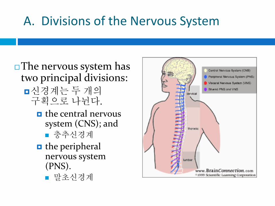
A. Divisions of the Nervous System
The nervous system has two principal divisions: 신경계는 두 개의 구획으로 나뉜다.
the central nervous system (CNS); and 충추신경계
the peripheral nervous system (PNS). 말초신경계

B. 뇌의 발생학적 단계 Embryological Levels of the Brain
The brain develops very rapidly during the first few years of life.
뇌는 인간의 인생 초기 몇 년 동안 매우 빨리 발달한다.
Growth is mainly due to an increase in the size of cells already present, proliferation and growth of neuroglia, development of synaptic contacts and dendritic branching, and myelination of various fiber tracts.
성장은 주로 이미 존재하는 뉴런 세포의 크기가 중가, 신경교의 확장과 성장, 뉴런 간의 시냅스 형성, 수상돌기 가지와 여러 신경줄기의 수초화의 발달에 의해 일어난다.

1. 중추신경계외 말초신경계의 발달 Development of the CNS and PNS
The development of the NS begins at about the third week of life with a thickening of the ectoderm of the neural plate. 신경계의 발달은 신경판의 외배엽이 두꺼워지면서 생후 3주에 시작된다.
The plate folds inward and forms a longitudinal groove, called the neural groove. 신경판은 안쪽으로 접혀 들어가고 긴 홈을 형성하는데 이는 신경홈이라 칭한다.
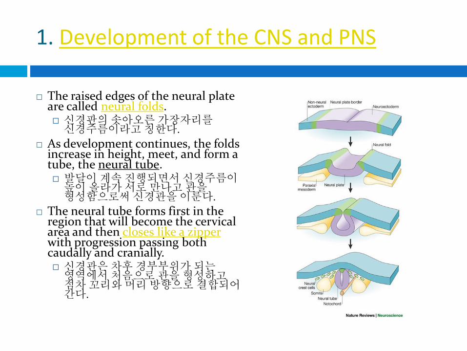
1. Development of the CNS and PNS
The raised edges of the neural plate are called neural folds. 신경판의 솟아오른 가장자리를 신경주름이라고 칭한다.
As development continues, the folds increase in height, meet, and form a tube, the neural tube. 발달이 계속 진행되면서 신경주름이 높이 올라가 서로 만나고 관을 형성함으로써 신경관을 이룬다.
The neural tube forms first in the region that will become the cervical area and then closes like a zipper with progression passing both caudally and cranially. 신경관은 차후 경부부위가 되는 영역에서 처음으로 관을 형성하고 점차 꼬리와 머리 방향으로 결합되어 간다.

1. Development of the CNS and PNS
The opening at the cranial end is called the anterior neuropore.
머리 쪽의 열림은 앞신경구멍
The opening at the caudal end is called the posterior neuropore.
꼬리 쪽의 열림은 뒤신경구멍
Closure of the anterior neuropore takes place by day 24 and the posterior neuropore by day 26.
앞신경구멍의 닫힘은 24일 경에 일어나고 뒤신경구멍은 26일 경에 일어난다.
The primitive CNS is now a hollow tubular structure closed at both ends.
이 시기의 원시 중추신경계는 양끝이 막힌 가운데가 텅 빈 튜브형태이다.
1. Development of the CNS and PNS
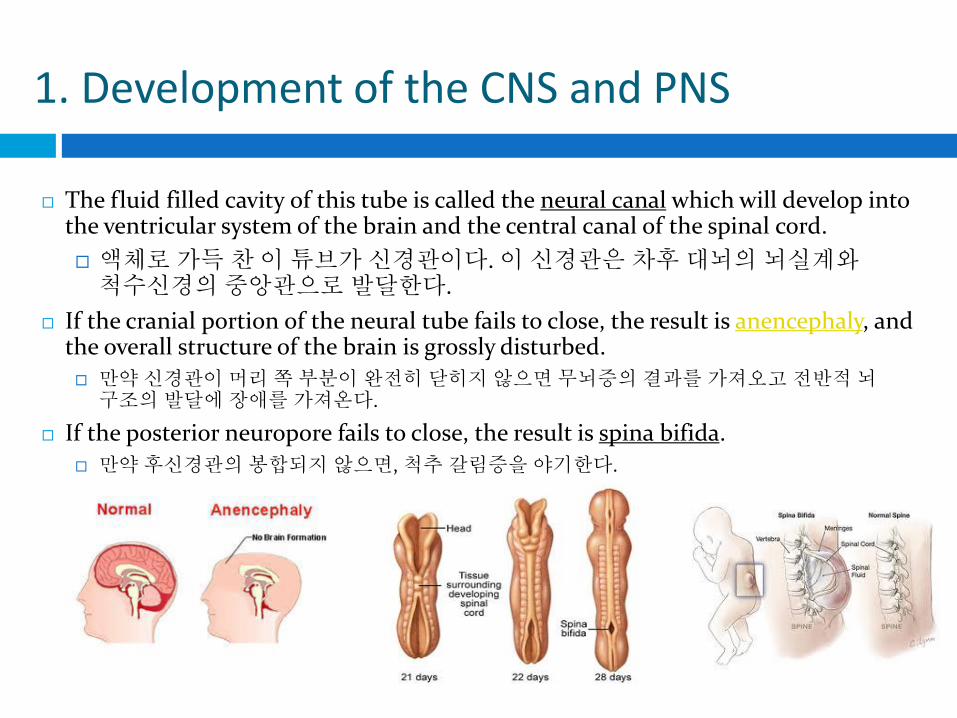
The fluid filled cavity of this tube is called the neural canal which will develop into the ventricular system of the brain and the central canal of the spinal cord.
액체로 가득 찬 이 튜브가 신경관이다. 이 신경관은 차후 대뇌의 뇌실계와 척수신경의 중앙관으로 발달한다.
If the cranial portion of the neural tube fails to close, the result is anencephaly, and the overall structure of the brain is grossly disturbed.
만약 신경관이 머리 쪽 부분이 완전히 닫히지 않으면 무뇌증의 결과를 가져오고 전반적 뇌 구조의 발달에 장애를 가져온다.
If the posterior neuropore fails to close, the result is spina bifida.
만약 후신경관의 봉합되지 않으면, 척추 갈림증을 야기한다.

2. Development of the Cerebral Vesicles 뇌포의 발달
By the fourth week, three distinct bulges, the primary vesicles, appear in the anterior neuropore. 4주차에 세 개의 뚜렷한 팽창(융기)인 일차 수포들이 앞신경구멍에서 나타난다.
From the top down, we have the prosencephalon (forebrain), the mesencephalon (midbrain), and the rhombencephalon (hindbrain). 위에서부터 전뇌, 중뇌, 후뇌라고 명명되어진다.
2. Development of the Cerebral Vesicles
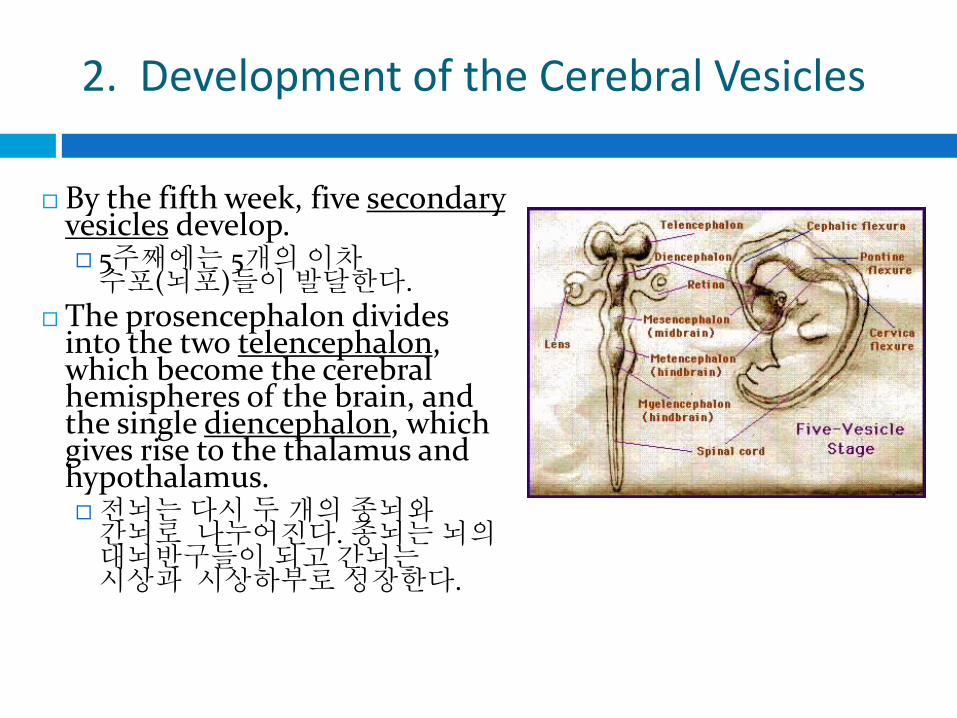
By the fifth week, five secondary vesicles develop. 5주째에는 5개의 이차 수포(뇌포)들이 발달한다.
The prosencephalon divides into the two telencephalon, which become the cerebral hemispheres of the brain, and the single diencephalon, which gives rise to the thalamus and hypothalamus. 전뇌는 다시 두 개의 종뇌와 간뇌로 나누어진다. 종뇌는 뇌의 대뇌반구들이 되고 간뇌는 시상과 시상하부로 성장한다.

2. Development of the Cerebral Vesicles
The mesencephalon remains unchanged and becomes the midbrain.
중뇌는 변하지 않으며 중뇌가 된다.
The rhombencephalon divides into the metencephalon, which becomes the pons and cerebellum, and the myelencephalon, which becomes the medulla oblongata.
후뇌는 소뇌(후뇌)와 수뇌(척수뇌)로 나누어진다. 소뇌는 뇌교와 소뇌가 되고 수뇌는 연수가 된다.
2. Development of the Cerebral Vesicles
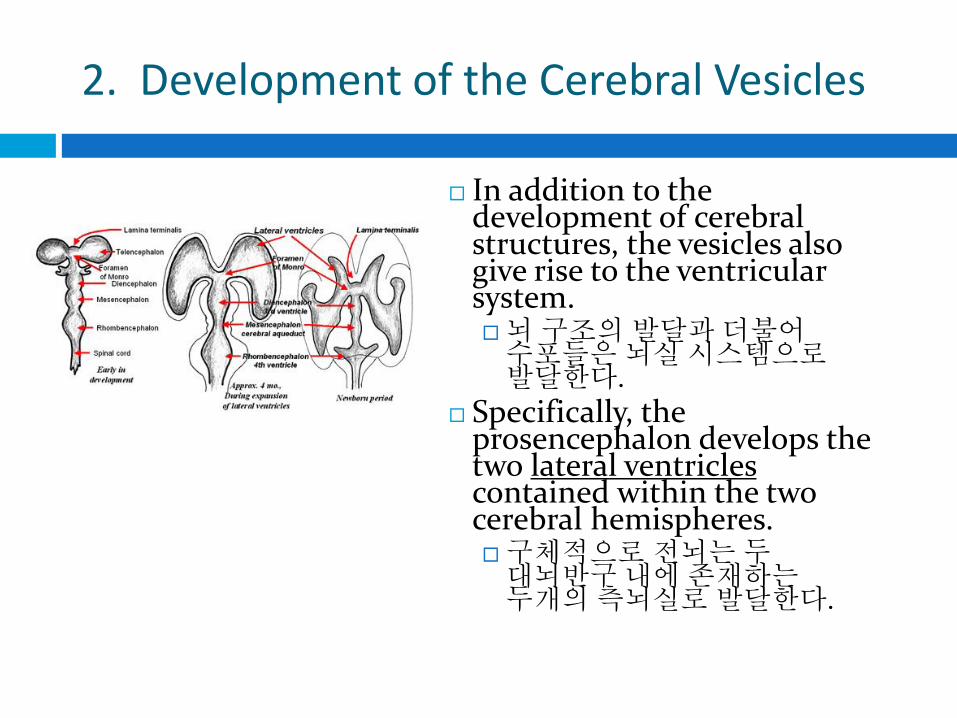
In addition to the development of cerebral structures, the vesicles also give rise to the ventricular system. 뇌 구조의 발달과 더불어 수포들은 뇌실 시스템으로 발달한다.
Specifically, the prosencephalon develops the two lateral ventricles contained within the two cerebral hemispheres. 구체적으로 전뇌는 두 대뇌반구 내에 존재하는 두개의 측뇌실로 발달한다.

2. Development of the Cerebral Vesicles
The third ventricle develops within the diencephalon. 제 3 뇌실은 간뇌에서 발달한다.
The mesencephalon gives rise to the cerebral aqueduct. 중뇌에서는 중뇌수도가 발달한다.
The rhombencephalon gives rise to the fourth ventricle. 후뇌에서는 제4 뇌실이 발달한다.

B. Embryological Levels of the Brain
The ventricles are a continuous series of fluid-filled spaces extending through all major divisions of the CNS.
뇌실들은 충추신경계의 거의 모든 부위에 걸쳐져 있으며 액체로 채워진 하나의 연결된 공간이다.
Each lateral ventricle communicates with the 3rd ventricle through the intraventricular foramen.
측뇌실은 내실 내 소공(구멍)을 통해 제 3뇌실과 연결되어 있다.
The 3rd ventricle communicates with the 4th ventricle through the cerebral
aqueduct of the midbrain. 제 3뇌실은 중뇌의 중뇌수도관을 통해 제
4뇌실과 연결되어 있다.
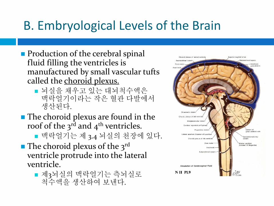
B. Embryological Levels of the Brain
Production of the cerebral spinal fluid filling the ventricles is manufactured by small vascular tufts called the choroid plexus. 뇌실을 채우고 있는 대뇌척수액은 맥락얼기이라는 작은 혈관 다발에서 생산된다.
The choroid plexus are found in the roof of the 3rd and 4th ventricles. 맥락얼기는 제 3.4 뇌실의 천장에 있다.
The choroid plexus of the 3rd ventricle protrude into the lateral ventricle. 제3뇌실의 맥락얼기는 측뇌실로 척수액을 생산하여 보낸다.
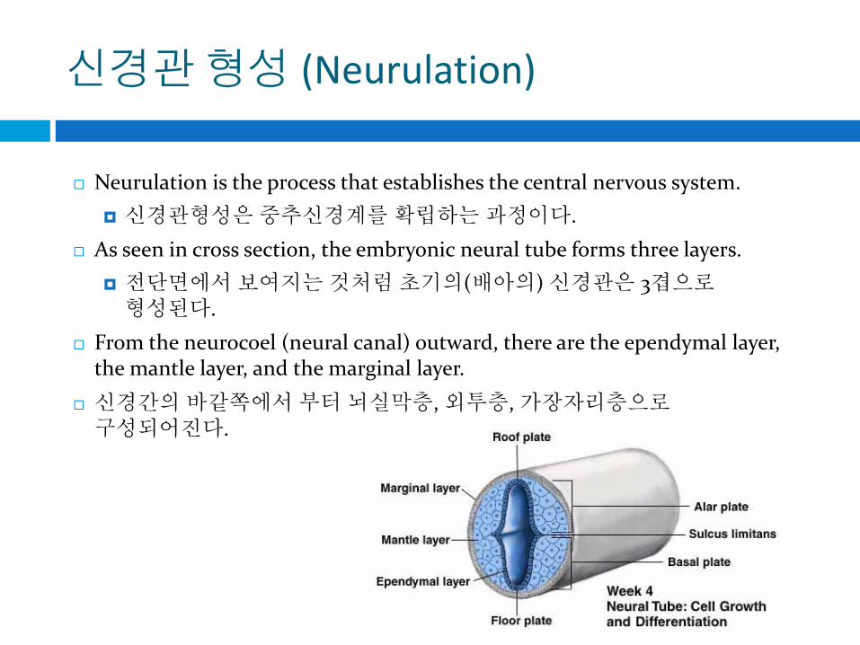
신경관 형성 (Neurulation)
Neurulation is the process that establishes the central nervous system.
신경관형성은 중추신경계를 확립하는 과정이다.
As seen in cross section, the embryonic neural tube forms three layers.
전단면에서 보여지는 것처럼 초기의(배아의) 신경관은 3겹으로 형성된다.
From the neurocoel (neural canal) outward, there are the ependymal layer, the mantle layer, and the marginal layer.
신경간의 바같쪽에서 부터 뇌실막층, 외투층, 가장자리층으로 구성되어진다.

Neurulation
Some of the cells of the ependymal layer remain in place to become the thin, ciliated lining of the adult central canal, but most migrate outward to join mantle cells in forming both neurons and neuroglia. 뇌실막층의 여러 세포들은 성인 중심관의 얇은 섬모세포 내벽이 되지만, 대부분의 세포들은 바깥쪽으로 이동하여 외투층과 결합하여 뉴런과 신경교세포가 된다.
These will be the gray matter of the adult. 이 세포들은 성인의 회백질이 된다.

Neurulation
With cell migration, the mantle layer develops the characteristic “butterfly” shape.
세포의 이동과 더불어 외투층은 “나비” 모양으로 발달한다.
The lateral walls of the tube thicken and maturing neurons clump into two different plates.
두꺼워지고 성숙해진 뉴런의 신경관 측벽은 다른 두개의 판으로 구분되어 진다.

Neurulation
The two plates are divided by a shallow, longitudinal groove called the sulcus limitans.
두 개의 판은 경계고랑이라는 얕고 긴 홈에 의해 나뉜다.
The sulcus limitans separates the developing gray matter into a dorsal alar plate and a ventral basal plate.
경계고랑은 자라나는 회백질을 등쪽 날개판과 배쪽 바닥판으로 구분한다.

Neurulation
These plates signal the future locations of sensory and motor functions, respectively. 이 판들은 각각 감각기능과 운동기능의 향후 위치를 알려준다.
Alar and basal plates become dorsal and ventral horns, respectively, while intermediate regions develop interneurons, mixed nerves. 중간지역이 중간뉴런과 혼합신경으로 발달하는 반면, 날개판과 바닥판은 각각 배각과 전각이 된다.

Neurulation
Finally, cells from the marginal layer mature by growing out their processes (axons and dendrites).
가장자리층의 세포들은 축삭과 수상돌기로 성장한다.
This layer is penetrated by nerve fibers growing out of the deeper layers.
더 깊은 층에서 자라나오는 신경섬유들이 이 층을 관통한다.
It becomes the white matter of the adult cord.
이것은 성인척수의 백질이 된다.

Neurulation
The brainstem develops in a manner similar to the spinal cord.
뇌간은 척수와 비슷한 방식으로 발달한다.
From the medulla through the midbrain, alar and basal plates form sensory and motor columns of cells that supply cranial nerves.
연수와 중뇌에 분포한 날개판과 바닥판이 대뇌신경을 공급하는 감각과 운동을 담당하는 세포기둥를 형성한다.
However, the organization of alar and basal plates in the brainstem differ from those of the spinal cord.
하지만, 뇌간의 날개판과 바닥판 조직은 척수의 구조와 다르다.
Neurulation
In the 6 mm embryo, the thin ependymal roof of the neural tube, the spinal cord, becomes even thinner as the ventricle of the neural tube begins to widen in the early stages of the development of the 4th ventricle.
6mm 배아에서, 신경관의 뇌실이 제 4뇌실 발달의 초기단계에서 넓어지기 시작하면서 신경과의 얇은 뇌실막층은 더욱 더 얇아진다.
With continued development, alar and basal plates shift laterally and become located in the floor of the ventricle.
계속적으로 발달하면서, 날개판과 바닥판은 측면으로 꺽이고 뇌실의 바닥에 놓이게 된다.

Neurulation
The sulcus limitans continues to be identifiable helping to mark the boundary between sensory and motor areas. 경계고랑은 점점 더 감각과 운동영역 사이의 경계를 구분하기 쉽게한다.
In the medulla and pons, the alar plate comes to lie lateral to the basal plate, not dorsal to it. 연수와 뇌교에서 날개판은 바닥판의 배측이 아닌 측면에 놓이게 된다.
The basal plate forms the motor nuclei of the cranial nerves, medial to the sulcus limitans in the ventricular floor. 바닥판은 뇌신경의 운동핵을 형성하고 뇌실바닥에서 경계고랑의 안쪽에 형성된다.

Neurulation
Lateral to the sulcus, the alar plate forms sensory relay nuclei. 경계고랑의 측면에서 날개판은 감각중계핵을 형성한다.
Rostral to the midbrain, the diencephalon and cerebral hemispheres develop from the alar plate. 중뇌의 입쪽에서 간뇌와 대뇌엽들이 바닥판으로부터 발달한다.
The cerebellum also develops from alar plate. 소뇌 또한 날개판으로 부터 발달한다.
Portions of the alar plate migrate ventrally and form the inferior olivary nucleus, a cerebellar relay nucleus. 날개판의 부분은 배쪽으로 이동하고 아래 올리브핵과 소뇌중개핵이 된다.

Differences in Male & Female Brains
The majority of brain development that determines sex-specific circuits happens during the first 18 weeks of pregnancy.
Until eight weeks old, every fetal brain looks female—female is nature’s default gender setting.
If you were to watch a female and male brain developing via time-lapse photography, you would see that some of the neural connections are being laid out according to a blueprint drafted by both genes and sex hormones (Brizendine, 2010).

Differences in Male & Female Brains
A huge testosterone surge beginning in the eighth week will turn this unisex brain male by killing off some cells in the communication centers and growing more cells in the sex and aggression centers.
If the testosterone surge doesn’t occur, the female brain continues to grow unperturbed.
Scientists agrees that when cells in various areas of the male and female brains are stimulated by hormones such as testosterone and estrogen, they turn on and off different genes.

Differences in Male & Female Brains
For boys, the genes that turn on will trigger the urge to track and chase moving objects, hit targets, test their own strength, and play at fighting off enemies.
For girls, the genes that turn on will enhance female brain circuits and centers for observation, gut feelings, even tending and caring and her fetal brain cells will continue to sprout more connections in the communication centers and areas that process emotion.