Embed Size (px)
Citation preview

Case Report Open Access
Morais, J Clin Exp Cardiolog 2014, 5:2DOI: 10.4172/2155-9880.1000290
Volume 5 • Issue 2 • 1000290J Clin Exp Cardiolog
ISSN: 2155-9880 JCEC, an open access journal
Cardiopulmonary Disorders-2nd Edition
*Corresponding authors: Humberto Morais, Rua Pedro Miranda 40-42 Maianga, Republic of Angola, Tel: 00244923520934; E-mail: [email protected]
Received February 10, 2014; Accepted February 24, 2014; Published February 28, 2014
Citation: Morais H (2014) Submitral Aneurysm: An Echocardiography Study in a Tertiary Center in Angola. J Clin Exp Cardiolog 5: 290. doi:10.4172/2155-9880.1000290
Copyright: © 2014 Morais H. This is an open-access article distributed under the terms of the Creative Commons Attribution License, which permits unrestricted use, distribution, and reproduction in any medium, provided the original author and source are credited.
Submitral Aneurysm: An Echocardiography Study in a Tertiary Center in AngolaHumberto Morais*Department of Cardiology, Hospital Militar Principal/Instituto Superior, Luanda, Republic of Angola
IntroductionSubmitral Aneurysms (SMA) are a relatively rare and poorly
understood cardiac condition, although there are multiple reports of its incidence in specific populations [1-5]. Clinically it is manifested by symptoms and signs of heart failure due to mitral regurgitation, and/or ventricular arrhythmias. Transthoracic echocardiography (TTE) plays a key role in the definitive diagnosis of this pathology. Transesophageal echocardiography is of paramount importance in assessing the rupture of the aneurysm into the left atrium [1].
In recent years, a number of clinical cases using others imaging modalities have been published including real-time three-dimensional echocardiography, nuclear magnetic resonance, Cardiac Tomography (CT ), as well as the use of different imaging modalities in the same patient [2,6-8]. The purpose of this paper is to present a brief review of the literature on the subject of SMA, regarding six patients studied in echocardiography laboratory of our Hospital in an eleven-year period.
MethodologyBased on database of the Echocardiography Laboratory of the
Hospital Militar Principal/Instituto Superior, demographics data, echocardiography features and data from follow-up of patients with the diagnosis of SMA, in the period from May 2001 to April 2012, were analyzed.
All patients underwent Transthoracic Echocardiography (TTE). Four patients underwent real time three-dimensional TTE (RT3DTTE), three patients underwent Transesophageal Echocardiography (TEE), two patient performed real time three-dimensional TEE (RT3DTEE). One patient underwent cardiac computed tomography angiography.
TTE and Doppler studies were performed in accordance with the recommendations of the American Society of Echocardiography [9,10]. TEE studies were performed in accordance with the recommendations of the European Society of Cardiology [11]. Real time three-dimensional echocardiography was performed as previously reported [12].
ResultsDemographic data, clinical presentation, NYHA functional
class and follow-up are shown in Table 1. Data from transthoracic echocardiography, cardiac complications and associated pathologies are presented in Table 2. During the review period six patients had a diagnostic of SMA. Four are male and two are female. The mean age was 27.3 ± 7.0. Two patients had rupture of the aneurysm into the left
atrium. One patient presented together with SMA an aneurysm of the right sinus of Valsalva dissecting into the interventricular septum and left ventricular noncompaction. Five of the six patients had severe mitral insufficiency. All patients were proposed for surgery: one was successfully operated in Angola, in Hospital Josina Machel, one refused surgery and died one year after the diagnosis was made, the remaining four patients were lost in follow-up.
DiscussionOnce SMA is a relative rare disease there are no data on the
prevalence and incidence of this condition in general population. However, there are few epidemiological data reported in the literature that it is worth highlighting. In a study by Gaultier et al. at Hospital de Miamex in Nigeria, SMA accounted for 0.04% of hospitalizations and 0.34% of cardiovascular disease [4]. Sliwa and Mocumbi found 10 cases of SMA in 5200 patients with heart failure diagnosed in the period 2006-2008 in Soweto, corresponding to 0.19% of cases [13]. Morais et al. in Angola found SMA in 4.1% of congenital heart disease diagnosed in patients aged greater than or equal to 15 years within 10 years [14].
Its etiology is still the subject of debate, although there is agreement that it can be congenital or acquired. The most common causes of acquired SMA are inflammatory or infectious diseases or trauma. Among infectious causes are syphilis, tuberculosis and infectious endocarditis [3,15]. It can also result from cardiomyopathy [4,16]. Congenital SMA arises from a defect of a valve ring and is sometimes associated with sinus of Valsalva aneurysm, which suggests a congenital weakness of aortic or mitral annulus insertion in the underlying
No Gender Age Symptoms Class NYHA Follow-up1 Male 21 Dyspnoea III Lost2 Male 27 Dyspnoea III Lost3 Male 22 Chest pain I Surgery4 Female 30 Dyspnoea I Lost5 Female 40 Dyspnoea II Lost6 Male 24 Dyspnoea III Refused Surgery
Table 1: Demographic data, clinical presentation, NYHA functional class, and follow up.
Table 2: Date of diagnosis, and findings on echocardiography, LA: left atrium, SVA: sinus of Valsalva aneurysm, LVNC: left ventricular non-compaction.
NO Year Systolic Function Mitral regurgitation Complications Other findings
1 2001 Good Severe Rupture into LA None2 2004 Good Severe None None3 2008 Good Mild None None4 2010 Good Severe None None5 2010 Good Severe Rupture into LA None
6 2011 Severe Depressed Severe None SVA; LVNC
Journal of Clinical & Experimental CardiologyJo
urna
l of C
linica
l & Experimental Cardiology
ISSN: 2155-9880
Citation: Morais H (2014) Submitral Aneurysm: An Echocardiography Study in a Tertiary Center in Angola. J Clin Exp Cardiolog 5: 290. doi:10.4172/2155-9880.1000290
Page 2 of 4
Volume 5 • Issue 2 • 1000290J Clin Exp Cardiolog
ISSN: 2155-9880 JCEC, an open access journal
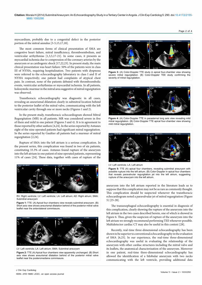
aneurysm into the left atrium reported in the literature leads us to suppose that this complication may not be as rare as commonly thought. This complication should be suspected whenever the transthoracic echocardiogram noted a paravalvular jet of mitral regurgitation (Figure 5) [25-28].
The transesophageal echocardiography is essential in diagnosis of this complication, clearly showing the rupture of the aneurysm into the left atrium in the two cases described herein, one of which is showed in Figure 6. Thus, given the suspicion of rupture of the aneurysm into the left atrium we strongly recommend performing TEE whenever possible. Multidetector cardiac CT may also be useful in this context [28].
Recently, real-time three-dimensional echocardiography has been shown to be superior to conventional echocardiography in the evaluation of SMA [6,25]. In our experience, the real-time three-dimensional echocardiography was useful in evaluating the relationship of the aneurysm with other cardiac structures including the mitral valve and its leaflets, the anatomical characterization of the aneurysm. Moreover in one patient, real-time three-dimensional echocardiography has allowed the identification of a bilobular aneurysm with two necks communicating with the left ventricle, providing additional data
myocardium, probably due to a congenital defect in the posterior portion of the mitral annulus [3-5,15,17,18].
The most common forms of clinical presentation of SMA are congestive heart failure, mitral insufficiency, thromboembolism, and ventricular arrhythmias [1,3,5,17-21]. In some cases, it presents as myocardial ischemia due to compression of the coronary arteries by the aneurysm or as cardiogenic shock [17,22,23]. In present study, the main clinical presentation was heart failure: 50% of the patients were in class III of NYHA, requiring hospitalization. Two patients with dyspnoea were referred to the echocardiography laboratory in class I and II of NYHA respectively; one patient had complaints of atypical chest pain. In contrast, none of the patients debuted with thromboembolic events, ventricular arrhythmias or myocardial ischemia. In all patients, holosystolic murmur in the mitral area suggestive of mitral regurgitation was observed.
Transthoracic echocardiography was diagnostic in all cases, revealing an aneurismal dilatation clearly in submitral location behind to the posterior leaflet of the mitral valve, communicating with the left ventricular cavity through one or more necks (Figures 1 and 2).
In the present study, transthoracic echocardiogram showed Mitral Regurgitation (MR) in all patients. MR was considered severe in five of them and mild in one patient (Figures 3 and 4). It is in agreement to those reported by other authors [4,24]. In the series reported by Antunes eight of the nine operated patients had significant mitral regurgitation. In the series reported by Gaultier all patients had a murmur of mitral regurgitation [2,24].
Rupture of SMA into the left atrium is a serious complication. In the present series, this complication was found in two of six patients, constituting 33.3% of cases. Antunes found rupture of the aneurysm into the left atrium in one patient of nine operated patients, representing 11% of cases [24]. These data, together with cases of rupture of the
RV: Right ventricle; LV: Left ventricle; LA: Left atrium; AD: Right atrium; SMA: Submitral aneurysmFigure 1: TTE: (A) Apical four chambers view reveals submitral aneurysm. (B) Short axis view shows aneurismal dilatation behind of the posterior mitral valve leaflet near the anterolateral commissure.
LV: Left ventricle; LA: Left atrium; SMA: Submitral aneurysmFigure 2: TTE (A) Apical four chambers view apparently unchanged. (B) Short axis view shows aneurismal dilatation behind of the posterior mitral valve leaflet near the posteromediana commissure.
Figure 3: (A) Color-Doppler TTE study in apical four-chamber view showing severe mitral regurgitation. (B) Color-Doppler TEE study confirming the severity of mitral regurgitation.
Figure 4: (A) Color-Doppler TTE in parasternal long axis view revealing mild mitral regurgitation. (B) Color-Doppler TTE apical four-chamber view showing mild mitral regurgitation.
LV: Left ventricle; LA: Left atriumFigure 5: TTE (A) apical four chambers, revealing submitral aneurysm with possible rupture into the left atrium. (B) Color-Doppler in apical four chambers that reveals paravalvular regurgitation jet into the left atrium, suggesting rupture of the aneurysm into the left atrium.

Citation: Morais H (2014) Submitral Aneurysm: An Echocardiography Study in a Tertiary Center in Angola. J Clin Exp Cardiolog 5: 290. doi:10.4172/2155-9880.1000290
Page 3 of 4
Volume 5 • Issue 2 • 1000290J Clin Exp Cardiolog
ISSN: 2155-9880 JCEC, an open access journal
to two-dimensional echocardiography. Real-time 3DTEE provided unambiguous images in case of rupture of the aneurysm into the left atrium (Figures 7 and 8).
Average age found in present series (27.3 ± 7.0) is similar to those reported by Antunes (28 ± 3), but slightly higher than reported by Gaultier (20 years). In the present study a 3:1 male: female ratio was found it is higher than reported by Antunes who found a 1.25:1 ratio [4,24].
Definitive treatment of SMA requires surgery [23,24,29-32]. The technique that is now more frequently utilized was described by Antunes in 1987 where the aneurysm is approached through the atrium [24]. This technique allows excluding the aneurysm and often preserving the mitral valve [22]. When this is not possible, the valve should be replaced.
ConclusionsSubmitral aneurysm, although uncommon, should always be
entered in the list of differential diagnosis in young patients presenting with a murmur suggestive of mitral insufficiency or signs and symptoms
of heart failure or thromboembolic events. The definitive diagnosis is made by transthoracic echocardiography in the presence of an aneurismal dilatation in submitral location behind the posterior leaflet that communicated with the left ventricular cavity through one or more necks. The Doppler study frequently reveals mitral regurgitation that is often severe.
In the present series the most frequent complication was rupture of the aneurysm into the left atrium. Transesophageal echocardiography was fundamental in the diagnosis of this complication in both cases, and we strongly recommend performing TEE whenever possible. Contrast-enhanced Cardiac CT may also be useful in this context, clearly revealing the passage of the contrast from the aneurysm into the left atrium through the point of rupture. Cardiac CT angiography also allows evaluating the coronary arteries.
Real-time three-dimensional echocardiography is not necessary for the diagnosis of SMA. However it is very useful in the evaluation of the relationship of the aneurysm with the other cardiac structures. In the evaluation of the anatomical characteristics of the aneurysm allowing the identification of one or more apertures through which aneurysm communicates with the left ventricle, providing additional data to two-dimensional echocardiography, and improving the plan for surgery. Real-time 3DTEE provides clear and unique images in cases of the rupture of the aneurysm into the left atrium.
References
1. Morais H, Branco LM, Cunha R, Martins T (2007) Rupture of a submitral ventricular aneurysm into the left atrium diagnosed by transesophageal echocardiography. Rev Port Cardiol 26: 367-372.
2. Chen CC, Hsiung MC, Wei J, Chang WT, Yin WH, et al. (2005) Mitral annular subvalvular left ventricular aneurysm. Echocardiography 22: 434-437.
3. Ribeiro PJF, Mendes RGG, Vicente WVA, Menardi AC, Évora PRB (2001) Aneurisma ventricular subvalvar mitral. Case presentations and surveys of published Brazilian cases. Arq Bras Cardiol 76: 395-398.
4. Gaultier Y, Cénac A, Aoua HO, Touré I (1989) [Idiopathic annular submitral aneurysm. Contribution of echography apropos of 5 cases]. Arch Mal Coeur Vaiss 82: 897-902.
5. Damasceno A, Hausse AO, Ferreira B, Teixeira R (1993) [Mitral subvalvular aneurysm]. Rev Port Cardiol 12: 963-971, 902.
6. Hotta VT, Cruz CB, Rassi Ddo C, Vieira ML, Mathias W Jr, et al. (2010) Subvalvular mitral pseudoaneurysm evaluated by three-dimensional echo. Echocardiography 27: 473-475.
7. Warren O, Athanasiou T, Massey R, Hamady M, Stanbridge R (2006) Large annular subvalvular left ventricular aneurysm: diagnostic evaluation using computed tomographic angiography. Tex Heart Inst J 33: 529-531.
8. Kharwar RB, Sethi R, Sanguri R, Singh V, Narain VS (2014) Multimodality imaging of submitral left ventricular aneurysm. Echocardiography 31: E44-47.
9. Henry WL, DeMaria A, Gramiak R, King DL, Kisslo JA, et al. (1980) Report of the American Society of Echocardiography Committee on Nomenclature and Standards in Two-dimensional Echocardiography. Circulation 62: 212-217.
10. Quiñones MA, Otto CM, Stoddard M, Waggoner A, Zoghbi WA; Doppler Quantification Task Force of the Nomenclature and Standards Committee of the American Society of Echocardiography (2002) Recommendations for quantification of Doppler echocardiography: a report from the Doppler Quantification Task Force of the Nomenclature and Standards Committee of the American Society of Echocardiography. J Am Soc Echocardiogr 15: 167-184.
11. Flachskampf FA, Decoodt P, Fraser AP, Daniel WG, Roelandt JRTC (2001) Guidelines from working group. Recommendation for performing transesophageal echocardiography. For the subgroup on transesophageal echocardiography and valvular heart disease, on behalf of working group on echocardiography of the European Society of Cardiology. Eur J Echocardiography 2: 8-21.
MV: Mitral valve; SMA: Submitral aneurysmFigure 7: RT3DTTE (A) view from the left ventricle in diastole showing submitral bilobular aneurysm. (B) The same view in systole.
Figure 8: RT3DTEE (A) “en face” view from the left atrium in systole, showing the rupture of the aneurysm into the left atrium (blue arrow). (B) The same view in diastole.
LV: Left ventricle; LA: Left atrium; SMA: Submitral aneurysmFigure 6: (A) TEE revealing submitral aneurysm with rupture into the left atrium. (B) Color-Doppler study showing regurgitation jet from the aneurysm into the left atrium, confirms the rupture of the aneurysm into the left atrium.

Citation: Morais H (2014) Submitral Aneurysm: An Echocardiography Study in a Tertiary Center in Angola. J Clin Exp Cardiolog 5: 290. doi:10.4172/2155-9880.1000290
Page 4 of 4
Volume 5 • Issue 2 • 1000290J Clin Exp Cardiolog
ISSN: 2155-9880 JCEC, an open access journal
12. Morais H, Martins T (2010) Ecocardiografia transesofágica tridimensional em tempo real: Experiência inicial em Angola An HMP/IS 1: 13-19.
13. Sliwa K, Mocumbi AO (2010) Forgotten cardiovascular diseases in Africa. ClinRes Cardiol 99: 65-74.
14. Morais H, Martins T Cunha R (2014) Spectrum of Congenital Heart Disease inAdolescents and Adults in Angola: An Echocardiography Study “2001-2009”.
15. Deshpande J, Vaideeswar P, Sivaraman A (2000) Subvalvular left ventricular aneurysms. Cardiovasc Pathol 9: 267-271.
16. Cenac A, Chaigneau C, Sueur JM, Orfila J (2002) [Submitral annular aneurysm, postpartum cardiomyopathy, and anti-Chlamydia pneumoniae antibodies inNiamey (Niger)]. Med Trop (Mars) 62: 81-84.
17. Chockalingam A, Gnanavelu G, Alagesan R, Subramaniam T (2004) Congenital submitral aneurysm and sinus of valsalva aneurysm. Echocardiography 21:325-328.
18. Mohanty A, Saxena A (2003) Submitral aneurysm: unusual echocardiographicfeatures. Heart 89: 552.
19. Esposito F, Renzulli A, Festa M, Cerasuolo F, Caruso A, et al. (1996) Submitral left ventricular aneurysm. Report of 2 surgical cases. Tex Heart Inst J 23: 51-53.
20. Moisés VA, Vieira Filho JP, Andrade JL, Leão LE, Martinez Filho EE (1993) [Submitral left ventricular aneurysm in a Brazilian Indian]. Arq Bras Cardiol 60:343-345.
21. Chi NH, Yu HY, Chang CI, Lin FY, Wang SS (2004) Clinical surgical experience of congenital submitral left ventricular aneurysm. Thorac Cardiovasc Surg 52:115-116.
22. Skoularigis J, Sareli P (1997) Submitral left ventricular aneurysm compressingthe left main coronary artery. Cathet Cardiovasc Diagn 40: 173-175.
23. Terzi CB, Pomerantzeff PM, Monachini MC, Kopel L, Medeiros CC, et al. (1996) [Mitral subvalvular aneurysm of the left ventricle]. Arq Bras Cardiol 67: 351-353.
24. Antunes MJ (1987) Submitral left ventricular aneurysms. Correction by a newtransatrial approach. J Thorac Cardiovasc Surg 94: 241-245.
25. Peters F, Essop R (2011) Congenital submitral aneurysm with rupture into theleft atrium: assessment by 2D and 3D transesophageal echocardiography.Echocardiography 28: E121-124.
26. Simpson L, Duncan JM, Stainback RF (2006) Perforated submitral leftventricular aneurysm resulting in severe mitral annular regurgitation. Tex Heart Inst J 33: 492-494.
27. Sai Krishna C, Naresh Kumar PV, Panigrahi NK, Suman K (2007) Submitral aneurysm with left atrial communication. Eur J Cardiothorac Surg 32: 547-549.
28. Duggal B, Inania R, Panchani N (2012) Rupture of a submitral aneurysm intothe left atrium. Pediatr Cardiol 33: 854-856.
29. Pektok E, Cikirikcioglu M, Didier D, Kalangos A (2008) Submitral left ventricular aneurysm: a rare but challenging pathology to treat. J Card Surg 23: 533-535.
30. Sharma A, Kulkarni V, Barde S, Bobade S, Bhargava V, et al. (2009) Submitralaneurysmorraphy with mitral valve replacement A case report. IJTCVS 25: 118-120.
31. Singh S, Agarwal S, Dutta N, Mishra S, Upreti L, et al. (2012) Surgical repairof a submitral aneurysm in a three-year-old child. J Card Surg 27: 238-240.
32. Ran JN, Gaijar T, Desai N (2011) Submitral left ventricular aneurysm – ourexperience Indian J Thorac Cardiovasc Surg 27: 91-95.
Citation: Morais H (2014) Submitral Aneurysm: An Echocardiography Study in a Tertiary Center in Angola. J Clin Exp Cardiolog 5: 290. doi:10.4172/2155-9880.1000290