Embed Size (px)
Citation preview
Rodrigues et al. BMC Pediatrics 2014, 14:252http://www.biomedcentral.com/1471-2431/14/252
CASE REPORT Open Access
Hypotonic male infant and MCT8 deficiency - adiagnosis to think aboutFilipa Rodrigues1*, Joana Grenha1, Carlos Ortez1,2, Andrés Nascimento1,2, Beatriz Morte2,3, Monica M-Belinchón2,3,Judith Armstrong4 and Jaume Colomer1,2
Abstract
Background: Thyroid hormone is crucial in the development of different organs, particularly the brain. MCT8 is aspecific transporter of triiodothyronine (T3) hormone and MCT8 gene mutations cause a rare X-linked disordernamed MCT8 deficiency, also known as Allan-Herndon-Dudley syndrome, characterized by psychomotor retardationand hypotonia. Typically, elevation of T3 and delayed myelination in cerebral magnetic resonance imaging are found.
Case presentation: We present a 24-month-old boy, born from non-consanguineous healthy parents, with severemotor and cognitive delay and global hypotonia, being unable to hold head upright or sit without support. Deeptendon reflexes were absent bilaterally at the ankles. T3 was elevated and thyroxine slightly decreased, consistentwith MCT8 deficiency. Genetic studies confirmed the diagnosis.
Conclusions: Although a rare disease (MCT8 mutations have been reported in about 50 families all around theworld), we illustrate the importance of excluding Allan-Herndon-Dudley syndrome in the evaluation of floppy maleinfants with development delay, without history of perinatal asphyxia. The simple evaluation of thyroid status,including T3, T4 and TSH can guide the diagnosis, avoiding a number of useless, expensive and invasive investigationsand allowing appropriate genetic counseling to the affected families.
Keywords: Hypotonic infant, Thyroid hormones, MCT8, Allan-Herndon-Dudley syndrome
BackgroundThyroid hormone (TH) plays a major role in the growthand development of multiple tissues, in particular thebrain [1,2]. The effects of TH are determined by theintracellular concentration of triiodothyronine (T3) avail-able to bind to its nuclear receptor [1,3-5]. Recently,monocarboxylate transporter 8 (MCT8) has been identi-fied as an active and specific TH transporter that plays acritical role in the transport of T3 across the blood–brain barrier and in T3 uptake into neuronal cells [3,6,7].Different mutations in MCT8 are responsible for a rareX-linked condition, Allan-Herndon-Dudley syndrome(AHDS) that is characterized by global hypotonia thatprogresses into spasticity with severe psychomotor delay[8]. All affected males present with elevated serum levels
* Correspondence: [email protected] Unit, Neurology Department, Fundación Sant Joan de Déu,Hospital Materno-Infantil Sant Joan de Déu, Passeig Sant Joan de Déu, 2.08950 Esplugues de Llobregat, Barcelona, SpainFull list of author information is available at the end of the article
© 2014 Rodrigues et al.; licensee BioMed CentCommons Attribution License (http://creativecreproduction in any medium, provided the orDedication waiver (http://creativecommons.orunless otherwise stated.
of T3, low to below normal serum levels of prohormonethyroxine (T4) and thyroid stimulating hormone (TSH)in normal range [8,9].The authors describe a 24 month-old-boy with severe
hypotonia during the first year of life, emphasizing theclinical, laboratory and neuroradiological findings thatprompted the genetic study that confirmed the mutationin MCT8 gene.
Case presentationWe present a 24-month-old male patient, second childof non-consanguineous parents. During pregnancy thy-roid hormone levels of the mother showed low free T4
level (8.75 pmol/L; normal values 9.39 - 28.31) and nor-mal TSH level (3.17 mUI/L, normal values 0.25 - 5.0).Delivery was uneventful. Gestational age was 38 weeksand the Apgar score in the first and fifth minutes was 9and 10 respectively. His birth weight and length werenormal and the head circumference (38 cm) was above2SD. Neonatal period was normal.
ral Ltd. This is an Open Access article distributed under the terms of the Creativeommons.org/licenses/by/4.0), which permits unrestricted use, distribution, andiginal work is properly credited. The Creative Commons Public Domaing/publicdomain/zero/1.0/) applies to the data made available in this article,
Rodrigues et al. BMC Pediatrics 2014, 14:252 Page 2 of 5http://www.biomedcentral.com/1471-2431/14/252
The mother’s homozygote twin sister has a 6-year-oldson with the diagnosis of cerebral palsy with no historyof any risk factors (prenatal, gestational or postnatal).The father and paternal grandmother have a sensori-neural hearing loss, which occurred at around the age offorty.From the age of 3 months global hypotonia and poor
head control became evident. A rehabilitation programwas started without any satisfactory improvement. Atthe age of 12 months the severe hypotonia persisted,without any improvement in head control. The childwas able to grasp objects and bring both hands to themidline, but with significant difficulty. No pyramidaltract involvement was described. At this age, thepediatrician described the child as active and reactiveto stimuli, with good visual contact, normal social smileand appropriate interaction between the child and hisparents. At that time, cerebral magnetic resonance im-aging (MRI) was performed and revealed an enlargementof the subarachnoid spaces, without any description ofwhite matter disorder. Brainstem auditory evoked poten-tial was normal. Assessment of visual evoked potentialsshowed evidence of a conduction delay in central opticalpathways; fundoscopy was normal. No alterations weredetected in the basic laboratory and metabolic investiga-tions performed (serum electrolytes, renal and hepaticfunction, creatine kinase, biotinidasis, urate, lactate andpyruvate, ammonia, urinary organic acid, plasma andurinary ammino acid).By the age of 21 months the patient was referred to
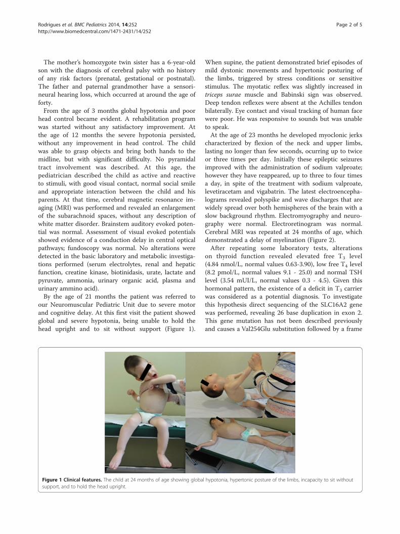
our Neuromuscular Pediatric Unit due to severe motorand cognitive delay. At this first visit the patient showedglobal and severe hypotonia, being unable to hold thehead upright and to sit without support (Figure 1).
Figure 1 Clinical features. The child at 24 months of age showing globalsupport, and to hold the head upright.
When supine, the patient demonstrated brief episodes ofmild dystonic movements and hypertonic posturing ofthe limbs, triggered by stress conditions or sensitivestimulus. The myotatic reflex was slightly increased intriceps surae muscle and Babinski sign was observed.Deep tendon reflexes were absent at the Achilles tendonbilaterally. Eye contact and visual tracking of human facewere poor. He was responsive to sounds but was unableto speak.At the age of 23 months he developed myoclonic jerks
characterized by flexion of the neck and upper limbs,lasting no longer than few seconds, ocurring up to twiceor three times per day. Initially these epileptic seizuresimproved with the administration of sodium valproate;however they have reappeared, up to three to four timesa day, in spite of the treatment with sodium valproate,levetiracetam and vigabatrin. The latest electroencepha-lograms revealed polyspike and wave discharges that arewidely spread over both hemispheres of the brain with aslow background rhythm. Electromyography and neuro-graphy were normal. Electroretinogram was normal.Cerebral MRI was repeated at 24 months of age, whichdemonstrated a delay of myelination (Figure 2).After repeating some laboratory tests, alterations
on thyroid function revealed elevated free T3 level(4.84 nmol/L, normal values 0.63-3.90), low free T4 level(8.2 pmol/L, normal values 9.1 - 25.0) and normal TSHlevel (3.54 mUI/L, normal values 0.3 - 4.5). Given thishormonal pattern, the existence of a deficit in T3 carrierwas considered as a potential diagnosis. To investigatethis hypothesis direct sequencing of the SLC16A2 genewas performed, revealing 26 base duplication in exon 2.This gene mutation has not been described previouslyand causes a Val254Glu substitution followed by a frame
hypotonia, hypertonic posture of the limbs, incapacity to sit without

Figure 2 Cerebral magnetic resonance image. Axial, T2-fast spin echo sequence (a) shows a significative alteration in white matter substancein the centrum semiovale, frontoparietal, parieto-occipital and subcortical regions, compatible with a marked delay in myelination; and cerebralmagnetic resonance spectrometry (b) demonstrates a low N-acetyl aspartic peak.
Rodrigues et al. BMC Pediatrics 2014, 14:252 Page 3 of 5http://www.biomedcentral.com/1471-2431/14/252
shift and a premature stop codon 24 aminoacids later(Exon 2 c.735_760dup p.Val254Glufs*24). His motherwas subsequently confirmed to be a carrier for this dupli-cation (Figure 3).
DiscussionThis phenotype of severe X-linked psychomotor delaywas first described in 1944 and was eponymously namedAllan-Herndon-Dudley syndrome [10]. Afterwards, in2004, the first mutations in MCT8 gene (SLC16A2)
Figure 3 Electropherograms. The duplication found in exon 2 of MCT8 inmother (bottom tracing), and the corresponding normal sequence (top tra
were discovered by two distinct investigation groups(Dumitrescu et al. and Friesema et al.), and neurologicalfindings of AHDS were explained by the resistance of T3
on entering the neuronal target cells, that leads to a clas-sical thyroid profile found in all affected patients [8,9].Currently, MCT8 mutations have been reported in about50 families worldwide [4,11].Diagnosis of AHDS in our patient is supported by: the
presence of an X-linked inheritance; the characteristicthyroid hormonal pattern; the marked delay of myelination
the patient (middle tracing), the sequence of the heterozygouscing).
Rodrigues et al. BMC Pediatrics 2014, 14:252 Page 4 of 5http://www.biomedcentral.com/1471-2431/14/252
of the central nervous system (CNS) found in the MRI andthe presence of the pathogenic mutation in the MCT8gene. AHDS shows a broad and heterogeneous clinicalspectrum according to the type of mutation and its impacton the protein synthesis stage. Our patient fits well withthe most severe phenotype [12].One of the main features of AHDS, also known as
MCT8 deficiency, is the marked global hypotonia anddifficulties maintaining the head up right, recognized as”limber neck” (Figure 1), evident since the first monthsof life [13]. In the presence of a floppy infant, evaluationof signs of CNS involvement is essential but not alwayseasy during the first year of life. In this case, normal eyecontact and social interaction by the age of 12 months,in addition to the lack of deep tendon ankle reflexes,made peripheral neuromuscular involvement an import-ant hypothesis to exclude. The absence of signs of CNSinvolvement during the first year of life could have re-sulted in a muscle biopsy in order to exclude a congeni-tal myopathy or other muscle disorder. However, thepresence of spasticity and abnormal movements duringthe second year of life make it clear that this investiga-tion was not warranted.During the evaluation of a hypotonic infant, the cere-
bral MRI is a helpful tool for detecting CNS abnormal-ities [11]. The difficulty in interpreting a cerebral MRIduring the first year of life leads to different descriptionsof the white matter abnormalities ranging from delayedmyelination to hypomyelination, which can delay thediagnosis [11,12,14,15]. Despite the static or even deteri-orating clinical picture for these patients, several studiesconfirm that the major neuroradiological feature ofthis disease is the marked delayed myelination [14,15].Many of these children were described as a “Pelizaeus–Merzbacher–Like Disease” because they had a similarphenotype without the identification of PLP1 mutation.This is a differential diagnosis that needs to be consideredand the cerebral MRI is an important tool. In Pelizaeus–Merzbacher disease we find a hypomyelination (same pat-tern of deficient myelination on 2 MRIs at least 6 monthsapart in a child older than 1 year) while in AHDS, a correctMRI interpretation will show a progression of myelination,even if slow [11,14]. In our case the relatively low N-acetylaspartic peak in the cerebral magnetic resonance spectros-copy also supports the hypothesis of delayed myelination(Figure 2) [16].The neurological manifestations of this disease are
quite complex. The axial hypotonia persists throughoutadulthood, while the hypotonia of the limbs progressesto spasticity and dystonic posturing [3,13]. The age ofonset of extrapyramidal symptoms is not clear in theliterature and it seems essential for us to suspect ofAHDS even before these signs appear [13]. Other im-portant features are the inability to sit, stand or walk
independently, the severe mental retardation with lack ofspeech development and rudimentary communicativeskills [13,17]. A quarter of the patients will have seizuresthat are usually responsive to anticonvulsant therapy [13].As already mentioned all affected males with MCT8 mu-
tations exhibit a typical thyroid profile which makes it cru-cial to request these hormones, including T3 early in theinvestigation [3,8,9]. Mean serum T4 and free T4 are nor-mal or slightly decreased, TSH is normal or mildly in-creased and serum T3 and free T3 are markedly elevated[3,8,9,14]. This will help the clinician to avoid unnecessarytests and to pursue a specific genetic analysis of SLC16A2for a definitive diagnosis. There is another medical condi-tion with the same thyroid hormonal pattern, that can befound in a hypotonic infant and that is due to a mutationin the thyroid hormone receptor α gene (TRα1). Thiscondition has a similar presentation in the first months oflife; however older infants and children present witha phenotype very different from AHDS, developing theclassic features of hypothyroidism (growth retardation,skeletal dysplasia, reduced muscle tone, constipation) andonly a mild cognitive impairment [18].Some AHDS patients have been treated with TH sup-
plementation, propylthiouracil or diiodothyropropionicacid; the last two result in better thyroid function tests,but without any improvement of motor or cognitive skills[19-21]. Up to this moment, we can only offer symptom-atic treatment to these patients, such as rehabilitationtherapies, antispasticity, antidystonic and anticonvulsantmedication, nutritional and orthopedic management[13,20,22]. Genetic counseling should be offered to thefamily as soon as the diagnosis is recognized as is the casewith all other X-linked recessive conditions: if the motherhas SLC16A2 mutation, boys will have a 50% risk of beingaffected, whereas girls will have a 50% chance of being acarrier of the mutation [13,22]. The genetic test was sug-gested to be undertaken by the patient’s cousin but unfor-tunately his parents have systematically refused.
ConclusionThrough this clinical report we demonstrate the import-ance of excluding AHDS in the initial laboratory evaluationof floppy male infant, without history of perinatal asphyxia,by the simple exploration of the thyroid hormone status,including T3. In spite of the absence of an effective treat-ment for this disease, an early diagnosis can avoid a num-ber of useless, expensive and invasive investigations andpermit correct genetic counseling to the affected families.
ConsentWritten informed consent was obtained from the parentsof the patient for publication of this Case report and anyaccompanying images. A copy of the written consent isavailable for review by the Editor of this journal

Rodrigues et al. BMC Pediatrics 2014, 14:252 Page 5 of 5http://www.biomedcentral.com/1471-2431/14/252
AbbreviationsTH: Thyroid hormone; T3: Triiodothyronine hormone; MCT8: Monocarboxylatetransporter 8; AHDS: Allan-Herndon-Dudley syndrome; T4: Thyroxine;TSH: Thyroid stimulating hormone; MRI: Magnetic resonance imaging;CNS: Central nervous system; TRα1: Thyroid hormone receptor α gene..
Competing interestsThe authors declare that they have no competing interests.
Authors' contributionsFR: data analysis, literature search, manuscript preparation; JG: literaturesearch, manuscript preparation, CO: provided medical care, manuscriptreview; AN: provided medical care, data analysis, manuscript review; BM:literature search, carried out the molecular genetic studies; MMB: carried outthe molecular genetic studies; JA: provided medical care, data analysis; JC:provided medical care, data analysis, manuscript review, literature search.All authors read and approved the final manuscript.
AcknowledgementsAll those who contributed in the preparation and revision of the article andwho provided medical care to the patient make part of the authors’ list.
Author details1Neuromuscular Unit, Neurology Department, Fundación Sant Joan de Déu,Hospital Materno-Infantil Sant Joan de Déu, Passeig Sant Joan de Déu, 2.08950 Esplugues de Llobregat, Barcelona, Spain. 2Center for BiomedicalResearch on Rare Diseases (CIBERER), ICIII, Madrid, Spain. 3Instituto deInvestigaciones Biomédicas Alberto Sols, Consejo Superior de InvestigacionesCientíficas - Universidad Autónoma de Madrid, Madrid, Spain. 4BiochemicalGenetics&Rett Unit, Laboratory Department, Hospital Materno-Infantil SantJoan de Déu, Barcelona, Spain.
Received: 28 June 2014 Accepted: 1 October 2014Published: 4 October 2014
References1. Bernal J, Guadaño-Ferraz A, Morte B: Perspectives in the study of thyroid
hormone action on brain development and function. Thyroid 2003,13:1005–1012.
2. Morreale de Escobar G, Obregon MJ, Escobar del Rey F: Role of thyroidhormone during early brain development. Eur J Endocrinol 2004,151:U25–U37.
3. Heuer H, Visser TJ: Minireview: pathophysiological importance of thyroidhormone transporters. Endocrinology 2009, 150:1078–1083.
4. Visser WE, Friesema EC, Visser TJ: Minireview: thyroid hormonetransporters: the knowns and the unknowns. Mol Endocrinol 2011,25:1–14.
5. Kersseboom S, Visser TJ: Tissue-specific effects of mutations in the thyroidhormone transporter MCT8. Arq Bras Endocrinol Metabol 2011, 55:1–5.
6. Ceballos A, Belinchon MM, Sanchez-Mendoza E, Grijota-Martinez C,Dumitrescu AM, Refetoff S, Morte B, Bernal J: Importance of monocarboxylatetransporter 8 for the blood–brain barrier-dependent availability of 3,5,3'-triiodo-L-thyronine. Endocrinology 2009, 150:2491–2496.
7. Wirth EK, Roth S, Blechschmidt C, Hölter SM, Becker L, Racz I, Zimmer A,Klopstock T, Gailus-Durner V, Fuch H, Wust W, Naumann T, Bräuer A, Angelis MH,Köhrle J, Grüters A, Schweizer U: Neuronal 3',3,5-tri-iodothyronine (T3) uptakeand behavioral phenotype of mice deficient in Mct8, the neuronalT3 transporter mutated in Allan-Herndon-Dudley syndrome. J Neurosci2009, 29:9439–9449.
8. Dumitrescu AM, Liao XH, Best TB, Brockmann K, Refetoff S: A novelsyndrome combining thyroid and neurological abnormalities isassociated with mutations in a monocarboxylate transporter gene.Am J Hum Genet 2004, 74:168–175.
9. Friesema ECH, Grueters A, Biebermann H, Krude H, Moers A, Reeser M,Barrett TG, Mancilla EE, Svensson J, Kester MH, Kuiper GG, Balkassmi S,Uitterlinder AG, Koehrle J, Rodien P, Halestrap AP, Visser TJ: Associationbetween mutations in a thyroid hormone transporter and severeX-linked psychomotor retardation. Lancet 2004, 364:1435–1437.
10. Allan W, Herndon CN, Dudley FC: Some examples of the inheritance ofmental deficiency: apparently sex-linked iodicy and microcephaly. Am JMent Defic 1944, 48:325–334.
11. López-Marín L, Martín-Belinchón M, Gutiérrez-Solana LG, Morte-Molina B,Duat-Rodríguez A, Bernal J: MCT8-specific thyroid hormone cell transporterdeficiency: a case report and review of the literature. Rev Neurol 2013,56:615–622 (in Spanish).
12. Jansen J, Friesema EC, Kester MH, Schwartz CE, Visser TJ: Genotype-phenotype relationship in patients with mutations in thyroid hormonetransporter MCT8. Endocrinology 2008, 149:2184–2190.
13. Schwartz CE, Stevenson RE: The MCT8 thyroid hormone transporter andAllan-Herndon-Dudley syndrome. Best Pract Res Clin Endocrinol Metab2007, 21:307–321.
14. Tonduti D, Vanderver A, Berardinelli A, Schmidt JL, Collins CD, Novara F,Genni AD, Mita A, Triulzi F, Brunstrom-Hernandez JE, Zuffardi O, Balottin U,Orcesi S: MCT8 deficiency: extrapyramidal symptoms and delayedmyelination as prominent features. J Child Neurol 2013, 28:795–800.
15. van der Knaap MS, Wolf NI: Hypomyelination versus delayed myelination.Ann Neurol 2010, 68:115.
16. Gika AD, Siddiqui A, Hulse AJ, Edward S, Fallon P, Mcentagart M, Jan W,Josifova D, Lerman-Sagie T, Drummond J, Thompson E, Refetoff S,Bönnemann CG, Jungbluth H: White matter abnormalities and dystonicmotor disorder associated with mutations in the SLC16A2 gene. Dev MedChild Neurol 2010, 52:475–482.
17. Filho HC, Marui S, Manna TD, Brust ES, Radonsky V, Kuperman H,Dichtchekenian V, Setian N, Damiani D: Novel mutation in MCT8gene in a Brazilian boy with thyroid hormone resistance and severeneurologic abnormalities. Arq Bras Endocrinol Metabol 2011, 55:60–66.
18. Bochukova E, Schoenmakers N, Agostini M, Schoenmakers E, Rajanayagam O,Keogh JM, Henning E, Reinemund J, Gevers E, Sarri M, Downes K, Offiah A,Albanese A, Halsall D, Schwabe JWR, Bain M, Lindley K, Muntoni F, Vargha-Khadem F, Dattani M, Farooqi IS, Gurnell M, Chatterjee K: A Mutation in thethyroid hormone receptor alpha gene. N Engl J Med 2012, 366:243–249.
19. Zung A, Visser TJ, Uitterlinden AG, Rivadeneira F, Friesema EC: A child witha deletion in the monocarboxylate transporter 8 gene: 7-year follow-upand effects of thyroid hormone treatment. Eur J Endocrinol 2011,165:823–830.
20. Wémeau JL, Pigeyre M, Proust-Lemoine E, Gottrand F, d’Herbomez M,Jansen J, Visser TJ, Ladsous M: Beneficial effects of propylthiouracilplus L-thyroxine treatment in a patient with a mutation in MCT8.J Clin Endocrinol Metab 2008, 93:2084–2088.
21. Verge CF, Konrad D, Cohen M, Cosmo CD, Dumitrescu AM, Marcinkowski T,Hameed S, Hamilton J, Weiss RE, Refetoff S: Diiodothyropropionic Acid(DITPA) in the treatment of MCT8 deficiency. J Clin Endocrinol Metab 2012,97:4515–4523.
22. Dumitrescu AM, Fu J, Dempsey MA, Refetoff S: MCT8-Specific ThyroidHormone Cell-Membrane Transporter Deficiency. In GeneReviews™[Internet]. Edited by Pagon RA, Adam MP, Bird TD, et al. Seattle (WA):University of Washington, Seattle; 2010. 1993–2013. Available from:http://www.ncbi.nlm.nih.gov/books/NBK26373/.
doi:10.1186/1471-2431-14-252Cite this article as: Rodrigues et al.: Hypotonic male infant and MCT8deficiency - a diagnosis to think about. BMC Pediatrics 2014 14:252.
Submit your next manuscript to BioMed Centraland take full advantage of:
• Convenient online submission
• Thorough peer review
• No space constraints or color figure charges
• Immediate publication on acceptance
• Inclusion in PubMed, CAS, Scopus and Google Scholar
• Research which is freely available for redistribution
Submit your manuscript at www.biomedcentral.com/submit