Embed Size (px)
Citation preview

HYPOTHERMIA AND TRAUMA
by
Andreas Wladis
Stockholm, 2001
From the Department of Surgery, Söder Hospital, Karolinska Institute, Stockholm, Sweden,
and the Laboratory of Experimental Traumatology, Department of Defense Medicine, Swedish
Defense Research Agency, Stockholm, Sweden

Hypothermia and Trauma
by Andreas Wladis
Stockholm, Sweden, 2001
Printed by Karolinska University Press, 2001
ISBN 91-628-4574-8

Panta rei! Herakleitos of Ephesus (540-480 B.C.)
To Isabella & Simon

4
LIST OF PUBLICATIONS
This thesis is based on the following papers, which will be referred to in the text by their
Roman numerals:
I. Wladis A, Hjelmqvist H, Brismar B, Kjellström BT. Acute metabolic and endocrine
effects of induced hypothermia in hemorrhagic shock - an experimental study in the
pig. J Trauma 45: 527-533, 1998.
II. Wladis A, Hahn R, Hjelmqvist H, Brismar B, Kjellström BT. Acute hemodynamic
effects of induced hypothermia in hemorrhagic shock - an experimental study in the
pig. Shock 15:60-64, 2001.
III. Wladis A, Hahn R, Brismar B, Kjellström BT. Effects of induced hypothermia after
soft-tissue injury. (Submitted)
IV. Wladis A, Hahn R, Brismar B, Kjellström BT. Induced hypothermia after high-energy
soft-tissue injury and subsequent hemorrhagic shock. (In press)
V. Heinius G, Wladis A, Hahn R, Kjellström BT. Induced hypothermia and rewarming
after hemorrhagic shock. (Submitted)

5
ABSTRACT
Hypothermia and trauma
Background Accidental hypothermia (HT) has been found to increase morbidity and
mortality in trauma patients. In contrast, HT has been induced in certain surgical procedures
for several decades because of its cerebroprotective properties. HT has even been used
therapeutically in patients with traumatic brain injury. In recent years, a number of
experimental studies have suggested beneficial effects of induced HT in hemorrhagic shock
(HS), but just how induced HT affects the organism subjected to both HS and trauma has been
unknown, hitherto.
Methods In papers I and II, animals were exposed to 50% exsanguination during 25 min.
In paper III, a standardized gunshot wound was inflicted on the right hind-leg. In paper IV,
animals were subjected to the combination of these insults. In paper V, the hemorrhage
amounted to 40% of the blood volume and was achieved in 3-5 min. Core temperature,
electrolytes, arterial blood gases, blood cell counts, Hb, and central hemodynamics were
monitored in all the studies. Catecholamines were analyzed in papers I-IV. IL-6 was studied
in papers III-IV. Thromboelastography was used to evaluate coagulation abnormalities in
paper V. In this paper, animals were rewarmed after cooling.
Results Paper I: Catecholamine levels in plasma increased in response to the hemorrhage,
but gradually decreased with cooling. Serum potassium levels increased in the controls, but
decreased transiently in HT animals. Paper II: HR increased markedly after the hemorrhage,
while CO and MAP were reduced. With HT, HR decreased and CO and MAP were further
depressed. Leukocyte counts decreased in HT animals. Paper III: HR, MAP, neutrophil
granulocyte counts and plasma adrenaline levels were lower in the HT group. Cardiac index
decreased slightly in both groups. Serum potassium increased with normothermia, but was not
affected in HT pigs. Paper IV: HR, VO2, ER, serum potassium, and creatinine levels were
lower with cooling. Paper V: VO2 was reduced in HT animals. Serum levels of potassium
were transiently stabilized with HT. The formation of blood clots was delayed, but once
formed, the clot strength was unaffected by HT. Effects of HT were reversed with rewarming.
Conclusions In HS and/or soft-tissue trauma, HT reduced plasma catecholamine levels and
transiently stabilized serum levels of potassium. Central hemodynamics after the combination
of the insults was affected by HT to a remarkably small extent, while VO2 and ER decreased.
In the presence of rewarming hemodynamics, VO2 and ER regained baseline levels.

6
CONTENTS
ABBREVIATIONS DEFINITIONS INTRODUCTION
Historical notes
Hemorrhage, shock and trauma
The treatment of hemorrhagic shock
Thermophysiology and hypothermia
AIMS OF THE STUDIES METHODOLOGICAL CONSIDERATIONS
PRINCIPAL RESULTS
Paper I
Paper II
Paper III
Paper IV
Paper V
GENERAL DISCUSSION CONCLUSIONS
ACKNOWLEDGMENTS REFERENCES PAPERS I-V

7
ABBREVIATIONS
BW Body weight
CaO2 Arterial oxygen content
CO Cardiac output
CI Cardiac index
CvO2 Mixed venous oxygen content
CVP Central venous pressure
DO2 Oxygen delivery
ER Oxygen extraction ratio
GSW Gunshot wound
Hb Hemoglobin concentration in blood
HR Heart rate
HS Hemorrhagic shock
HT Hypothermia, hypothermic
IL-6 Interleukin-6
i.v. Intravenous(ly)
i.m. Intramuscular(ly)
MAP Mean arterial pressure
MPAP Mean pulmonary artery pressure
NT Normothermia, normothermic
PCWP Pulmonary capillary wedge pressure
PO2 Arterial oxygen tension
SaO2 Arterial oxygen saturation
SEM Standard error of the mean
SV Stroke volume
SvO2 Mixed venous oxygen tension
VO2 Oxygen uptake
8
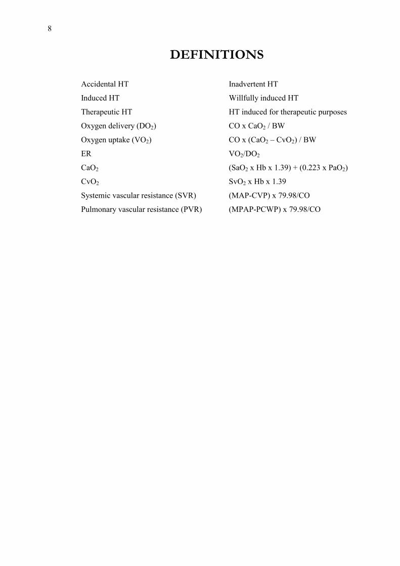
DEFINITIONS Accidental HT Inadvertent HT
Induced HT Willfully induced HT
Therapeutic HT HT induced for therapeutic purposes
Oxygen delivery (DO2) CO x CaO2 / BW
Oxygen uptake (VO2) CO x (CaO2 � CvO2) / BW
ER VO2/DO2
CaO2 (SaO2 x Hb x 1.39) + (0.223 x PaO2)
CvO2 SvO2 x Hb x 1.39
Systemic vascular resistance (SVR) (MAP-CVP) x 79.98/CO
Pulmonary vascular resistance (PVR) (MPAP-PCWP) x 79.98/CO

9
INTRODUCTION
Trauma is, by definition, an injury of any sort, but it is usually referred to as a somatic
injury severe enough to pose a threat to life or limb1. Such injuries often require immediate
attention or else they worsen or even cause death. In Sweden and other countries in the West,
trauma represents a major cause of preventable death in persons under the age of 44 and
causes a greater loss of life years than do cardiovascular ailments and cancer taken together2.
A reduction of the body core temperature, HT, either preserves life or kills3. The exact
mechanism behind these two main effects remains unknown. Before any of these effects are
reached, HT will have altered a broad range of physiological parameters in different
directions depending on how and when HT is achieved and for how long it is maintained.
This thesis is focused on the effects of induced HT in three forms of trauma: HS, soft-
tissue injury, and the combination of these. It also deals with how the HT-induced effects on
HS are altered during rewarming.
Historical notes
Accidental hypothermia
One of the oldest accounts of HT can be found in the Old Testament of the Bible (1
Kings, 1:1-2):
When King David was old and well advanced in years, he could not keep
warm even when they put covers over him. So his servants said to him, "Let
us look for a young virgin to attend the king and take care of him. She can
lie beside him so that our lord the king may keep warm.�
Throughout history, accidental HT has frequently been reported in conjunction with
military battles, and cold weather has altered the course of many of them. Accordingly,
Hannibal is said to have lost about 20,000 soldiers in 218 B.C. while en route through the
Alps. A month after the Swedish King Carolus XII had fallen in the battle of Frederikshald,
Norway, in 1718, about 5000 of his men attempted to return to Sweden. They got caught in
blizzard and around 3000 soldiers succumbed due to HT and/or frostbite.
Moreover, in 1812, the French Emperor Napoleon I lost most of what remained of his
Grande Armée due to the cold while attempting to invade Moscow. Baron Dominique Jean

10
Larrey, Emperor Napoleon�s surgeon-in-chief made the observation in his memoirs that
soldiers sitting closest to the fire died �mysteriously�, which may be the first account of shock
induced by external rewarming of HT subjects 4.
More recently, the French lost more than 1000 soldiers in 1845-55 in the Crimean War
due to HT. The battle of Stalingrad in 1942-43 was not only one of the greatest and most
atrocious struggles in the history of warfare, but also a turning point in World War II. It cost
more lives than any other battle: 1.1 Million Russians and 800,000 Germans. Many of the
German casualties were due to HT and frostbite.
Therapeutic hypothermia
The use of cold for therapeutic purposes is described in the most ancient medical text
known hitherto, the Edwin Smith Papyrus (ca. 3500 B.C.)5. Furthermore, the Chinese surgeon
Hua T�O (ca. 200 A.D.) used general HT for chronic fever5. Baron Larrey, Emperor
Napoleon�s surgeon-in-chief, is known to have advocated the use of snow massage as a means
of treating local HT (frostbite). This treatment, as paradoxical as it may sound, was widely
adopted and was in use in the Royal Swedish Army well in to the 20th century. In 1932, a
textbook from the medical services of the Royal Swedish Army recommended not only snow
massage but, indeed, a bath in ice-cold water to treat general HT6.
Induced hypothermia
Induced HT was introduced in clinical practice close to 50 years ago to provide whole-
body protection, and cerebral protection in particular, while enabling successful surgical
correction of cardiac anomalies in children. Initially, deep HT was achieved by means of
surface cooling or core cooling, or a combination of these. The aim was to decrease the core
temperature and cerebral metabolism enough to allow periods of circulatory arrest without
incurring cerebral injury. These periods were kept as short as possible to minimize morbidity
and mortality. Presently, the use of HT circulatory arrest has expanded to include surgical
interventions for cerebral and complex aortic aneurysms in adults, reoperations for cardiac
valve replacements, hepatic tumor resection, and operations for tumors within the venae
cavae7.
Hemorrhage, shock and trauma
Shock arises when the perfusion of vital organs is not sufficient to satisfy the metabolic
demands of cells. Shock is the clinical manifestation of a trauma too severe to be immediately

11
compensated for by the body�s regulatory mechanisms and may result in death without
expeditious treatment. Seven categories of shock have been proposed: hypovolemic,
vasogenic, neurogenic, septic, cardiogenic, obstructive, and traumatic8. Weil and Shubin, in
turn, defined four categories of circulatory shock, namely: hypovolemic, cardiogenic,
obstructive, and distributive9.
Except for septic shock, shock is characterized by a decrease in systemic oxygen
transport due to hypovolemia and myocardial or respiratory failure. In septic shock, there is a
maldistribution of nutritive organ blood flow owing to microcirculatory disturbances in spite
of normal or even increased CO. Common to all forms of shock is a severe derangement of
local perfusion because of activated leukocytes and the release of humoral mediators which
cause changes in organs not primarily affected by the initiating insult10. Two of the forms of
shock, hypovolemic and traumatic shock, are relevant in this context and will be discussed
below although it is recognized that hypovolemic shock in real life is oftentimes compounded
by e.g. neurogenic, cardiogenic, or obstructive shock.
Hypovolemic and hemorrhagic shock
In most cases, hypovolemic shock is caused by hemorrhage and the expression is
therefore often interchangeable with HS. However, hypovolemia may also arise from other
conditions, such as severe gastrointestinal, renal, or cutaneous fluid loss. It can also be caused
by excessive ascites formation. Moreover, a situation with relative hypovolemia may result
from vasodilation. For the sake of clarity, only the expression HS will therefore be used in the
following.
It is noteworthy that HS may arise without overt bleeding, such as when blood
accumulates in the intestine, muscles, or retroperitoneum. Thus, HS may be due to either
external or internal bleeding. Some common examples of externally induced HS are traumatic
amputation of a limb, disruption of blood vessels, excessive blood loss during surgery, while
internal causes of HS include extrauterine pregnancies, esophageal and gastrointestinal
hemorrhage, traumatization of parenchymatous organs, and fractures of major bones.
Exsanguinating hemorrhage is the cause of death in 31% of civilian and 47% of military
trauma victims 11. Half of all trauma deaths occur within minutes of the injury. They are
usually due to severe injuries to the brain or major vessels and are only rarely susceptible to
curative treatment. Approximately 30% of trauma-related mortality occurs within the first few
hours and is caused by neurological injuries or various kinds of hemorrhage 2. The concept of

12
a �golden hour� following injury arises from considerations of this group. But in spite of
standard resuscitative measures, the mortality rate remains high.
Pathophysiology of hemorrhagic shock
The balance between oxygen delivery and oxygen demand is maintained as long as
tissue oxygen extraction can increase in cases of reduced blood flow. At a certain point, tissue
perfusion becomes inadequate for the cellular oxygen needs. Oxygen debt thus materializes,
resulting in anaerobic metabolism, cellular acidosis, and lactic acidosis.
Acute hemorrhage causes redistribution of the blood flow to preserve perfusion and the
viability of vital organs. Accordingly, the blood flow to the skin, musculature, and the
splanchnic region is considerably reduced. The high adrenergic innervation of the splanchnic
vascular region will cause the blood flow to the intestine and its mucosa, in particular, to be
severely compromised when the sympathetic nervous system is activated in response to the
hemorrhage. In a study on monkeys in HS, the intestines still remained underperfused 2 h
after the blood volume had been restored 12. This intestinal ischemia may cause endotoxemia
and bacterial translocation and trigger massive production and release of hepatic cytokines,
which, in turn, might eventually contribute to the development of multiple organ failure.
However, the data on bacterial translocation after HS are inconsistent and its clinical
significance remains unclear8.
The physiological hemodynamic response to major hemorrhage is aimed at preserving
the blood circulation to vital organs, restoring blood loss, and limiting the hemorrhage. In
principle, this response often evolves as follows:
1) Decreasing circulating blood volume is detected within seconds by
arterial baroreceptors which sense decreased stretching of the arterial
wall. This releases the chronic inhibition imposed by the baroreceptors,
which causes the nucleus tractus solitarius in the medulla oblongata to
reduce the tonic inhibition of the HR. Also, by virtue of activation of the
sympathetic outflow with noradrenaline release from postganglionic
sympathetic fibers, constriction of venous capacitance vessels is
achieved. This, in turn, increases the venous return to the heart and thus
maintains the CO and MAP13, 14. In addition to the sympatho-adrenal
axis, both vasopressin and angiotensin II may contribute within 10 min to
an hour to maintaining perfusion during hemorrhage14, 15. The magnitude

13
of the neuroendocrine response to hemorrhage depends on the magnitude
of the decrease in effective circulating volume and on the rate at which
the blood loss occurs. The neuroendocrine reaction may also be modified
by associated injuries or conditions, drugs, prescribed medication, pre-
existing illness, drug withdrawal, the age of the patient, pain, and
psychological factors such as fear and stress. Moreover, repeated insults
may also potentiate the response8.
2) The posthemorrhage activation of the sympathetic nervous system also
causes a noradrenaline-mediated constriction of precapillary arterioles,
which decreases the capillary hydrostatic pressure. Up to 0.5L of fluid
from the interstitial space can be mobilized in the adult and drawn into
the capillaries to compensate for parts of the blood loss in this fashion.
Adrenaline released into the circulation stimulates hepatic
glycogenolysis, leading to hyperosmotic hyperglycemia, which may
mobilize another 0.5L of cellular water into the bloodstream16.
Eventually, the compensatory mechanisms are exhausted and the venous
return to the heart decreases. At this point, maintenance of normal CO is
no longer possible and the MAP decreases. With decreasing MAP, the
coronary blood flow decreases and thus a vicious circle is entered.
3) The ischemic hypoxia that comes with hemorrhage and the release of
circulating cytokines and other mediators cause swelling of the
endothelial cells, which impedes perfusion by narrowing the vessel
lumen. This sludging phenomenon is aggravated by the adherence of
activated leukocytes, mainly neutrophils, to the vascular endothelium10.
HS is a dynamic process in which the relevant variables change depending on the
degree of hemorrhage and achieved resuscitation. In order to simplify and stratify the
characterization and treatment of HS, some authors have divided it into a number of stages.
AC Guyton, for one, described three stages, the first one named nonprogressive or
compensated, the second progressive or decompensated, and the third irreversible 14. The first
one is called so because the neuroendocrine responses are sufficient to compensate for the
hypovolemia. If this compensation is insufficient, organ and cellular dysfunction will follow

14
and decompensated chock will occur. Organ and cellular dysfunction can still be reversed by
appropriate volume resuscitation. It is not known exactly what causes the transition from the
compensated to the decompensated stage, but local factors such as acidosis and adrenergic
activity are known to be able to alter the microvascular response. Consequently, vasodilation
occurs with a decrease in peripheral vascular resistance and a decrease of venous return to the
heart with a further decline in CO and increasing tissue acidosis. If volume loss continues or
fluid resuscitation is inadequate, irreversible shock will occur insidiously. This is usually
defined in retrospect8.
Peter Baskett has chosen another way of classifying HS based on the estimated volume
of blood lost with four, partly overlapping, stages as a guideline for the treatment of patients
in HS17. According to this system, patients with a blood loss up to 15% of the calculated
blood volume (i.e. up to 750 ml in a 70kg patient) belong to class I. The corresponding figures
for class II is 15-30%, for class III, 30-40%, and, for class IV, >40%.
Trauma and traumatic shock
Traumatic shock should be considered a separate entity, as it comprises components of
several of the aforementioned types of shock. In its purest form, however, it involves HS in
combination with soft-tissue trauma and/or bone fractures. Consequently, the study of pure
HS has limited relevance to the pathophysiological condition of traumatic shock. Pulmonary
complications, e.g. after HS, are uncommon in clinical practice, while in traumatic shock they
are common, probably due to post-injury release of various proinflammatory mediators8. Such
a release is more intense in traumatic shock than in pure HS.
The treatment of hemorrhagic shock
The treatment of HS is directed toward restoring organ perfusion with adequately
oxygenated blood. This is achieved by rapidly securing an airway, replacement of the blood
volume, and by prompt control of the hemorrhage. The current dogma dictates resuscitation
with asanguinous fluids in addition to packed red blood cells18. The choice of asanguinous
fluid, be it crystalloid or colloid, is a matter of scientific discussion and lies beyond the scope
of this thesis16. This is also true of hypertonic saline solutions.
In uncontrolled hemorrhage, some authors advocate that i.v. fluid be withheld until the
patient has arrived in the operating room. This stance is based on data showing increased
survival with fluid restriction in the field after penetrating thoracic injuries19. It is believed
that the differences between the groups develops because of rebleeding in patients receiving

15
i.v. fluid before surgery. These notions are not yet generally accepted and apply, at any rate,
only to patients with penetrating traumas, ruptured abdominal aortic aneurysms, or
gastrointestinal bleeding. Patients subjected to blunt trauma and possibly head injury should
still receive standard resuscitative treatment8.
Thermophysiology and hypothermia
Thermophysiology
Man is homeothermic (warm-blooded) as are most mammals, which means they strive
to maintain a constant body core temperature within a narrow range regardless of changes in
the ambient temperature. A few animals hibernate, which causes their basal metabolic rate to
be considerably reduced for a period of time.
Maintenance of the core temperature is governed by the hypothalamus, which receives
input concerning the thermal condition of the body. This information, which originates from
thermosensors in the skin, various core regions, and locally in the hypothalamus, is compared
with a set point signal, after which appropriate output commands are activated. A number of
factors affect the regulated set point: time of day, sleep state, activity levels, presence of
pyrogens, ambient temperature, and satiety. Information about the ambient temperature is
extremely important, if not the most important, for temperature homeostasis20. Consequently,
the skin temperature has a big impact on the behavioral thermoregulatory responses, but a
smaller effect on autonomic thermoregulation. However, a low skin temperature in
conjunction with immersion in cold water can also elicit a strong and more or less immediate
response from the sympathetic nervous system21.
The preoptic anterior hypothalamic area of the hypothalamus (POAH) has been
identified as the center for autonomic thermoregulation20. A decrease in the temperature of
blood flowing in the POAH will elicit vasoconstriction and increase metabolic heat
production. Conversely, adding heat to the POAH will stimulate vasodilation and sweating
and thus decrease the body core temperature. The magnitude of the thermoregulatory response
is proportional to the displacement of the POAH temperature from the threshold temperature.
The blood circulation in the skin is maintained by two types of vasculature. These are
(1) nutritive vessels consisting of arteries, veins, and capillaries and (2) heat exchange vessels
consisting of venous plexuses and arteriovenous anastomoses. While the nutritive vasculature
is found uniformly in the skin, the heat exchange vessels are found only in the hairless skin of
the hands, feet, ears, and face. It has been estimated that the blood flow in to the venous

16
plexuses can range from near zero to as much as 30% of the total CO. With maximal
vasoconstriction of these vessels, the body core is insulated from the environment22.
Normally, behavioral adjustment and autonomous subcutaneous heat exchange are
sufficient to maintain thermal homeostasis in man. Heat for thermoregulation is lost by
sweating and produced by two mechanisms, shivering and non-shivering thermogenesis20, 23.
The latter is only seen in infant humans, making shivering thermogenesis the main heat
producer. Shivering is the action of muscular agonists and antagonists acting in a non-
synchronized manner. It leads to tremor but little other movement of the body. In a resting
human, about 20% of the heat is generated by the muscles. With maximum shivering,
however, the muscular heat production can increase manifold, causing the metabolic rate to
increase two to five times and will increase O2 consumption by 40-100%23-25.
The human or porcine body is thermally compartmentalized. The thermal core consists
of the organs that generate heat, whereas the thermal periphery are tissues engaged in heat
exchange and tissues underlying the body surface. Such tissues can include subcutaneous fat
and skeletal muscle. One of the functions of these tissues is to buffer the thermal core from
environmental attempts to lower the body core temperature22. Differences in this insulation
can determine the occurrence of HT and the rate of heat loss. At low ambient temperatures,
the thermal core, which is not an anatomical entity, can shrink and eventually only encompass
critical organs. The central nervous system, being the center of thermoregulation, is the most
protected of the core organs and thus the last region of the body to lose NT. In contrast, the
normal temperature of the skin is approximately 22-23°C.
Hypothermia
HT is commonly, but arbitrarily, defined as a general decrease of the body core
temperature below 35°C, regardless of whether the temperature decrease is accidental or
induced26, 27. The severity of human HT is classified in different ways depending on the
patient�s core temperature. The most common classification defines mild HT as being
between 32 and 35°C, while moderate HT is between 28 and 32°C and severe HT is below
28°C27. This classification mainly follows some physiologically important steps. Accordingly,
shivering is most intense at 35°C and it ceases at about 32°C. Cardiac arrhythmias usually
appear around a core temperature of 30 to 28°C.
The physical laws governing heat loss are critical to the understanding of the
development, management, and prevention of HT. Hence, heat loss occurs in four ways:

17
conduction, the transfer of heat by direct contact, particularly important in cold water
immersion where thermal conductivity is 32 times that of air; convection, the transfer of heat
by particles of air or water that have been heated by contact with the body; radiation, the
transfer of heat by non-particulate means, such as heat from unprotected skin; and
evaporation of water.
As mentioned previously, HT either kills or preserves life3. The latter effect is created
by the HT-induced decrease in metabolic activity, and hence in VO2. The survival time of HT
mammals is rather long in mild HT, but it is greatly shortened when the core temperature is
profoundly reduced.
The abnormal physiology of HT depends on many factors, such as how it is attained, at
what speed this occurs, and the depth and duration of HT. The general condition of the
organism will also have influence on the effects of HT. Infants and the elderly, e.g., are more
sensitive to HT and do not have the same capability to cope with a decrease in the core
temperature. Likewise, several drugs and medical conditions, such as diabetes, hypothyreosis,
burn injuries, injuries to the central nervous system, HS, and trauma, can adversely effect the
thermoregulatory response. Moreover, it is a well-known fact that there are species
differences in the reaction to HT. Small animals are more sensitive to low ambient
temperatures and become HT easier, but, paradoxically, appear to be more tolerant to HT28.
Some of the major physiological reactions to HT in man are listed below:
Metabolic rate
If shivering is prevented, tissue VO2 and the metabolic rate will decrease progressively
with decreasing core temperatures. Hence, at a core temperature of 32°C, the metabolic rate
will have fallen by 25%. At 28°C the corresponding figure is 50%23. In cases of uninhibited
shivering, however, the metabolic rate has been reported to increase by up to 600%, but more
recent studies suggest that this increase is normally around 100% in shivering patients
postoperatively29, 30.
Central nervous system
Drowsiness and dysarthria will occur at around 33°C. At 30°C, the patient is usually
stuporous, but can be readily aroused23. At 26°C and below, the patient will often fail to

18
respond to any stimulus. At this level, patients will often also lose their normal reflexes.
Electroencephalographic activity will disappear completely at 20-15°C24.
Hemodynamics
The HR and MAP will often increase somewhat initially in response to cold24. This is
due to the immediate sympatho-adrenal reaction to cold exposure and may also result from
shivering. HT reduces the myocardial work, but the myocardial efficiency is increased so that
the bradycardia is still sufficient for myocardial function. The myocardial work is, however,
increased by the vigorous systemic vasoconstriction that accompanies HT. Consequently, CO
will be reduced because of bradycardia and increased peripheral resistance. There is a
prolongation of the isovolumetric relaxation phase and the conduction velocity. The decrease
in CO during HT is proportional to the decrease in myocardial oxygen consumption31.
Heart rhythmicity
With progressing HT, higher centers of rhythmicity are suppressed, causing lower
pacemakers to act23. Hence, sinus rhythm may be followed by atrial flutter or fibrillation.
Ventricular fibrillation may appear at core temperatures below 30°C. This often fatal
arrhythmia may be precipitated by physical handling of the patient, such as endotracheal
intubation27. The cause of ventricular fibrillation in HT remains unknown. Around a core
temperature of 20°C, asystole will occur.
At around 31°C, the QRS complex will begin to widen at the base and the QT interval
will be prolonged. Eventually, an inversion of the T-wave may develop, often accompanied
by the characteristic J-wave or J-deflection, which is a secondary wave at the QRS-ST
junction24. These ECG findings are, however, often inconsistent.
Renal function and fluid balance
HT has a depressant effect on all aspects of renal function and, at 30°C, the renal blood
flow has decreased by as much as 50%, but the HT-induced reduction of tubular reabsorption
of sodium and water may be the dominant effect and it is due to the reduced oxidative
activity23. This may cause polyuria, especially in conjunction with so-called cold diuresis,
which is due to an increased blood volume centrally after peripheral vasoconstriction27.
Consequently, hypovolemia will develop. In addition to this, increased levels of

19
catecholamines may cause a translocation of fluid to the interstitial space, further exacerbating
the hypovolemia24.
Blood chemistry
Electrolyte changes appear to vary greatly depending on the circumstances around the
HT. Either hyperkalemia or hypokalemia has been described, whereas hyponatremia is seen
most frequently24.
Platelets and leukocytes are sequestered in the spleen or the liver, causing
thrombocytopenia and leukopenia27. HT-induced bone marrow depression may enhance this
effect.
The adrenergic response to cold stress appears to be inconsistent. Some authors report
that cold exposure in humans causes sympatho-adrenal response with increasing levels of
plasma catecholamines21, 32, 33. Lehot and coworkers also reported an increase in plasma
catecholamines in patients on cardiopulmonary bypass, but the increase in the NT group was
twice as high34.
In contrast, plasma catecholamines decreased after the induction of HT in some animal
studies35-37.
Respiratory function and acid-base status
The respiratory rate may increase transiently as an immediate reaction to cold exposure,
but it soon falls progressively with the core temperature. Below 28°C, the respiratory rate may
be less than 4 breaths/min24, 27.
The oxyhemoglobin dissociation curve shifts to the left with decreasing temperature and
PO2 and CO2 decrease about 5% per °C decrease in core temperature, which is in line with the
decrease in the metabolic rate in HT27. In accidental HT, however, when shivering is
uninhibited, acidosis may develop because of depression of the respiratory drive in
conjunction with deteriorated peripheral circulation, tissue hypoxia, lactate formation and
ketogenesis in the liver.
Coagulation and blood viscosity: The function of coagulation factors and platelets
decreases progressively with HT below 34-35°C. As mentioned above, thrombocytopenia
often occurs in HT and contributes to the coagulopathy as well. Also, blood viscosity

20
increases gradually with decreasing core temperature, but it is also dependent on the blood
flow27.
Studies concerning these effects are often difficult to analyze as some of them were
performed in vitro and in many of them the blood tests were analyzed at 37°C. One study by
Schmied and coworkers, found the intra- and postoperative blood loss to be greater in
inadvertently HT patients undergoing total hip arthroplasty38. In contrast, Johansson et al.
failed to demonstrate increased blood loss in mildly HT patients subjected to the very same
procedure39. Furthermore, Marion and coworkers did not find increased bleeding or clinically
important coagulopathies in patients with traumatic brain injury treated with 33°C HT40.
Moreover, another study on trauma patients evaluated with thromboelastography found
hypercoagulability in patients with mild HT down to 34°C, while coagulopathy was described
in core temperatures below 34°C41.
Hypothermia in clinical practice and research
In civilian life, accidental HT usually occurs involuntarily in conjunction with accidents
and in the hospital setting during surgery. Accidental HT is usually regarded as harmful and
measures are normally taken to prevent or counteract it. In trauma patients, the severity of HT
has been found to correlate with negative outcomes42. Between 21 and 50% of severely
injured trauma patients become HT43. This is due to exposure in the field or the hospital with
inadequate clothing, hypovolemia that impairs thermoregulation, common standard treatment
with infusion of cold fluids and opening of body cavities, and limited heat production due to
anesthetic agents, which may decrease heat production by as much as 20%43, 44.
As mentioned previously, induced HT was introduced in clinical practice close to 50
years ago to facilitate and enable certain cardiothoracic surgical procedures in children
because of its cerebroprotective properties45. Since then, the use of induced HT has become a
normal and unchallenged part of many cardiothoracic procedures. In the most common
cardiothoracic procedures, such as coronary artery bypass grafting, HT at around 32°C is
used, but HT down to 18°C is used routinely in conjunction with circulatory arrest in the
resection of aortic arch aneurysms46. It has also been used in certain interventions in pediatric
surgery and neurosurgery47, 48.
The therapeutic use of HT has been studied since the first decades of the 20th century.
Most experiments were performed on animals, but Nazi doctors also studied the effects of HT
on prisoners incarcerated in, for instance, the Dachau concentration camp. In the 1940�s, some

21
clinicians also tried HT unsuccessfully as a therapeutic tool in the treatment of cancer and
psychosis28. Nowadays, therapeutic HT is mainly used topically as a local anesthetic in minor
surgical procedures, such as the removal of birthmarks.
During the past two decades, an increasing amount of experimental data has suggested
that HT may be beneficial in HS and cerebral ischemia. In humans, HT has been shown to
improve survival in sepsis-related adult respiratory distress syndrome49 and in a randomized,
controlled study on 82 patients with traumatic brain injury, induction of moderate HT (33°C)
seemed to hasten neurological recovery and improve the outcome40. However, this conclusion
was questioned in a multicenter study of 392 patients exposed to closed head injuries and
treated with HT in a similar fashion50. One possible explanation of the results in the latter
study may be the fact that HT patients were found to be significantly more intoxicated with
alcohol, which is known to cause vasodilation with secondary heat loss as a result, and they
were given significantly more i.v. fluid.
The effects of HT on homeothermic mammals thus appear to be complex and
inconsistent. When evaluating these effects, one must consider the general health of the
subject exposed to HT. The modality of HT and its depth and duration have considerable
influence on the metabolic response. In experimental conditions, anesthetic compounds may
also inhibit the thermoregulatory mechanisms.
The treatment of accidental hypothermia
When HT occurs inadvertently, it can be treated by external and/or internal warming.
The method of choice depends not only on the degree of HT but also on the available
facilities. In conscious patients with core temperatures above 31°C, external, passive
rewarming with insulating blankets, a high ambient temperature, and warm fluids is usually
sufficient. In patients with core temperatures below 31°C, active warming should be
instigated. This can be done externally with warm blankets and by bathtub heating. It can also
be achieved internally in many ways: warm air inhalation, warmed i.v. fluids, irrigation of the
gastrointestinal tract, pleural or peritoneal lavage, and cardiopulmonary bypass24.
The rationale for inducing hypothermia in shock and trauma
In HS, there is an inherent imbalance between the supply and demand of oxygen. As
described above, this will induce anaerobic metabolism, acidosis and, ultimately, death. The
standard resuscitative approach has been to uphold tissue perfusion and control the
22
hemorrhage. This is often, but far from always, successful. Theoretically, a reduction of the
metabolic rate would alleviate the metabolic imbalance by decreasing the demand for oxygen.
The disturbance of the microvascular perfusion, caused by leukocyte interaction with the
endothelium, is one of the main reasons for this imbalance10. Thus, if the oxygen demand and
the leukocyte count were decreased, it is conceivable that the imbalance could be shifted in a
favorable direction.
23
AIMS OF THE STUDIES
The overall aim of this thesis was to investigate the effects of induced moderate HT
superimposed on pigs in HS with and without attendant soft-tissue injury. In reality, HT often
occurs after serious trauma with subsequent hemorrhage, but there are no published studies to
answer the important questions raised by this clinical dilemma. Does moderate HT under
these circumstances ultimately worsen the chances for survival?
Specifically, the following questions were addressed in the studies:
I. What effect does induced moderate HT have on the acute metabolic and endocrine
response to HS? Will HT exacerbate or improve the metabolic reaction to the shock?
In what way will HT influence the catecholamine response to the shock?
II. What effect does induced moderate HT have on the acute central hemodynamic
response to HS? Will induced HT improve cardiovascular variables? Will induced HT
even improve survival rates?
III. What are the acute effects of induced moderate HT on hemodynamic, metabolic and
endocrine parameters of soft-tissue trauma?
IV. What are the acute hemodynamic, metabolic, and endocrine effects of moderate HT
when induced after soft-tissue trauma and immediately subsequent HS? Will the
attendant trauma diminish the effects of induced HT? Will HT affect survival?
V. What effects does induced mild HT have on the general response to HS and how is
this response altered by rewarming? Will HT-induced changes in studied variables
return to baseline with rewarming? How effective is rewarming with an HT bed and
bladder irrigation? Will HT negatively affect the formation and strength of blood
clots? Will HT and/or rewarming affect survival?

24
METHODOLOGICAL CONSIDERATIONS
The experimental procedures and relevant technical details are presented and referred to
in the separate papers. Therefore, only specific aspects concerning the animals, the surgical
preparation, the hemorrhage, the induction of HT and rewarming, the trauma model, and the
hemodynamic measurements will be commented on.
Animals
Porcine models have been used extensively in the past decades to study hemodynamics
in general and HS in particular 51. Earlier canine models have gradually been replaced with
porcine equivalents in many laboratories since porcine hemorrhage models appear to be
superior to canine models in terms of human applicability. Most circulatory functions in the
pig are fully developed at birth, making the use of 2 to 3-month-old piglets suitable as models
for hemorrhage and shock as well as for newly developed resuscitation procedures51.
All pigs were of Swedish landrace weighing 17-27 (22,1) kg and were obtained from a
commercial breeder. The experimental protocols were all approved by the Ethics Committee
on Animal Research in Umeå, Sweden.
Fifteen of the 24 animals in papers I and II were used jointly.
Anesthesia & ventilation
Ketamine, which is known to increase HR and cardiac work in man and experimental
animals alike, was chosen as the main anesthetic during the experiments to avoid the
vasodilatory effects of general anesthesia52. Another reason for choosing ketamine was the
determination to minimize polypharmacy while securing analgesia and anesthesia in the
animal.
All animals were premedicated with i.m. injections (40mg/kg BW) of ketamine
hydrochloride (50mg/ml) while in the pen. This dosis is relatively high, but it was deliberately
chosen to diminish the need for other sedatives53. After 5-15 min of tranquility, the pigs were
immobilized and sedated enough to be brought into the operating room and an i.v. cannula
was inserted into a superficial auricular vein. This allowed i.v. administration of atropine and
pentobarbital sodium to facilitate endotracheal or tracheal intubation. The typical dose of
pentobarbital was 10 mg/kg BW, which is less than half of what some authors reported54. In
25
addition to this, 2.5 mg diazepam was given i.m. to animals described in papers I, II, and III.
This dose of diazepam is rather low compared to the suggested dose of 2mg/kg i.m.53.
After preparation, the maintenance dose of ketamine averaged approximately 10-12
ml/h, which corresponds to 500-600 mg/h and thus 25-30mg/kg BW/h (in a pig wighing 20
kg). This is also higher than what is reported elsewhere55.
The animals were ventilated at a rate of 25 breaths per min and at tidal volumes of 10
ml/kg BW regardless of acid-base changes. This equals a minute volume of 5000 ml in a 20
kg pig, which is about 33% lower than the 7500 ml suggested in Flecknell�s Laboratory
Animal Anesthesia for a pig of this BW53. This volume was chosen to avoid excessive
respiratory compensation of the expected acidosis resulting from the HS. In paper V, VO2 was
calculated by means of indirect calorimetry using a gas exchange and metabolic monitor. This
integrated facility of the Elvira ventilator enables continuous measurement of VO2 by
calculating the difference between inspired and expired oxygen.
Hemorrhage model and estimations of blood volume
In experimental research on HS, two basic models are at hand. One is isovolumetric and
involves the withdrawal of a specified blood volume based on BW. The other is isobaric and
was first described by Wiggers and is therefore often called the Wiggers model56. In the latter
model, animals are bled to a certain predetermined blood pressure. We chose the former
model because it seemed more realistic when compared to the latter, although both of them
have obvious drawbacks in simulating a real-life hemorrhage. Experimental hemorrhage can
also be controlled or uncontrolled. In this project, we chose controlled hemorrhage to obtain
data that are comparable and would depend on the variation of one variable only, namely core
temperature.
Pigs may, however, contract their spleen to a certain degree in response to hemorrhage,
which has caused some authors to splenectomize their pigs some time prior to the
experiments57. This seems, however, to induce increased heart frequency in response to
hemorrhage, compared to non-splenectomized controls. The spleen is therefore often retained
in studies based on porcine models. The fact that the total body mass of red blood cells,
corrected for BW, is similar to that of man supports this notion, although splenic contraction
may contribute up to 20-25% of the red cell volume in pigs58. Furthermore, it has been shown
that the total circulating blood volume is virtually the same in splenectomized and non-
splenectomized pigs57. For these reasons, our animals were not splenectomized before the

26
experiments. Furthermore, a surgical event before the main experiments would constitute a
confounding factor.
Before exsanguination began, the total blood volume was estimated to be 65 ml/kg
BW57. In papers IV-V, the change in blood volume during the experiments was estimated by
calculations of the blood hemoglobin concentration59.
Papers I, II, IV: After preparation, 30 min were allowed for stabilization. Then baseline
values were recorded and 50% of the individually calculated blood volume was withdrawn at
a constant rate in 20-25 min using a roller pump. In order to prevent clotting of the roller
pump, 1000 U of heparin was given i.v. immediately before exsanguination.
Paper V: When the animals were deemed to be in steady state after 30 min of rest and
baseline values were noted, 40% of the individually calculated blood volume was withdrawn
in 3-5 min by syringe aspiration without previous administration of heparin. In this study, the
extraction of circulating blood amounted to only 40%, because the withdrawal was completed
in only 3-5 min, which results in a greater sympatho-adrenal and hemodynamic response. This
change in the HS- model stemmed from a desire to more closely imitate the hemodynamic
insult created by uncontrolled hemorrhage in an aortotomy model in swine60.

27
Fig. 1 The Swedish Missile Trauma Model
Trauma model studies (papers III and IV)
To achieve a significant and standardized soft-tissue injury, the Swedish Missile
Trauma Model was used61. With this trauma model, a well-directed and reproducible injury
can be inflicted while minimizing the risk of collateral injuries. When using this very model
on pigs of similar size, Riddez and coworkers estimated the total blood loss from the injury in
NT animals to be 22 ± 3 ml (unpublished data). The point of aim of the weapon was the
posterolateral aspect of the right suspended thigh (Fig. III:2). The exit velocity of the
projectile was set at 1500 m/s. Since the muzzle-to-target distance was only about 60
centimeters, it is safe to assume that the muzzle and impact velocities were nearly the same.
In paper IV, exsanguination began within a min of the injury.
Hemodynamic measurements and blood sampling
The left external jugular vein was catheterized (Portex Ltd., Hythe, Kent, England) for
infusion of ketamine after a paramedian incision on the neck. Through the same incision, a
catheter was introduced into the left common carotid artery for blood sampling, recording of
arterial pressures and exsanguination. A right paramedian neck incision was performed in
order to introduce a flow-directed, multichannel thermodilution catheter (CritiCath, SP5105H,
size 5F, Spectramed, Inc., Oxnard, CA (I-IV), Swan-Ganz, Edward labs, Santa Ana, CA (V))
into the pulmonary artery by way of the right external jugular vein. The accurate position of

28
the catheter was determined by repeated measurements of the pulmonary capillary wedge
pressure, after which registrations of MPAP, CO and SvO2 (V) were performed. The
catheters in the left common carotid artery and the pulmonary artery were connected to a
Sirecust 1280 (Siemens Medical Systems, Inc., Danvers, MA) for continuous pressure
monitoring. The MPAP was measured via the pulmonary artery catheter, the tip of which was
located in the pulmonary artery. The CVP was measured in the right atrium through a side-
hole in the catheter. CO was estimated with thermodilution by rapid injection in triplicate of 3
ml of saline through the catheter, the injection port being 5 cm from the tip. The temperature
of the injectate was similar to that of the ambient temperature. In cases where the difference
between these two temperatures was 8°C or less, cooled saline with temperatures of 2-5°C
was used as an injectate instead. CO was determined using an Oximetrix 3 computer system
(Abbott Critical Care, Abbott Laboratories, North Chicago, IL) and was defined as the
average of the three measurements. The thermodilution method has been found to be valid for
use even during HT62.
Samples of arterial blood for analyses of blood gases, Hb, and electrolytes were
extracted through the arterial line and measured using a BGE analysator and CO-Oximeter
482 (Instrumentation Laboratory Srl, Milan, Italy) (I-III) or a Gem Premier Plus
(Instrumentation Laboratory Srl, Milan, Italy) (IV-V). We refrained from correcting arterial
blood gases for core temperature and analyzed them at 37°C. Since neutral pH rises with
cooling, so should the blood pH. This approach ensures intracellular electrochemical
neutrality, which optimizes enzyme and protein function by maintaining a degree of relative
alkalinity in the blood. Hence, it appears that the HT patient is metabolically most stable at
slight alkalosis24, 27, 63.The increase in arterial PO2 in HT animals in the present study results
from this strategy. This increase will also affect the calculations of arterial oxygen content and
DO2, but the effect is so small relative to the other factors in the calculation that there are no
detectable differences between the groups.
CO and the oxygen content of arterial (CaO2) and venous blood (CvO2) were used to
calculate DO2 and VO2 as given in the definitions. However, VO2 in paper IV was estimated
by the ventilator through indirect calorimetry, in which an analysis of expired air allows the
determination of VO2 and the production of CO2.
Counts of blood cells and analyses of lactate, lactate dehydrogenase, blood glucose,
creatine kinase and creatine kinase (muscle/brain) were performed using standard clinical kits.
Plasma concentrations of catecholamines and IL-6 were measured using high-pressure liquid
chromatography64 and a bioassay using a B-cell hybridoma cell line65, respectively.

29
Thromboelastography was used in paper V to evaluate the effects of HT on coagulation
and fibrinolysis66.
Hypothermia protocol, studies I-IV
The HT bed (Auto Hypotherm, Heljestrands, Sweden) was developed for use in patients
with neurosurgical conditions. It looks and works like a big incubator, allowing circulation of
air at any given temperature around the subject placed in it and under the plexiglass hood. The
HT bed was chosen in this project for practical reasons and because it has been in clinical
use67.
Animal preparation took place on the HT bed, minimizing the risk of technical
disturbances while moving the anesthetized, catheterized, and intubated animal. All pigs were
covered with a blanket to maintain their normal core temperature (38.5-39.5°C), which was
determined with the aid of the pulmonary artery catheter. After inflicting the trauma (HS
and/or GSW), randomization took place. Animals randomized to NT retained the blanket,
while animals randomized to HT were immediately relieved of the blanket, whereupon the
plexiglass hood was put in place to cover the animals. The ambient air temperature in the HT
bed was lowered to about 4°C within minutes after starting the bed�s compressor. Small
windows in the plexiglass allowed tubes and catheters to exit the animal without interruption.
Active cooling was terminated at 31-32°C to allow some afterdrop to the target core
temperature of 30°C. At this point, the hoods were removed and the animal were thus exposed
to the ambient air with temperatures at 23-25°C. The target core temperature of 30°C was
chosen to achieve a significant reduction of metabolism while avoiding such complications of
HT as cardiac arrhythmias.
Pigs normally have a core temperature of 38.5-39.5°C. The exact definition of porcine
HT remains to be established. In this thesis, it has been assumed to be similar to that of
humans since the physiology of these two species is in many aspects very much alike.
It is sometimes questioned what the true thermal core is, but it is now commonly
accepted that the temperature in the hypothalamus constitutes this entity20. For this reason,
some authors employ tympanic temperature recordings. We used the temperature probe in the
tip of the pulmonary artery catheter for recordings of the core temperature. The deviations of
the temperatures in the heart and lungs from that of the brain are known to be so miniscule
that they have no practical implications. For instance, Marion et al. reported that in 95% of

30
over 4000 simultaneous measurements, rectal and brain temperatures did not vary by more
than 0.5°C40.
Fig. 2 The Hypothermic bed
Hypothermia and rewarming protocol, paper V
In study V, HT was induced as described above, but cooling was stopped when the core
temperature reached 33.5°C since the target temperature in this study was 32°C. The reason
for increasing the target core temperature to 32°C in this study was to evaluate whether or not
the possibly protective metabolic alterations found at 30°C would be maintained with a wider
margin from core temperatures at which various complications have been described.
Rewarming was initiated after 30 min at the target temperature and was achieved by
instilling Ringer�s acetate in the urinary bladder and simultaneous circulation of warm air
(42°C) in the HT bed. The exchange of fluid in the bladder was maintained at a rate to keep
the vesical temperature at 42°C. Warm Ringer�s acetate (<42°C) was also given i.v. After
regaining the baseline core temperature, the animals were observed for another two hours.
The HT bed was used for rewarming for practical reasons and because it allowed the
combination of active core and topical rewarming.

31
Controls
To specifically study the metabolic effects of shivering in this model, five pigs were
exposed to our usual preparation and, subsequently, a standardized GSW, as described above,
with similar energy transmission levels. HT was induced in all of them, and in addition to the
continuous infusion of ketamine, the animals were given repeated i.v. injections of
pancuronium for muscle relaxation. The animals were followed for 4 h after the injury.
To isolate the effects of HT and the preparation and handling of the animals, 10 pigs
were randomized to HT or NT, but they were not exposed to any other trauma. The animals
were followed for 4 h.
Table 1. Study design, papers I-V
Note: Hemorrhage in all relevant studies was completed at or near time 0 min, at which point
HT was induced.
Statistics
Results are expressed as the mean and standard error of the mean (SEM). Repeated-
measures analysis of variance (ANOVA) was used for statistical evaluations within each
group and a two-way ANOVA was employed for comparisons between groups. Differences
were considered statistically significant if P<0.05.
In paper I, the Wilcoxon matched-pairs test was used to evaluate differences within
each group and the Wilcoxon rank-sum test was used for comparisons between the groups.
When appropriate, Student�s paired t-test was performed as well.

32
PRINCIPAL RESULTS
I. Acute Metabolic and Endocrine Effects of Induced Hypothermia in
Hemorrhagic Shock: An Experimental Study in the Pig.
Withdrawal of 50% of the individually calculated blood volume in anesthetized piglets
during 25 min resulted in a persistent increase of plasma catecholamine levels in the NT
controls (Fig. I:2,3). In HT animals, this increase was transient and plasma adrenaline levels
decreased faster than with plasma noradrenaline, eventually going below baseline levels.
Simultaneously, serum potassium levels increased gradually in the controls, but decreased
transiently in the HT group (Fig. I:4). PO2 increased by 55% (mean) in the HT group but was
largely unchanged in the controls. Levels of pH decreased similarly within normal limits
regardless of the core temperature. Plasma levels of lactate more than doubled in both groups
in course of the experiment.
II. Acute Hemodynamic Effects of Induced Hypothermia in Hemorrhagic
Shock: an Experimental Study in the Pig.
A reduction of the circulating blood volume by 50% in anesthetized piglets during 25
min resulted in a prompt increase in HR and a simultaneous reduction of CO, CI, SV, MAP,
and VO2 in both groups. In the NT controls, these changes were persistent. With the induction
of HT, however, HR decreased to levels below baseline (Fig. II:2). MAP and CO also
continued to decrease in HT animals, while SV was not affected by cooling (Fig. II:3-5). DO2
in both groups decreased after exsanguination, but was further reduced with cooling (Fig.
II:6). The systemic vascular resistance increased in both groups after the hemorrhage, but
gradually returned to baseline levels irrespective of the core temperature.
The total blood leukocyte count and polymorphonuclear leukocyte count increased in
both groups in response to the hemorrhage, but decreased gradually in HT animals one hour
and onwards after the hemorrhage (Fig. II:7). The platelet count decreased in both groups, but
only significantly so with cooling. The Hb decreased significantly after the hemorrhage by a
mean of 26% in the HT group and 20% in the controls. This difference between the groups
was not significant.

33
Three NT animals succumbed to HS, while all HT pigs survived.
III. Effects of Induced Hypothermia after Severe Soft-Tissue Injury
Infliction of a standardized soft-tissue injury in anesthetized piglets followed by cooling
to a core temperature of 30°C caused a reduction of HR and MAP (Fig. III:3, 4). In the
controls, these parameters decreased as well but not to the same extent creating significant
differences between the groups. In contrast, CI and SV decreased similarly regardless of core
temperature (Fig. III:5). There were no significant changes in either group regarding CVP,
MPAP, PCWP, and the systemic and pulmonary vascular resistances.
Serum potassium levels decreased transiently with HT, but increased by approximately
34% (mean) in the controls (Fig. III:6). The difference between the groups was significant.
The serum activity of creatine kinase and lactate dehydrogenase increased in both
groups after the GSW, but more so in HT animals (Fig. III:7, 8). Serum levels of lactate
increased transiently in both groups after the insult.
Plasma levels of adrenaline and noradrenaline decreased in both groups over time, but
this was more pronounced for both hormones with cooling (Fig. III:9, 10).
DO2 decreased gradually and similarly in both groups (Fig. III:13).
A reduction of blood leukocyte counts was found with HT, while the leukocyte counts
increased with normal core temperature (Fig. III:14). In contrast, the counts of
polymorphonuclear leukocytes (PMN) was not affected by HT, but had increased by 61%
(mean) in the controls two hours after the GSW. Platelet counts gradually decreased with
cooling, but were unaltered in NT animals. The Hb fluctuated around the baseline level in the
HT group, whereas a gradual decrease occurred in the controls (Fig. III:15). The difference
between the groups was statistically significant for counts of leukocytes and PMN.
There was no mortality in either group.
IV. Induced Hypothermia after High-Energy Soft-Tissue Trauma and
Subsequent Hemorrhagic Shock
A standardized soft-tissue injury in anesthetized piglets and immediately subsequent
withdrawal of 50% of the blood volume in 25 min followed by either HT or NT resulted in a
reduction of CO and MAP by approximately 50% in both groups (Figs. IV:2-3). HT markedly
reduced the VO2, but did not affect DO2 as compared to the controls (Fig. IV:5-6). In NT

34
animals, VO2 decreased slightly during the hemorrhage, but baseline levels were attained
within two hours. As a result, the ER increased from approximately 35% in both groups to
about 75% after the insults (Fig. IV:7). With the HT-induced reduction of VO2, ER decreased
to 50%, while it remained high in the controls. HR and plasma catecholamines increased in
both groups as a result of the insults, but eventually returned to near baseline levels in the
course of HT. CO, CI, and SV all decreased dramatically in both groups without any
differences. The CVP and MPAP decreased in response to the insults and remained low in
both groups. The systemic vascular resistance increased transiently after the insults in the
controls and then returned to baseline levels. In animals exposed to cooling, this resistance
continued to increase reaching a maximum nearly 50% higher than in the controls two hours
after the insults.
The insults caused a reduction of arterial BE and pH within normal limits, core
temperature notwithstanding. Arterial PO2 and SaO2 were largely unaffected in the controls,
but increased with cooling. Levels of pH decreased within normal limits in both groups.
Plasma levels of lactate more than doubled in both groups after the insults. There were no
significant differences between the groups.
Levels of both serum potassium and creatinine both increased in HT animals and
controls alike after the insults (Figs. IV:9,10). Cooling caused a decrease in both parameters.
In NT animals, however, both serum potassium and creatinine increased to levels
approximately 30% above that of HT pigs, reaching the upper echelon of normality.
At the end of the study, the calculated blood volume was 44% of baseline values in the
controls. The corresponding figure in the HT group was 47%. Plasma levels of IL-6 were
significantly lower in HT animals at the end of the experiment. Hb decreased by about 16% in
both groups during the course of the study. Blood leukocyte counts decreased similarly in
both groups after the insults. One hour into the study, counts of leukocytes started increasing
in the controls while they decreased in the HT animals. The counts of polymorphonuclear
leukocytes mimicked that of the total leukocyte counts, as did monocyte counts (Fig. IV:11).
Platelet counts decreased gradually and similarly in both groups, but the decrease was
significant in HT animals only.
All HT animals survived, but one NT pig died of HS.

35
V. Induced Hypothermia and Rewarming after Hemorrhagic Shock
Withdrawal of 40% of the individually calculated blood volume in anesthetized piglets
within a few minutes, followed by fluid resuscitation and either HT or NT, and, eventually,
rewarming in the HT group resulted in the depression of MAP, CI and SV in both groups
(Figs. V:3,4,5). In both groups, DO2 decreased in response to the exsanguination, increased
during resuscitation and decreased again during the rewarming phase. In contrast, VO2
decreased significantly more in HT animals, causing the ER to decrease significantly below
levels in the controls (Fig. V:6). With rewarming, the two groups eventually regained similar
levels of VO2 and ER. Serum potassium levels increased transiently in both groups after the
hemorrhage (Fig. V:7). With cooling, serum potassium decreased but increased again during
rewarming to reach hyperkalemic levels at the end of the experiment. In the controls, serum
potassium levels increased continuously, reaching nearly the same levels as those in HT
animals.
HT induced a slight prolongation of the time until clot formation started and the time
needed for formation of the clot (Fig. V:8). These changes were reversed during rewarming.
Clot formation was not affected in the NT group. The strength of the fibrin clot was not
affected by HT, but some values were reduced among the controls (Fig. V:9). The only
significant difference found between the groups was that of clot lysis, which was significantly
reduced during HT but remained unaffected in the NT group (Fig. V:10; P<0.05).
The blood platelet count decreased gradually by 22% (mean) in the HT animals and by
31% in the controls during the course of the experiment, but the difference was not
significant.
One HT pig and three of the controls died during the experiment.

36
GENERAL DISCUSSION
Plasma catecholamines and serum potassium
In man and most of the common experimental model animals, the most conspicuous
hormonal response to hemorrhage appears to be a considerable increase in the secretion of
plasma catecholamines from the adrenal medulla51, 68, 69. These hormones are known to lower
levels of plasma potassium70, 71.
When these beings are exposed to HT there is instead no uniformity in the hormonal
response. Consequently, when these two insults are superimposed on each other, the response
is likely to vary. Furthermore, the hormonal response will vary depending on any anesthetics
being used, the degree, type, and longevity of the HT, and, possibly, the species.
It is possible that when HT is superimposed on HS, as in paper I, the catecholamine
response, which, as far as plasma catecholamines are concerned, is mainly modulated by the
adrenal medulla, has already been exhausted. This would explain why there is only a small
increase, soon followed by a decrease, of plasma catecholamines when surface HT is induced.
The decrease is found immediately after the induction of HT in study IV. This consistent
decrease may be due, not only to exhausted catecholamine reserves but also to gradual
downregulation of the metabolism in the adrenal medulla (studies I and IV). It is noteworthy
that plasma levels of noradrenaline were approximately three times higher 30 min after the
termination of the hemorrhage in study I as compared to plasma levels of this hormone in
study IV at the same point in time. Plasma concentrations of adrenaline were, however,
comparable in both of these studies. There are no obvious explanations for this disparity in
plasma noradrenaline response.
In study III, the induction of HT following the gunshot only caused a slight increase in
the plasma catecholamine levels (III:9, 10), but they decreased with time. These findings
conform with those of Derek Maclean and Donald Emslie-Smith, who describe a sharp
decline in the medullary output of both catecholamines after the initial surge when HT is
induced23.
In studies I, III and IV it was consistently found that plasma adrenaline levels decreased
before levels of noradrenaline. Furthermore, the decrease in plasma adrenaline appeared to
correlate quite well with the decrease in core temperature (I, III, IV). AC Guyton notes that
approximately 80% of the secretion from the adrenal medulla is adrenaline and 20% is
noradrenaline. However, he also reports that the relative proportions of these catecholamines

37
�change considerably under different physiological conditions�72. This variability is well in
line with our findings, where the increase in plasma noradrenaline was most pronounced in
study I and that of adrenaline was most pronounced in studies III and IV. In contrast, a 30%
hemorrhage at a rate of 1% blood volume per minute in a series of 104 conscious swine (BW
20 kg) caused an increase of over 300% (mean) of plasma adrenaline and of nearly 200% of
plasma noradrenaline levels73. Moreover, it is possible that the use of ketamine anesthesia in
HS further stimulates the sympathetic nervous system and causes an added increase of plasma
catecholamines74. Hence, the explanation for the discrepancy found in plasma catecholamine
response in our studies is multifactorial, but it is clear that, with our model, HT results in a
significant reduction of plasma adrenaline and noradrenaline alike. This reduction explains
some of the cardiovascular effects noted in this thesis and it may save the myocardium from
metabolic exhaustion75.
Potassium homeostasis is under the influence of many forces. Normally, the acute
regulation of extracellular potassium levels is achieved by alterations in the distribution of
potassium between the extracellular and intracellular fluids. The movement of potassium is
largely under hormonal control. Hence, insulin, catecholamines, and aldosterone promote
cellular uptake of potassium. This occurs mainly through activation of the sodium-potassium
pump mechanism which creates a high intracellular potassium concentration.
Levels of serum potassium appear to vary in HS. In dogs, serum potassium has been
reported to increase following hemorrhage76. Hannon and Bossone reported a small, but
significant, reduction in plasma potassium levels from 4.5 to 3.7 mmol/L in conscious pigs
subjected to 50% blood loss during 60 min77. Moreover, pigs subjected to isobaric
hypovolemia often show electrolyte changes during hypotension that are just the opposite to
those seen during fixed-volume hemorrhage. The potassium increase may be due to
deterioration of sodium/potassium pump activity51.
HT, in turn, also seems to have a variable impact on serum potassium levels, but
Maclean and Emslie-Smith have concluded that there is a lack of convincing evidence in man
that HT by itself significantly alters the serum potassium concentration23. In contrast,
Boelhouwer and coworkers showed a significant correlation between the decrease in serum
potassium and the fall in rectal temperature78.
Destruction of skeletal muscle causes a leakage of substances such as potassium into
intravascular and extracellular spaces, which probably influenced serum potassium levels
(studies III and IV).

38
With regard to the above, it is obvious that in our experimental settings a number of
forces acted simultaneously to increase and decrease serum potassium levels. In the absence
of tissue destruction, HT caused a transient reduction of serum potassium (paper I). In the NT
controls, serum potassium levels increased gradually, reaching hyperkalemic levels after
bleeding. The one pig that died of HS in this study had a terminal serum potassium
concentration of 6.9 mmol/L. These observations conform with data from a study by Johnson
et al. on rats in HS in which a temperature-dependent rate of rise in plasma potassium was
demonstrated, suggesting that HT applied late in the compensatory phase would slow the loss
of cellular potassium79.
In studies III and IV, the GSW to the right hindlimb caused significant destruction of
muscle tissue. This made the serum potassium concentration increase significantly in the
controls, whereas a transient, but significant, reduction was noted in HT animals after the
GSW (Fig. III:6, IV:9). In paper V, a similar reaction was found although no trauma was
inflicted on the animals (Fig. V:7). However, during the extended experiment described in
paper V, serum potassium levels in the HT group not only returned to baseline levels but
eventually increased to reach hyperkalemic levels comparable to those in the controls. This
suggests that in HS with or without soft-tissue trauma, HT has a transiently stabilizing effect
on serum potassium.
In study IV, it appeared that serum potassium levels were inversely correlated with
those of plasma adrenaline. Accordingly, plasma adrenaline levels increased markedly in
response to the GSW and hemorrhage in this paper. The tissue destruction and local ischemia
secondary to the GSW explains the initial increase of serum potassium levels in spite of
elevated plasma adrenaline concentrations. With HT, plasma adrenaline levels slowly
decreased and, 2 h into cooling, baseline levels were reached. Simultaneously, the seemingly
depressant effect of plasma adrenaline on serum potassium appeared to be extinguished as
levels of this electrolyte in serum started to increase again. The persistently high levels of
both plasma catecholamines and serum potassium in the NT controls indicate that another
mechanism as well was at work elevating serum concentrations of this electrolyte.
In studies II and III, the adrenaline-potassium correlation was similar. Initially, elevated
plasma adrenaline levels caused a quick reduction of serum potassium. In the course of HT,
plasma adrenaline was slowly reduced and, concomitantly, serum plasma increased. In study
V, however, hypokalemia was supplanted by gradual hyperkalemia during the course of
normalization of the core temperature. The lack of data on plasma catecholamines in this

39
paper, makes it impossible to draw conclusions about the relationship between these
hormones and serum potassium impossible regarding this paper.
When 10 animals where randomized to either HT (n=5) or NT (n=5) after our standard
preparation, serum potassium levels decreased with cooling, while they increased somewhat
in the controls. However, the changes in both groups were within normal limits (unpublished
data). There is no information on plasma catecholamines from this series of experiments
either.
In summary, of all the driving forces that may affect serum potassium levels in our
experimental settings, plasma adrenaline appears to be of major importance in cases of HT.
Plasma levels of this hormone depend on the core temperature. Consequently, when HT is
firmly established, plasma adrenaline levels will decrease and so will the depressive effect of
this hormone on serum potassium levels. Our data corroborate findings in previous studies on
the effect of HT on serum potassium and show that this effect is transient and temperature-
dependent. The persistently high levels of both plasma catecholamines and serum potassium
in the controls suggest, however, that the flow of potassium in these studies is under the
influence of another driving force as well. There are no data in the present study to determine
whether this force is, in fact, the combined forces of reduced renal clearance due to HS
reducing renal circulation and the glomerular filtration rate, the shock itself with concomitant
disturbance of the microcirculation or tissue distruction due to the GSW (papers III and IV),
or only one of these forces. Considering that the acid-base status in the present papers
remained fairly unaffected, this would seem to be an unlikely explanatory mechanism.
Hemodynamics and metabolism
By virtue of the significant hemorrhage in studies I, II, and IV, all animals were in
decompensated shock as no fluid resuscitation was carried out in these studies. Only standard
fluid maintenance comprising 10 ml/kg Ringer�s acetate was provided throughout the
experiments. In contrast, pigs in study V were resuscitated with Ringer�s acetate in a volume
of three times the volume of withdrawn blood. In addition to that, the exsanguination in study
V occurred much faster (3-5 min vs. 25 min) and, after the fluid resuscitation, the animals
were given Ringer�s acetate at the above-mentioned maintenance rate. Standard fluid
resuscitation was carried out 10 min after the hemorrhage to imitate a clinical situation.
There were no laboratory signs suggestive of splenic contraction secondary to the
hemorrhage in any of the studies. The hematocrit and Hb levels decreased uniformly in both

40
groups after the hemorrhage. Leukocyte counts increased, however, during the first hour after
the exsanguination, but this is more likely an effect of catecholamine-induced recruitment
from the marginal pool80.
HT pigs seemed to compensate for the diminished circulating blood volume with less
vigor, which probably was due to diminished metabolic demands, indicated by the marked
decrease in VO2 in this group. The hemodynamic compensatory mechanism seemed to be
particularly weak in HT animals exposed to hemorrhage only (study II). Thus, in the HT
group, MAP and CO decreased significantly below the levels in the controls (Fig. II:3-4),
while SV did not differ from that in the NT animals. Also, CO in the HT animals described in
this paper seemed to follow the decrease in core temperature rather closely. In contrast, MAP,
CO, SVR, and DO2 were strikingly similar in both NT and HT animals when the hemorrhage
was preceded by a high-energy GSW (study IV). Compared to the hemodynamics described
in paper II, the attendant soft-tissue trauma in paper IV produced remarkably few effects on
the central hemodynamics. These findings are in sharp contrast to those reported by Rady et
al.81. In Rady�s study, skeletal muscle injury in both hind legs of similarly sized pigs followed
by a 40% blood volume extraction caused a reduction of tolerance to hemorrhage as
compared to hemorrhage alone. This discrepancy may be explained by differences in study
design. Accordingly, as outlined in paper IV, Rady et al. ventilated the pigs with isoflurane,
which is a potent vasodilator. Furthermore, the animals in that study had both femoral arteries
ligated, which could induce considerable ischemia and secondary cardiovascular depression.
At any rate, the differences warrant further investigation of how soft-tissue trauma modulates
HS.
The hemorrhage in studies I and IV caused a reduction of pH within normal limits in
both groups, while plasma lactate increased markedly after the hemorrhage regardless of the
core temperature. These results suggest that the hemorrhage induced lactic acidosis, which
was not reduced by metabolism in the liver, presumably because of shock-induced
malfunction of this organ. The data also suggest that the ventilation of the animals was
efficient. The lack of differences between the groups indicate that HT does not deteriorate the
metabolic situation. There is also no basis for concluding from these data that the induction of
HT either ameliorates or deteriorates the metabolic imbalance induced by the shock. In
contrast, the reduction of VO2 and the resultant decrease in ER in study IV constitute indirect
evidence that tissue oxygenation in HS is enhanced by HT despite the shivering. In studies II
to V, DO2 in HT pigs decreased similarly (studies III to V) or somewhat more (paper II) than
in the controls. Although there is no data on tissue oxygenation, this suggests that the left shift

41
of the oxyhemoglobin dissociation curve only has a small effect on oxygen delivery in this
experimental setting. Gutierrez and coworkers arrived at the same conclusion when studying
the effects of HT on oxygenation in dogs subjected to hypoxic hypoxia82. In a review of the
pathophysiology of HT, James B. Reuler explained that the left shift of the oxyhemoglobin
dissociation curve is not clinically relevant because of the concomitant decrease in oxygen
requirements26. Furthermore, in studies IV and V, HT caused a significant reduction of VO2.
In study V, this reduction coincided well with both the fall and rise of the core temperature.
The GSW alone, followed by HT, caused significant reductions of HR and MAP, but no
further decrease in CI, SV, and DO2 compared to the NT controls (study III). Moreover, in a
series of animals (n=10) exposed to nothing but the experimental preparation and then
randomized to either HT (n=5) or NT (n=5), the most conspicuous hemodynamic event was
the decrease in CI, which was 47% (mean) in HT animals while it increased by 14% (mean)
in the controls (unpublished data). These changes in HT pigs are well explained by a decrease
over time in both HR and SV.
In study V, the hemodynamic influence of HT was small, probably due to the
concomitant fluid resuscitation. Hence, two hours into this study when the animals� blood
volume was replenished, rewarming was started. The absence of deleterious effects of HT in
this setting is nevertheless noteworthy with regard to the current dogma on HT in shock and
trauma.
To summarize the hemodynamic changes found in the present studies, HT, per se,
appeared to depress most hemodynamic parameters, except in study V. When two insults
(GSW and hemorrhage) were inflicted consecutively within minutes of each other, this
depressive effect was not equally apparent except for HR, which decreased, and SVR, which
increased as compared to the controls. This surprising finding is difficult to explain and
warrants further investigation. However, it is conceivable that the combined insults described
in study IV had such a profound depressing impact on the cardiovascular function of the
animals that the depressive effect of HT per se was concealed. Furthermore, the animals in
study I responded with higher catecholamine levels than those in study IV, as mentioned
previously, which makes the cardiovascular effects in these papers even more surprising and
bewildering. Accordingly, after two hours, CO in the HT animals in study III (Fig. III:5) was
approximately 3.5 L/min (mean), while, in study IV, it was not even half of that and, in study
II, it was even lower (Table 2). It is indeed remarkable that the HT animals in studies II and IV
managed to survive with such a low CO for hours on end.

42
The ultimate end-point in studies on HS and trauma is mortality. As indicated in Table
2, only one HT animal died despite the severely compromised hemodynamics in this group,
while, altogether, seven of the NT controls succumbed to HS. Although the numbers may be
too small to allow reliable statistical inferences and the study protocols differ somewhat, they
are suggestive of a protective effect induced by HT. According to Fisher�s exact one-tailed
test, the difference in mortality between all HT and NT animals in papers I-V is significant
(P=0.03).
Table 2. Two h into the studies, the target temperature was usually reached and the
following parameters were altered as described below (means). Data on overall death are also
noted.
HR CO MAP SV Death
(bpm) (L/min) (mmHg) (ml/beat)
HT NT HT NT HT NT HT NT HT NT
Papers I & II (hemorrhage) 104 169 0,9 2 38 56 9,7 11 0 3
Paper III (GSW) 93 109 3,5 4 55 74 38 37 0 0
Paper IV (hemorrhage & GSW) 138 165 1,6 2,2 60 60 12 14 0 1
Paper V (rapid hemorrhage) 96 98 3,3 2,6 64 60 36 26 1 3
Controls (HT vs NT) 104 87 2,3 2,5 87 84 21 29 0 0
Blood cells, coagulation and immunocompetence
As mentioned previously, induction of HT is known to cause both thrombocytopenia
and leukopenia through sequestration of these blood cells in the spleen and in the bone
marrow. In studies II-IV, HT universally caused a reduction of blood platelets. Although this
decrease was not statistically significant as compared to the controls, there was an obvious
trend. Furthermore, leukocytes decreased consistently and significantly in studies II to IV
after the initial increase, which is a physiological catecholamine-induced response to the
hemorrhage. In study V, platelets decreased significantly and similarly in both groups, while
blood leukocytes fluctuated around baseline levels in both groups.
Clinically, the HT-induced changes in blood cell counts found in studies I-IV may
materialize as coagulopathy and increased susceptibility to infections, but the available data
are inconsistent. As indicated above, some reports suggest that HT in patients undergoing
surgery causes increased bleeding38, 83. In contrast, Marion et al. failed to detect increased

43
bleeding in patients subjected to induced HT after traumatic brain injury40. Furthermore,
Johansson et al. did not find any differences in bleeding between NT and mildly HT patients
operated on for total hip replacement39. In studies I-V, the platelet counts decreased
significantly in animals exposed to cooling, but the decrease was not significant in the
controls and the difference between the groups was not significant either, as mentioned above.
The animals were subjected to the trauma of the surgical preparation and/or the GSW. Some
of the animals in studies I and II also underwent a thoracotomy. There were no signs of
bleeding from the wounds in any animal.
The ultimate strength of the clot in study V was not affected by HT, which is in line
with data on patients undergoing liver transplantation and coronary revascularization84, 85.
However, Watts et al. found a significant reduction of clot strength in trauma patients with
core temperatures below 34°C. The strength of blood clots depends on the interaction of
platelets and fibrin, and severe and uncontrolled non-surgical trauma possibly alters the
quality and quantity of these two factors unfavorably. Fibrinolytic activity also seemed to be
slowed down by HT, suggesting a temperature-dependant inhibition of fibrinolytic enzymes.
The fact that few significant differences between the groups in TEG-related data was found is
in itself interesting, but must be interpreted cautiously. Overall, the TEG results in study V
suggest that induction of HT in trauma patients may not promote rebleeding once the clot is
formed, but if rebleeding occurs, hemorrhage may be more prolonged.
Development of adult respiratory syndrome (ARDS) and multiple organ failure are both
due to an overreaching immunological response to injuries. The immunological effects of HT
are not yet fully understood, which is indicated by the variability in results presented by
different researchers. Some clinical investigations have demonstrated increased susceptibility
to infections in surgical patients inadvertently exposed to HT44. Experimental data seem to
corroborate this86. In contrast, Villar and Slutsky have demonstrated improved oxygen
extraction and survival in patients with septic ARDS when treated with HT49. In study IV, but
not in study III, the HT animals had significantly lower levels of plasma IL-6. This difference
in IL-6 evolved slower than in blood leukocytes. Although Lin et al. and Biffl et al. have
shown that IL-6 activates polymorphonuclear leukocytes and postpones their apoptosis87, 88,
the reduction of leukocytes in our studies (I-IV) is probably attributable primarily to
sequestration of these cells in the bone marrow and reduced recruitment from the marginal
pool because of decreasing catecholamine levels and not to lower levels of plasma IL-6 in HT
animals. However, since plasma levels of IL-6 are proportional to the severity of the injury87,
our data would indicate that HT mitigated the early response to the injury. The HT-induced
44
reduction of blood leukocytes in our studies may also be due to trapping of these cells in, for
example, the pulmonary vascular bed, which could instigate ARDS. If, however, further
research shows this not to be the case, our findings could be an expression of HT mitigating
the immunolgical response to injuries.
In summary, the present thesis provides some new views on the effects of induced HT
in HS and/or soft tissue injury. Using the HT bed to induce HT is advantageous in that it is
non-invasive. A major drawback with this method, however, is the the speed with which core
cooling and rewarming can be achieved. Clinically, various forms of cardio-pulmonary
bypass with a heat exchanger provides an expeditious means of controlling core temperature.
The invasivity of this latter method will hardly constitute a problem if the method is ever used
in trauma patients since they will have to be subjected to invasive monitoring and treatment at
any rate.
Forthcoming studies in this field will have to determine not only the best means of
controlling core temperature, but what the optimum core temperature in traumatized and/or
hemorrhaged subjects is. This will likely occur around 33-34°C, where most of the beneficial
effects of HT are in place but few of the detrimental ones.
With regard to our findings of reduced counts of various leukocytes in blood and also
lower plasma IL-6 levels in HT animals, another area for future exploration is to further
characterize the immunological implications of HT in HS and/or trauma.

45
CONCLUSIONS
♦ Induced moderate or mild HT does not increase mortality when imposed in pigs after HS,
soft-tissue injury, or the combination of the two. To the contrary, induction of HT seems
to reduce mortality in these studies despite severely compromised central hemodynamics.
Induced HT appears to exert a protective effect after these traumatic insults by reducing
the metabolic demands and thus creating a metabolically wider margin to cope with the
reduced perfusion. These effects are reversed by elevating the core temperature to baseline
levels. The exact mechanism behind the protective effect of induced HT remains to be
identified.
♦ The hemorrhage causes a rapid increase in plasma catecholamines, which, in turn,
depresses serum levels of potassium in the presence of HT. HT slowly reduces levels of
plasma catecholamines, which exstinguishes the effects of these hormones on serum
potassium. Serum levels of this electrolyte will then be subjected to the same elevating
driving force that influences the serum levels of potassium in NT controls.
♦ The increase in plasma catecholamines mobilizes blood leukocytes from the marginal
pool. With cooling, plasma levels of catecholamines will diminish and so will the counts
of blood leukocytes.
♦ The closing overall conclusion is that with the models used in this project, the induction of
moderate or mild HT in HS, with or without soft-tissue trauma, has not been found to be
harmful. On the contrary, a number of potentially beneficial effects have been
demonstrated.

46
ACKNOWLEDGMENTS
Successful scientific work is rarely one man�s work. On the contrary, any modern
researcher relies on the assistance of many people and institutions. Personally, I wish to
express my profound gratitude to the following:
B. Thomas Kjellström, my main tutor, who has been instrumental to the project, for
his generous and invaluable support and continuous encouragement.
Robert G. Hahn, my co-tutor, for tremendous helpfulness and for generously sharing
his vast scientific knowledge with me.
Bo Brismar, my co-tutor, for recruiting me into the field of science and for being very
supportive.
Inga-Lisa Larsson and Elisabeth Malm for their skillful and indefatigable assistance
before, during, and after the experiments and for being cheerful and loyal all
along.
Göran Heinius for good camaraderie and scientific collaboration.
Anders Sondén for his friendship, fruitful and fun discussions on many topics, not all
of them science-related.
The Medical Library, Söder Hospital, for continuously providing me with seemingly
endless amounts of articles and books.
Christian Löwbeer and Roland Ekström, Department of Clinical Chemistry,
Huddinge Hospital, for helping me analyze thousands of blood specimens.
Caroline Fossum and coworkers, Department of Veterinary Microbiology,
Biomedical Center, Uppsala, for helping me analyze porcine cytokines.
Thomas Ihre, former head of the Department of Surgery, for his generous support in
the early phase of my surgical and scientific career.
Staffan Törngren, head of the Department of Surgery, for giving me the opportunity to
do research.
Friends and colleagues at the Department of Surgery, Söder and Ersta Hospitals, and at
the Defense Research Agency, Stockholm, Sweden.

47
The Kingdom of Norway & Försäkringskassan for private financial support, without
which this thesis could not have been concluded.
The pigs of Swedish landrace without whom this thesis certainly would not have
materialized.
Ann, my wife, for her love, encouragement, self-sacrifice, and understanding.
Isabella and Simon, my two wonderful children, for giving so much love and joy and
for forcing me, day and night, to think about what really makes life worthwhile.

48
REFERENCES
1. Grossman MD. Introduction to trauma care. In: Peitzman AB, Rhodes M,
Schwab CW, Yealy DM, eds. The trauma manual. Philadelphia, PA, USA:
Lippincott-Raven Publishers, 1998:1-5.
2. Trunkey DD. Trauma. Sci Am 1983; 249:28-35.
3. Gentilello LM, Jurkovich GJ, Stark MS, Hassantash SA, O'Keefe GE. Is
hypothermia in the victim of major trauma protective or harmful? A randomized,
prospective study. Ann Surg 1997; 226:439-47; discussion 447-9.
4. Larrey DJ. Surgical memoirs of the campaigns of Russia, Germany and France.
Philadelphia: Lea & Carey, 1832:72-95.
5. Swan H. Clinical hypothermia: a lady with a past and some promise for the future.
Surgery 1973; 73:736-58.
6. Lärobok i hälsovård och sjukvårdstjänst för manskap vid armén. Stockholm,
Sweden: Lantförsvarets kommandoexpeditions bokförlag, 1932:59.
7. Ein SH, Shandling B, Williams WG, Trusler G. Major hepatic tumor resection
using profound hypothermia and circulation arrest. J Pediatr Surg 1981; 16:339-
42.
8. Peitzman AB, Billiar TR, Harbrecht BG, Kelly E, Udekwu AO, Simmons RL.
Hemorrhagic shock. Curr Probl Surg 1995; 32:925-1002.
9. Weil MH, Shubin H, Carlson R. Treatment of circulatory shock. Use of
sympathomimetic and related vasoactive agents. Jama 1975; 231:1280-6.
10. Kreimeier U, Messmer K. Regional blood flow and oxygen delivery in shock. In:
Risberg B, ed. Trauma care - an update. Gothenburg: Pharmacia & Upjohn,
1996:104-117.
11. Bellamy RF, Maningas PA, Vayer JS. Epidemiology of trauma: military
experience. Ann Emerg Med 1986; 15:1384-8.
12. Zinner MJ, Gurll NJ, Reynolds DG. The effect of hemorrhagic shock and
resuscitation on regional blood flow in cynomulgus monkeys. Circ Shock 1977;
4:291-6.
13. Guyton AC. Arterial pressure regulation: I. Rapid pressure control by nervous
reflexes and other mechanisms. In: Guyton AC, ed. Textbook of medical
physiology. Philadelphia, PA: W.B. Saunders, 1986:244-256.

49
14. Guyton AC. Circulatory shock and the physiology of its treatment. In: Guyton
AC, ed. Textbook of medical physiology. Philadelphia, PA: W.B. Saunders,
1986:326-335.
15. Ullman JE, Hjelmqvist H, Lundberg JM, Rundgren M. Tolerance to haemorrhage
during vasopressin antagonism and/or captopril treatment in conscious sheep.
Acta Physiol Scand 1992; 146:457-65.
16. Haljamäe H. Crystalloids vs colloids. In: B. R, ed. Trauma care - an update.
Gothenborg: Pharmacia & Upjohn, 1996:129-141.
17. Baskett PJ. ABC of major trauma. Management of hypovolaemic shock [see
comments]. Bmj 1990; 300:1453-7.
18. Surgeons ACo. Shock. Advanced trauma life support for doctors. Chicago, Ill:
American College of Surgeons, 1997:93-107.
19. Bickell WH, Wall MJ, Jr., Pepe PE, et al. Immediate versus delayed fluid
resuscitation for hypotensive patients with penetrating torso injuries [see
comments]. N Engl J Med 1994; 331:1105-9.
20. Guyton AC. Body temperature, temperature regulation, and fever. In: Guyton
AC, ed. Textbook of medical physiology. Philadelphia, PA: W.B. Saunders,
1986:849-860.
21. Johnson DG, Hayward JS, Jacobs TP, Collis ML, Eckerson JD, Williams RH.
Plasma norepinephrine responses of man in cold water. J Appl Physiol 1977;
43:216-20.
22. Grahn D. The physiology of mammalian temperature homeostasis. 10th annual
trauma anesthesia and critical care symposium and world exposition. Baltimore,
MD, USA, 1997. International Trauma Anesthesia and Critical Care Society.
23. Maclean D, Emslie-Smith D. Accidental hypothermia. Oxford, UK: Blackwell
Scientific Publications, 1977.
24. Brändström H. Hypothermia. In: Risberg B, ed. Trauma care - an update.
Gothenburg: Pharmacia & Upjohn, 1996:389-408.
25. Frank SM. Pathophysiology and consequences of hypothermia. 10th annual
trauma anesthesia and critical care symposium and world exposition. Baltimore,
MD, USA, 1997. International Trauma Anesthesia and Critical Care Society.
26. Reuler JB. Hypothermia: pathophysiology, clinical settings, and management.
Ann Intern Med 1978; 89:519-27.

50
27. Wilson RF. Temperature-related (nonburn) injuries. In: Wilson RF, Walt AJ, eds.
Management of trauma : pitfalls and practice. Baltimore: Williams & Wilkins,
1996:777-786.
28. Wong KC. Physiology and pharmacology of hypothermia. West J Med 1983;
138:227-32.
29. Hervey GR. Proceedings: Physiological changes encountered in hypothermia.
Proc R Soc Med 1973; 66:1053-8.
30. Frank SM, Fleisher LA, Olson KF, et al. Multivariate determinants of early
postoperative oxygen consumption in elderly patients. Effects of shivering, body
temperature, and gender. Anesthesiology 1995; 83:241-9.
31. Tveita T. Circulation during hypothermia and rewarming. Departments of medical
physiology and anesthesiology, University of Tromsö, Tromsö. 1998.
32. Lamke LO, Lennquist S, Liljedahl SO, Wedin B. The influence of cold stress on
catecholamine excretion and oxygen uptake of normal persons. Scand J Clin Lab
Invest 1972; 30:57-62.
33. Frank SM, Higgins MS, Breslow MJ, et al. The catecholamine, cortisol, and
hemodynamic responses to mild perioperative hypothermia. A randomized
clinical trial. Anesthesiology 1995; 82:83-93.
34. Lehot JJ, Villard J, Piriz H, et al. Hemodynamic and hormonal responses to
hypothermic and normothermic cardiopulmonary bypass. J Cardiothorac Vasc
Anesth 1992; 6:132-9.
35. Hanhela R, Hollmen A, Huttunen P, Hirvonen J. Plasma catecholamines,
corticosterone, glucose and fatty acids concentrations and mean arterial pressure
and body temperature in haemorrhagic hypovolaemia, hypothermia and a
combination of these in the rabbit. Acta Physiol Scand 1990; 139:441-9.
36. Hume DM, Egdahl RH. Effect of hypothermia and of cold exposure on adrenal
cortical and medullary secretion. Ann NY Acad Sci 1959; 80:435-444.
37. Mohri H, Pitts CL, Sands MP, Manhas DR, Dillard DH, Merendino KA. Effects
of surface-induced hypothermia and circulatory occlusion on plasma
catecholamines. Surgery 1972; 72:596-603.
38. Schmied H, Kurz A, Sessler DI, Kozek S, Reiter A. Mild hypothermia increases
blood loss and transfusion requirements during total hip arthroplasty. Lancet
1996; 347:289-92.

51
39. Johansson T, Lisander B, Ivarsson I. Mild hypothermia does not increase blood
loss during total hip arthroplasty. Acta Anaesthesiologica Scandinavica 1999;
43:1005-1010.
40. Marion DW, Penrod LE, Kelsey SF, et al. Treatment of traumatic brain injury
with moderate hypothermia. N Engl J Med 1997; 336:540-6.
41. Watts DD, Trask A, Soeken K, Perdue P, Dols S, Kaufmann C. Hypothermic
coagulopathy in trauma: effect of varying levels of hypothermia on enzyme speed,
platelet function, and fibrinolytic activity. J Trauma 1998; 44:846-54.
42. Jurkovich GJ, Greiser WB, Luterman A, Curreri PW. Hypothermia in trauma
victims: an ominous predictor of survival. J Trauma 1987; 27:1019-24.
43. Tisherman SA. Hypothermia, cold injury, and drowning. In: Peitzman AB,
Rhodes M, Schwab CW, Yealy DM, eds. The trauma manual. Philadelphia:
Lippincott-Raven, 1998:394-400.
44. Kurz A, Sessler DI, Lenhardt R. Perioperative normothermia to reduce the
incidence of surgical-wound infection and shorten hospitalization. Study of
Wound Infection and Temperature Group. N Engl J Med 1996; 334:1209-15.
45. Lewis FJ, Taufic T. Closure of atrial septal defects with the aid of hypothermia;
experimental accomplishments and the report of one successful case. Surgery
1953; 33:52-59.
46. King RC, Kron IL, Kanithanon RC, Shockey KS, Spotnitz WD, Tribble CG.
Hypothermic circulatory arrest does not increase the risk of ascending thoracic
aortic aneurysm resection. Ann Surg 1998; 227:702-5; discussion 705-7.
47. Kunkel R, Hagl S, Richter JA, Habermeyer P, Sebening F. The effects of deep
hypothermia and circulatory arrest on systemic metabolic state of infants
undergoing corrective open heart surgery: a comparison of two methods. Thorac
Cardiovasc Surg 1979; 27:168-77.
48. Guegan Y, Scarabin JM, Le Guilcher C, Guillou L, Logeais Y, Pecker J.
Extracorporeal circulation with deep hypothermia and circulatory arrest in the
treatment of intracranial arterial aneurysms. Surg Neurol 1985; 24:441-8.
49. Villar J, Slutsky AS. Effects of induced hypothermia in patients with septic adult
respiratory distress syndrome. Resuscitation 1993; 26:183-92.
50. Clifton GL, Miller ER, Choi SC, et al. Lack of effect of induction of hypothermia
after acute brain injury. N Engl J Med 2001; 344:556-63.
52
51. Hannon JP. Hemorrhage and hemorrhagic shock in swine: a review. In: Swindle
MM, Moody DC, Phillips LD, eds. Swine as models in biomedical research.
Ames: Iowa State University Press, 1992:197-245.
52. Thurmon JC, Nelson DR, Christie GJ. Ketamine anesthesia in swine. J Am Vet
Med Assoc 1972; 160:1325-30.
53. Flecknell PA. Laboratory animal anaesthesia : a practical introduction for
research workers and technicians: London : Academic Press, 1996:126.
54. Booth NH. Anesthesia in the pig. Fed Proc 1969; 28:1547-52.
55. Rawlins M, Gullichsen E, Kuttila K, Peltola O, Niinikoski J. Central
hemodynamic changes in experimental muscle crush injury in pigs. Eur Surg Res
1999; 31:9-18.
56. Wiggers CJ. Experimental hemorrhagic shock. Physiology of shock Carl J.
Wiggers: New York : Commonwealth Fund, 1950:121-146.
57. Hannon JP, Bossone CA, Rodkey WG. Splenic red cell sequestration and blood
volume measurements in conscious pigs. Am J Physiol 1985; 248:R293-301.
58. Wade CE, Hannon JP. Confounding factors in the hemorrhage of conscious
swine: a retrospective study of physical restraint, splenectomy, and hyperthermia.
Circ Shock 1988; 24:175-82.
59. Hahn RG. A haemoglobin dilution method (HDM) for estimation of blood volume
variations during transurethral prostatic surgery. Acta Anaesthesiol Scand 1987;
31:572-8.
60. Riddez L, Johnson L, Hahn RG. Early hemodynamic changes during uncontrolled
intra-abdominal bleeding. Eur Surg Res 1999; 31:19-25.
61. Schantz B. Is the missile wound a model suitable for general trauma studies? Acta
Chir Scand Suppl 1982; 508:159-66.
62. Merrick SH, Hessel EA, 2nd, Dillard DH. Determination of cardiac output by
thermodilution during hypothermia. Am J Cardiol 1980; 46:419-22.
63. Rahn H, Reeves RB, Howell BJ. Hydrogen ion regulation, temperature, and
evolution. Am Rev Respir Dis 1975; 112:165-72.
64. Hallman H, Farnebo LO, Hamberger B, Johnsson G. A sensitive method for the
determination of plasma catecholamines using liquid chromatography with
electrochemical detection. Life Sci 1978; 23:1049-52.
65. Aarden LA, De Groot ER, Schaap OL, Lansdorp PM. Production of hybridoma
growth factor by human monocytes. Eur J Immunol 1987; 17:1411-6.

53
66. Mallett SV, Cox DJ. Thrombelastography. Br J Anaesth 1992; 69:307-13.
67. Gerlach J, Jensen H-P, Koos W, Kraus H. Künstliche Hypothermie. Pädiatrische
Neurochirugie. Stuttgart: Georg Thieme Verlag, 1967:198-201.
68. Chien S. Role of the sympathetic nervous system in hemorrhage. Physiol Rev
1967; 47:214-88.
69. Ronning G, Busund R, Revhaug A, Sager G. Effect of haemorrhagic shock and
intraosseous resuscitation on plasma and urine catecholamine concentrations and
urinary clearance in pigs. Eur J Surg 1995; 161:387-94.
70. Brown MJ, Brown DC, Murphy MB. Hypokalemia from beta2-receptor
stimulation by circulating epinephrine. N Engl J Med 1983; 309:1414-9.
71. Rosa RM, Silva P, Young JB, et al. Adrenergic modulation of extrarenal
potassium disposal. N Engl J Med 1980; 302:431-4.
72. Guyton AC. The autonomic nervous system; the adrenal medulla. In: Guyton AC,
ed. Textbook of medical physiology. Philadelphia, PA: W.B. Saunders, 1986:686-
697.
73. Weiskopf RB, Bogetz MS, Reid IA, Roizen MF, Keil LC. Cardiovascular,
endocrine and metabolic responses of conscious swine to hemorrhage. In:
Tumbleson ME, ed. Swine in biomedical research. Vol. 3. New York: Plenum
Press, 1986:1405-141.
74. Weiskopf RB, Bogetz MS, Roizen MF, Reid IA. Cardiovascular and metabolic
sequelae of inducing anesthesia with ketamine or thiopental in hypovolemic
swine. Anesthesiology 1984; 60:214-9.
75. Mann DL, Kent RL, Parsons B, Cooper G. Adrenergic effects on the biology of
the adult mammalian cardiocyte. Circulation 1992; 85:790-804.
76. Hagberg S, Haljamäe H, Rockert H. Shock reactions in skeletal muscle. 3. The
electrolyte content of tissue fluid and blood plasma before and after induced
hemorrhagic shock. Ann Surg 1968; 168:243-8.
77. Hannon JP, Bossone CA. The conscious pig as a large animal model for studies of
hemorrhagic hypotension. In: Tumbleson M, ed. Swine in Biomedical Research.
New York, N.Y.: Plenum, 1986:1413-1428.
78. Boelhouwer RU, Bruining HA, Ong GL. Correlations of serum potassium
fluctuations with body temperature after major surgery. Crit Care Med 1987;
15:310-2.

54
79. Johnson KB, Wiesmann WP, Pearce FJ. The effect of hypothermia on potassium
and glucose changes in isobaric hemorrhagic shock in the rat. Shock 1996; 6:223-
9.
80. Drobin D, Hahn RG. Leukocytosis after fluid loading and induction of epidural
anesthesia. J Anesth 1995; 9:235-238.
81. Rady MY, Kirkman E, Cranley J, Little RA. A comparison of the effects of
skeletal muscle injury and somatic afferent nerve stimulation on the response to
hemorrhage in anesthetized pigs. J Trauma 1993; 35:756-61.
82. Gutierrez G, Warley AR, Dantzker DR. Oxygen delivery and utilization in
hypothermic dogs. J Appl Physiol 1986; 60:751-7.
83. Bernabei AF, Levison MA, Bender JS. The effects of hypothermia and injury
severity on blood loss during trauma laparotomy. J Trauma 1992; 33:835-9.
84. Douning LK, Ramsay MA, Swygert TH, et al. Temperature corrected
thrombelastography in hypothermic patients. Anesth Analg 1995; 81:608-11.
85. Kettner SC, Kozek SA, Groetzner JP, et al. Effects of hypothermia on
thrombelastography in patients undergoing cardiopulmonary bypass. Br J Anaesth
1998; 80:313-7.
86. Sheffield CW, Sessler DI, Hunt TK. Mild hypothermia during isoflurane
anesthesia decreases resistance to E. coli dermal infection in guinea pigs. Acta
Anaesthesiol Scand 1994; 38:201-5.
87. Lin E, Calvano SE, Lowry SF. Inflammatory cytokines and cell response in
surgery. Surgery 2000; 127:117-26.
88. Biffl WL, Moore EE, Moore FA, Barnett CC, Carl VS, Peterson VN. Interleukin-
6 delays neutrophil apoptosis. Arch Surg 1996; 131:24-9; discussion 29-30.