Embed Size (px)
Citation preview

In continuation of our search for safe and potent hypoten-sive constituents from indigenous plants,1,2) current work de-scribes the pharmacological and chemical analysis of Opun-tia dillenii. Opuntia—a large genus of family Cactaceae,3) isnow categorized in Opuntiae.4) The plants belonging to Op-untia are succulent shrubs of Western countries, which havebeen naturalized widely to the warmer regions of world in-cluding Pakistan. Medicinal and edible value of Opuntiaspecies is obvious from the fact that they were brought toEastern countries in eighteen century as a vegetable by Euro-pean Travellers to prevent them from scurvy during the longvoyages.3)
Opuntia species are rich source of dietry fibers, naturalcolorants and antioxidant vitamins5,6) and therefore used as afood7) and fodder.8) It is because of their edible fruit that theyare known in vernacular as prickly pears. Pharmacologicalevaluation of Opuntia has shown its efficacy as antihyper-lipidemic,9) antiatherosclerotic,10) antiviral,11) anti-inflamma-tory,12) antidiabetic,13) antioxidant and antiulcerogenic14)
agent. It has also been reported to protect nerve cells andused for the treatment of Alzheimer’s disease, Parkinson’sdisease and stroke.15)
Opuntia dillenii (Nagphana), the specie under investiga-tion, is not much explored. However, its analgesic, anti-in-flammatory,4) radical scavenging activity16) and antispermato-genic effect17) have so far been reported. In folkloric systemof medicine Opuntia dillenii is considered as a good remedyfor asthma, whooping and spasmodic cough and in hepaticcongestion. Its cladodes are used in the treatment of scorbu-tic ulcers and ophthalmia.18) Chemical constituents isolatedso far include glycosides of quercetin, rhamnetin3,16) andkaempferol19); opuntiol and its glucoside.16,20)
This is the first time that plant has been studied for its hy-potensive activity, toxicity and histopathology. Opuntioside-Iwhich showed potent hypotensive activity in present studies,has been isolated simply by Preparative Thin Layer Chro-matography (PTLC) of the extract in 0.327% of weight ofcladodes.20) During the course of chemical studies on its stem(cladodes), isolation of opuntioside-I was reported in 0.078%through solvent fractionation of the extract followed by re-
versed phase column chromatography.16)
RESULTS AND DISCUSSION
In a bioassay directed hypotensive evaluation of Opuntiadillenii, intravenous administration of methanolic extract ofits cladodes (OM) showed decrease in blood pressure of nor-motensive rats in a dose dependent manner. It caused 28%and 54% fall in the Mean Arterial Blood Pressure (MABP) atthe doses of 1 mg/kg and 10 mg/kg respectively (Table 1).Hypotensive effect of the former dose lasted for less than oneminute while that of higher dose remained for a fairly longertime period of 37 min. A group of rats treated interaperi-toneally with 1000 mg/kg/d of OM (vide Experimental)caused 32% fall in MABP (Table 2).
Four bands (OM-1—OM-4) obtained by PTLC of OM(vide Experimental), showed different potencies of hypoten-sive action. OM-1 caused maximum decrease of 62% inMABP at 10 mg/kg. However, at higher dose of 30 mg/kg in-stead of causing more decline, hypotensive effect was re-duced to 35% (Table 1). Duration of activity remains same atboth doses which was about 7 min. PTLC of band OM-1(0.084 g) resulted in the isolation of opuntiol (0.070 g,83.33%) which could not be examined for hypotensive activ-ity due to solubility problem. Isolation of more than 80% ofopuntiol from OM-1 suggests that the molecule responsiblefor the hypotensive activity of band OM-1 may be opuntiol.
Band OM-2 which was identified as opuntioside-I by spec-troscopic and chemical analysis,16,20) caused 25% decrease in
1844 Biol. Pharm. Bull. 28(10) 1844—1851 (2005) Vol. 28, No. 10
∗ To whom correspondence should be addressed. e-mail: [email protected] © 2005 Pharmaceutical Society of Japan
Hypotensive Activity, Toxicology and Histopathology of Opuntioside-I andMethanolic Extract of Opuntia dillenii
Rubeena SALEEM,*,a,b Mohammad AHMAD,b Aisha AZMAT,b Syed Iqbal AHMAD,b Zareen FAIZI,c
Lubna ABIDI,d and Shaheen FAIZId
a Faculty of Pharmacy, Hamdard University; b Dr. HMI Institute of Pharmacology & Herbal Sciences, HamdardUniversity; Karachi-74600, Pakistan: c Batool General Hospital; A-197, Block-5, Gulistan-e-Jauhar, Karachi: and d HEJResearch Institute of Chemistry, International Center for Chemical Sciences, University of Karachi; Karachi-75270,Pakistan. Received March 25, 2005; accepted June 22, 2005
Methanolic extract of Opuntia dillenii cladodes and its pure compound aa-pyrone glycoside, opuntioside-Ishowed potent hypotensive activity in normotensive rats. Both the extract and opuntioside-I showed comparableeffect of 44—54% fall in Mean Arterial Blood Pressure (MABP) at the dose of 10 mg/kg. No mortality was ob-served in rats even at the doses of 1000 mg/kg/d and 900 mg/kg/d per oral of extract and opuntioside-I respec-tively. However, histopathology revealed adverse effects of high doses on liver and spleen of the experimental ani-mals.
Key words hypotensive activity; Opuntia dillenii cladodes; Opuntiae; a-pyrone glycoside; toxicology; histopathology

MABP of rats at the dose of 3 mg/kg through intravenousroute. It showed comparable activity at higher doses of 10mg/kg and 30 mg/kg by reducing 44% and 43% of bloodpressure (Table 1). Duration of effect at each dose remainedclose to one and half minutes. A group of four rats treatedorally with 100 mg/kg/d of opuntioside-I for 7 d (vide Experi-mental) caused 28% fall in MABP (Table 2). Activity oftetraacetyl derivative of opuntioside-I could not be deter-mined due to solubility problem.
Like OM-2 (opuntioside-I), band OM-3 also showed com-parable hypotensive effect (27, 23%) at 10 mg/kg and 30mg/kg, while, band OM-4 caused 15% and 36% reduction inMABP, in a dose dependent manner at the above mentioneddoses (Table 1).
None of these substances caused any change in MABP ofrats pretreated with 10�4
M atropine sulphate. The behaviouris indicative of the fact that mode of action is similar to thatof acetylcholine and Muscarinic M2 receptors on cardiacmuscles and vascular dilation by endothelium derived relax-ing factor may contribute to the hypotensive activity of thesesubstances.
Fall in the MABP of rats by OM and opuntioside-1 bythree different routs (intravenous, intraperitoneal, and oral) ofadministration is significant (Tables 1, 2). However, changein mode of administration, affects the intensity of action. In-traperitoneal administration of the 1000 mg/kg of OM pro-duced hypotensive effect of 32%. Since this effect is compa-rable to its effect displayed by intravenous administration (at1 mg/kg, Table 1), it may be tentatively assumed that only0.1% (1 mg/kg/d) of drug is probably absorbed into bloodplasma to lower the blood pressure of rat when it is adminis-tered intraperitoneally.
Opuntioside-I also showed significant hypotensive effectthrough oral route. At the dose of 100 mg/kg/d, it caused28% decrease in MABP, which is comparable to the effect at3 mg/kg (i.v., Table 1) and is 63% of the maximum effectrecorded by intravenous administration of its 10 mg/kg.Hence it may be supposed that at least 3% of the orally givena-pyrone glycoside is remained unaltered and absorbed toproduce 28% hypotension. However, if it is metabolized inthe acidic atmosphere of stomach, the ultimate hydrolyzedproduct is a-pyrone, i.e. opuntiol, which is the main compo-nent of active band OM-1 (Table 1). It may therefore be hy-pothesized that either in glucosidic (opuntioside-I) or agluco-sidic (opuntiol) from, a-pyrone produces significant hy-potensive effect in rats.
Toxicology Toxicology of active components, OM andopuntioside-I was carried out in two species of rodents, miceand rats. OM was administered only to rats at the dose of1000 mg/kg/d both orally and intraperitoneally. Oral adminis-tration of OM did not cause any change in the physical be-havior or motor activity of animals. However, intraperitonealadministration of the same dose caused hyperemia, red paws,crams, tail erection, enophthalmos, increased respiratorydepth, ataxia, decreased motor activity, loss of grip, cornersitting, hind limb abduction and chromatourea in early 2 h ofdosing. Like OM, oral administration of opuntioside-I didnot produce any apparent change in rats. However, in miceintraperitoneal administration of higher dose (1000 mg/kg/d)caused ataxia, decreased motor activity, corner sitting, tailerection and palpebral ptosis in early 2 h after dosing. OMand opuntioside-I did not cause any mortality in rats. How-ever, one male mouse was expired in each group of mice(group-VII and XI) after taking six oral doses of 100 mg/kg/d
October 2005 1845
Table 1. Effect of Methanolic Extract of Opuntia dillenii Cladodes and Its Fractions on Mean Arterial Blood Pressure (MABP) of Normotensive RatsThrough Intravenous Route
SampleDose MABP (mmHg�S.E.M.) MABP (mmHg�S.E.M.) % fall Duration
(mg/kg) before treatment after treatment (mmHg) (min)
OM 1 119.60�3.75 86.53�8.47 27.65* 0.72�0.23(Methanolic extract of Opuntia 10 123.27�4.62 56.56�6.04 54.12* 37.20�1.61
dillenii cladodes)OM-1 (First band of OM) 10 135.33�1.63 51.67�9.23 61.72* 7.0�2.08
30 155.33�2.51 100.66�3.61 35.20* 6.71�1.67OM-2 (Opuntioside-I) 3 108.44�11.98 81.17�10.23 25.15* 1.33�0.22
10 121.44�6.95 68.00�9.97 44.00* 1.58�0.2830 99.34�2.3 56.33�2.33 43.29* 1.30�0.12
OM-3 (Third band of OM) 10 163.55�3.21 119.33�7.51 27.03* 0.61�0.0530 132.17�9.19 101.17�9.91 23.45* 0.46�0.11
OM-4 (Fourth band of OM) 10 156.3�16.15 132.33�18.40 15.33* 0.70�0.0330 118.50�25.69 75.83�21.92 36.01* 1.13�0.12
Acetylcholine 10�6 a) 131.42�2.74 69�3.97 47.49* 0.44�0.10
Values shown represent�S.E.M. of four determinations. a) mg/kg ; ∗ p�0.001.
Table 2. Effect of OM and Opuntioside-I on MABP of Rats after Oral and Intraperitoneal Administration
Number of Mode of DurationMABP MABP % change in
Sampleanimals
Doseadministration (d)
(mmHg�S.E.M.) (mmHg�S.E.M.) MABP of control rats treated rats (mmHg)
OM 5 1000 mg/kg Intraperitoneal 15 177.00�8.70 119.60�10.39 �32.43*Opuntioside-I 4 100 mg/kg Oral 7 158.00�4.72 114.00�1.13 �27.85*
� Decrease in MABP. ∗ p�0.001.

and three intraperitoneal doses of 1000 mg/kg/d of opuntio-side-I. None of the groups examined showed significantchange in body weights.
Tissue Analysis No change in the morphology of organscould be detected in rats treated orally with OM and opuntio-side-I. However, animals treated intraperitoneally showedmany changes. OM treated rats exhibited urinary bladderfilled with chromatourea (Figs. 1A, B), red spot on lungs(Figs. 2A, B) and faded and enlarged liver with slight patcheson it. One of the mice treated orally with opuntioside-I (atthe dose of 100 mg/kg/d) was found to have red spot on liver(Fig. 3) while spleen of some of the mice seemed larger thanthe control size (Fig. 4).
Opuntioside-I made some significant changes in theweights of vital organs of mice and rats at the doses of 100mg/kg/d and 900 mg/kg/d respectively (Table 3). It might beinteresting to note that irrespective the significance of
change, liver and spleen of most of the treated animalsshowed histopathological changes while the histology ofhearts and kidneys of all animals examined seems unaf-fected.
Histopathology. Liver Intraperitoneal administrationof 1000 mg/kg/d of OM showed complete disorganization inthe structure of liver (Figs. 5A, B). Laminae and sinusoidsseem to be disintegrated while cell boundaries are not prop-erly seen. Nuclei have been shrinked and structure insidethem is disturbed.
Opuntioside-I also made several changes in the liver ofrats and mice. Rats, at the dose of 100 mg/kg/d (p.o.) showedabnormal appearance of surviving liver cells with lot of infil-tration, disintegration of many hepatic cells, and irregularshape of cells with nuclei showing atrophy (Fig. 5C). At 900mg/kg/d (p.o.) these infiltration became more pronouncedand further areas of necrosis, hemorrhage, and irregular
1846 Vol. 28, No. 10
Fig. 1. (A) Urinary Bladder Showing Normal Coloured Urine in Control Rat and (B) Chromatourea in Rat Treated with 1000 mg/kg (i.p.) of OM
Fig. 2. (A) Lungs of Control Rat and (B) Lungs of Rat Treated with 1000 mg/kg (i.p.) of OM Showing Red Spots
Fig. 3. Liver of Mice Treated with 100 mg/kg (p.o.) of Opuntioside-1Showing Red Spot
Fig. 4. Larger Spleen in Mice Treated with 100 mg/kg (p.o.) of Opuntio-side-1 (Right Side) than Control (Left Side)

October 2005 1847
Tabl
e3.
Eff
ect o
f E
xtra
ct (
OM
) an
d P
ure
Opu
ntio
side
-I o
n W
eigh
ts o
f V
ital
Org
ans
of R
ats
and
Mic
e
Dos
eM
ode
of
Dur
atio
nA
nim
alW
eigh
t of
hear
t (g�
S.E
.M.)
Wei
ght o
f liv
er (
g�S
.E.M
.)W
eigh
t of
kidn
ey (
g�S
.E.M
.)W
eigh
t of
sple
en (
g�S
.E.M
.)S
ampl
e(m
g/kg
)ad
min
is-
(d)
spec
ies
trat
ion
Con
trol
Tre
ated
% c
hang
eC
ontr
olT
reat
ed%
cha
nge
Con
trol
Tre
ated
% c
hang
eC
ontr
olT
reat
ed%
cha
nge
OM
1000
p.o.
15R
ats
0.70
�0.
020.
66�
0.02
�5.
716.
14�
0.15
6.40
�0.
25�
4.23
0.63
�0.
180.
65�
0.16
�3.
170.
61�
0.25
0.61
�0.
190.
00O
M10
00i.p
.15
Rat
s0.
75�
0.18
0.67
�0.
3�
10.6
76.
72�
0.46
8.85
�1.
25�
31.7
00.
78�
0.05
0.68
�0.
02�
12.8
50.
73�
0.36
0.85
�0.
07�
16.4
4O
punt
iosi
de-I
100
p.o.
7R
ats
0.80
�0.
140.
78�
0.57
�2.
506.
18�
0.37
5.38
�0.
38�
13.0
80.
75�
0.05
0.70
�0.
05�
6.67
0.79
�0.
280.
78�
0.17
�1.
27O
punt
iosi
de-I
100
p.o.
7M
ice
0.13
�0.
010.
15�
0.01
�15
.38*
1.41
�0.
151.
74�
0.09
�23
.40*
*0.
21�
0.02
0.24
�0.
01�
14.2
80.
24�
0.03
0.38
�0.
05�
58.3
3*O
punt
iosi
de-I
900
p.o.
3R
ats
0.82
�0.
140.
72�
0.04
�12
.20
6.18
�0.
374.
98�
0.16
�19
.42*
0.75
�0.
060.
79�
0.05
�5.
330.
79�
0.02
0.49
�0.
07�
37.9
7*O
punt
iosi
de-I
1000
i.p.
3M
ice
0.12
�0.
010.
15�
0.02
�25
.01.
21�
0.16
1.30
�0.
15�
7.44
0.21
�0.
020.
18�
0.02
�14
.29
0.33
�0.
050.
25�
0.03
�24
.24
p.o.
, per
ora
l; i.
p., i
ntra
peri
tone
al; �
, inc
reas
e in
wei
ght;
�, d
ecre
ase
in w
eigh
t: ∗
p�0.
05, ∗
∗p�
0.01
.
(A)
(B)
(C)
(D)
Fig. 5. Liver of Control Rat (�20) (A) and Liver of Rat after Treatmentwith (B) 1000 mg/kg (i.p.) of OM (�20), (C) 100 mg/kg (p.o.) of Opuntio-side-1 (�20), (D) 900 mg/kg (p.o.) of Opuntioside-1 (�20)

structure of cells are seen (Fig. 5D).The liver of mice at the dose of 100 mg/kg/d (p.o.) of op-
untioside-I, was found to contain distended sinusoids (Figs.6A, B). The red spot observed during tissue analysis, re-vealed the centrilobular necrosis with hemorrhage and abnor-mal appearance of surviving cells (Fig. 6B). In the center ofnecrotic area degenerating liver cells are seen and there is anearly fibrous tissue formation around it. At the dose of 1000mg/kg/d (i.p.) most of the nuclei seem to be disintegratedwhile others vary in size. Areas of fibrosis are also visible(Fig. 6C).
Spleen At 100 mg/kg/d of opuntioside-I, rats showed se-vere atrophy of spleenic cells destroying whole architectureof spleenic cords along with extravasations (Figs. 7A, B). Athigher dose of 900 mg/kg/d, it exhibited similar effects with
more areas of infiltration and extravasation (Fig. 7C). Spleenof rats treated with OM could not be studied histologically.
In case of mice there is severe hypertrophy of spleeniclymphoid cells at the dose of 100 mg/kg/d (p.o.) (Figs. 8A,B). Sinusoids and their lining cells become very prominent.Siderotic area and haemorrhage is also visible. At 1000mg/kg/d (i.p.) spleenic cords of mice though retain theirstructure but cells became atrophic. Further, disintegration ofcords, extravasation of blood and infiltration can be clearlyseen in many areas (Fig. 8C).
Effect of OM and Opuntioside-I on Serum Cholesterol,Glucose, Bilirubin and Total Protein Levels of Rats OMand opuntioside-I both decreased the serum cholesterol, glu-cose and total protein levels (Table 4) however, significantdifference was observed only in glucose and total protein lev-
1848 Vol. 28, No. 10
(A)
(B)
(C)
Fig. 6. Liver of Control Mice (�20) (A) and Liver of Mice after Treat-ment with (B) 100 mg/kg (p.o.) of Opuntioside-1 (�20), (C) 1000 mg/kg(i.p.) of Opuntioside-1 (�20)
(A)
(B)
(C)
Fig. 7. Spleen of Control Rat (�20) (A) and Spleen of Rat after Treat-ment with (B) 100 mg/kg (p.o.) of Opuntioside-1 (�20), (C) 900 mg/kg(p.o.) of Opuntioside-1 (�20)
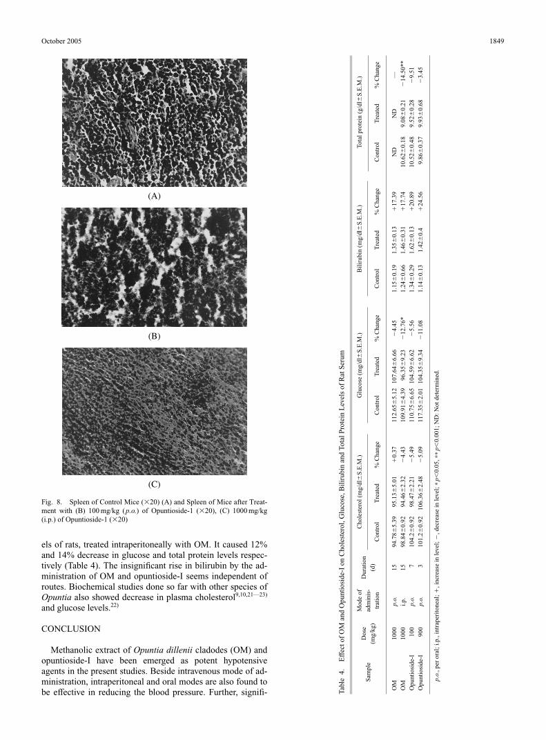
els of rats, treated intraperitoneally with OM. It caused 12%and 14% decrease in glucose and total protein levels respec-tively (Table 4). The insignificant rise in bilirubin by the ad-ministration of OM and opuntioside-I seems independent ofroutes. Biochemical studies done so far with other species ofOpuntia also showed decrease in plasma cholesterol9,10,21—23)
and glucose levels.22)
CONCLUSION
Methanolic extract of Opuntia dillenii cladodes (OM) andopuntioside-I have been emerged as potent hypotensiveagents in the present studies. Beside intravenous mode of ad-ministration, intraperitoneal and oral modes are also found tobe effective in reducing the blood pressure. Further, signifi-
October 2005 1849
(A)
(B)
(C)
Fig. 8. Spleen of Control Mice (�20) (A) and Spleen of Mice after Treat-ment with (B) 100 mg/kg (p.o.) of Opuntioside-1 (�20), (C) 1000 mg/kg(i.p.) of Opuntioside-1 (�20)
Tabl
e4.
Eff
ect o
f O
M a
nd O
punt
iosi
de-I
on
Cho
lest
erol
, Glu
cose
, Bil
irub
in a
nd T
otal
Pro
tein
Lev
els
of R
at S
erum
Mod
e of
C
hole
ster
ol (
mg/
dl�
S.E
.M.)
Glu
cose
(m
g/dl
�S
.E.M
.)B
ilir
ubin
(m
g/dl
�S
.E.M
.)To
tal p
rote
in (
g/dl
�S
.E.M
.)S
ampl
eD
ose
adm
inis
-D
urat
ion
(mg/
kg)
trat
ion
(d)
Con
trol
Tre
ated
% C
hang
eC
ontr
olT
reat
ed%
Cha
nge
Con
trol
Tre
ated
% C
hang
e C
ontr
olT
reat
ed%
Cha
nge
OM
1000
p.o.
1594
.78�
5.39
95.1
3�5.
01�
0.37
112.
65�
5.12
107.
64�
6.66
�4.
451.
15�
0.19
1.35
�0.
13�
17.3
9N
DN
D—
OM
1000
i.p.
1598
.84�
0.92
94.4
6�2.
32�
4.43
109.
91�
4.39
96.3
5�9.
23�
12.7
6*1.
24�
0.66
1.46
�0.
31�
17.7
410
.62�
0.18
9.08
�0.
21�
14.5
0**
Opu
ntio
side
-I10
0p.
o.7
104.
2�0.
9298
.47�
2.21
�5.
4911
0.75
�6.
6510
4.59
�6.
62�
5.56
1.34
�0.
291.
62�
0.13
�20
.89
10.5
2�0.
489.
52�
0.28
�9.
51O
punt
iosi
de-I
900
p.o.
310
1.2�
0.92
106.
36�
2.48
�5.
0911
7.35
�2.
0110
4.35
�9.
34�
11.0
81.
14�
0.13
1.42
�0.
4�
24.5
69.
86�
0.37
9.93
�0.
68�
3.45
p.o.
, per
ora
l; i.
p., i
ntra
peri
tone
al; �
, inc
reas
e in
leve
l; �
, dec
reas
e in
leve
l; ∗
p�0.
05, ∗
∗p�
0.00
1; N
D: N
ot d
eter
min
ed.

cant reduction in serum glucose level by OM has also beenrecorded. Since O. dillenii is an edible plant, its extract OM,as well as opuntioside-I did not kill any rat during studies,however, they left adverse effects on liver and spleen of ex-perimental animals at higher doses. Opuntioside-I has alsocaused the expiry of one mouse while survived mice werefound to have pathetic liver and spleen. Hence further inves-tigations on lower but effective doses of OM and opuntio-side-I in variety of animal species especially in non rodentsare required to determine the exact margin of safety for thesesubstances so as to make the study beneficial for human con-sumption.
MATERIALS AND METHODS
Chemistry. Plant Material Cladodes of Opuntia dil-lenii were collected in the month of October, 2001 from Uni-versity of Karachi campus and authenticated and depositedby Dr. Surraya, Dept. of Botany, University of Karachi withvoucher specimen No. KUH GHS 68218.
Instrumentation Ultraviolet spectra were recorded inMeOH on Hitachi-U3200 and infrared spectra were mea-sured in CHCl3 on JASCO-A-302, spectrophotometers. Theelectron impact (EI) mass spectra were recorded on a Finni-gan MAT-112 instrument while recording of High-resolutionmass spectra were carried out on a JMS HX-10 spectrometer.The 1H- and 13C-NMR spectra were run in CDCl3 on aBruker Aspect AM-500 spectrometer operating at 500 MHzfor 1H and 125 MHz for 13C nuclei, with spectra referencedto residual solvent signals. For PTLC silica gel 60 GF254
(Merck) was used.Extraction and Isolation of Chemical Constituents
from Opuntia dillenii Fresh and undried cladodes of Op-untia dillenii (3.34 kg) were cut into small cubes and ex-tracted four times with methanol at room temperature for 3 d.The combined extracts were concentrated on rotavapourunder reduced pressure to give a residue (OM, 203 g). Ex-tract (OM, 6.74 g) was subjected to preparative thin layerchromatography (PTLC) (silica gel, CHCl3 : MeOH 8.0 : 2.0)which gave four bands (OM-1—OM-4). The band OM-2(0.36 g) showed single spot on TLC (CHCl3 : MeOH 8 : 2, Rf0.44) and identified as opuntioside-I through detailed spec-tral studies including UV, IR, MS, NMR, 2D-NMR andchemical transformation.20) The band OM-1 (0.084 g) wasfurther purified through PTLC (silica gel, CHCl3 : MeOH,9.5 : 05, Rf 0.36) to give pure opuntiol (0.070 g). Spectraldata of both opuntioside-I and opuntiol were comparable tothe literature values.16,24) Opuntioside-I furnished its acetylderivative on treatment with acetic anhydride and pyridine atroom temperature. The remaining bands OM-3 (0.32 g) andOM-4 (0.83 g) were found to be the mixture of several com-pounds on TLC.
Acetylation of Opuntioside-I To the solution of opun-tioside-I (0.04 g) in pyridine (1 ml) acetic anhydride (1 ml)was added and left overnight at room temperature. Afterusual work up of reaction mixture, it gave pure tetraacetylderivative of opuntioside-I (0.05 g).
Pharmacology. Animals and Drugs Animals used inthis study were Sprague–Dawley rats (200—250 g) andNMRI mice (20—30 g) . They were housed at the AnimalHouse of Dr. HMI Institute of Pharmacology & Herbal Sci-
ences, Hamdard University and were given a standard dietand tap water ad libitum. Drugs used were Acetylcholine andSodium chloride from E. Merck, Atropine sulfate fromBoehringer Ingelheim, and Pentothal® sodium from AbbottKarachi. Acetylcholine (1 mg/kg) and saline (0.9% NaC1)were used as positive and negative controls respectively.
Hypotensive Activity Normotensive Sprague–Dawleyrats (either sex) were anaesthetized with pentothal® sodium(50 mg/kg i.p.). The trachea was exposed and cannulated tofacilitate spontaneous respiration. Drugs were injected (vol.0.2—0.25 ml) through a polyethylene cannula inserted intothe right external jugular vein followed by a saline flush(0.2 ml). The arterial blood pressure was recorded from thecarotid artery via polyethylene arterial cannula connected toa Research Grade Blood pressure Transducer (Harvard, 60-3003) coupled with four channel Harvard Universal Oscillo-graph (Curvilinear, 50-9307). The temperature of the animalswas maintained at 37 °C by use of over head lamp. Animalswere allowed to equilibrate for at least 15 min before admin-istration of any drug. Mean arterial blood pressure (MABP)was calculated as sum of the diastolic blood pressure plusone-third pulse width. Changes in blood pressure were ex-pressed as the percent of control values, obtained immedi-ately before the administration of test substance.
Toxicology Toxicity of OM was measured in four groups(I—IV) of rats (either sex) containing 10 animals eachgroup. The dose of 1000 mg/kg/d of OM was given orally(p.o.) and intraperitoneally (i.p.) to group-I and II respec-tively for fifteen consecutive days. Group-III and IV servingas control were given saline through oral and intraperitonealroutes respectively. Toxicology of opuntioside-I was studiedin both rats and mice. Two sets of rats (Group-V, VI) andmice (Group-VII, VIII) containing 10 animals (either sex)each group were treated orally for 7 d. Group-V and VII weretreated with opuntioside-I at the dose of 100 mg/kg/d whileGroup-VI and VIII serving as corresponding control weregiven saline.
Two more sets of rats (Group-IX, X) containing 4 animalseach group and two groups of mice (Group-XI, XII) contain-ing 10 animals (either sex) were subjected to toxicology for3 d. Group-IX was treated orally with opuntioside-I at thedose of 900 mg/kg/d while group-X served as its control.Group-XI of mice was given opuntioside-I intraperitoneallyat the dose of 1000 mg/kg/d and group-XII serving as itscontrol was injected (i.p.) with saline. Number of animals ingroup-IX, and duration of toxicology for groups-IX—XIIhave been reduced due to the paucity of opuntioside-I. Vol-ume of each dose in rats and mice were 0.6—0.8 ml and0.20—0.35 ml respectively.
All the animals were kept under observation for early 2 hafter the administration of dose, for any change in behaviouror physical activities. Number of expired animals were notedat the end of study period. Survived animals of both rats andmice were anaesthetized with 1.5—2.0 ml and 0.04 ml re-spectively of pentothal sodium (50 mg/ml, i.p.) for cannula-tion, withdrawing of blood and tissue analysis. Blood col-lected from rats was incubated and centrifuged for biochemi-cal study, while heart, liver, kidneys, and spleen of both ratsand mice were removed, blotted and weighed immediately onelectronic balance.
Effect of OM and Opuntioside-I on MABP of Rats
1850 Vol. 28, No. 10

after Oral and Intraperitoneal Administration All sur-vived animals of groups II, IV and V, VI at the end of theirtoxicity trials were anaesthetized with pentothal sodium andcannulated through trachea and carotid artery in the samemanner as described earlier to record their blood pressure.
Effect of OM and Opuntioside-I on Some Biochemicalsof Rat Serum Blood drawn (2—4 ml each rat) from alltreated and control rats was left at room temperature for 20min. Then incubated at 37 °C for 30 min and centrifuged sep-arately in (BHG) Herml Z230 (Germany) at the speed of3000 rpm for 20 min.
Serum obtained (1—1.5 ml) was subjected for the study ofserum cholesterol, glucose, bilirubin and total protein levelsby using commercial assay kits. Kits used were Ecoline® 25by CHOD-PAP method for cholesterol, Ecoline® 1000 byGOD-PAP method for glucose, Mercko test® for bilirubinand Mercko test® by biuret method for total protein. All thesekits were purchased from diagnostica Merck (Germany). U-2000 spectrophotometer (Hitachi) was used to measure theabsorbance of light.
Histology Heart, liver, spleen and kidneys were fixed in10% formalin. After usual processes of dehydration, clearingand infiltration, tissues were embedded in paraffin wax andsectioned into 7-mm slices through Leica RM 2145-RotationMicrotom. The tissues were stained with haematoxylin andeosin. The slides were studied and photographed throughNikon Advance Trincocular Research Microscope OP-TIPHOT Model X2T-21E equipped with Nikon Micropho-tography system; Model UFX-DX-35 and phase contrast Nplan.
Statistical Analysis Changes in blood pressure, serumbiochemical levels, body weights and weights of vital organswere compared using analysis of variance followed by Stu-dent’s t-test. Values of p�0.05, p�0.01 and p�0.001 wereconsidered to be significant.
Acknowledgements The authors are grateful for finan-cial assistance provided by Hamdard University, Karachi,Pakistan. One of the authors, Lubna Abidi appreciatesHigher Education Commission Islamabad, Pakistan for its fi-nancial support during her Ph.D. studies.
REFERENCES
1) Saleem R., Ahmad S. I., Ahmed M., Faizi Z., Rehman S. Z., Ali M.,
Faizi S., Biol. Pharm. Bull., 26, 41—46 (2003).2) Saleem R., Faizi S., Siddiqui B. S., Ahmed M., Hussain S. A., Qazi A.,
Dar A., Ahmad S. I., Qazi M. H., Akhtar S., Hasnain S. N., PlantaMed., 67, 757—760 (2001).
3) Zaheer S. H., “The Wealth of India,” Vol. VII, National Institute ofScience Communication, CSIR, New Delhi, 1997, pp. 100—104.
4) Loro J. F., Rio I. D., Santana L. P., J. Ethnopharmacol., 67, 213—218(1999).
5) Saenz C., Acta Horticulturae, 581, 253—263 (2002) [Chem. Abstr.,138, 186525 (2003)].
6) Castellar R., Obon J. M., Alacid M., Fernandez-Lopez J. A., J. Agric.Food Chem., 51, 2772—2776 (2003).
7) Saenz C., Estevez A. M., Fontanot M., Pak N., Acta Horticulturae,581, 275—278 (2002) [Chem. Abstr., 138, 203991 (2003)].
8) Tegegne F., Acta Horticulturae, 581, 343—346 (2002) [Chem. Abstr.,138, 186744 (2003)].
9) Choi J., Lee C. K., Lee Y. C., Moon Y. I., Park H. J., Han Y. N.,Saengyak Hakhoechi., 33, 230—237 (2002) [Chem. Abstr., 138,106060 (2003)].
10) Choi J. W., Lee C. K., Moon Y. I., Park H. J., Han Y. N., SaengyakHakhoechi., 33, 238—244 (2002) [Chem. Abstr., 138, 106061 (2003)].
11) Ahmed A., Davies J., Randall S., Skinner G. R. B., Antiviral Res., 30,75—85 (1996).
12) Park E. H., Kahng J. H., Lee S. H., Shin K. H., Fitoterapia, 72, 288—290 (2001).
13) Karsten K. A. S., S. African ZA 93 04523 (Cl. A61K) [Chem. Abstr.,124, 270550 (1996)].
14) Galati E. M., Mondello M. R., Giuffrida D., Dugo G., Miceli N., Per-golizzi S., Taviano M. F., J. Agric. Food Chem., 51, 4903—4908(2003).
15) Lee Y. S., Park H., Jin C., Kim H. J., Cho J., Park M., Song Y., PCTInt. Appl. WO 03 37324 (Cl. A61K31/352) [Chem. Abstr., 138,348735 (2003)].
16) Qiu Y., Chen Y., Pei Y., Matsuda H., Yoskikawa M., Chem. Pharm.Bull., 50, 1507—1510 (2002).
17) Gupta R. S., Sharma R., Sharma A., Chaudhdery R., Bhatnager A. K.,Dobhal M. P., Joshi Y. C., Sharma M. C., Pharm. Biology, 40, 411—415 (2002).
18) Nadkarni’s K. M., “Indian Materia Medica,” Popular Prakashan, Bom-bay, 1976, pp. 872—873.
19) Qiu Y. K., Chem Y. J., Pei Y. P., Hisashi M., Masayuki Y., J. ChinesePharm. Sci., 12, 1—5 (2003).
20) Faizi S., Abidi L., Isolation and Structure Elucidation of New a-Py-rones from Prickly Pear. Poster Presented in the 3rd International and13th National Conference held at Karachi from Dec. 28—31 2002.
21) Fernanduz M. L., Lin E. C. K., Trejo A., McNamara D. J., Diet. J.Nutr., 124, 817—824 (1994).
22) Wolfram R. M., Kritz H., Efthimiou Y., Stomatopoulos J., SinzingerH., Wiener Klinische Wochenschrift., 114, 840—846 (2002) [Chem.Abstr., 138, 49742 (2002)].
23) Kang M. S., Kang J. S., Hanguk Yongyang Hakhoechi, 34, 141—149(2001) [Chem. Abstr., 134, 352722 (2001)].
24) Ganguly A. K., Govindachari T. R., Mohamed P. A., Tetrahedron, 21,93—99 (1965).
October 2005 1851


![[Toxicology] toxicology introduction](https://img.dokumen.tips/doc/110x75/55c46616bb61ebb3478b4643/toxicology-toxicology-introduction.jpg)