Embed Size (px)
Citation preview

Hypopituitarism: An
Update
Karen K. Miller, M.D.
Chief, Neuroendocrine Unit, Massachusetts General
Hospital (MGH)
Director, MGH Neuroendocrine and Pituitary Tumor
Clinical Center
Professor of Medicine, Harvard Medical School
Dr. Miller has no relevant conflicts to declare.

Hypopituitarism Case
• 72-year-old man admitted to outside hospital with abdominal pain, nausea, vomiting, fever, headache and hypotension
• Treated with antibiotics, IV fluids and pressors
• Developed obtundation, hyponatremia (Na+ 124 meq/L), meningismus, and cranial nerve III, IV and VI palsies
• Transferred to MGH

Hypopituitarism Case
• Intubated, placed on antibiotics for presumed basilar meningitis
• Head CT: remarkable for a sellar mass
• Cortisol = 1.8 mcg/dl (50 nmol/L)
• Stress dose steroids administered
• Pressors were discontinued, and the patient was extubated
• A head MRI was performed

NORMAL MRI
PITUITARY
PITUITARY
ADENOMA
HEMORRHAGE

Hypopituitarism Case
• Diagnosis: pituitary apoplexy with hemorrhage in a macroadenoma
• Treatment: Transphenoidal surgical decompression
• Pathology: NF pituitary adenoma
• Clinical course:
– Cranial nerve palsies resolved
– Panhypopituitarism developed
– Doing well on hormone replacement therapy

Topics
• Causes of Hypopituitarism
• Central Hypothyroidism
• Central Adrenal insufficiency
• Central Hypogonadism
• Growth Hormone Deficiency

Topics
• Causes of Hypopituitarism
• Central Hypothyroidism
• Central Adrenal insufficiency
• Central Hypogonadism
• Growth Hormone Deficiency

Etiologies Of Hypopituitarism• Pituitary tumors and treatment
• Suprasellar/hypothalamic tumors– Craniopharyngioma
• Infiltrative disease– Sarcoid
– Lymphocytic hypophysitis
• Infarction– Sheehan’s syndrome
– Apoplexy
• Congenital
• Post-trauma/SAH
• Ipilimumab
• Opioids

Etiologies Of Hypopituitarism• Pituitary tumors and treatment
• Suprasellar/hypothalamic tumors– Craniopharyngioma
• Infiltrative disease– Sarcoid
– Lymphocytic hypophysitis
• Infarction– Sheehan’s syndrome
– Apoplexy
• Congenital
• Post-trauma/SAH
• Ipilimumab
• Opioids

Traumatic Brain Injury (TBI) and
Subarachnoid Hemorrhage (SAH): Pooled
Data from 19 Studies (1137 Patients)
• Prevalence of hypopituitarism chronically
– TBI: 27.5%
– SAH: 47%
– Adrenal insufficiency: 8% (TBI); 21% (SAH)
• Associated with impaired QOL and metabolic abnormalities
Schneider, HJ et al, JAMA 2007

Ipilimumab
• Anti-cytotoxic T-lymphocyte antigen-4
(CTLA-4) therapy
• Complicated by autoimmune
hypophysitis with panhypopituitarism
in 17% of cases
– Likely an underestimate
• Much lower incidence with nivolumab
and/or pembrolizumab (0.5%)

Treatment of Ipilimumab-
Induced Hypopituitarism
• Hormone replacement
– No need to d/c ipilimumab
• High-dose glucocorticoids not needed to
treat hypophysitis but often used to treat
autoimmune colitis
• Development of hypophysitis positively
predicts survival (19.4 vs. 8.8 mos)
• Recovery of pituitary function in only 24%
Faje, A et al, J Clin Endocrinol Metab 2014

Opioid-Induced Hypopituitarism
Systematic Review and Meta-analysis
• 52 studies including 18,428 subjects
• Patients with chronic pain, on maintenance for
addiction treatment, and healthy volunteers
• Hypogonadism in 63% (99.5% of studies in
males only)
• Adrenal insufficiency 15-24% (depending on
diagnostic criteria used)
• Thyroid and GH axes do not appear to be
affected
De Vries, F et al, J Clin Endocrinol Metab 2019

Topics
• Causes of Hypopituitarism
• Central Hypothyroidism
• Central Adrenal insufficiency
• Central Hypogonadism
• Growth Hormone Deficiency

Free T4 Target in Treatment of
Central Hypothyroidism
As we cannot use the TSH to target
therapy, what should be the free T4 level
target?

Free T4 Target in Treatment of
Central Hypothyroidism
• Data suggest that central hypothyroidism is often
under-treated
– Doses are lower than in primary hypothyroidism, resulting
in low-normal free T4 levels
– Higher doses (~ 1.6 μg/kg in one study) may improve BMI
and lipid profile
• A 2018 study in patients with primary hypothyroidism
suggests that increasing L-T4 dose within the normal
range does not result in objective benefit in weight,
body composition quality of life, mood or cognition
Goller K et al., Exp Clin Endocrinol 2004; Samuels et al. JCEM
2018; Slawik et al., JCEM 2007;

Data Suggest We Should be
Cautious about Free T4 Levels in
the High-Normal Range
• 10,318 participants of the Rotterdam Study
were included – not a study of hypopituitarism
• Higher free T4 levels, even within the normal
range, were associated with an increased risk of
sudden cardiac death (hazard ratio, 2.28 per 1
ng/dL free T4, 95% CI, 1.31-3.97)
• The absolute 10-year risk of sudden cardiac
death increased from 1-4% in euthyroid patients
with higher free T4 levels
• Goal: free T4 in the mid-normal range?Chaker L et al., Circulation 2016

What is an Appropriate Free T4
Goal?
• None of these studies were in hypopituitary
patients
– TSH not elevated, and therefore T4 to T3 conversion is
less than in primary hypothyroidism
• Goal: free T4 in the mid-normal range?
– Except in those with contraindications such as cardiac
issues or anxiety disorders
Chaker L et al., Circulation 2016

Apoplexy Case:
Central Hypothyroidism• Timing: 6 weeks after apoplexy
• Lab results
– TSH: 1.35 mIU/L (0.5-5 mIU/L)
– Free T4: 0.7 ng/dl (0.9-1.8 ng/dl) (9.0 with a normal range of 11.6-23.2 pmol/L)
– T3: 63 ng/dl (60-181 ng/dl) (0.97 with a normal range of 0.92-2.79 nmol/L)
• Replacement therapy: L-thyroxine
– Mid-normal free T4
– TSH <0.01 mIU/L (0.5-5 mIU/L)

Topics
• Causes of Hypopituitarism
• Central Hypothyroidism
• Central Adrenal insufficiency
• Central Hypogonadism
• Growth Hormone Deficiency

Diagnostic Issues
•“Low-dose” 1 mcg cort-stim
– In theory, superior for diagnosing mild
central adrenal insufficiency
– Controversial and technically challenging
– 2 meta-analyses using mostly same data
•Opposite conclusions
– If center has experience with 1 mcg test,
likely clinically equivalent to 250 mcg
•We use traditional 250 mcg testDorin RI et al., Ann Intern Med, 2003
Kaslauskaite R et al., J Clin Endocrinol Metab 2008

Central Adrenal
Insufficiency: Diagnosis
• Insulin Tolerance Test (ITT)
– Gold standard
– Hypoglycemia stimulates entire HPA axis
•Useful to diagnosis acute and chronic adrenal insufficiency, as well as growth hormone deficiency
– Contraindicated in elderly, cv disease, seizures, serious psychiatric disease
– Physician must be present
– We do not use it any longer
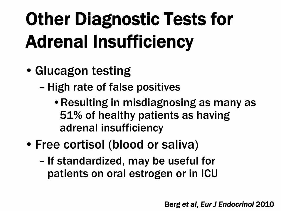
Other Diagnostic Tests for
Adrenal Insufficiency
• Glucagon testing
– High rate of false positives
•Resulting in misdiagnosing as many as 51% of healthy patients as having adrenal insufficiency
• Free cortisol (blood or saliva)
– If standardized, may be useful for patients on oral estrogen or in ICU
Berg et al, Eur J Endocrinol 2010

Other Diagnostic Tests for
Adrenal Insufficiency• Recent studies suggest that a cut-off
serum free cortisol by LC/MS/MS and equilibrium dialysis of 1 mcg/dl is roughly equivalent to a total cortisol of 18 mcg/dl
– May be a guide in patients with impaired liver synthetic capacity in particular, but use with caution
Dichtel et al, J Clin Endocrinol Metab 2019

Central Adrenal
Insufficiency: Treatment
• Differs from 1 adrenal insufficiency
– Mineralocorticoid (fludrocortisone) replacement not necessary
– Glucocorticoid requirements may be lower, and therefore a once-daily medium-acting glucocorticoid can be used

What is the Proper
Replacement Dose and
Timing of Dosing?
• Probably lower than we think
• Cortisol production rate
– 9.9 ± 2.7 mg/day (0.027 ± 0.007 mol/day)
– 5.7 mg/m2/day (0.016 mol/m2/day)
Esteban NV et al, J Clin Endocrinol Metab 1991

Esteban NV et al, J Clin Endocrinol Metab 1991
Diurnal Variation

Hydrocortisone (HC) 30 mg in
Divided Doses is Supraphysiologic
Behan et al, Clin Endo, 2011

HC 10 mg (8 am)/5 mg (2 pm)
more Closely Mimics Physiologic
Cortisol Secretion
Behan et al, Clin Endo, 2011

Important to Individualize
Regimen
• Significant range in individual glucocorticoid
requirements: incompletely understood
– Degree of glucocorticoid deficiency
– Endogenous variability in metabolism (which
can also be affected by medications, e.g.
anticonvulsants)
– Differences in glucocorticoid sensitivity
• Glucocorticoid receptor polymorphisms
Quax RA et al, Nature Reviews 2013

Treatment Options other than
Hydrocortisone (HC)
• Once daily medium half-life glucocorticoid
(e.g. prednisone or prednisolone 2-5 mg
daily)
– Advantage: afternoon dose is not necessary
for patients with central adrenal insufficiency

Is a Prednisone/Prednisolone
Less Safe than Hydrocortisone?• 2 studies
– No difference in CV risk markers: mean 3.7 mg dose
prednisolone vs. 20.5 mg hydrocortisone
– Higher LDL and total cholesterol, but no difference in
HbA1c, triglycerides, BMI, blood pressure or waist
circumference: mean 5.0 mg dose prednisolone vs.
21.5 mg hydrocortisone
• Dose likely the important variable
– Very long-acting compounds (eg dexamethasone)
may be more likely to cause iatrogenic Cushing’s
Smith et al., Endocr Connect, 2017; Quinkler et al., Endocr Connect, 2017

Once daily dual-release
hydrocortisone
•Approved in Europe
•Equivalent dose given once daily
resulted in lower BMI, blood
pressure and glucose metabolism
compared with same dose given in
3 divided doses
Johannsson, G. et al., J Clin Endocrinol Metab 2012

Johannsson G et al, J Clin Endocrinol Metab 2012
Once-Daily HC (solid line) vs. TID
Comparable Dosing (dotted line)• Avoids peak blood levels
• Not available in U.S.
• Expensive where available

Increased mortality observed (in patients
with non-functioning adenomas) receiving ≥
30 mg/day
Zueger T et al, J Clin Endocrinol Metab 2012
2017 study suggests
there may be an
increased mortality in
patients receiving
>20 mg/day
hydrocortisone
Hammarstrand C et al EJE
2017

Therefore, minimizing
glucocorticoid doses is
important.However, so is appropriate
supplementation.
Question: When should we
supplement?

What is the Increased Death
Rate in Hypopituitarism Due To?
Burman P et al, J Clin Endocrinol Metab 2013
Most deaths coded as “infectious disease” in
origin were GI infections complicated by shock

Therefore, Although Minimizing
Daily Glucocorticoid Doses is
Important• It is also very important prepare patients
for emergency stress-dosing
– Especially for GI illnesses (fever, surgeries,
hospitalizations, accidents also)
– Prescribe 100 mg hydrocortisone for self-
administration if severe GI illness
– Instructions to page MD and go to
emergency room
– Medic Alert bracelet to be worn at all times

Apoplexy Case: Adrenal
Insufficiency
• Serum cortisol while hypotensive: 1.8 mcg/dl (50
nmol/L), diagnostic of adrenal insufficiency
• Receives prednisone 3 mg daily (a decrease from
the 5 mg daily I prescribed when he experienced
apoplexy >20 years ago)
– Mineralocorticoid replacement not necessary, as
adrenal insufficiency is of central origin
• Monitored regularly for signs/symptoms of adrenal
insufficiency or iatrogenic Cushing’s syndrome

Topics
• Causes of Hypopituitarism
• Central Hypothyroidism
• Central Adrenal insufficiency
• Central Hypogonadism
• Growth Hormone Deficiency

Central Hypogonadism:
Treatment•Gonadotropin therapy if fertility desired
•Testosterone replacement for most men
–IM testosterone esters for men, less expensive option
–Peak serum T supraphysiologic →
HCT, prostate stimulation
–Testosterone patches and gels
–Study suggests that IM testosterone may be associated with a greater risk of cardiovascular events and deaths compared with gels (Layton JB et al., JAMA Intern
Med 2015)

Other Treatment Options
•Testosterone pellets inserted every 4-6 months
•Testosterone undecanoate injected every 10 weeks (after an initial 4-week injection)
–Black box warning for pulmonary-oil microembolism (POME) (0.2%) and anaphylaxis (0.05%)
•Testosterone nasal gel
–Three times daily administration

FDA-Approved March, 2019: Long-Acting
Testosterone Undecanoate Preparation
• Oral twice-daily, fatty meal not required
• 87% of patients achieved normal testosterone levels
• Black box warning: drug can cause blood pressure to
rise, increasing the risk of heart attack, stroke and
cardiovascular death
• Increase in PSA and Hct, and decrease in HDL
observed
• Approved for men with hypopituitarism or genetic
causes of hypogonadism, not age-related declines in
testosterone levels

Is Testosterone Therapy
Safe?• A retrospective study showed that normalization
of testosterone levels was associated with a
reduction in mortality, MI and stroke in veterans
with frankly low T levels
• Prospective trials in elderly men and those with
preexisting cv disease who were treated for
declines in testosterone levels due to aging have
shown that higher T levels associated with
markers of CV disease and increased CV events
Sharma R et al, Eur Heart J 2015, Ruige JB et al, J Clin Endocrinol Metab,
2013; Yeap BB et al, J Clin Endocrinol Metab, 2013; Basaria S et al, NEJM,
2010; Vigen R et al, JAMA 2013; Finkle WD et al, PLOS ONE 2014

New Studies Published in 2019• Testosterone therapy is associated with increased risk of
venous thromboembolism among men with (OR 2.32) and
without (OR 2.02) hypogonadism
– Case-crossover study in 39,622 men (claims data)
• Testosterone therapy prevents progression from prediabetes to
diabetes over 8 years in men with hypogonadism
– 8-year observational study in which 229 men received
testosterone undecanoate
•None progressed to overt diabetes; 90% normalized
hgbA1c < 5.7%
– 87 untreated control subjects
•40% progressed to diabetes (hgbA1c >6.5%)
Walker RF et al, JAMA Intern Med, 2019; Yassin A et al, Diabetes Care, 2019

Testosterone Therapy Prevents Progression
to Diabetes in Observational Study
Walker RF et al, JAMA Intern Med, 2019

Testosterone: Replace?
Optimal Window?• Few studies in patients with hypopituitarism, many of
whom have profound hypogonadism and prepubertal
levels
– Severe hypogonadism is associated with increased cv risk,
increased visceral adiposity, reduced muscle mass,
osteoporosis, fatigue, anemia, depression, sexual
dysfunction
• Recommend replacing testosterone, esp in those with
very low levels who do not have contraindications
• Conservative serum testosterone level targets,
especially in elderly patients and those with cv disease
or history of venous thromboembolism, is warranted

Central Hypogonadism:
Treatment
•Estrogen/progestin replacement for women of reproductive age
–No data
–WHI results cannot be extrapolated to hypopituitary women of reproductive age
–Goal: restoration of “normal” hormonal milieu
–Gonadotropin therapy if fertility desired

Central Hypogonadism:
Treatment
•Women of postmenopausal age
–Decision re: HRT similar as for eupituitary women, but hot flashes rare and some intracranial tumors have estrogen receptors

Apoplexy Case: Central
Hypogonadism
• Testosterone 135 ng/dl (normal range
267-916 ng/dl) (468 nmol/L, with a
normal range of 926-3176 nmol/L)
•“Normal” LH, FSH and prolactin
• Doing well on physiologic testosterone
therapy using a topical preparation
– Hct and PSA measured annually

Androgen Deficiency In Women
•Effects of androgen replacement in
men
–Bone: bone density
–Body composition: visceral fat,muscle
mass and strength
–Quality of life: libido and mood

Androgens in Women
• Testosterone levels:
– 1/10 to 1/20th male levels
• Important for libido and quality of life,
body composition, bone density at the
low concentrations present in women?
• Or are androgen levels too low to play
an important biologic role in women?

Androgen Deficiency In Women
In Hypopituitarism
• Hypopituitarism is characterized by
hypogonadism and/or hypoadrenalism,
which affects critical sources of
androgen production in women
•Testosterone and DHEAS are both low in
women with hypopituitarism

Randomized Trial: Free
Testosterone Levels Increased
Increased to Normal Female Range
0 1 3 6 9 12
Months
Fre
e T
esto
ste
ron
e (
pg
/ml)
Miller KK et al, J Clin Endocrinol Metab 2006

BMD (Hip and Radius) Increased
with Testosterone Replacement
-2.5
-2
-1.5
-1
-0.5
0
0.5
1
1.5
2
Placebo Testosterone
Hip Radius PA Spine
* *
% C
han
ge i
n B
MD
* p < 0.02
Miller, KK et al., J Clin Endocrinol Metab 2006

Thigh Muscle Area Increased
with Testosterone
Replacementp = 0.013
% C
ha
ng
e in
Cro
ss
-Se
cti
on
al
Mu
sc
le A
rea
at
Mid
-Fe
mu
r
*
Placebo Testosterone
Miller, KK et al, J Clin Endocrinol Metab 2006

Mood Improved with
Testosterone Replacement
Baseline Post- Baseline Post-
Treatment Treatment
Placebo Testosterone
*
0
5
10
15
Miller, KK et al, J Clin Endocrinol Metab 2006

Sexual Function Improved with
Testosterone Replacement
20
25
30
35
40
45
50
Orgasm Arousal Behavior/
Experience
Cognition/
Fantasy
Drive/Rela
-tionship
Total
*
**
De
rog
ati
s T
Sco
res a
fter
Tre
atm
en
t
Miller, KK et al, J Clin Endocrinol Metab 2006

2019 Systematic Review and
Meta-analysis
• 46 reports of 36 RCT, including 8480
postmenopausal participants
• Testosterone improved sexual function
• Oral testosterone increased LDL and decreased
TC, HDL and triglycerides
• No significant effects on muscle mass or
cognitive endpoints (limited by small N)
• Increase in acne and hair growth but no serious
adverse events

DHEA Replacement Therapy in
Women with Adrenal Insufficiency
•Meta-analysis (10 studies total)
•“DHEA may improve, in a small and perhaps trivial manner, health-related quality of life and depression….”
–No significant effect on anxiety or sexual well-being
•“The evidence appears insufficient to support the routine use of DHEA in women with adrenal insufficiency.”
Alkatib A et al, J Clin Endocrinol Metab, 2010

Possible Explanations for
Lack of Stronger Effect
• Effect is weak, nonexistent or only present for an uncharacterized subset
• Methodologic issues with some of the DHEA studies– Some studies used very low DHEA doses
– Some studies combined data from men and women
– Multiple different instruments used
– Questionnaires not disease-specific

Clinical Issues: Androgen
Administration in Women
• Risks
– Hyperandrogenism
•Low incidence in short-term studies
– Long-term effects (greater than 24 months) of androgen use in women are not known

No Government-Agency Approved
Testosterone Preparation for
Women in the U.S. or Europe
•Pharmacokinetics of gels that are
designed for men cannot be reliably
dosed for women
•Compounding pharmacy preparations
may result in variable levels and not
FDA-monitored

Global Consensus Position
Statement 2019
• The only evidence-based indication for the use
of testosterone in women is for the treatment
of postmenopausal women who have been
diagnosed as having Hypoactive Sexual Desire
Disorder
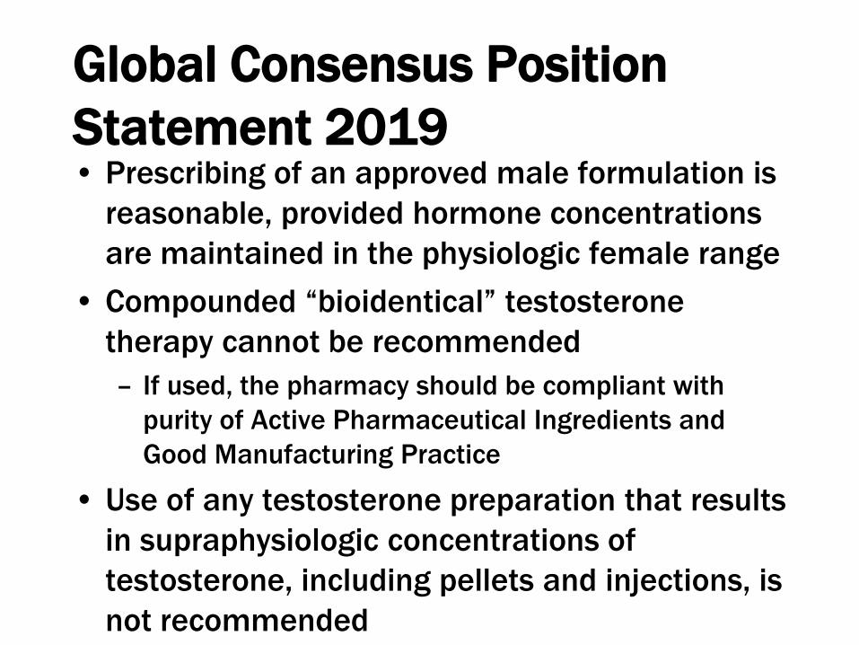
Global Consensus Position
Statement 2019• Prescribing of an approved male formulation is
reasonable, provided hormone concentrations
are maintained in the physiologic female range
• Compounded “bioidentical” testosterone
therapy cannot be recommended
– If used, the pharmacy should be compliant with
purity of Active Pharmaceutical Ingredients and
Good Manufacturing Practice
• Use of any testosterone preparation that results
in supraphysiologic concentrations of
testosterone, including pellets and injections, is
not recommended

Global Consensus Position
Statement 2019
• A baseline total testosterone concentration
should be measured before commencement,
with a repeat level 3–6 weeks
• Patients should be monitored for their clinical
response to treatment and signs of androgen
excess with a serum testosterone level every 6
months
• If no benefit is experienced by 6 months,
treatment should be ceased

DHEA is a Dietary Supplement in U.S., with Little FDA Oversight
•Contain 0 to >100% of declared amount
•Oral → in HDL (risk of oral androgens and pre-androgens)
•Converted to androgens and estrogens

Topics
• Causes of Hypopituitarism
• Central Hypothyroidism
• Central Adrenal insufficiency
• Central Hypogonadism
• Growth Hormone Deficiency
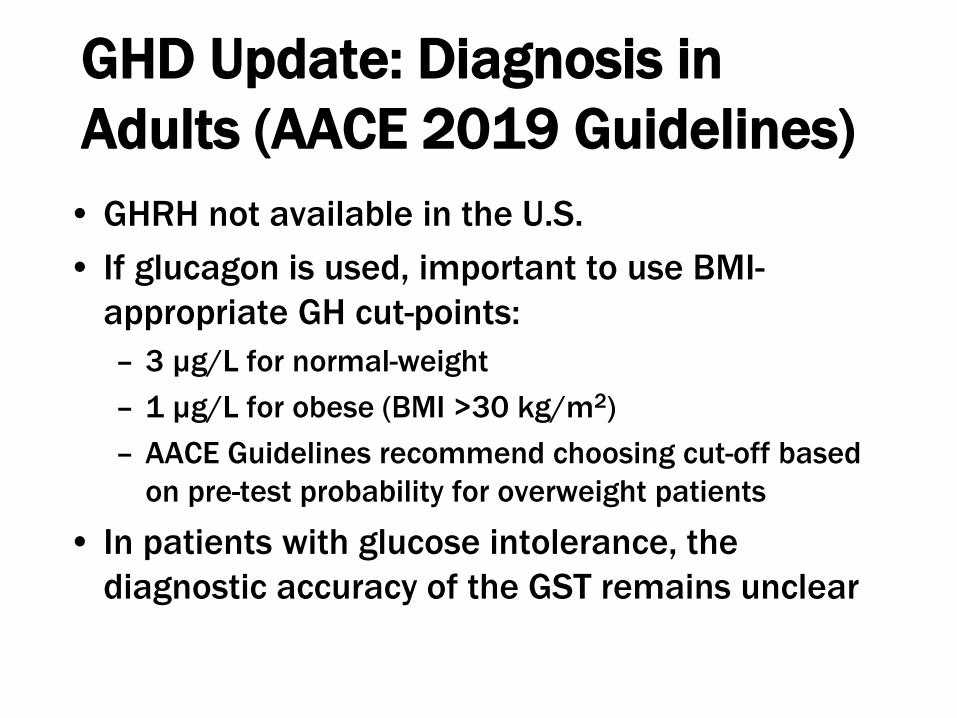
GHD Update: Diagnosis in
Adults (AACE 2019 Guidelines)
• GHRH not available in the U.S.
• If glucagon is used, important to use BMI-
appropriate GH cut-points:
– 3 µg/L for normal-weight
– 1 µg/L for obese (BMI >30 kg/m2)
– AACE Guidelines recommend choosing cut-off based
on pre-test probability for overweight patients
• In patients with glucose intolerance, the
diagnostic accuracy of the GST remains unclear

Macimorelin: Orally Active GH
Secretagogue• FDA approved this test for use as a
diagnostic test for adult GHD in
December, 2017
• Oral, very well tolerated, 90-minute test
• Expensive and variably covered by
insurance
• Cut-point is 2.8 mg/L
• BMI-adjusted peak GH cut-points for this
test are needed for overweight and obese
patients

Long-Acting GH
• The frequency of daily injections is one of
the major factors contributing to
nonadherence with rhGH therapy
• Weekly long-acting GH (LAGH)
preparations are currently under
development
• Not currently available in the U.S. market
– Several preparations in later phase trials

Hypopituitarism: Future
Directions • Perfect hormone replacement regimens
• Androgen replacement in women and GH
deficiency and replacement
– To whom should we prescribe?
– Effects on cardiovascular and cancer risk?
• Other hormone deficiencies?
– Low plasma oxytocin levels and increased
psychopathology in hypopituitary men with
diabetes insipidus demonstrated
– Obesity, quality of life largely unexplainedAulinas A. et al., J Clin Endocrinol Metab 2019