Embed Size (px)
DESCRIPTION
método mulligan no tratamento de epicondilite lateral do cotovelo
Citation preview

2003; 83:374-383.PHYS THER. VicenzinoAatit Paungmali, Shaun O'Leary, Tina Souvlis and BillEpicondylalgiaMobilization With Movement for Lateral Hypoalgesic and Sympathoexcitatory Effects of
http://ptjournal.apta.org/content/83/4/374found online at: The online version of this article, along with updated information and services, can be
Collections
Pain Manual Therapy
Injuries and Conditions: Elbow in the following collection(s): This article, along with others on similar topics, appears
e-Letters
"Responses" in the online version of this article. "Submit a response" in the right-hand menu under
or click onhere To submit an e-Letter on this article, click
E-mail alerts to receive free e-mail alerts hereSign up
by guest on April 17, 2015http://ptjournal.apta.org/Downloaded from by guest on April 17, 2015http://ptjournal.apta.org/Downloaded from

Hypoalgesic and SympathoexcitatoryEffects of Mobilization WithMovement for Lateral Epicondylalgia
Background and Purpose. Mulligan has proposed the use of mobiliza-tion with movement for lateral epicondylalgia. In this study, mobiliza-tion with movement for the elbow was examined to determine whetherthis intervention was capable of inducing physiological effects similarto those reported for some forms of spinal manipulation. Participants.Seven women and 17 men (mean age�48.5 years, SD�7.2) withchronic lateral epicondylalgia participated in the study. Methods. Aplacebo, control, repeated-measures study was conducted to evaluatewhether mobilization with movement at the elbow produced concur-rent hypoalgesia and sympathoexcitation. Results. The treatment dem-onstrated an initial hypoalgesic effect and concurrent sympathoexcita-tion. Improvements in pain resulted in increased pain-free grip forceand pressure pain thresholds. Sympathoexcitation was indicated bychanges in heart rate, blood pressure, and cutaneous sudomotor andvasomotor function. Discussion and Conclusion. This study showed thata mobilization with movement treatment technique exerted a physio-logical effect similar to that reported for some spinal manipulations.[Paungmali A, O’Leary S, Souvlis T, Vicenzino B. Hypoalgesic andsympathoexcitatory effects of mobilization with movement for lateralepicondylalgia. Phys Ther. 2003;83:374–383.]
Key Words: Lateral epicondylalgia, Manual therapy, Mechanism, Pain, Tennis elbow.
Aatit Paungmali, Shaun O’Leary, Tina Souvlis, Bill Vicenzino
374 Physical Therapy . Volume 83 . Number 4 . April 2003
Rese
arch
Repo
rt �
�������������������������������������������������������������������������������������������������������������������������������������������������������������������
������
������
������
������
������
������
������
������
������
������
������
������
������
������
������
������
������
������
������
by guest on April 17, 2015http://ptjournal.apta.org/Downloaded from

Mulligan1 has recently described an inter-vention in which a therapist applies apassive glide mobilization to a joint (usu-ally an accessory motion) and sustains it
while the client performs a physical task involving thelimbs. The techniques, called “mobilization with move-ments” (MWM), are claimed to bring about improve-ments in pain and function immediately following theirapplication in the clinic,1 but there is a lack of experi-mental data reported in peer-reviewed publications. TheMWM group of techniques are claimed to achieve thisrapid improvement in persistent musculoskeletal painstates that have been recalcitrant to other forms oftherapy.2,3 To date, the postulated mechanism(s) ofaction of this treatment approach has focused onmechanical effects such as the restoration of bony posi-tional faults.4,5 The physiological effects have largelybeen ignored.
Much of the research about manipulation over the pastdecade has not focused on evaluating the subluxationtheory of spinal manipulation, but rather such researchhas concentrated on elucidating the physiological effectsof spinal manipulations.6–13 Most of this research, how-ever, has not dealt with the effects of the MWM tech-nique. Several authors6–13 have reported data that havebeen interpreted as reflecting possible neurophysiologicmechanism(s) for hypothesized actions. Some studies6,7
have shown that passive mobilization treatments of thecervical spine, techniques frequently used by physicaltherapists, may produce an initial hypoalgesia and con-current excitation in the motor system and the sympa-thetic nervous system (SNS).6,7
Any concurrent initial hypoalgesic and physiologicaleffects have not been studied in manipulation of periph-eral joints. Our study represents an initial investigationof the effect of a MWM treatment technique at the elbow
A Paungmali, PT, MPT, is a doctoral student, Department of Physiotherapy, The University of Queensland, St Lucia, Queensland, Australia.
S O’Leary, PT, MPT, MAPA, is a doctoral student, Department of Physiotherapy, The University of Queensland.
T Souvlis, PT, BPT (Hons), MAPA, MPAA, is Lecturer, Physiotherapy Department, The University of Queensland. She was a doctoral student,Department of Physiotherapy, The University of Queensland, at the time of this study.
B Vicenzino, PT, PhD, MAPA, MPAA, is Senior Lecturer, Department of Physiotherapy, Director, Musculoskeletal Pain and Injury Research Unit,The University of Queensland, St Lucia, Queensland, Australia, 4072 ([email protected]). Address all correspondence to DrVicenzino.
All authors provided concept/research design, writing, data collection and analysis, project management, and subjects. Ms Souvlis and DrVicenzino provided fund procurement and facilities/equipment. The authors acknowledge Angela Chang for consultation (including review ofmanuscript before submission).
Ethical approval for the study was obtained from the Institutional Review Board (Medical Research Ethics Committee) of The University ofQueensland.
This study was awarded the best paper prize (plenary oral presentation) at the Musculoskeletal Physiotherapy Australia 12th Biennial Conference;November 21–24, 2001; Adelaide, South Australia, Australia.
This article was submitted August 15, 2002, and was accepted October 8, 2002.
Physical Therapy . Volume 83 . Number 4 . April 2003 Paungmali et al . 375
������
������
������
������
������
������
������
������
������
������
������
������
������
������
������
������
������
by guest on April 17, 2015http://ptjournal.apta.org/Downloaded from

on both pain and the SNS simultaneously. Recently, aMWM treatment technique for chronic lateral epicondy-lalgia has been described in the peer-reviewed andnon–peer-reviewed literature.1–3,14 These authors1–3,14
contended that a MWM treatment technique applied atthe elbow produced substantial and rapid pain reliefimmediately following application of the technique.
The aim of our study was to describe the physiologicaleffects of a MWM treatment technique for chroniclateral epicondylalgia by measuring concurrent changesin measurements of pain and SNS function. We did notset out to determine the benefit of the intervention onfunction or other outcomes over a course of severaltreatments.
MethodsA placebo, control, repeated-measures study was used toevaluate the initial pain-relieving effects and changes inSNS function during and immediately following theapplication of MWM for chronic lateral epicondylalgia.
ParticipantsTwenty-four participants, 7 female and 17 male (meanage�48.5 years, SD�7.2), with unilateral lateral epicon-dylalgia of 8.9 months’ (SD�8.4) duration participatedin the study. All participants were right-handed, and83.33% of the participants (n�20) had lateral epicon-dylalgia on the right-hand side. Participants wererecruited from the metropolitan and suburban areas ofBrisbane, Australia, by media releases and on referralfrom local health care practitioners. This sample size wasdetermined a priori on the basis of a pilot study(alpha�.05, power�80%, effect size�0.44).14 The effectsize for pressure pain threshold was chosen because itwas considered to be a more conservative choice thanone based on pain-free grip force, which had a largereffect size.14
We defined lateral epicondylalgia as pain over the lateralside of the elbow that was provoked by palpation of thelateral epicondyle region and gripping tasks. In addi-tion, pain had to be experienced over the lateral epicon-dyle during at least one of the following: resisted staticcontraction of the wrist extensors or extensor carpiradialis brevis muscle or stretching of the forearm exten-sor muscles.15,16 Volunteers were excluded from thestudy if they had cervical spine or upper-limb problems(eg, referred pain, conditions other than lateral epicon-dylalgia). Other exclusion criteria included neurologicalimpairments, neuromuscular diseases, cardiovasculardiseases, health conditions that would have precludedtreatment (eg, osteoporosis, malignancies, hemophilia,diabetes), recent steroid injection, using prescriptionmedications such as beta-adrenoceptor blocking agentsor anti-inflammatory or analgesic drugs, aversion to
manual contact, and previous therapy for the elbow joint(to minimize expectation bias). All participants providedwritten consent prior to participation.
Experimental ConditionsThe experimental conditions were the treatment condi-tion (elbow MWM), a placebo condition, and a controlcondition. All conditions were administered by a physi-cal therapist with 8 years of clinical experience andpostgraduate tertiary qualification in manipulative phys-ical therapy. The treatment condition involved a lateralglide MWM technique for the elbow as described byMulligan.1 To apply this treatment, the physical therapistused one hand to stabilize the distal end of the humeruson the lateral side just proximal to the elbow joint linewhile using the other hand to apply a laterally directedglide of the proximal ulna and radius (Fig. 1). The handapplying the lateral glide was situated just distal to theelbow joint line on the medial side of the ulna. The glidewas painlessly applied and sustained for approximately 6seconds while the participant performed a pain-freegripping action. The gliding pressure was then main-tained until the participant completely released the grip.Ten repetitions of the treatment technique wereapplied, with approximately 15-second rest intervalsbetween repetitions.3 The placebo condition was appliedby the same physical therapist and consisted of a firmmanual contact with both hands over the participant’selbow while the participant performed a pain-free grip-ping action. The therapist was told to take particularcare not to cause loading across the elbow joint like thatapplied during the MWM. The control conditioninvolved the pain-free gripping action by the participantin the identical upper-limb position to that in the
Figure 1.The mobilization with movement treatment technique for the elbow joint.The therapist’s right hand is applying a lateral glide across the elbow jointwhile the left hand is stabilizing the distal humerus on the lateral side. Theparticipant’s right arm is in medial (internal) rotation at the shoulder jointand in pronation at the forearm. The dynamometer is included as a wayof reproducing the participant’s pain, but only to threshold levels.
376 . Paungmali et al Physical Therapy . Volume 83 . Number 4 . April 2003 by guest on April 17, 2015http://ptjournal.apta.org/Downloaded from

treatment and placebo conditions, but with no manualforce being applied.
At no time during the application of the experimentalconditions were the participants supposed to experienceany pain or discomfort other than that transiently expe-rienced when performing the tests of pain-related mea-sures. No such symptoms were reported.
Outcome MeasuresThere were 2 categories of outcome measures: those thatmeasure pain threshold and those that measure SNSfunction.
Pain-related measures. The pain-related measures wereall pain threshold measures consisting of pain-free gripforce (PFGF), pressure pain threshold (PPT), and ther-mal pain threshold (TPT).
Pain-free grip force is a measure of the grip forcerequired to produce the onset of pain. Pain-free gripforce has been used as an outcome measure in labora-tory and clinical studies because it is purported to reflectthe degree of impairment associated with lateral epicon-dylalgia among other pathologies.16–18 An electronicdigital dynamometer* that was factory calibrated to �1N and checked at the commencement of each experi-ment session was used to measure PFGF over 3 repeti-tions with 30-second rest intervals. The test was per-formed with the participant’s arm placed by his or herside with the elbow extended and forearm pronated.Stratford et al19 have conducted a study of the intratesterreliability and validity of data obtained with the PFGFmeasure in 32 people with lateral epicondylalgia. Thereliability of the measurements was evaluated over 2trials within 4 days apart, and a coefficient of .87 wasreported, indicating an acceptable level of repeatabilityof PFGF measurements. Stratford et al19 also studied theconstruct validity of data obtained with the PFGF mea-sure and its sensitivity to detect change over time in theparticipants’ condition. The PFGF measurements corre-lated with self-perceived pain-free function as measuredby a questionnaire (R�.68) and with function levels asmeasured by a visual analog scale (R�.66), and theycorrelated moderately with pain as measured on a visualanalog scale (R��.47). The data implied sound con-struct validity for PFGF as a measure used in lateralepicondylalgia. In terms of detecting change over timein lateral epicondylalgia, PFGF and the pain free func-tion rating were the most sensitive of all measuresevaluated in this study (eg, maximum grip strength ofthe affected and unaffected arms, visual analog scales forpain and function).19 Thus, PFGF is an appropriate and
relevant measure to use in detecting change followingtreatment in patients with lateral epicondylalgia.7,15
Pressure pain threshold was measured with an electronicalgometer (strain-gauge type I†). This measure is some-what akin to the manual palpation often performed byphysical therapists in that it measures the amount ofpressure required to cause pain. This is done by applyingthe algometer probe tip over the most sensitive point ofthe lateral epicondyle.15 The pressure stimulus wasapplied at a rate of 40 kPa/s. Pressure pain threshold wasmeasured 3 times, with a rest interval of approximately30 seconds between measurements. The algometer isfactory calibrated to �3% of readout and is regularlyrecalibrated in the laboratory with a 100-kPa calibratingweight before experimentation. Although PPT has beenused in evaluating outcomes in a number of studies oflateral epicondylalgia,7,14,15,18 there have been no studiesin this patient population that have specificallyaddressed its reliability and validity. Ohrbach and Gale20
studied the reliability and validity of measurements ofPPT in 45 participants with unilateral muscle painassociated with temporomandibular joint dysfunction.Repeated trials of the measure were done and found toyield reliable measurements, with correlation coeffi-cients ranging from .79 to .89. The validity of dataobtained with the measure was evaluated by testing themeasure’s ability to distinguish between normal musclein otherwise pain-free control subjects and affectedmuscle in patients with myogenic pain as well as bytesting its ability to differentiate between the affectedand unaffected muscle within the same patient. Ohrbachand Gale20 reported that there was a difference betweenthese groups and concluded that the PPT measure hadstrong discriminating validity. Studies in our laboratoryhave consistently shown the differences PPT between theaffected elbow and the unaffected elbow.7,14,15
Thermal pain threshold also was measured 3 times with30-second rest intervals at the lateral epicondyle using aThermotest System.‡ Each participant was instructed topress a hand-controlled switch when the heat sensationfirst became painful.6 The analog signals of TPT werecollected on an IBM-compatible PC.§ The ThermotestSystem is factory calibrated to �0.2°C with a controlresolution of greater than 0.2°C. Park et al21 evaluatedthe reliability of repeated measurements of heat painthreshold at a site on the volar aspect of the left forearmin 19 otherwise pain-free participants. Using a modifiedBland-Altman plot,22 Park et al21 reported that TPTdemonstrated good reliability, with difference scoresexpressed as a percentage of the mean of the values
* MIE Medical Research Ltd, 6 Wortley Moor Rd, Leeds, LS12 4JF, UnitedKingdom.
† Somedic Production, Box 14162, Stockholm, S-10441, Sweden.‡ Somedic AB, Frestavagen 69, Box 519, Sollentuna, S-19205, Sweden.§ Rosh-Tech, 17 Duncan St, West End, Queensland 4101, Australia.
Physical Therapy . Volume 83 . Number 4 . April 2003 Paungmali et al . 377
������
������
������
������
����
by guest on April 17, 2015http://ptjournal.apta.org/Downloaded from
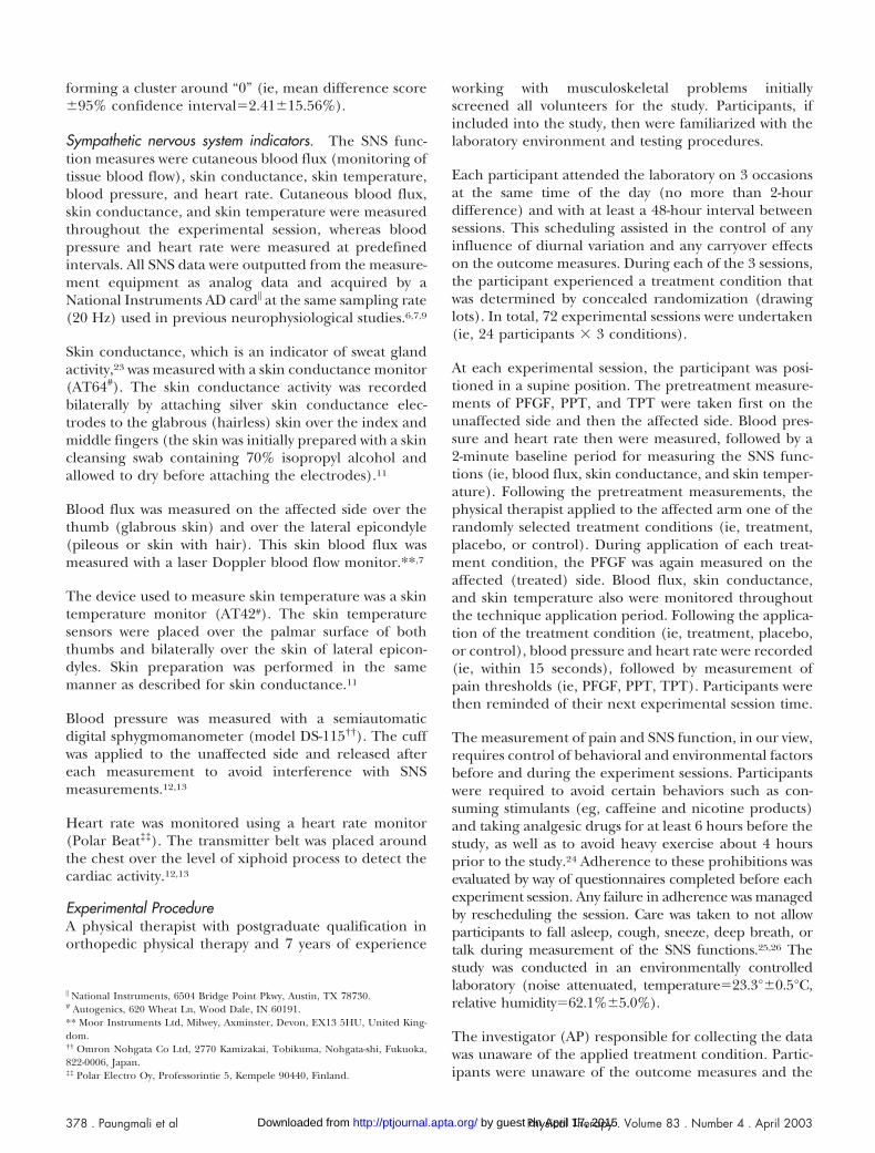
forming a cluster around “0” (ie, mean difference score�95% confidence interval�2.41�15.56%).
Sympathetic nervous system indicators. The SNS func-tion measures were cutaneous blood flux (monitoring oftissue blood flow), skin conductance, skin temperature,blood pressure, and heart rate. Cutaneous blood flux,skin conductance, and skin temperature were measuredthroughout the experimental session, whereas bloodpressure and heart rate were measured at predefinedintervals. All SNS data were outputted from the measure-ment equipment as analog data and acquired by aNational Instruments AD card� at the same sampling rate(20 Hz) used in previous neurophysiological studies.6,7,9
Skin conductance, which is an indicator of sweat glandactivity,23 was measured with a skin conductance monitor(AT64#). The skin conductance activity was recordedbilaterally by attaching silver skin conductance elec-trodes to the glabrous (hairless) skin over the index andmiddle fingers (the skin was initially prepared with a skincleansing swab containing 70% isopropyl alcohol andallowed to dry before attaching the electrodes).11
Blood flux was measured on the affected side over thethumb (glabrous skin) and over the lateral epicondyle(pileous or skin with hair). This skin blood flux wasmeasured with a laser Doppler blood flow monitor.**,7
The device used to measure skin temperature was a skintemperature monitor (AT42#). The skin temperaturesensors were placed over the palmar surface of boththumbs and bilaterally over the skin of lateral epicon-dyles. Skin preparation was performed in the samemanner as described for skin conductance.11
Blood pressure was measured with a semiautomaticdigital sphygmomanometer (model DS-115††). The cuffwas applied to the unaffected side and released aftereach measurement to avoid interference with SNSmeasurements.12,13
Heart rate was monitored using a heart rate monitor(Polar Beat‡‡). The transmitter belt was placed aroundthe chest over the level of xiphoid process to detect thecardiac activity.12,13
Experimental ProcedureA physical therapist with postgraduate qualification inorthopedic physical therapy and 7 years of experience
working with musculoskeletal problems initiallyscreened all volunteers for the study. Participants, ifincluded into the study, then were familiarized with thelaboratory environment and testing procedures.
Each participant attended the laboratory on 3 occasionsat the same time of the day (no more than 2-hourdifference) and with at least a 48-hour interval betweensessions. This scheduling assisted in the control of anyinfluence of diurnal variation and any carryover effectson the outcome measures. During each of the 3 sessions,the participant experienced a treatment condition thatwas determined by concealed randomization (drawinglots). In total, 72 experimental sessions were undertaken(ie, 24 participants � 3 conditions).
At each experimental session, the participant was posi-tioned in a supine position. The pretreatment measure-ments of PFGF, PPT, and TPT were taken first on theunaffected side and then the affected side. Blood pres-sure and heart rate then were measured, followed by a2-minute baseline period for measuring the SNS func-tions (ie, blood flux, skin conductance, and skin temper-ature). Following the pretreatment measurements, thephysical therapist applied to the affected arm one of therandomly selected treatment conditions (ie, treatment,placebo, or control). During application of each treat-ment condition, the PFGF was again measured on theaffected (treated) side. Blood flux, skin conductance,and skin temperature also were monitored throughoutthe technique application period. Following the applica-tion of the treatment condition (ie, treatment, placebo,or control), blood pressure and heart rate were recorded(ie, within 15 seconds), followed by measurement ofpain thresholds (ie, PFGF, PPT, TPT). Participants werethen reminded of their next experimental session time.
The measurement of pain and SNS function, in our view,requires control of behavioral and environmental factorsbefore and during the experiment sessions. Participantswere required to avoid certain behaviors such as con-suming stimulants (eg, caffeine and nicotine products)and taking analgesic drugs for at least 6 hours before thestudy, as well as to avoid heavy exercise about 4 hoursprior to the study.24 Adherence to these prohibitions wasevaluated by way of questionnaires completed before eachexperiment session. Any failure in adherence was managedby rescheduling the session. Care was taken to not allowparticipants to fall asleep, cough, sneeze, deep breath, ortalk during measurement of the SNS functions.25,26 Thestudy was conducted in an environmentally controlledlaboratory (noise attenuated, temperature�23.3°�0.5°C,relative humidity�62.1%�5.0%).
The investigator (AP) responsible for collecting the datawas unaware of the applied treatment condition. Partic-ipants were unaware of the outcome measures and the
� National Instruments, 6504 Bridge Point Pkwy, Austin, TX 78730.# Autogenics, 620 Wheat Ln, Wood Dale, IN 60191.** Moor Instruments Ltd, Milwey, Axminster, Devon, EX13 5HU, United King-dom.†† Omron Nohgata Co Ltd, 2770 Kamizakai, Tobikuma, Nohgata-shi, Fukuoka,822-0006, Japan.‡‡ Polar Electro Oy, Professorintie 5, Kempele 90440, Finland.
378 . Paungmali et al Physical Therapy . Volume 83 . Number 4 . April 2003 by guest on April 17, 2015http://ptjournal.apta.org/Downloaded from

true intent of the study in that they were only madeaware of the ethical implications of the study withoutrevealing that a treatment was being evaluated.
Data Management and Analysis
Data management. All 24 participants completed thestudy, and their data were analyzed in the assignedconditions as allocated, using the SPSS statistical pack-age (version 10.0).§§ The triplicate measurements ofPFGF, PPT, and TPT were averaged prior to furtheranalysis. The single measurements of heart rate, systolicblood pressure, and diastolic blood pressure takenimmediately before and immediately after the applica-tion were used in the statistical analyses.
The indicator of sympathetic effect that was used in thisstudy, as in previous studies, was the maximum effect,which was the maximum increase or decrease of SNSresponse based on the relative direction of the response.7,10
The maximum value for each measure was derived fromthe acquired data by the data acquisition program.
Statistical procedures. A 2-way, within-subjects analysisof variance (ANOVA) was used to evaluate the differ-ences in outcome measurements between pretreatmentand posttreatment measurement times (ie, independentvariable of time) for the 3 treatment conditions (ie,treatment, placebo, and control). Pain-free grip forcevaried from this model in that the time factor had 3levels: before, during, and after treatment.
Because skin conductance and skin temperature weremeasured on both upper limbs, an additional factor, side(affected, unaffected), was included in the ANOVA forthese sympathetic indicators, making it a 3-way, within-subjects ANOVA.
Tests of simple effects were used to further evaluate anysignificant 2-way or 3-way interaction effects.27 Thisinvolved post hoc analyses with the Tukey honestly signif-icant difference (HSD) test for PFGF, which requiredcomparisons among 3 measurement times (ie, before,during, and after treatment). Paired t tests with Bonfer-roni corrections for type I error rate were used formultiple comparisons of pretreatment and posttreat-ment data. For example, the alpha level was set at .017for 3 pair-wise comparisons (pretreatment and posttreat-ment comparisons for treatment, placebo, and control),and the alpha level was set at .0083 for the 6 pair-wisecomparisons of measures of SNS function.
To evaluate the assumption of repeated-measures designthat each individual participant was the same betweenlevels of the primary factor being evaluated (ie, treat-
ment, placebo, and control), we conducted 2 ANOVAs,both on the pretreatment data, with one ANOVA com-paring differences among conditions (ie, treatment,placebo, and control) and the other ANOVA comparingdifferences among days (ie, days 1, 2, and 3).
ReliabilityCommensurate with the primary aims of the study and inthe context of the study design, the reliability of theoutcome measures was evaluated from repeated mea-surements taken on the same day before the applicationof the experimental conditions and with only one inves-tigator taking all measurements from all participants inthis study. Intraclass correlation coefficients (ICCs) andstandard errors of measurement (SEMs) were used asindicators of reliability and error, respectively. The ICCand SEM for PFGF were .95 and 2.5 N, respectively.Pressure pain threshold had an ICC of .90 and a SEM of9.4 kPa, and TPT had an ICC and SEM of .85 and 0.39°C,respectively. The ICCs for heart rate, systolic bloodpressure, and diastolic blood pressure were reasonableat .68, .84, and .74, respectively, whereas the SEMs werelow at 2.5 beats per minute (bpm), 1.6 mm Hg, and 1.8mm Hg, respectively. Blood flux, skin conductance, andtemperature were stable across the pretreatment period,with very high ICCs (.98, .88, .99, respectively) and smallSEMs (0.008 flux units/min, 0.011 �siemens, and0.0002°C, respectively).
ResultsThe results of the within-subjects ANOVA and the meansand standard deviations for all data are presented inTables 1 through 5. Interaction plots are presented inFigures 2 through 5. The pretreatment data for alloutcome measures were not different between condi-tions (treat, placebo, or control) or between experimen-tal session days (days 1, 2, and 3).
Pain Measures
Pain-free grip force. The PFGF increased from 127.1 Nto 166.2 N during the treatment and further increased to174.1 N immediately after the treatment (Tab. 2). Theseincreases in PFGF represented approximate meanincreases of 37.0% and 47.5%, respectively (calculatedon a per individual basis). The change in placebo andcontrol was negligible both during and after their appli-cation. The ANOVA showed that there was a condi-tion � time interaction effect for PFGF (Tab. 1, Fig. 2).Post hoc pair-wise comparisons with the Tukey HSD testrevealed that PFGF increased during and after theapplication of the MWM treatment technique, but notduring or following the placebo or control (Tab. 2).
Pressure pain threshold. Pressure pain thresholdchanged from 281.4 kPa to 300.8 kPa following the
§§ SPSS Inc, 233 S Wacker Dr, Chicago, IL 60606.
Physical Therapy . Volume 83 . Number 4 . April 2003 Paungmali et al . 379
������
������
������
������
����
by guest on April 17, 2015http://ptjournal.apta.org/Downloaded from

treatment application, whereas PPT did not change inthe placebo condition and decreased in the control condi-tion (Tab. 3). The ANOVA showed a condition (ie, treat-ment, placebo, and control) � time (ie, before and aftertreatment) interaction effect for PPT (Tab. 1, Fig. 2).There was no mean percentage increase in PPT (calculatedon a per individual basis) following the MWM treatmentcompared with the placebo and control conditions.
Thermal pain threshold. Thermal pain threshold didnot change following the treatment application or in theplacebo condition; however, there was a reduction inTPT in the control condition (Tabs. 1 and 3). There wasa mean percentage reduction of 4.1% for the controlcondition as compared with an increase of 0.8% and areduction of 1.1% for the treatment and placebo condi-tions, respectively.
Sympathetic Nervous System-Related Measures
Heart rate and blood pressure. The treatment tech-nique produced an increase in heart rate from 66.3 bpmto 69.0 bpm, as well increases in systolic blood pressurefrom 117.9 mm Hg to 122.1 mm Hg and in diastolicblood pressure from 77.6 mm Hg to 80.1 mm Hg(Tab. 4). These mean increases were approximately4.1% for heart rate, 3.5% for systolic blood pressure, and3.1% for diastolic blood pressure. There were no placebo-or control-induced changes (Tabs. 1 and 4, Fig. 3).
Skin conductance, cutaneous blood flux, and temperature.On the affected side, the cutaneous SNS functions(ie, skin temperature, cutaneous blood flux, and skinconductance) were all activated (sympathoexcitation) inthe treatment condition, but not in the placebo orcontrol condition (Tabs. 1 and 5, Figs. 4 and 5). Thetreatment technique induced reductions in hand skintemperature (�1.1%) and hand blood flux (�72.4%)and a increases in skin conductance (55.0%), elbow skintemperature (2.1%), and elbow blood flux (123.7%).
Discussion and ConclusionsOur study showed that MWM for chronic lateral epicon-dylalgia is capable of producing concurrent hypoalgesiceffects during and following its application, as well asaltering SNS function. These findings are consistent withthose of previous studies of spinal manipulation,6,7
which implies to us that there is a multisystem responseto manipulation regardless whether the spine or theelbow is manipulated.
In contrast to the apparent similarity in multisystemresponse produced by manipulation applied to the spineand the elbow, the MWM-induced response profile inthe pain system appears different from that shown inprevious studies of manipulation of the cervical spine for
Table
1.
Resu
ltsof
the
With
in-S
ubje
cts
Ana
lysi
sof
Varia
nce
(Inte
ract
ion
Effe
ctBe
twee
nTr
eatm
entC
ondi
tions
and
Tim
e)fo
rA
llO
utco
me
Mea
sure
s
Outc
om
eM
easu
resa
Trea
tmen
tPla
cebo
Contr
ol
Pb
Bef
ore
Duri
ng
Aft
erBef
ore
Duri
ng
Aft
erBef
ore
Duri
ng
Aft
er
XSD
XSD
XSD
XSD
XSD
XSD
XSD
XSD
XSD
Pain
-rela
ted
mea
sure
sPa
in-fr
eegr
ipfo
rce
127.
155
.916
6.2
56.7
174.
153
.114
6.8
74.3
145.
073
.714
4.9
70.9
150.
473
.414
6.5
69.1
143.
965
.6.0
01Pr
essu
repa
inth
resh
old
281.
414
2.6
300.
811
5.2
280.
812
3.0
280.
313
3.7
305.
513
5.3
279.
912
5.7
.01
Ther
mal
pain
thre
shol
d43
.83.
644
.13.
844
.43.
543
.93.
644
.53.
242
.73.
4.0
1
SNS-
rela
ted
mea
sure
sH
eart
rate
66.3
7.6
69.0
8.2
66.8
9.4
65.8
8.8
65.8
9.4
65.2
9.2
.001
Systo
licbl
ood
pres
sure
117.
910
.012
2.1
12.7
118.
810
.611
9.0
11.1
120.
511
.511
9.7
12.2
.001
Dia
stolic
bloo
dpr
essu
re77
.66.
680
.18.
077
.58.
377
.77.
677
.58.
276
.77.
9.0
1H
and
skin
tem
pera
ture
31.6
1.4
31.2
1.4
31.3
1.8
31.3
1.8
30.5
1.8
30.7
1.7
.001
Elbo
wsk
inte
mpe
ratu
re32
.40.
833
.20.
832
.40.
932
.80.
832
.61.
932
.81.
8.0
01Sk
inco
nduc
tanc
e1.
91.
03.
01.
52.
22.
12.
72.
61.
81.
22.
31.
7.0
02H
and
bloo
dflu
x22
7.9
125.
858
.062
.816
5.9
117.
078
.559
.116
4.8
89.7
78.8
47.0
.001
Elbo
wbl
ood
flux
49.9
35.6
108.
152
.449
.553
.075
.452
.057
.043
.377
.143
.6.0
01
aPa
in-fr
eegr
ipfo
rce
(in
new
ton
s),
pres
sure
pain
thre
shol
d(i
nki
lopa
scal
s),
ther
mal
pain
thre
shol
d(i
nde
gree
sC
elsi
us),
hea
rtra
te(i
nbe
ats
per
min
ute)
,bl
ood
pres
sure
(in
mill
imet
ers
ofm
ercu
ry),
skin
tem
pera
ture
(in
degr
ees
Cel
sius
),bl
ood
flux
(in
flux
unit
spe
rm
inut
e),
skin
con
duct
ance
(in
mic
rosi
emen
s).
bD
egre
esof
free
dom
�2,
46,
exce
ptfo
rpa
in-fr
eegr
ipfo
rce
(df�
4,92
).
380 . Paungmali et al Physical Therapy . Volume 83 . Number 4 . April 2003 by guest on April 17, 2015http://ptjournal.apta.org/Downloaded from
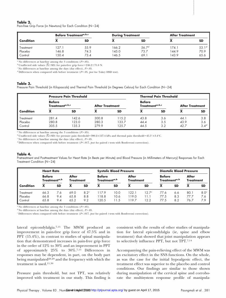
lateral epicondylalgia.7,15 The MWM produced animprovement in pain-free grip force of 47.5% and inPPT (15.4%), in contrast to studies of spinal manipula-tion that demonstrated increases in pain-free grip forcein the order of 12% to 30% and an improvement in PPTof approximately 25% to 30%.7,15 Differences inresponses may be dependent, in part, on the body partbeing manipulated28,29 and the frequency with which thetreatment is used.11,30
Pressure pain threshold, but not TPT, was relativelyimproved with treatment in our study. This finding is
consistent with the results of other studies of manipula-tion for lateral epicondylalgia (ie, spine and elbowtreatment) that showed that joint manipulation appearsto selectively influence PPT, but not TPT.7,14
Accompanying the pain-relieving effect of the MWM wasan excitatory effect in the SNS functions. On the whole,as was the case for the initial hypoalgesic effect, thetreatment effect was superior to the placebo and controlconditions. Our findings are similar to those shownduring manipulation of the cervical spine and corrobo-rate the multisystem response profile of manipula-
Table 2.Pain-free Grip Force (in Newtons) for Each Condition (N�24)
Condition
Before Treatmenta,b,c During Treatment After Treatment
X SD X SD X SD
Treatment 127.1 55.9 166.2 56.7d 174.1 53.1d
Placebo 146.8 74.3 145.0 73.7 144.9 70.9Control 150.4 73.4 146.5 69.1 143.9 65.6
a No differences at baseline among the 3 conditions (P�.05).b Unaffected side values (X�SD) for pain-free grip force�246.2�71.0 N.c No differences at baseline among the days (day effect), P�.05.d Differences when compared with before treatment (P�.05, post hoc Tukey HSD test).
Table 3.Pressure Pain Threshold (in Kilopascals) and Thermal Pain Threshold (in Degrees Celsius) for Each Condition (N�24)
Condition
Pressure Pain Threshold Thermal Pain Threshold
BeforeTreatmenta,b,c After Treatment
BeforeTreatmenta,b,c After Treatment
X SD X SD X SD X SD
Treatment 281.4 142.6 300.8 115.2 43.8 3.6 44.1 3.8Placebo 280.8 123.0 280.3 133.7 44.4 3.5 43.9 3.6Control 305.5 135.3 279.9 125.7 44.5 3.2 42.7 3.4d
a No differences at baseline among the 3 conditions (P�.05).b Unaffected side values (X�SD) for pressure pain threshold�390.2�127.4 kPa and thermal pain threshold�45.5°�3.4°C.c No differences at baseline among the days (day effect), P�.05.d Differences when compared with before treatment (P�.017, post hoc paired t tests with Bonferroni correction).
Table 4.Pretreatment and Posttreatment Values for Heart Rate (in Beats per Minute) and Blood Pressure (in Millimeters of Mercury) Responses for EachTreatment Condition (N�24)
Condition
Heart Rate Systolic Blood Pressure Diastolic Blood Pressure
BeforeTreatmenta,b
AfterTreatment
BeforeTreatmenta,b
AfterTreatment
BeforeTreatmenta,b
AfterTreatment
X SD X SD X SD X SD X SD X SD
Treatment 66.3 7.6 69.0 8.2c 117.9 10.0 122.1 12.7c 77.6 6.6 80.1 8.0c
Placebo 66.8 9.4 65.8 8.8 118.8 10.6 119.0 11.1 77.5 8.3 77.7 7.6Control 65.8 9.4 65.2 9.2 120.5 11.5 119.7 12.2 77.5 8.2 76.7 7.9
a No differences at baseline among the 3 conditions (P�.05).b No differences at baseline among the days (day effect), P�.05.c Differences when compared with before treatment (P�.017, post hoc paired t tests with Bonferroni correction).
Physical Therapy . Volume 83 . Number 4 . April 2003 Paungmali et al . 381
������
������
������
������
����
by guest on April 17, 2015http://ptjournal.apta.org/Downloaded from

tion.6,7,15 Sato et al31 studied cats and showed thatsustained end-range positions of the knee (eg, rotation)altered heart rate and blood pressure and that decorti-cation attenuates this stimulation-induced sympatho-excitatory effect. The manipulation of the cat knee in that
study, we contend, is somewhat similar to the MWMbecause both involved application of a manually inducedsustained pressure across a joint. On the basis of thepattern of response in the SNS in our study and evidencefrom animal studies about the control mechanisms of theseSNS functions, it appears likely that, as a part of itsresponse, the MWM may activate some centers in theneuraxis.
An important finding of our study is that the effects onexperimentally induced pain and the SNS were pro-duced by the MWM treatment technique, but not by theplacebo or control conditions. This finding, in our view,confirms that any treatment effects cannot easily beexplained as placebo effects or the natural history of acondition. The purpose of our study was to examinewhether physiological effects similar to those seen withspinal manipulation occurred with MWM, and they didoccur. We were not attempting to determine whetherMWM provided a beneficial clinical outcome as a treat-ment (eg, improved function, permanently reduceddisability or impairments).
Table 5.Hand and Elbow Skin Temperature (in Degrees Celsius), Hand andElbow Blood Flux (in Flux Units per Minute), and Skin Conductance(in Microsiemens) on the Affected (Treated) Side for TreatmentCondition (N�24)
Outcome Measures
BeforeTreatment
DuringTreatmenta
X SD X SD
Hand skin temperature 31.6 1.4 31.2 1.4Elbow skin temperature 32.4 0.8 33.2 0.8Skin conductance 1.9 1.0 3.0 1.5Hand blood flux 227.9 125.8 58.0 62.8Elbow blood flux 49.9 35.6 108.1 52.4
a Differences when compared with before treatment (P�.0083, post hoc pairedt tests with Bonferroni correction).
Figure 2.Plot of interaction between the treatment condition (treatment, placebo,control) and time for pain-free grip force (PFGF) (in newtons) over 3measurement times (before, during, and after treatment) and for pressurepain threshold (PPT) (in kilopascals) from before treatment to after treatment.
Figure 3.Plot of interaction between the treatment condition (treatment, placebo,control) and time (before and after application of the experimentalconditions) for heart rate (HR) (in beats per minute) and for systolic anddiastolic blood pressure (SBP and DBP) (in millimeters of mercury).
Figure 4.Plot of interaction between the treatment condition (treatment, placebo,control) and time (before and during application of the experimentalconditions) for skin conductance (SC) (in microsiemens) and for handand elbow skin temperature (HST and EST) (in degrees Celsius).
Figure 5.Plot of interaction between the treatment condition (treatment, placebo,control) and time (before and during application of the experimentalconditions) for hand and elbow blood flux (HBF and EBF) (in flux unitsper minute).
382 . Paungmali et al Physical Therapy . Volume 83 . Number 4 . April 2003 by guest on April 17, 2015http://ptjournal.apta.org/Downloaded from
The magnitude of changes of some sympathetic nervoussystem-related measures was relatively small. For exam-ple, heart rate, blood pressure, and cutaneous skintemperatures were induced changes of less than 5%;therefore, caution must be exercised when interpretingthese findings as to the clinical significance.
We did not address the possible involvement of localeffects at the elbow as a possible explanation of themechanism of action of the MWM technique beingevaluated. Local effects, such as possible changes in therelationships of bones and soft tissues about the jointand possible changes in local neural receptors in softtissues at the time of treatment, may account for thechanges we measured.4,5
In considering the mechanism of action we measuredfor MWM, the reader should consider that our data showthat there was a change in pain and SNS function duringthe application of the treatment technique and thatthere were changes in blood pressure, heart rate, vaso-motor function, and sudomotor function. These changescannot be fully explained by local mechanical effects.
References1 Mulligan B. Manual Therapy “NAGS,” “SNAGS,” “MWMSs,” etc. 3rd ed.Wellington, New Zealand: Plane View Service; 1995:78–88.
2 Abbott J, Patla C, Jensen R. The initial effects of an elbow mobilisa-tion with movement technique on grip strength in subjects with lateralepicondylalgia. Man Ther. 2001;6:163–169.
3 Vicenzino B, Wright A. Effects of a novel manipulative physiotherapytechnique on tennis elbow: a single case study. Man Ther. 1995;1:30–35.
4 Hsieh C, Vicenzino B, Yang CH, et al. Mulligan’s mobilisation withmovement for the thumb: a single case report using magnetic reso-nance imaging to evaluate the positional fault hypothesis. Man Ther.2002;7:44–49.
5 Kavanagh J. Is there a positional fault at the inferior tibiofibularjoints in patients with acute or chronic ankle sprains compared tonormals? Man Ther. 1999;4:19–24.
6 Sterling M, Jull G, Wright A. Cervical mobilisation: concurrent effectson pain, sympathetic nervous system activity and motor activity. ManTher. 2001;6:72–81.
7 Vicenzino B, Collins D, Benson H, Wright A. An investigation of theinterrelationship between manipulative therapy-induced hypoalgesiaand sympathoexcitation. J Manipulative Physiol Ther. 1998;21:448–453.
8 Zusman M, Edwards B, Donaghy A. Investigation of a proposedmechanism for the relief of spinal pain with passive joint movement.J Manual Med. 1989;4:58–61.
9 Petersen N, Vicenzino B, Wright A. The effects of a cervical mobil-isation technique on sympathetic outflow to the upper limb in normalsubjects. Physiotherapy Theory and Practice. 1993;9:149–156.
10 Vicenzino B, Collins D, Wright A. Sudomotor changes induced byneural mobilisation techniques in asymptomatic subjects. J ManualManipulative Ther. 1994;2:66–74.
11 Chiu T, Wright A. To compare the effects of different rates ofapplication of a cervical mobilisation technique on sympathetic out-flow to the upper limb in normal subjects. Man Ther. 1996;1:198–203.
12 McGuiness J, Vicenzino B, Wright A. Influence of a cervical mobil-isation technique on respiratory and cardiovascular function. ManTher. 1997;2:216–220.
13 Vicenzino B, Cartwright T, Collins D, Wright A. Cardiovascular andrespiratory changes produced by lateral glide mobilisation of thecervical spine. Man Ther. 1998;3:67–71.
14 Vicenzino B, Paungmali A, Buratowski S, Wright A. Specific manip-ulative therapy treatment for chronic lateral epicondylalgia producesuniquely characteristic hypoalgesia. Man Ther. 2001;6:205–212.
15 Vicenzino B, Collins D, Wright A. The initial effects of a cervicalspine manipulative physiotherapy treatment on the pain and dysfunc-tion of lateral epicondylalgia. Pain. 1996;68:69–74.
16 Haker E. Lateral epicondylalgia: diagnosis, treatment, and evalua-tion. Critical Reviews in Physical Rehabilitation Medicine. 1993;5:129–154.
17 Stratford P, Levy D, Gowland C. Evaluative properties of measuresused to assess patients with lateral epicondylitis at the elbow. Physio-therapy Canada. 1993;45:160–164.
18 Smidt N, Van-der Windt D, Assendelft W, et al. Corticosteroidinjections, physiotherapy, or a wait-and-see policy for lateral epicondy-litis: a randomised control trial. Lancet. 2002;359:657–662.
19 Stratford P, Levy D, Gauldie S, Levy K, Miseferi D. Extensor carpiradialis tendonitis: a validation of selected outcome measures. Physio-therapy Canada. 1987;39:250–255.
20 Ohrbach R, Gale E. Pressure pain thresholds, clinical assessment,and differential diagnosis: reliability and validity in patients withmyogenic pain. Pain. 1989;39:157–169.
21 Park R, Wallace M, Schulteis G. Relative sensitivity to alfentanil andreliability of current perception threshold vs von Frey tactile stimula-tion and thermal sensory testing. J Peripher Nerv Syst. 2001;6:232–240.
22 Bland J, Altman D. Statistical methods for assessing agreementbetween two methods of clinical measurement. Lancet. 1986;1:307–310.
23 Freedman L, Scerbo A, Dawson M, et al. The relationship of sweatgland count to electrodermal activity. Psychophysiol. 1994;31:196–200.
24 Koltyn K. Analgesia following exercise. Sports Med. 2000;29:58–98.
25 Delius W, Hagbarth K, Hongell A, Wallin B. Manoeuvers affectingsympathetic outflow in human skin nerves. Acta Physiol Scand. 1972;84:177–186.
26 Hallin R, Torebjork H. Single unit sympathetic activity in humanskin nerves during rest and various manoeures. Acta Physiol Scand.1974;92:303–317.
27 Winer B, Brown D, Michels K. Statistical Principles in ExperimentalDesign. 3rd ed. New York, NY: McGraw-Hill Inc; 1991:326–333.
28 Harris W, Wagnon J. The effects of chiropractic adjustments ondistal skin temperature. J Manipulative Physiol Ther. 1987;10:57–60.
29 Nansel D, Peneff A, Quitoriano J. Effectiveness of upper versuslower cervical adjustments with respect to the amelioration of passiverotational versus lateral-flexion end-range asymmetries in otherwiseasymptomatic subjects. J Manipulative Physiol Ther. 1992;15:99–105.
30 Mierau D, Cassidy J, Bowen V, et al. Manipulation and mobilisationof the third metacarpophalangeal joint: a quantitative radiographicand range of motion study. Man Med. 1988;3:135–140.
31 Sato A, Sato Y, Schmidt R. Changes in blood pressure and heart rateinduced by movements of normal and inflamed knee joints. NeurosciLett. 1984;52:55–60.
Physical Therapy . Volume 83 . Number 4 . April 2003 Paungmali et al . 383
������
������
������
������
����
by guest on April 17, 2015http://ptjournal.apta.org/Downloaded from
2003; 83:374-383.PHYS THER. VicenzinoAatit Paungmali, Shaun O'Leary, Tina Souvlis and BillEpicondylalgiaMobilization With Movement for Lateral Hypoalgesic and Sympathoexcitatory Effects of
References
http://ptjournal.apta.org/content/83/4/374#BIBLfor free at: This article cites 26 articles, 0 of which you can access
Cited by
http://ptjournal.apta.org/content/83/4/374#otherarticles
This article has been cited by 6 HighWire-hosted articles:
Information Subscription http://ptjournal.apta.org/subscriptions/
Permissions and Reprints http://ptjournal.apta.org/site/misc/terms.xhtml
Information for Authors http://ptjournal.apta.org/site/misc/ifora.xhtml
by guest on April 17, 2015http://ptjournal.apta.org/Downloaded from


![MWM - èñòîðèÿ, àãðåãàòû, ïðîåêòû [Ðåæèì ñîâìåñòèìîñòè]ges-ukraine.com/files/MWM-2008.pdf · mwm, которые нужны для ремонтных](https://img.dokumen.tips/doc/110x75/5c420b1593f3c338cd7a6f0f/mwm-enoidey-aadaaaou-idiaeou-daaeei-niaianoeiinoeges-.jpg)


