Embed Size (px)
Citation preview

8/7/2019 Hydroxyapatite Column Chromatography in Procedures
http://slidepdf.com/reader/full/hydroxyapatite-column-chromatography-in-procedures 1/9
ANALYTICAL BIOCHEM ISTRY 59, 555-563 (1974)
Hydroxyapatite Column Chromatography in Proceduresfor Isolation of Purified DNA
Institute of Biochemistry, Bulgarian Acutlemy of Scierlc es,
13 Sofirc, Bulgaria
Received. August. 2 1, 1973; accepted Piovember 16, 1973
Two procedures are described for the isolation of purified DNA from
mamm alian tissue s hy using hydroxyapatite chromatography. They avoid
enzyme treatment and are eas ily carrird out in one day. The first one is
a mo difica tion of the MFP (8 M urea-O.24 M sod ium phosp hate buffer)
method of Britkn et al. (2) in which some techn ical diffic? llties (clogging
of the colum n) are overcome and the yield and the purity of the DNA are
improved. The second procedure represent s a comb inniion of the cla ssi cal
rn:thods of lys is of nu c*!r i with hydroxy:tpatite chromatography of the DNA
and may be espec ially convenient for the isolation of high-molecular-weightDXA.
Most of the currelIt methods for the isolation of purified DXA require
enzyme treatment with ribonuclease and pronase, combined with many-
fold deproteinization, precipitation and redissolving. Such methods last
several days and lead to considerable loss of DNA.
The hydroxyapatitc (HA) chromatography offers new possibilit’ies
for the isolation of purified DNA. Double-stranded DNA has a much
higher affinity to HA t.han R’NA, prot’cins, carbohydrates and various
low-molecular-weight substances (1 I. In principle, this would permit
the isolation of DNA, free of contaminants, simply by loading the tissue
lysate onto a HA column and then cluting by phosphate buffers of appro-
priate concentrations.
Recently, Britten et nl. (2) developed a method based on this principle
for the isolation of purified DIVA from both prokaryotic and eukaryotic
cells. In their method, the starting material (fresh tissue, tissue frozen
in liquid nitrogen or dry ice, or isolated nuclei) is lyscd in 8 M urea-O.24 M
sodium phosphate (Nap), pH 6.8 COUP) ,I containing 1% sodium dodecyl
’ Abb reviations used : MUP. 8 M urcaa-0.24 M sodiu m phosp hate buffer; NaP ,
sodiu m phosp hate buffer (an equim olar mixture of iSaH?P O, and Na2HPO I, pH 6.8) ;
HA, hydrosyapatite; EDT A, ethylenediaminetetraacctic acid, sodium salt; SSC,
0.15 M NaCl-0.015 M sodium citrate; SDS, sodium dodecyl sulphate.
555
Copyr$rt. @ 1974 13~ Acad emic Press:. Inc..
Al l rights of rrproduction in any form reserved.
8/7/2019 Hydroxyapatite Column Chromatography in Procedures
http://slidepdf.com/reader/full/hydroxyapatite-column-chromatography-in-procedures 2/9
sulphatc (SDS) and 0.01 RI EDTA. The lysate is loaded onto a HA
column, the column is washed with MUP, and urea is removed by washingwith 0.014 M KaP, pH 6.8. DNA is eluted with 0.40 &XINaP, pH 6.8. Sev-
eral tnodifications have been proposed to improve the method: lysis of
previously isolated nuclei instead of whole tissue (31 ; batch procedures
(2,3) ; treatment of the lysate with an equal volume of chloroform-octanol
(20: 1) before loading (2).
The m&hod is simple, fast, permits processing of small amounts of
tissue and gives a high yield of DNA. However, in our hands, this pro-
cedure shon-ed some shoitcomings and technical difficulties. The firstone is the clogging of the column which strongly decreases the flow rate
and makes it necessary to apply vacuum or pressure. This leads to
channeling and incomplete retention of DNA. The difliculties caused
by clogging become critical when large amounts of material are loaded.
Secondly, the lysing medium decreases the ability of HA to adsorb
DNA. This effect is more pronounced in the presence of proteins in the
lgsate.
Thirdly, even the extensive washing of the column with MUP maynot e1ut.e completely the proteins and other substances, which may con-
taminate DKA in the 0.40 M fraction.
Al l these disadvantages prompted us to study in more details the
factors affecting H-4 chromatography as applied to the isolation and
purification of DNA from mammalian tissues. As a result, we worked
out two technically convenient procedures, which gave best results in
respect to yield and purity of DNA.
MATERIALS
Hydroxyapatite was prepared according t.o Miyazawa and Thomas
(4) under very slow stirring to obtain large crystals (1). Hydroxyapatite
Bio Gel HTP (Bio-Rad Labs, Richmond, CA) was also used with
similar results. The cellulose powder 123~ was a product of Schleicher and
Schuell, West Germany. The hemoglobin, crystalized four times, was
purchased from Reanal, Hungary, and the bovine serum albumin fromKoch-Light Laboratories, England. In some experiments DNA was iso-
lated from tissue labelled in viva for proteins with [l%]valine (95 mCi/
mmole), production of the Institute for research, production and uses of
isotopes, Prague, Czechoslovakia, and for RNA with [YJ]orotic acid
(15 mCi/mmole), production of the NAEC, Institute for isotopes, Buda-
pest. Hungary. Al l other reagents and chemicals were of analytica
grade.
8/7/2019 Hydroxyapatite Column Chromatography in Procedures
http://slidepdf.com/reader/full/hydroxyapatite-column-chromatography-in-procedures 3/9
ISOLATION OF DNA BY HA CHROXATO!:RAI’HT :xJ 1
Proteins were determined by the LolT-ry proccdurc (5) and RNA by
the orcinol reactio:l (6). Radioactivity was measured in :L model 33’20
Tricarb Packard scintil lation spectrometer and absorbnnry in a Zeiss
spectrophotometer VSU 2. Melting curves were recorded in an rnicam
SP 1800 spectrophotometer.
lSOLATION PROCEDURES
Procehe I: Ml;P Procedure
Lysis. The lysis was performed as described by Brittcn et nl. (2) and
Hell et al. (3). The starting material was suspendecl in 10 vol of lysing
medium (8 M urea-O.24 M NaP, pH 6.8-0.01 &I EDTA-1% SDS) and
blended in a filled sealed container to avoid foaming, using a MSE
homogenizer run at 14000 rpm. For complete lysis the blending was re-
peated 4-6 times for 30 set each with intervals of 1 min to cool the
lysate in an ice bath.Preliminary Pwification of the Lysate. The lysate was mixed with an
equal volume of chloroform-isoamyl alcohol-phenol (24: 1: 25)) vigor-
ously shaken for 15 min at room temperature and centrifuged for 10 min
at 5000 rpm. The supernatant was removed, treated once more in the
same way and then extracted three times with ether to remove the
phenol. The crude DNA preparation thus obtained was passed through
a column of cellulose powder whose volume was about three times the
volume of the initial tissue. The cellulose n-as previously boiled forseveral minutes in distilled water, poured into the column and equili-
brated with MUP. In some cases the material retained on the cellulose
decreased considerably the flow rate, which made it necessary to appIy
a peristaltic pump. After passing the liquid, the column was washed with
two bed volumes of MUP. The eflluents were combined and loaded on
a HA column.
Hydrozyapatite Chromutog~aph~. The dry HA was suspended in
0.01 M NaP, pH 6.8, heated to boiling, poured into the column andequilibrated with MUP. One gram of dry HA was sufficient, for 1.5-2 mg
of DNA. To achieve an optimal flow rate (0.5 ml/min by grarity) the
height of the HA layer should be 3-5 cm. After loading, the column was
washed with MUP until no absorbance at 260 and 280 nm rould be
recorded. In practice this was achieved after passing about 59-100 ml
of MUP for every 2-5 g of tissue.
The urea was removed next by washing the column with 0.014 M Nap,
8/7/2019 Hydroxyapatite Column Chromatography in Procedures
http://slidepdf.com/reader/full/hydroxyapatite-column-chromatography-in-procedures 4/9
which was monitored by refractive index mtasurcmc~nt of the effluents.
DNA was eluted with 0.48 M NaP, pH 6.8 and collected in 5 ml fractions.”It was found that under these conditions a certain amount of DNA
was not adsorbed on the column. To recover this portion of DNA, the
Iysate should be passed through a second HA column with a bed volume
about half that of the first one.
Collection of DNA. To precipitate the total DXA, al l fractions eluted
with 0.48 M NaP were combined and dialyzed against distilled wat’er for
18 hr at 4°C. The solution was made 0.2 M in respect to NaCl by adding
solid NaCl and DNA was precipit,at,cd with 2 vol of ethanol at -20°Cfor 18 hr.
If necessary, the precipitation of DNA from fractions with very low
concentration (0.5-0.05 A,,,/ml) could be achieved using the procedure
of Dessev and Grantcharov (7) in the following modification: the frac-
tions were diluted with an equal volume of distilled water to reduce
the concentration of the NaP, thus preventing further precipitation of
the sodium phosphates by ethanol and 0.1 vol of 0.5 M CaCl, was added
dropwise under continuous stirring. A voluminous precipitate of calciumphosphate was formed. An equal volume of ethanol was then added, the
suspension was shaken and centrifuged for 5 min at 5000 rpm. The pellet
containing the whole amount of DYA was dissolved in 1 M EDTA (half
the volume of CaCl,), salts were removed by dialysis and DXA was pre-
cipitated with two vol of ethanol after adding solid XaCl to 0.2 M.
Procedure II: Lysis in NaCMDS-EDTA Solution
Isolation of Nuclei and Lysis. The tissue was homogenized in 10 volof ice-cold 0.14 M NaCl-0.02 M EDTA, pH 7.0, and nuclei were pelleted
by centrifugation for 6 min at 8009 at 4°C. The nuclear pellet was lysed
by adding 1 M NaCl-0.1 M EDTA-2% SDS, pH 8.0, final volume 50%
of that of the homogenate. The lysing medium was warmed to dissolve
SDS, then cooled to about 30°C and added to the pellet with vigorous
stirring. The lysis was conducted with shaking at 60°C for 10 min. The
sample was then cooled quickly in an ice bath.
Preliminary Purification. An equal volume of chloroform-isoamylalcohol (24: 1) was added and shaking continued at 4°C for 10 min.
After centrifugation for 5 min at 5000 rpm the aqueous phase was col-
lected, treated with an equal volume of water saturated phenol, pH 8.0
a Whe n working with large amou nt of materia l the flow rate of the 0.48 M NaP
effluent decreases considerably due to the viscosity of DNA. In such case s the
elution can be performed as a hatch procedure. Hpdrosyapatite with adsorbed DNA
is transferred into a beaker, susp ende d in 0.48 M p\‘aP and centrifuged. Th is is
repeated3-4 timesand the supernatantscontainingDNA are combined.
8/7/2019 Hydroxyapatite Column Chromatography in Procedures
http://slidepdf.com/reader/full/hydroxyapatite-column-chromatography-in-procedures 5/9
ISOL ATIO T\’ OF DSA BP HA CHROh3ATOGRAPHT 559
in the same way and then extracted three times with ether to eliminate
the phenol. EDTA was neutralized by adding 0.5 M CaCl, to equimolaramount and the solution was butiered at pH 6.8 by adding 0.48 M NaP
to a final concentration of 0.18 31. Before loading onto the H-4 column,
the solution was passed through n cellulose column as described in pro-
cedure I.
Hydrozyapatite Chro?natoyraphy. The HA column was prepared as
in procedure I except that the HA was equilibrated with 0.18 M NaP.
After loading, the column was washed with 0.18 IVI NaP until no absorb-
ance at 260 and 280 nm was recorded in the effluent. Al l procedures wereperformed at 4°C. The adsorbed DXA was then eluted with 0.48 M
NaP at room temperature and precipitated as in procedure I.
Other Isolation Procedures
For comparison, DSA was isolated also by the original method of
Britten et al. (2) and by a modified procedure of Marmur (8) including
additional deproteinization with phenol and treatment with ribonuclease
(100 p.g/ml) and pronase (200 kLg/ml).
RESULTS AND DISCUSSION
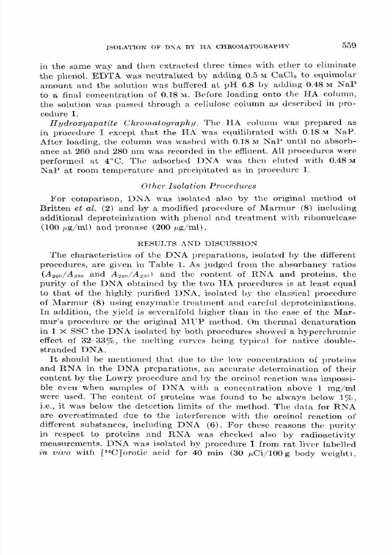
The characteristics of the DSA preparations, isolated by the different
procedures, are given in Table 1. As judged from the absorbancy ratios
(Az6”/Azg0 and A,,,/A,,,I and the content of RNA and proteins, the
purity of the DNA obtained by the two HA procedures is at least equal
to that of the highly purified DNA, isolated by the classical procedure
of ;19armur (8) using enzymatic treatment and rareful dcprotcinizations.In addition, the yield is severalfold higher than in the case of the Mar-
mur’s procedure or the original i\ll’P method. On thermal dcnaturation
in 1 X SW the DNA isolated by bot#hprocedures showed a hyperchromic
effect of 32-33%, t.he melting rurycs being t’ypicnl for native doublc-
stranded DNA.
It should be mentioned t,hat due to the low concentration of proteins
and RNA in the DNA preparations, an accurate determination of their
content by the Lowry procedure and by the orcinol reaction was impossi-ble even when samples of DNA with a concentration above I mg/ml
were used. The content of proteins was found to be always below 1%,
i.e., it was below the detection limits of the method. The data for RNA
are overestimated due to the interference with the orcinol reaction of
different substances, including DNA (6). For these reasons the purity
in respect to proteins and RXA was checked also by radioactivity
measurements. DNA was isolated by procedure I from rat liver labelled
in viva with [14C]orotic acid for 40 min (30 pCi/lOO g body weight).
8/7/2019 Hydroxyapatite Column Chromatography in Procedures
http://slidepdf.com/reader/full/hydroxyapatite-column-chromatography-in-procedures 6/9
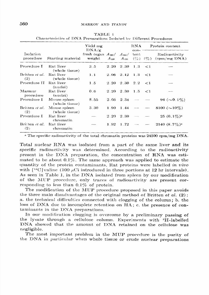
560 MARKOV AXD IVAXOV
TABLE 1
Characteristirs of TIN.4 Prevnrations Isolated bv Ijifferent Procedllres
Yield mg RNA Protein content
DNA/g con-
Isolat.ictn fresh organ A26nj A26,,/ tent. Radioactivity
procedrlre Starting mab erial weigh t -4 280 A130 (‘2) (%I,, (cpm/mgDNA)
Procedure I
Britten pf al.
(2)Procedure II
1llarmiir
procedure
Procedure I
Britten ct al.
(2)
Procedure I
Bri: t,en ct al.
(2,
Rat liver 3.5
(whole tissue)
Rat liver 1.1
(whole tissue)Rat liver 1.5
(nuclei)
Rat liver 0.6
(nuclei)
Moirse spleen 8.55
(whole tissue)
Mouse spleen 3.3c
(whole tissue)
Rat liver
chromatin1~at, liver -
chromatin
2.20 2.30 1.3 <1 -
2.06 2.12 1.3 <l -
2.20 2.30 2.2 <l -
2.20 2.30 1.5 <1 -
3.05 2.34 - - 94 (-o.lyo)
1.90 1.44 - - 8100 (-10%)
2.20 2.30 - - 25 (o.l%)a
1.92 1.72 - - 2140 (8.7%)”
a The spe cific radioactivity of the total chromatin proteins was 24500 cpm/mg DNA.
Total nuclear RNA was isolated from a part of the same liver and its
specific radioactivity was determined. According to the radioactivity
present in the DSA preparation, the concentration of RNA was esti-
mated to be about 0.1%. The same approach was applied to estimate thequantity of the protein rontaminants. Rat proteins were labelled in viva
with [W]valine (100 PCi introduced in three portions at 12 hr intervals).
As seen in Table 1, in the DNA isolated from spleen by our modification
of the MUP procedure, only traces of radioactivity are present cor-
responding to less than 0.1% of protein.
The modification of the MUP procedure proposed in this paper avoids
the three main disadvantages of the original method of Britten et al. (2) :
a. the technical difficulties connected with clogging of the column; b. theloss of DNA due to incomplete retention on HA; c. the presence of con-
taminants in the DNA preparations,
In our modifiration clogging is overcome by a preliminary passing of
the lysatc through a cellulose column. Experiments with 3H-labelled
DNA showed that the amount of DNA retained on the cellulose was
negligible.
The most important problem in the MUP procedure is tile purity of
the DNA in particular when whole tissue or crude nuclear preparations

8/7/2019 Hydroxyapatite Column Chromatography in Procedures
http://slidepdf.com/reader/full/hydroxyapatite-column-chromatography-in-procedures 7/9
8/7/2019 Hydroxyapatite Column Chromatography in Procedures
http://slidepdf.com/reader/full/hydroxyapatite-column-chromatography-in-procedures 8/9
562 ;MARI~ov AND 1vm0v
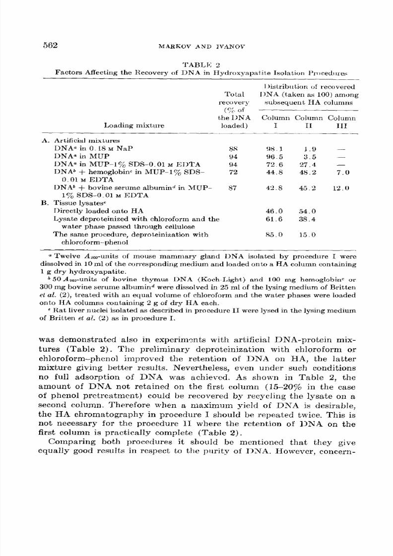
TABLK 2
Factors Affecting the Recovery of DNA in Hydroxyapatite Isolation Procedllres
Loading mixture
I )ist.ribut.ion of recovered
Total I)NA (taken as 100) among
recovery subsequent HA columns
(y;, of
t,he DNA Column Column Column
loaded) I II III-
A. Artificial mixtures
DNAa in 0.18 M NaPDNAa in MUP
__-.
DNAa in MUP-l’j$ SDS-O. 01 M EI>TA
DNA6 f hemoglobinc in MUP-170 SDS-
0.01 M EDTADNA* + bovine serume albumind in MUP-
1% SDS-O. 01 M EDTA
B. Tissue lysateseDirectly loaded onto HA
Lysate deproteinieed with chloroform and the
water phase passed through celluloseThe same procedure, deproteinization with
chloroform-phenol
8S 98.194 96.5
94 72.672 44.8
87 42.8
46.0
61.6
85.0
1.9 -3.5 -
27.4 -
48.2 7.0
45.2 12.0
54.0
38.4
15.0
u Twelve &,,-units of mouse mammary gland DNA isolated by procedure I were
dissolved in 10 ml of the corresponding medium and loaded onto a HA column containing
1 g dry hydroxyapatite.b 50A2~units of bovine thymus DNA (Koch-Light) and 100 mg hemoglobinc or
300 mg bovine serume albumind were dissolved in 25 ml of the lysing medium of Britten
et al. (2), treated with an equal volume of chloroform and the water phases were loaded
onto HA columns containing 2 g of dry HA each.c Rat liver nuclei isolated as described in procedure II were lysed in the lysing medium
of Britten et al. (2) as in procedure I.
was demonstrated also in experiments with artificial DNA-protein mix-
tures (Table 2). The preliminary deproteinization with chloroform or
chloroform-phenol improved the retention of DNA on HA, the latter
mixture giving better results. Nevertheless, even under such conditions
no full adsorption of DNA was achieved. As shown in Table 2, theamount of DNA not retained on the first column (15-2074 in the case
of phenol pretreatment) could be recovered by recycling the lysate on a
second column. Therefore when a maximum yield of DXA is desirable,
the HA chromatography in procedure I should be repeated twice. This is
not necessary for the procedure II where the retention of DNA on the
first column is practicaIly compIete (Table 2).
Comparing both procedures it should be mentioned that they give
equally good results in respect to the purity of DNA. However, concern-
8/7/2019 Hydroxyapatite Column Chromatography in Procedures
http://slidepdf.com/reader/full/hydroxyapatite-column-chromatography-in-procedures 9/9
ISOLATIO N OF DK.4 BY HA CHROMATOGRAPHY 563
ing other characteristics, there are some important differences which
should be taken into consideration. Procedure I has some technicaladvantages. It is carried out at room tempcraturc (except the blending).
Whole tissue (fresh or preliminary frozen) can bc used as starting ma-
terial, which improves the yield (up to 807% of the DNA). These ad-
vantages may be of importance especially when labelled DNA should
be isolated from a small amount of tissue or from tissues where the pre-
liminary isolation of nuclei is difficult. However, it should be emphasized
that the DNA obtained by this procedure is slightly sheared due to the
blending, so that it can not be used in experiments where high-molecular-weight DNA is needed. In such cases procedure II is preferable. Both
procedures are easily carried out in one day.
REFERENCES
1. BER XAR DI, G. (1971) in Methods in Enzymology (Colowick, S. P., and Kap lan,
N. O., eds.), Vol. 21, p. 95, Acad emic Press, New York.
2. BRITT EN, R. J., PAVICH, M., .&ND SMITH, J. (1969) Ca?xegie Inst. Wash ington
Yea&. 68, 400.
3. HEL L, A., BIRNIE , G. D., SLIMMIKG. T. K., AND PAU L, J. (1972) Ana l. Bioc hem .
48, 369.
4. MIYAZAWA, Y., AND THOMAS, C. A. (1965) 1. Mol. Bio l. 11, 223.
5. LOWRY, 0. H. (1857) in Methods in Enzymology (Colowick, S. P., and Kaplan,N. O., eds.). Vol. 4, p. 366, Acad emic Press, New York.
6. SCHN EIDER, W. C. (1957) in Methods in Enzymology (Colowick, S. P., and &plan,N. O., eds.), Vol. 3 , p. 680, Acad emic Press, New York.
7. DES SEV, G. N., AND GR.ANCHAROV, K. (1973) Anal. &o&em . 53, 269.
8. MARMUR, J. (lQ63) in. Methods in Enzymology (Colowick, S. P., and Kaplan, N. O.,eds.), Vol. 6, p. 726, Acad emic Press, New York.
Q. OIsHI, M. (1971) in Methods in Enzymology (Colowick, S. P., and Kaplan, N. O.,eds.), Vo l. 21, p. 140, Acad emic Press, New York.