Embed Size (px)
Citation preview
JOURNAL OF VIROLOGY, May 2011, p. 4386–4398 Vol. 85, No. 90022-538X/11/$12.00 doi:10.1128/JVI.01492-10Copyright © 2011, American Society for Microbiology. All Rights Reserved.
Hydrolyzable Tannins (Chebulagic Acid and Punicalagin) Target ViralGlycoprotein-Glycosaminoglycan Interactions To Inhibit Herpes
Simplex Virus 1 Entry and Cell-to-Cell Spread�
Liang-Tzung Lin,1† Ting-Ying Chen,2† Chueh-Yao Chung,3 Ryan S. Noyce,1 T. Bruce Grindley,4Craig McCormick,1 Ta-Chen Lin,5 Guey-Horng Wang,6
Chun-Ching Lin,2,3* and Christopher D. Richardson1*Department of Microbiology and Immunology, Dalhousie University, Halifax, Nova Scotia, Canada1; School of Pharmacy, College ofPharmacy, Kaohsiung Medical University, Kaohsiung, Taiwan2; Graduate Institute of Natural Products, College of Pharmacy,
Kaohsiung Medical University, Kaohsiung, Taiwan3; Department of Chemistry, Dalhousie University, Halifax, Nova Scotia, Canada4;Graduate Institute of Pharmaceutical Science and Technology, Central Taiwan University of Sciences and Technology, Taichung,
Taiwan5; and Department of Cosmetic Science, Chia Nan University of Pharmacy and Science, Tainan, Taiwan6
Received 16 July 2010/Accepted 31 January 2011
Herpes simplex virus 1 (HSV-1) is a common human pathogen that causes lifelong latent infection of sensoryneurons. Non-nucleoside inhibitors that can limit HSV-1 recurrence are particularly useful in treating immu-nocompromised individuals or cases of emerging acyclovir-resistant strains of herpesvirus. We report thatchebulagic acid (CHLA) and punicalagin (PUG), two hydrolyzable tannins isolated from the dried fruits ofTerminalia chebula Retz. (Combretaceae), inhibit HSV-1 entry at noncytotoxic doses in A549 human lung cells.Experiments revealed that both tannins targeted and inactivated HSV-1 viral particles and could preventbinding, penetration, and cell-to-cell spread, as well as secondary infection. The antiviral effect from either ofthe tannins was not associated with induction of type I interferon-mediated responses, nor was pretreatmentof the host cell protective against HSV-1. Their inhibitory activities targeted HSV-1 glycoproteins since bothnatural compounds were able to block polykaryocyte formation mediated by expression of recombinant viralglycoproteins involved in attachment and membrane fusion. Our results indicated that CHLA and PUGblocked interactions between cell surface glycosaminoglycans and HSV-1 glycoproteins. Furthermore, theantiviral activities from the two tannins were significantly diminished in mutant cell lines unable to produceheparan sulfate and chondroitin sulfate and could be rescued upon reconstitution of heparan sulfate biosyn-thesis. We suggest that the hydrolyzable tannins CHLA and PUG may be useful as competitors for glycos-aminoglycans in the management of HSV-1 infections and that they may help reduce the risk for developmentof viral drug resistance during therapy with nucleoside analogues.
Herpes simplex virus 1 (HSV-1) is an alphaherpesvirus thattypically causes mucocutaneous lesions in oral, perioral, andother mucosal sites in the body (58). The virus commonly usesthe oropharyngeal mucosa as a port of entry, and after primaryinfection, establishes a lifelong latent state in the host’s trigem-inal ganglia sensory neurons. Sporadic recurring infections oc-cur when HSV-1 is reactivated by various stimuli, such assunlight, immunosuppression, menstruation, fever, or stress(23). Although primary or reactivated HSV-1 infections can besubclinical or manifested by mild and self-limited diseases,severe cases of this viral infection may lead to complicationssuch as keratoconjunctivitis, meningitis, and encephalitis (3, 5).Importantly, corneal HSV-1 infection can lead to stromal ker-atitis, which remains one of the leading causes of blindness in
developing countries (37). More aggressive diseases due toHSV-1 are common in immunocompromised individuals (3, 5,23). To date, no treatment has been identified that eradicatesor resolves latent infections by this ubiquitous pathogen.
HSV-1 viral entry into cells is initiated by interaction ofviral envelope glycoproteins (gB and gC) with host cell sur-face proteoglycans (PGs) conjugated to glycosaminoglycans(GAGs) containing heparan sulfate (HS) or chondroitin sul-fate (CS) moieties. These initial interactions are sufficient forviral adsorption but not viral entry (67). Subsequently, higheraffinity interaction of gD with its receptors including herpesvi-rus entry mediator (HVEM; a member of tumor necrosis fac-tor receptor family), nectin-1 and nectin-2 (two members ofthe immunoglobulin superfamily), and/or 3-O-sulfated HS,leads to fusion of the viral membrane with either the plasma orendosomal membranes of the cell through further interactionswith gB, gH, and gL (29, 57, 67). Initial interaction of HSV-1with GAGs ensures a highly efficient infection, but infection ofcells deficient in HS or CS can still be achieved via the high-affinity receptors, albeit at lower efficiency. After transport ofthe viral capsid to the nucleus, where the HSV-1 genome isreleased, viral products are then expressed in a sequential andcoordinated fashion and are divided into three groups of virus-specific proteins designated as immediate-early (�) (ICP0 and
* Corresponding author. Mailing address for C.-C. Lin: School ofPharmacy, College of Pharmacy, Kaohsiung Medical University, No.100 Shih-Chuan First Road, Kaohsiung 807, Taiwan. Phone: 886-7-3121101, ext. 2122. Fax: 886-7-3135215. E-mail: [email protected] address for C. D. Richardson: Department of Microbiologyand Immunology, Dalhousie University, Halifax, Nova Scotia B3H1X5, Canada. Phone: (902) 494-6876. Fax: (902) 494-5125. E-mail:[email protected].
† L.-T.-L. and T.-Y.C. contributed equally to this study.� Published ahead of print on 9 February 2011.
4386
on April 5, 2019 by guest
http://jvi.asm.org/
Dow
nloaded from
ICP4)-, early (�) (ICP8, UL42, and thymidine kinase)-, andlate (�) (gB, gC, gD, and gH)-phase proteins (73). Althoughcellular innate immunity is activated upon virus infection,HSV-1 can produce one or more proteins that counteract thehost antiviral response (46, 49, 50).
Most anti-herpes drugs target the viral DNA polymeraseand include nucleoside or pyrophosphate analogues. Acyclovir(ACV), a guanosine analogue, has been the most importantclinical drug for the prophylactic or therapeutic treatment ofHSV infections, and represents the gold standard for anti-HSVtherapy (4, 62). However, extensive use of this drug has led toclinical problems with the emergence of ACV-resistant virusstrains, particularly in immunocompromised patients, includ-ing those who have had transplantation surgery or have beeninfected by the human immunodeficiency virus (HIV) (9, 21,24, 48, 70, 79). Subsequent management of ACV-resistant pa-tients using a different class of DNA polymerase-targeting in-hibitor, such as foscarnet, has also been hindered by drugresistance (21, 59). There is a need to identify alternativeantiviral therapies with different modes of action to improvethe treatment and control of HSV infections, especially inimmunocompromised individuals.
Terminalia chebula Retz. (T. chebula), a member of theCombretaceae family, is a traditional medicinal plant that isnative to India and Southeast Asian countries. The dried rip-ened fruit of T. chebula (Fructus Chebulae), often referred toas “myrobalans,” contains antioxidants (15) and is commonlyused as a broad-spectrum medicinal agent for the treatment ofdysentery, asthma, cough, sore throat, bloody stools, and dis-eases of the heart and bladder (30). T. chebula is rich intannins, which are polyphenolic secondary metabolites foundin higher plants (27, 32, 36). Tannins are characterized by theirrelatively high molecular mass (500 to 20,000 Da) and theunique ability to form insoluble complexes with proteins, car-bohydrates, nucleic acids, or alkaloids (27, 55, 63). The hydro-lyzable class of tannins possesses structures that generally con-sist of gallic or ellagic acid esters conjugated to a sugar moiety(28, 36). These polyphenols have high affinity for proteins andpolysaccharides and are thought to be the major bioactivecompounds found in the leaves and the fruit of T. chebula.
Antiviral activities from hydrolyzable tannins are well doc-umented and are generally thought to target viral adsorption tothe host cell membrane (for HSV and HIV), as well as reversetranscriptase activity of HIV (reviewed in references 8 and 63).We have previously identified several tannins of various plantsources that exhibit potent antiviral activities against HSV-1and HSV-2. These include 1,3,4,6-tetra-O-galloyl-�-D-glucose(77), casuarinin (10), ent-epiafzelechin-(4�3R8)-epiafzelechin(17), excoecarianin (16), geraniin (77), hippomanin A (76),prodelphinidin B-2 3�-O-gallate (11), prodelphinidin B-2 3,3�-di-O-gallate (12), pterocarnin A (13), and putranjivain A (14).Studies from other laboratories have also reported a series oftannins and related compounds capable of inhibiting HSVinfections (25, 56, 64, 71, 72). These earlier reports providestrong precedent for our studies and suggest that the tanninsconstitute an excellent focus for antiviral discovery, particularlyin the field of HSV therapeutics.
Identification of multiple drugs that can act on differentphases of the viral life cycle can be particularly useful in man-aging HSV-1 infection or reactivation in either immunocom-
promised individuals or cases of ACV resistance. To pursuethis goal, we extended our previous studies and concentratedour efforts on four chemically defined hydrolyzable tannins(Fig. 1), including chebulagic acid (CHLA), chebulinic acid(CHLI), punicalagin (PUG), and punicalin (PUN), which arepresent in T. chebula (39, 40, 78). Although an effect againstHSV-1 has been previously reported for CHLA, the mecha-nism of its activity was not elucidated (71). In the present studywe report that two of the tannins tested, specifically CHLA andPUG, were found to be most effective against HSV-1. Detailedstudies into their inhibitory action revealed that both drugsspecifically target HSV-1 particles, block virus entry into thecell, inhibit cell-to-cell spread of the virus, and reduce second-ary infection from released virions. The antiviral mechanism isattributed to the binding of CHLA and PUG to viral glycopro-teins that interact with cell surface GAGs. Their ability toeffectively control viral entry and spread, underscore the po-tential of these two hydrolyzable tannins for treating HSV-1infection and/or recurrence.
MATERIALS AND METHODS
Chemicals and reagents. Dulbecco modified Eagle medium (DMEM) andfetal calf serum (FCS) were supplied by Wisent Scientific (St-Bruno, Quebec,Canada). Gentamicin and amphotericin B (Fungizone) were purchased fromGibco-Invitrogen (Carlsbad, CA). ACV (acycloguanosine) was obtained fromCalbiochem (EMD Biosciences, Darmstadt, Germany). Foscarnet (FOS; so-dium phosphonoformate tribasic hexahydrate), dimethyl sulfoxide (DMSO),and an in vitro toxicology assay kit (XTT-based) were purchased from Sigma(St. Louis, MO).
Test compounds. Fructus Chebulae and dried leaves from T. chebula werecommercially obtained from Uchida Wakanyaku Co. (Tokyo, Japan) and anherbal market in Ping-Tung, Taiwan, respectively. Prior to extraction, both ma-terials were anatomically authenticated by C.-C. Lin. CHLA, CHLI, and PUGwere extracted from Fructus Chebulae, and PUN was derived from the leaves ofT. chebula. The tannins were isolated and purified as described previously (39,40). Before use, the structure of each compound was further confirmed byHPLC-UV/ESI-MS analyses, and their purities were checked by using high-pressure liquid chromatography with photodiode array detection (HPLC-PDA)as previously reported (34, 35). CHLA, CHLI, PUG, PUN, and ACV weredissolved in DMSO. FOS was dissolved in sterile double-distilled H2O. Allcompounds were diluted with cell culture medium before use. The final concen-tration of DMSO in the drug solution was below or equal to 1% at the effectivedoses used.
Plasmids. The pCAGGS/MCS vector and its derivative plasmids expressingHSV-1 gB (pPEP98), gD (pPEP99), gH (pPEP100), and gL (pPEP101) (54)were generously provided by Patricia G. Spear and Richard Longnecker (North-western University, Chicago, IL).
Cells and viruses. Vero (African green monkey kidney cells; ATCC CCL 81),HEL (human embryonic lung fibroblast; ATCC CCL 137), and A549 (humanlung carcinoma; ATCC CCL-185) cells were obtained from the American TypeCulture Collection (ATCC; Rockville, MD) and cultured in DMEM supple-mented with 10% FCS, 50 �g of gentamicin/ml, and 0.5 �g of amphotericin B/mlat 37°C in a 5% CO2 incubator. Mouse L cells (provided by Bruce W. Banfield,Queen’s University, Kingston, Ontario, Canada) and its mutant derivative celllines gro2C (26) (obtained from Gary H. Cohen and Roselyn J. Eisenberg,University of Pennsylvania, Philadelphia, PA) and sog9 (6) were cultured asdescribed above. Sog9-EXT1 cells were established as previously described (45)by transfecting sog9 cells with plasmid expressing the exostosin-1 (EXT1) geneand selecting in medium containing 700 �g of G418/ml. HSV-1 KOS strain (a giftfrom James R. Smiley, University of Alberta, Edmonton, Alberta, Canada),HSV-1 KOS strain with green fluorescent protein tag (HSV-1-GFP; provided byKaren L. Mossman, McMaster University, Hamilton, Ontario, Canada) (47), andvesicular stomatitis virus with green fluorescent protein tag (VSV-GFP; Indianaserotype, a gift from Brian D. Lichty, McMaster University, Hamilton, Ontario,Canada) (69) were propagated in Vero cells. HSV-1-GFP and VSV-GFP exhibitsimilar infectivity as their nontagged wild-type counterparts. Virus titers weredetermined by standard plaque assay. Overlay media containing 0.1% Gamunex(purified clinical human IgGs, provided by Andrew C. Issekutz, Dalhousie Uni-
VOL. 85, 2011 TANNINS INHIBIT ENTRY AND SPREAD OF HSV 4387
on April 5, 2019 by guest
http://jvi.asm.org/
Dow
nloaded from

versity, Halifax, Nova Scotia, Canada) or 2% methylcellulose were used fordetermination of virus titer for HSV-1 and VSV, respectively. The basal mediumfor the antiviral assays consisted of DMEM plus 2% FCS with antibiotics.
Cytotoxicity assay. The cytotoxic effects of CHLA, CHLI, PUG, and PUN onthe different cell types used in the present study were measured by thecalorimetric XTT (2,3-bis[2-methoxy-4-nitro-5-sulfophenyl]-5-phenylamino)-carbonyl]-2H-tetrazolium hydroxide) assay as described previously with somemodifications (16). Briefly, cells were seeded in 96-well plates (104 cells per well)and incubated overnight to form a monolayer. Increasing concentrations of thetest compounds were then applied to the culture wells in triplicate. After incu-bation at 37°C for 72 h, the medium on the plate was discarded, and the cellswere washed twice with phosphate-buffered saline (PBS). A volume of 100 �l ofassaying solution from the in vitro XTT-based toxicology assay kit was added toeach well. The plates were incubated for another 3 h to allow XTT formazanproduction. The absorbance was determined with an ELx800 Microplate reader(Bio-Tek Instrument, Inc., Winooski, VT) at a test wavelength of 492 nm and areference wavelength of 690 nm. The data were calculated as percentage ofsurviving cells using the following formula: cell viability (%) � At/As � 100%,where “At” and “As” refer to the absorbances of the test compounds and thesolvent control (DMSO), respectively. The concentration of 50% cellular cyto-toxicity (CC50) of the test compounds was determined as the drug concentrationthat yielded 50% cell death as previously described (19).
Antiviral plaque assay and drug dose-response analysis. A549 cells seeded in12-well plates (5 � 105 cells per well) were treated with serial dilutions of the testcompounds for 15 min at 37°C and then challenged with HSV-1 (50 PFU/well)for 1 h. The inocula and drugs were subsequently removed from the wells, andthe cells were washed with PBS twice and overlaid again with different dilutionsof the test compounds. After further incubation for 48 h, the supernatant wasremoved, and the wells were fixed with methanol and stained with Giemsa stainsolution (Sigma). Viral inhibition (%) was calculated as follows: [1 � (number of
plaques)exp/(number of plaques)control] � 100%, where “(number of plaques)exp”indicates the plaque counts from virus infection with test compound treatmentand “(number of plaques)control” indicates the number of plaques derived fromvirus-infected cells with control treatment (HSV-1 with DMSO only) (10). The50% effective concentration (EC50) for antiviral activity was defined as theconcentration of antiviral compound that produced 50% inhibition of the virus-induced plaque formation (19).
For dose-response determination, A549 cells seeded in 96-well plates wereinfected with HSV-1-GFP (MOI � 1) in the presence or absence of the testcompounds at various concentrations (0, 10, 20, 40, and 60 �M) for 24 h. Theplates were then scanned by the Typhoon 9410 variable mode imager (Amer-sham Biosciences; Baie d’Urfe, Quebec, Canada), and the data were analyzed byusing ImageQuant TL software (Amersham Biosciences). Viral infection (%)was calculated as follows: (fluorescenceexp – fluorescencecell control)/(fluores-cencevirus control � fluorescencecell control) � 100%, where “fluorescenceexp” in-dicates the GFP expression value from the virus-infected wells with drug treat-ment, “fluorescencecell control” signifies the GFP expression value of the cellcontrol (DMSO only), and “fluorescencevirus control” indicates the GFP expres-sion value derived from virus-infected cells with control treatment (HSV-1-GFPwith DMSO only). Values were obtained from three independent experimentswith each sample assay performed in triplicate. A standard curve was alsogenerated to ensure linear correlation between virus infection and GFP expres-sion at the multiplicity of infection (MOI) assessed.
Assays for effect of tannin treatment at different times. The effect of drugaddition over time was assessed according to a previously reported method withsome modifications (41). To assess the effect of pretreating cells with tannins,A549 cell monolayers seeded in 12-well plates were treated with CHLA (60 �M)and PUG (40 �M) for 24 h (long term) or 1 h (short term) and then washed withPBS before challenge with HSV-1 (50 PFU/well) in DMEM containing 2% FCS.To study the effect of adding tannins and virus concurrently, A549 cells were
FIG. 1. Chemical structures of chebulagic acid (CHLA), chebulinic acid (CHLI), punicalagin (PUG), and punicalin (PUN). Components of thetannins including galloyl, hexahydroxydiphenoyl (HHDP), gallagyl, and chebuloyl units are indicated.
4388 LIN ET AL. J. VIROL.
on April 5, 2019 by guest
http://jvi.asm.org/
Dow
nloaded from

treated simultaneously with HSV-1 (50 PFU/well) and CHLA (60 �M) or PUG(40 �M). After incubation for 1 h at 37°C, the virus-drug mixture was removed,and cells were washed prior to overlay with media. To evaluate whether thetannins had any effects after viral entry, A549 cells were challenged with HSV-1(50 PFU/well) for 1 h, and after removal of the virus inoculum, infected cellswere washed and subsequently overlaid with media containing CHLA (60 �M)or PUG (40 �M). For the continuous drug treatment, cells were pretreated for1 h with the tannins, challenged with HSV-1 in the presence of the drugs, andoverlaid with media containing the test compounds after viral entry. For all of theabove experiments, viral plaques were stained and counted following a totalincubation of 48 h postinfection (p.i.). DMSO (0.1%) treatment was included ascontrol in each condition.
VSV plaque reduction assay for host innate immunity. To evaluate whetherCHLA and PUG induced host innate immune response, a VSV plaque reductionassay was performed. Briefly, A549 cells were seeded in 12-well plates (5 � 105
cells per well) and then pretreated with CHLA (60 �M), PUG (40 �M), or alphainterferon (IFN-�) from human leukocytes (1,000 U/ml; Sigma) or with mediumand DMSO (0.1%) controls for 24 h. Cell monolayers were washed with PBStwice and subsequently infected with VSV-GFP at an MOI of 0.01 for 1 h beforethe overlay media containing 2% FCS and 2% methylcellulose were applied. Theplates were scanned by a Typhoon 9410 variable mode imager to visualizefluorescent plaques at 48 h p.i.
Viral inactivation assay. A viral inactivation assay was performed as previouslydescribed with some modifications (41). HSV-1 (104 PFU/ml) was mixed withCHLA (60 �M) or PUG (40 �M) and then incubated at 37°C for 1 h. The testcompound-virus mixture was then diluted 50-fold (final virus concentration, 50PFU/well) with DMEM containing 2% FCS to yield a subtherapeutic concen-tration of the drug, and the virus inocula were subsequently added to monolayersof A549 cells seeded in 12-well plates. As a comparison, HSV-1 was mixed withtest compounds, diluted immediately to 50-fold (no incubation period), andadded to A549 cells for infection. The 50-fold dilution served to titrate the drugsbelow their effective doses and prevent meaningful interactions with the host cellsurface. After adsorption for 60 min at 37°C, the diluted inocula were discarded,and the cells were washed with PBS twice. An overlay medium (DMEM con-taining 2% FCS) was applied to each well, and the plates were further incubatedat 37°C for 48 h before being subjected to plaque assay as described above. Viralplaques were counted, and plaque numbers obtained from infections in thepresence of drug compounds were compared to the 0.1% DMSO control.
Viral attachment assay by ELISA and flow cytometry. Viral attachment to cellsurface was assessed at 4°C, which allows for HSV-1 binding but excludes entry(41), using cellular enzyme-linked immunosorbent assay (ELISA), as well as flowcytometry. The ELISA binding assay was performed as previously described withsome modifications (2, 31). Briefly, 96-well plates were seeded with A549 cells(2 � 104 cells per well) and allowed to grow overnight. The cell monolayers wereprechilled at 4°C for 1 h and subsequently challenged with HSV-1 (MOI � 5)inoculum in the presence of CHLA, PUG, or the DMSO (1%) and heparincontrols at various concentrations for 3 h at 4°C. Cells inoculated with DMSO(1%) alone served as a mock control. After infection, the wells were washedtwice with ice-cold PBS to remove unbound virus, fixed with prechilled 4%paraformaldehyde (PFA) in PBS for 1 h on ice, and then blocked with 5% bovineserum albumin (BSA) at 4°C. To detect bound virus by ELISA, samples wereincubated at 37°C for 1 h with a polyclonal rabbit anti-HSV-1 antibody (DakoCanada, Inc., Mississauga, Ontario, Canada) at a 1:7,500 dilution in a PBSsolution with 0.1% Tween 20 (PBST) and containing 5% BSA. The wells weresubsequently washed two times with PBST containing 5% BSA and two timeswith PBST only, with each wash performed at 5-min interval on a plate shaker tominimize background effects. The secondary antibody consisted of a goat anti-rabbit IgG conjugated with horseradish peroxidase (HRP; Invitrogen) and wasadded to the samples at a 1:100,000 dilution in PBST containing 5% BSA. Afterincubation at 37°C for 1 h, the wells were washed as previously described, anddeveloped with a TMB (3,3�,5,5�-tetramethylbenzidine) two-component micro-well peroxidase substrate kit (KPL, Gaithersburg, MD) at room temperature for20 min before stopping the reaction with 1 M phosphoric acid (H3PO4). Theabsorbance was immediately determined at 450 nm with an ELx800 Microplatereader. Values are expressed as the fold change of absorbance relative to themock infection control.
For the viral binding assay using flow cytometry analysis, A549 cells (106 cellsper well) were first dissociated using a nonenzymatic cell dissociation buffer(Sigma). Cells were infected with HSV-1 (MOI � 1) in the presence or absenceof CHLA (60 �M) or PUG (40 �M) for 3 h at 4°C. DMSO (0.1%) was used asa negative control and heparin (100 �g/ml) was included as a positive control.Cells were subsequently washed twice with ice-cold fluorescence-activated cellsorting (FACS) buffer (1� PBS, 2% FCS, and 0.1% sodium azide), blocked with
5% FCS for 30 min on ice, and then stained with a fluorescein isothiocyanate(FITC)-conjugated polyclonal rabbit anti-HSV-1 antibody (1:500; Dako Canada,Inc). Stained samples were washed twice with FACS buffer and then fixed with1% PFA before being subjected to standard flow cytometry analysis. Normalrabbit serum was included in the experiments as an isotype control (1:250;Abcam). The data acquisition and flow cytometry analysis were performed on aCyan flow cytometer (Dako Canada, Inc.).
Viral penetration assay. The viral penetration assay was performed as previ-ously described (41) with minor modifications. A549 cell monolayers grown in12-well plates were prechilled at 4°C for 1 h and subsequently incubated withHSV-1 (100 PFU/well) for 3 h at 4°C to allow for viral adsorption. The infectedcell monolayers were then incubated in the presence of CHLA (60 �M), PUG(40 �M), heparin (100 �g/ml), or DMSO (0.1%) for an additional 20 min at 37°Cto facilitate HSV-1 penetration. At the end of the incubation, extracellular viruswas inactivated by citrate buffer (pH 3.0) (18) for 1 min, and then cells werewashed with PBS and overlaid with DMEM containing 2% FCS. After 48 h ofincubation at 37°C, viral plaques were stained and counted.
Effect of tannin addition on viral RNA expression at different times postentry.The effects of tannin addition on HSV-1 RNA expression within the cell wasperformed by reverse transcriptase PCR (RT-PCR) analysis. A549 cells wereinfected with HSV-1 (MOI � 1) for 1 h and then treated with low pH citratebuffer (pH 3.0) to inactivate extracellular viral particles. Cells were then overlaidwith medium containing CHLA (60 �M), PUG (40 �M), or DMSO control(0.1%). At 4, 8, and 12 h p.i., total cellular RNA was isolated by using TRIzolReagent (Invitrogen), treated with DNase I (Qiagen, Inc., Mississauga, Ontario,Canada) to remove genomic DNA, and purified by phenol-chloroform accordingto the protocols of the manufacturers. Aliquots of 1 �g of RNA were used togenerate cDNA with a high-capacity cDNA reverse transcription kit (ABI, Fos-ter City, CA). The cDNA (10%) was then subjected to standard PCR amplifi-cation using the following primers against HSV-1 immediate-early (ICP27), early(TK), and late (gD) genes and also against the cellular glyceraldehyde-3-phos-phate dehydrogenase (GAPDH) gene: ICP27 forward primer, 5�-GCCGCGACGACCTGGAATCG-3�; ICP27 reverse primer, 5�-TGTGGGGCGCTGGTTGAGGATC-3� (216 bp); TK forward primer, 5�-AGGTATCGCGCGGCCGGGTAG-3�; TK reverse primer, 5�-ATGGCTTCGTACCCCTGCCA-3� (533 bp); gDforward primer, 5�-ATGGGAGGCAACTGTGCTATCC-3�; gD reverse primer,5�-CTCGGTGCTCCAGGATAAAC-3� (250 bp); GAPDH forward primer, 5�-GCCTCCTGCACCACCAACTG-3�; and GAPDH reverse primer, 5�-ACGCCTGCTTCACCACCTTC-3� (349 bp). For comparison, A549 cells were also in-fected with HSV-1 (MOI � 1) in the presence of CHLA (60 �M), PUG (40 �M),or DMSO (0.1%) at zero time (co-addition). After incubation for 1 h at 37°C, theinocula and tannins were removed, and the cells were washed with PBS beforebeing overlaid with DMEM containing 2% FCS. Again, at 4, 8, and 12 h p.i., totalcellular RNA was harvested and subjected to RT-PCR analysis as describedabove.
Effect of tannins on HSV-1 secondary infection and cell-to-cell spread. Toassess the effect of the tannins on secondary viral infections, confluent A549 cellswere seeded in 12-well plates and then infected with HSV-1-GFP (200 PFU/well)for 1 h. After virus adsorption and penetration, extracellular virus was inactivatedby citrate buffer (pH 3.0) for 1 min, followed by PBS washes. The cells were thentreated with CHLA (60 �M), PUG (40 �M), heparin (100 �g/ml), or DMSO(0.1%) following one round of HSV-1 replication (12 h). Any increase in plaqueformation due to released virions from secondary infections could be monitoredover the subsequent incubation period. At 24 and 48 h after the initial infection,the plates were scanned by using a Typhoon 9410 variable mode imager, andfluorescent viral plaques were counted by using ImageQuant TL software todetermine whether any new plaques were formed due to virus progeny. Valueswere obtained from three independent experiments with each sample beingperformed in duplicate and plotted on a bar graph.
For the analysis of virus cell-to-cell spread, experiments were performed asdescribed above, except that the drugs were added to HSV-1-GFP-infected-cellsat 24 h p.i. The overlay media also contained neutralizing Gamunex antibodies(0.1%), which were used to prevent secondary infection from released HSV-1virions. The antibodies do not affect lateral spread of virus between cells viaintercellular junctions. This allowed the monitoring of cell-to-cell spread of virusin the presence or absence of tannins. Drugs were added at 24 h p.i., and a visualcomparison of viral plaque size was made at the endpoint of the experiment (48h p.i.). Photomicrographs were taken at �100 magnification (Leica Microsys-tems, Wetzlar, Germany) between 24 and 48 h p.i.
Virus-free cell fusion assay. To examine whether the compounds interactedwith HSV-1 glycoproteins to inhibit glycoprotein-mediated fusion events, a virus-free cell fusion assay was performed (54). A549 cells were seeded in six-welldishes and transfected with plasmid DNA expressing the individual HSV-1 glyco-
VOL. 85, 2011 TANNINS INHIBIT ENTRY AND SPREAD OF HSV 4389
on April 5, 2019 by guest
http://jvi.asm.org/
Dow
nloaded from

proteins (gB, gD, gH, and gL) or with the control vector using Arrest-In (OpenBiosystems, Huntsville, AL). The total amount of plasmid DNA per well was 4�g. After 6 h of incubation at 37°C, the transfection mixture was discarded, andthe cells were washed with PBS before treatment with CHLA (60 �M), PUG (40�M), heparin (100 �g/ml), or DMSO (0.1%). After incubation for another 24 h,cells were fixed with methanol and then stained with Hoechst dye (Sigma) orGiemsa stain solution. Photomicrographs were then taken at �200 magnification(Leica Microsystems). Polykaryocytes with 10 nuclei from the dishes were alsocounted.
Effect of tannins using plaque assays in GAG-deficient cell lines. For antiviralanalysis in GAG mutant cell lines, confluent mouse L, gro2C, sog9, and sog9-EXT1 cells in 12-well dishes were infected with HSV-1-GFP at 200 PFU/well(MOI � 0.0004), reflecting virus titers obtained in each cell line, in the presenceof CHLA (60 �M), PUG (40 �M), or DMSO (0.1%). After incubation for 1 hat 37°C, the virus-drug mixture was removed, and the cells were washed with PBSbefore overlaying them with DMEM containing 2% FCS. After further incuba-tion for 48 h, fluorescent plaques were scanned as described above and countedusing the ImageQuant TL software. The data were expressed as the percent viralinfection � [(number of plaques)exp/(number of plaques)control] � 100%.
RESULTS
Inhibition of HSV-1 infection by the hydrolyzable tannins.CHLA, CHLI, PUG, and PUN (Fig. 1) have been reported toexhibit antiviral activities. We investigated whether these hy-drolyzable tannins could inhibit HSV-1 infection. In order toensure that the tannin concentrations were not cytotoxic, atoxicity analysis was carried out in A549 cells by using a XTTcell viability assay. Our results indicated that these four tanninsdid not have apparent cytotoxic effects below 100 �M in A549cells, while a dose-dependent cytotoxic effect was seen whenconcentration 100 �M was used (data not shown). The 50%cellular toxicity indices (CC50) of CHLA, CHLI, PUG, andPUN were 316.87 9.01, 330.83 9.07, 318.84 4.98, and310.85 1.99 �M, respectively (summarized in Table 1). Forcomparison, toxicity in primary human fibroblast (HEL) cellswas also assessed, and similar results were observed (data notshown).
We then evaluated the antiviral effects of these four naturalcompounds against HSV-1 infection by using a plaque assay.ACV and FOS were used as positive controls, and DMSO wasincluded as a negative control. FOS is the treatment of choicein the clinical setting if resistance develops against ACV. Allfour tannins could inhibit viral plaque formation, after in-oculation of 50 PFU, in a dose-dependent manner, and their
50% effective concentration (EC50) values were 17.02 2.82 (CHLA), 20.85 2.40 (CHLI), 10.25 1.13 (PUG), and21.33 1.77 �M (PUN) (Table 1). The selectivity index (SI),which measures the preferential antiviral activity of a drug inrelation to its cytotoxicity, was calculated according to theirCC50 and EC50. The SIs of CHLA, CHLI, PUG, and PUNwere 18.62, 15.87, 31.11, and 14.57, respectively (Table 1).Given their higher SI values, CHLA and PUG were chosen forsubsequent analyses.
To obtain a more accurate dose-response curve for thesetwo hydrolyzable tannins, A549 cells were infected with HSV-1-GFP (MOI � 1) in the presence of the tannins, and fluores-cent signals were quantified. The HSV-1-GFP was susceptibleto the antiviral effects of the tannins. Both CHLA and PUGdisplayed anti-HSV-1 activity in a dose-dependent manner(Fig. 2), and the concentrations of CHLA at 60 �M and PUGat 40 �M, which provided near complete protection against thevirus infection at an MOI of 1, were chosen for all subsequentexperiments.
Antiviral activities of CHLA and PUG depend upon thepresence of HSV-1, and inhibition is not due to activation ofhost cell innate immunity. To better understand the antiviralmechanism and the stage of HSV-1 infection affected by thesetwo T. chebula tannins, we added the compounds at differenttimes of the virus life cycle (pre-entry, entry, and postentry). Inorder to study pre-entry, more specifically the effect of thecompounds on the cell itself prior to virus addition, A549 cellswere pretreated with CHLA and PUG for long-term (24 h) orshort-term (1 h) periods and then washed prior to HSV-1infection. For effects on the viral entry stage, virus and thedrugs were simultaneously applied to the cells. To investigateevents after virus entry, A549 cells were first infected withHSV-1 for 1 h and then treated with the tannins. For compar-ison, the tannins were also maintained throughout the exper-imental period.
Pretreating A549 cells with CHLA and PUG (both longterm and short term) did not protect against HSV-1 infection.
FIG. 2. Dose response for inhibition of HSV-1 infection in A549cells by CHLA and PUG. A549 cells were seeded into 96-well platesand then infected with HSV-1-GFP (MOI � 1) in the presence orabsence of the tannins at various concentrations (0, 10, 20, 40, and 60�M) for 24 h. DMSO (0.1%) served as a negative control. Viralinfection was quantified by measuring GFP fluorescence using aTyphoon 9410 variable mode imager. The data shown are means thestandard errors of the mean (SEM) of three independent experiments,with each tannin treatment being performed in triplicate.
TABLE 1. Cytotoxicity and anti-HSV-1 activity of CHLA, CHLI,PUG, and PUN in A549 cellsa
Compound Mean CC50b
(�M) SEM
Anti-HSV-1 effect
Mean EC50c
(�M) SEM SId
CHLA 316.87 9.01 17.02 2.82 18.62CHLI 330.83 9.07 20.85 2.40 15.87PUG 318.84 4.98 10.25 1.13 31.11PUN 310.85 1.99 21.33 1.77 14.57ACV 2,000 14.45 1.71 138.41FOS 2,000 183.37 25.18 10.91
a The values shown are means from three independent experiments with eachtreatment performed in triplicate.
b Cytotoxic effects were evaluated by XTT assay to determine the concentra-tion of 50% cellular cytotoxicity (CC50) of the tested compounds.
c Antiviral effects were evaluated by plaque assay to determine the effectiveconcentration that achieved 50% inhibition (EC50) against HSV-1 infection.
d SS, selectivity index. SI � CC50/EC50.
4390 LIN ET AL. J. VIROL.
on April 5, 2019 by guest
http://jvi.asm.org/
Dow
nloaded from
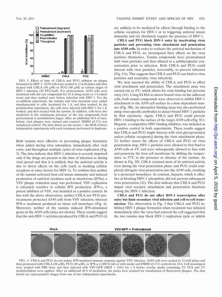
Both tannins were effective in preventing plaque formationwhen added during virus adsorption, immediately after viralentry, and throughout multiple cycles of virus replication (Fig.3). The data indicate that HSV-1 infection is severely impairedonly if the drugs are present at the time of infection or duringviral spread and that it is unlikely that the antiviral activity isdue to direct effects on the cells (such as masking cellularreceptors or entry factors for HSV-1). To confirm that neitherof the tannins activated host cell innate immunity and inducedproduction of antiviral cytokines such as interferons (IFNs), aVSV plaque reduction assay was performed. VSV replicationis extremely sensitive to cellular IFN production. IFN-�, apotent inhibitor of VSV, was included as a positive control. Inline with the above observation, neither CHLA nor PUG pre-treatments protected A549 cells from VSV infection, whereasIFN-� treatment produced an intact cell monolayer (Fig. 4).Moreover, neither of the tannins induced IFN-stimulatedgenes in the A549 cells (data not shown). These results suggestthat the anti-HSV-1 activities produced by CHLA and PUG (i)
are unlikely to be mediated by effects through binding to thecellular receptors for HSV-1 or in triggering antiviral innateimmunity and (ii) absolutely require the presence of HSV-1.
CHLA and PUG block HSV-1 entry by inactivating virusparticles and preventing virus attachment and penetrationinto A549 cells. In order to evaluate the antiviral mechanism ofCHLA and PUG, we investigated their effects on the virusparticles themselves. Tannin compounds were preincubatedwith virus particles and then diluted to a subtherapeutic con-centration prior to infection. Both CHLA and PUG couldinteract with virus particles, irreversibly, to prevent infection(Fig. 5A). This suggests that CHLA and PUG can bind to virusparticles and neutralize virus infectivity.
We next assessed the ability of CHLA and PUG to affectviral attachment and penetration. The attachment assay wascarried out at 4°C, which allows for virus binding but preventsentry (41). Using ELISA to detect bound virus on the adherentcells, both tannin compounds were observed to inhibit HSV-1attachment to the A549 cell surface in a dose-dependent man-ner (Fig. 5B). An alternative binding assay was also performedusing virus-specific antibodies to detect bound HSV-1 particlesby flow cytometry. Again, CHLA and PUG could preventHSV-1 binding to the surface of the target A549 cells (Fig. 5C).Heparin, a competitive HSV binding inhibitor, was included asa positive control in both experiments. These results suggestthat CHLA and PUG might interact with viral glycoprotein(s)and/or cellular receptor(s) during the virus attachment phase.To further assess the effects of CHLA and PUG on viruspenetration step, HSV-1 particles were allowed to first bind toA549 cells at 4°C and were subsequently allowed to fuse withand penetrate the host cell membrane by shifting the temper-ature to 37°C in the presence or absence of the tannins. Asshown in Fig. 5D, CHLA retained most of its antiviral activityeven during the viral penetration phase and PUG could com-pletely abrogate virus penetration into the A549 cells, resultingin a protected monolayer. In contrast, heparin, which is effec-tive at blocking HSV-1 adsorption, did not prevent subsequentvirus penetration (42). The data indicate that CHLA and PUGimpair viral receptor attachment and penetration functionsduring the HSV-1 infection.
CHLA and PUG do not affect HSV-1 transcription afterentry but limit secondary viral infection and cell-to-cell trans-mission. The observation in Fig. 3 that CHLA and PUG in-hibited HSV-1 plaque formation when treatment was initiatedimmediately after the virus had entered the cell suggested thatthe two tannins may block HSV-1 replication cycle or inhibit
FIG. 3. Effect of time of CHLA and PUG addition on plaqueformation by HSV-1. A549 cells were seeded in 12-well plates and thentreated with CHLA (60 �M) or PUG (40 �M) at various stages ofHSV-1 infection (50 PFU/well). For pretreatment, A549 cells wereincubated with the test compounds for 24 h (long term) or 1 h (shortterm) and then washed once before infection with HSV-1. For theco-addition experiment, the tannins and virus inoculum were addedsimultaneously to cells, incubated for 1 h, and then washed. In thepostinfection experiment, the cells were infected with HSV-1 for 1 h,washed, and then treated with the tannins. In addition, cells were alsoincubated in the continuous presence of the test compounds frompretreatment to postinfection stages. After an additional 48 h of incu-bation, viral plaques were stained and counted. DMSO (0.1%) wasincluded as control. The data shown are the means the SEM of threeindependent experiments with each treatment performed in duplicate.
FIG. 4. CHLA and PUG do not induce IFN-mediated immune response against VSV infection. A549 cells were seeded in 12-well plates andthen pretreated with CHLA (60 �M), PUG (40 �M), or IFN-� (1,000 U/ml) or with media and DMSO (0.1%) controls for 24 h. Cell monolayerswere washed with PBS twice and then infected with VSV-GFP (MOI � 0.01) for 1 h before overlay media containing 2% FCS and 2%methylcellulose were applied. After an additional 48 h of incubation, the plates were scanned for visualization of fluorescent plaques. The datashown are representative images from one of two independent experiments.
VOL. 85, 2011 TANNINS INHIBIT ENTRY AND SPREAD OF HSV 4391
on April 5, 2019 by guest
http://jvi.asm.org/
Dow
nloaded from

HSV-1 secondary infection and/or cell-to-cell spread in theensuing incubation period. To specifically address these possi-bilities, we first evaluated the effects of CHLA and PUG onHSV-1 mRNA expression after virus entry. A549 cells wereinfected with HSV-1 for 1 h; extracellular virus was then inac-tivated by citrate buffer treatment and washed away; andCHLA, PUG, or DMSO was subsequently added to the cells.
For comparison, CHLA and PUG were also added simultane-ously with HSV-1. Total cellular RNA was isolated from allsamples at various time points after viral infection. Our resultsclearly indicated that CHLA and PUG did not affect HSV-1mRNA expression after virus penetration, since levels of im-mediate-early (ICP27), early (TK), and late (gD) viral genetranscripts were unaffected by the compounds (Fig. 6A). On
FIG. 5. CHLA and PUG inhibit HSV-1 entry by inactivating viral particles and preventing virus binding and internalization into A549 cells.(A) Viral inactivation assay. HSV-1 (104 PFU/ml) was mixed with CHLA (60 �M) or PUG (40 �M) for 1 h at 37°C and then diluted 50-fold (finalvirus concentration, 50 PFU/well) before infecting A549 cells. As a control, the same amount of virus was also mixed with the tannin, but dilutedimmediately and applied to the A549 cells. After a 48-h incubation period, viral plaques were stained and counted. DMSO (0.1%) was includedas a negative control. The data shown are means the SEM of three independent experiments with each treatment performed in duplicate.(B) Viral attachment analysis using ELISA. Adherent A549 cell monolayers in 96-well plates were prechilled at 4°C for 1 h and then inoculatedwith HSV-1 (MOI � 5) in the presence of CHLA, PUG, or the DMSO (1%) and heparin controls at various concentrations for another 3 h at4°C. Wells were washed to remove unadsorbed virus, subsequently fixed with 4% PFA, and then blocked with 5% BSA. ELISA was performedwith a primary polyclonal rabbit anti-HSV-1 antibody (1:7,500) and a secondary goat anti-rabbit IgG conjugated with HRP (1:100,000), followedby development with TMB substrate and reaction termination with 1 M H3PO4. The absorbance was immediately determined at 450 nm. Valuesare expressed as the fold change of absorbance relative to the mock infection control (cells � 1% DMSO), which is indicated by the dashed line.The data shown are means the SEM of three independent experiments with each treatment performed in triplicate. (C) Viral binding assay usingflow cytometry analysis. Dissociated A549 cells were infected with HSV-1 (MOI � 1) in the presence or absence of 60 �M CHLA or 40 �M PUGfor 3 h at 4°C. Cells were then washed, blocked, and stained with FITC-conjugated polyclonal rabbit anti-HSV-1 antibody (1:500). Stained sampleswere washed and fixed with 1% PFA before being subjected to standard flow cytometry analysis. DMSO (0.1%) was used as a negative control,and heparin (100 �g/ml) was included as a positive control. Color indication for different treatments: gray, mock � DMSO; red, HSV-1 � DMSO;blue, HSV-1 � CHLA; green, HSV-1 � PUG; and purple, HSV-1 � heparin. The data shown are representative of three independentexperiments. (D) Viral penetration analysis by plaque assay. A549 cells were prechilled at 4°C for 1 h before inoculation with HSV-1 (100PFU/well) for 3 h at 4°C. The cells were then treated in the presence or absence of CHLA (60 �M), PUG (40 �M), or heparin (100 �g/ml) andfurther incubated for an additional 20 min with the temperature shifted to 37°C to facilitate viral penetration. At the end of the incubation,extracellular virus was inactivated by citrate buffer (pH 3.0) for 1 min and then washed with PBS twice before overlaying with medium. After 48 hof incubation at 37°C, viral plaques were stained and counted. DMSO (0.1%) was included as a negative control. The data shown are means the SEM of three independent experiments with each treatment performed in duplicate.
4392 LIN ET AL. J. VIROL.
on April 5, 2019 by guest
http://jvi.asm.org/
Dow
nloaded from

the other hand, both tannins suppressed HSV-1 mRNA syn-thesis when added together with the virus at the same time(Fig. 6B). These findings suggest that neither CHLA nor PUGinhibit HSV-1 transcription following penetration of the hostcell.
We next examined whether CHLA and PUG inhibitedHSV-1 secondary infection and/or cell-to-cell spread. A fluo-rescent plaque assay was performed using HSV-1-GFP. Afterviral inoculation of A549 cells, CHLA and PUG were added tothe overlay media at 12 or 24 h p.i. in the presence or absenceof HSV-1 neutralizing antibodies, and fluorescent viral plaqueswere quantified or photographed over the subsequent courseof infection (total of 48 h after the initial challenge). In thisassay, neutralizing antibodies coat viral particles released frominfected cells and prevent secondary infection of surroundinguninfected cells. Thus, the only route of cell-to-cell transmis-sion in the presence of neutralizing antibody is via intercellularjunctions between infected and uninfected cells. Furthermore,prior to drug addition (12 and 24 h p.i.), HSV-1 was permittedto complete at least one round of replication cycle, allowingdrug effects on postentry infection to be monitored in theensuing incubation period. As expected, in the absence ofneutralizing antibodies, there was an increase in viral plaquesin the DMSO control group due to secondary infections (Fig.7A). However, addition of CHLA, PUG, or heparin substan-
tially reduced the number of viral plaques formed and limitedits increase in comparison to DMSO (Fig. 7A). Similarly, withrespect to viral spread, treatment with CHLA and PUGprevented viral plaque growth. Incubation with the tanninsyielded plaques with considerably reduced size comparedto the DMSO control after the 48 h of infection (Fig. 7B).Interestingly, heparin, which has limited inhibitory activity onHSV-1 postattachment, also exhibited some protective effectagainst viral spread albeit at a lower efficiency compared to thetannins. Taken together, the data indicated that once HSV-1entered the cells and completed at least one cycle of infection(12 to 24 h), any subsequent de novo infections and viral spreadvia intercellular contacts were restricted upon addition ofCHLA and PUG. Tannin-mediated inhibition of viral attach-ment and fusion, as observed earlier (Fig. 5B to D), confirmedthese results, and may be responsible for their effects in neu-tralizing secondary infections and restricting cell-to-cellspread, respectively.
CHLA and PUG target HSV-1 glycoproteins that mediateglycosaminoglycan-specific interactions. HSV-1 viral glyco-proteins are known to mediate HSV-1 binding, internalization,and cell-to-cell spread. From the preceding data, it appearsthat the hydrolyzable tannins CHLA and PUG target viralglycoprotein(s), which would explain the need for virus to bepresent during inhibition and their effect on virus entry andspread. In an attempt to elucidate the underlying mechanism,we checked whether the two tannins interacted with HSV-1glycoproteins in order to block entry-associated events. Usinga virus-free system, we overexpressed HSV-1 glycoproteinsthat have been shown to mediate cell fusion (occurs duringentry and cell-to-cell spread) by transfecting the individual gB,gD, gH, and gL genes into A549 cells, followed by treatmentwith the tannins. Expression of all four genes induced cellfusion resulting in polykaryocyte formation (10 nuclei),which is absent after transfection with the empty vector con-trol. The two tannins and heparin each blocked polykaryocyteformation, suggesting that CHLA and PUG interact withHSV-1 glycoproteins to prevent virus attachment, entry, andcell-to-cell spread (Fig. 8).
Several HSV-1 glycoproteins are known to interact with cellsurface GAGs. To further explore the virus-host interactionsthat are being targeted by the tannins, we used a series of celllines known to possess defects in surface HS and CS synthesis.The relative infectivities of HSV-1 (KOS) are ca. 10% forHS-deficient gro2C cells and 0.5% for HS/CS-deficient sog9cells compared to parental mouse L cells (6). Stable expressionof the EXT1 gene in sog9 cells (sog9-EXT1) restores HS bio-synthesis and susceptibility to HSV-1 infection (43). To eval-uate the effects of the drugs in the presence or absence ofGAG expression, each cell line was infected with differentdilutions of HSV-1 sufficient to achieve 200 PFU/well (MOI �0.0004) in the presence of the tannins. CHLA and PUG effec-tively protected the parental mouse L cells and sog9-EXT1cells from infection, but antiviral effects were diminished inHS-deficient gro2C cells, and almost completely abolished inHS/CS-deficient sog9 cells (Fig. 9). Similar results were ob-tained in experiments using different MOIs (data not shown).These observations strongly suggest that CHLA and PUGtarget interactions between HSV-1 glycoproteins and
FIG. 6. CHLA and PUG do not affect HSV-1 transcription, follow-ing entry into the host cell. (A) A549 cells were infected with HSV-1(MOI � 1) for 1 h, treated with low-pH citrate buffer (pH 3.0) toinactivate noninternalized extracellular viral particles, and subse-quently overlaid with medium containing CHLA (60 �M), PUG (40�M), or DMSO control (0.1%). At 4, 8, and 12 h p.i., total cellularRNA was isolated, subjected to first-strand synthesis by reverse tran-scription, and then amplified by standard PCR procedures with prim-ers against HSV-1 immediate-early (ICP27), early (TK), and late (gD)gene products. GAPDH was included as a control. (B) A549 cells werecoincubated with HSV-1 (MOI � 1) and CHLA (60 �M), PUG (40�M), or DMSO control (0.1%) for 1 h. Cells were washed with PBSbefore overlay media without the tannins was applied. Total cellularRNA was isolated for RT-PCR analysis as in panel A. Representativedata shown are from one of two independent experiments.
VOL. 85, 2011 TANNINS INHIBIT ENTRY AND SPREAD OF HSV 4393
on April 5, 2019 by guest
http://jvi.asm.org/
Dow
nloaded from

GAGs. CHLA inhibition also appeared to be more sensitive tocell surface deficiency in GAGs compared to that of PUG.
Taken together, the data indicate that CHLA and PUGfunction as GAG competitors to inhibit the initial events ofHSV-1 infection (adsorption and penetration) and the cell-to-cell spread of virus. The interaction of HSV-1 glycoproteinswith cellular GAGs plays a critical role in viral infections, andthe hydrolyzable tannins could offer a primary means of de-fense against HSV-1 infections.
DISCUSSION
There is currently no cure that completely resolves latentinfections caused by alphaherpesviruses. Therefore, the devel-opment of small molecules capable of inhibiting infection byreactivated virus represents an attractive therapeutic strategy,particularly in immunocompromised individuals who are oftenat risk of generating ACV-resistant HSV-1 strains. In a searchfor such molecules, we report that CHLA and PUG, two hy-drolyzable tannins isolated from the fruits of T. chebula, effec-tively inhibited HSV-1 infection in A549 cells without signifi-cantly reducing cell viability. In addition, our results suggestthat CHLA and PUG specifically targeted HSV-1 particles bybinding to viral glycoproteins that interact with cellular GAGs,rendering the virus incapable of adsorbing, penetrating, andspreading throughout the cell monolayer. These features un-derscore the potential of tannins as HSV-1 entry inhibitors.
Our data show that entry events, including primary and
secondary infection, viral attachment and/or penetration,and cell-to-cell spread are inhibited only when the tannins andHSV-1 glycoproteins are in contact with each other. Pretreat-ment of host cells with the tannins, followed by washes toremove unadsorbed compounds, had no effects upon HSV-1replication. This indicated that masking cell surface receptorsor entry factors for HSV-1 by the tannins is unlikely. Viralbinding assays using ELISA and flow cytometry analyses re-vealed that the tannins blocked viral attachment to the hostcell. While CHLA and PUG could inactivate the HSV-1 par-ticles, we do not believe that a direct lysis effect of the viralmembrane is responsible for their effects, since HSV-1 infec-tion of GAG-deficient mutant cell lines was still observed, evenin the presence of these compounds (Fig. 9). Given their largemolecular weights (CHLA, 954; and PUG, 1,084) and highaffinity for proteins and sugars, the two hydrolyzable tanninsare thought to bind to HSV-1 glycoproteins on the infectiousvirions making them inert, impairing glycoprotein function,and preventing successful attachment and entry of the hostcell. These tannins could also bind to viral glycoproteins on theinfected-cell surface, rendering them unavailable to mediatethe cell-to-cell spread of virus.
HSV-1 entry into epithelial cells, which express the cellularreceptors (HS, HVEM, nectin-1, and nectin-2) for HSV (67),requires an ordered and concerted effort from the viral glyco-proteins. Specifically, the glycoproteins gB, gC, gD, gH, and gLinteract with host cell receptors and are involved in penetra-tion of the plasma membrane through a membrane fusion
FIG. 7. CHLA and PUG can limit HSV-1 secondary infection and cell-to-cell spread of the virus. A549 cells were infected with HSV-1-GFP(200 PFU/well) for 1 h and then treated with citrate buffer (pH 3.0) to inactivate noninternalized extracellular viral particles. Cells were overlaidwith medium alone (A) or medium containing 0.1% neutralizing antibody (B). After a p.i. incubation period of 12 h (A) or 24 h (B), the infectedcells were treated with CHLA (60 �M), PUG (40 �M), heparin (100 �g/ml), or DMSO (0.1%), before further incubation for a total of 48 h afterthe initial infection. Over the course of infection subsequent to the drug addition, the plates were scanned and quantified for fluorescent viralplaques (A) or photographed using an inverted fluorescence microscope at �100 magnification (B). (A) Number of fluorescent plaques countedbetween 24 and 48 h p.i. with drug treatment initiated at 12 h after viral challenge. The data shown are means the SEM of three independentexperiments with each treatment performed in duplicate. (B) Comparison of viral plaque size between 24 and 48 h p.i. with drug treatment initiatedat 24 h after viral challenge. Scale bars, 100 �m. Representative images are from one of two independent experiments.
4394 LIN ET AL. J. VIROL.
on April 5, 2019 by guest
http://jvi.asm.org/
Dow
nloaded from

process (29, 57, 67). While viral entry and spread require aparticular combination of viral surface proteins, several HSV-1glycoproteins are repeatedly involved in both processes. Im-portantly, gB and gD function in viral binding and fusion andare also engaged during cell-to-cell transmission in culturedepithelial cells (22, 29, 33, 38, 57, 60, 61, 67). The associa-tions between viral glycoproteins that mediate HSV-1 at-tachment and entry represent a complex scenario when con-sidering CHLA and PUG and their mechanism of action. Thecandidate targets of the tannins likely involve viral glycopro-teins that interact with host cell surface GAGs and participatein adsorption, membrane fusion, and cell-to-cell spread. Theobservation that both tannins blocked virus attachment to cells,as did heparin, suggests that interaction of gC and gB withheparan sulfate proteoglycans (HSPGs) is targeted. However,the drugs also prevented virus internalization into cells in thepostbinding phase. At this point, the interaction with HSPGs
should be irrelevant, since virions now interact with a gDreceptor and become resistant to removal by heparin (42) (Fig.5D). One explanation is that the tannins bind to gB and blockits interaction with HSPG while also interfering with its sub-sequent role in membrane fusion during virus entry (in whichgC is not involved). Alternatively, the tannins may be impedingthe activity of additional glycoproteins (gD and/or gH/gL) orworking by some other mechanism(s) to prevent successfulentry into the A549 cells. Finally, there is the possibility thatviral glycoproteins may still be accessible to the tannins, evenwhen these viral proteins are bound to the host cell or areexpressed in the intercellular junctions. This could explain whythe considerably larger heparin (molecular weight, 17,000 to19,000) can bind free gB but is unable to interact efficientlywith the shielded glycoprotein that is needed in order to inhibitviral penetration or cell-to-cell transmission. Additional bind-ing experiments using glycoprotein-deficient HSV-1 mutants,
FIG. 8. CHLA and PUG can prevent HSV-1 glycoprotein-mediated cell fusion events. A549 cells were transfected with plasmids expressing theindividual HSV-1 glycoproteins (gB, gD, gH, and gL). After 6 h of transfection, cells were washed with PBS and treated with CHLA (60 �M), PUG(40 �M), heparin (100 �g/ml), or DMSO (0.1%). After further incubation for 24 h, cells were fixed with methanol and stained with Hoechst dye(A) or Giemsa (B). Photomicrographs were then taken at �200 magnification. (A) Phases (upper panels) and respective fluorescence picturesdisplaying the Hoechst dye-stained nuclei (bottom panels). (B) Giemsa-stained cells in a similar experiment. Representative pictures shown arefrom one of three independent experiments. Vector, empty vector; GP, HSV-1 glycoproteins; scale bars, 100 �m. (C) The total number ofpolykaryocytes (10 nuclei) from each treatment was quantified. The data shown are means the SEM of three independent experiments.
VOL. 85, 2011 TANNINS INHIBIT ENTRY AND SPREAD OF HSV 4395
on April 5, 2019 by guest
http://jvi.asm.org/
Dow
nloaded from

as well as soluble recombinant HSV-1 glycoproteins could helpelucidate the targeting specificity of the tannins. We speculatethat the two natural compounds can bind to all GAG-interact-ing glycoproteins, including gB, gC, and/or gD, and neutralizetheir functions. The ability of CHLA and PUG to effectivelyblock virus membrane penetration, as well as virus attachment,could explain their higher efficacy in restricting the spread ofHSV-1 compared to the inhibitory effects of heparin.
In the case of HSV-1, the interactions between several of itsglycoproteins and cell surface GAGs are critical for ensuringefficient viral entry, as well as viral spread (6, 26, 44, 52, 53, 65,66, 68). CS can confer susceptibility to HSV-1 infection in theabsence of HS (7, 26), but HS is still the preferred substrate forviral attachment. GAG deficiency renders cell surfaces rela-tively resistant to HSV-1 binding and residual infectivity relieson stable attachment receptors (6). Earlier studies have shownthat the HS/CS-deficient sog9 cells are insensitive to inhibitoryeffect of soluble HS on HSV-1 infection (6). We observed thatwhereas the absence of HS in gro2C cells weakened the tan-nins’ inhibitory effects, the absence of HS and CS on sog9 cellsseverely limited their antiviral activities (Fig. 9). Overexpres-sion of the EXT1 gene, which restores HS biosynthesis in EXT1mutant sog9 cells, rescued the antiviral effects from bothCHLA and PUG to 90%. The results suggest that the tanninsinterfere with GAG interactions with viral glycoproteins whichcan involve attachment and may also affect downstream pen-etration of the host cell. Additional experiments are requiredto clarify whether the drugs are able to inhibit binding and/orfusion of HSV-1 on the GAG-deficient cells.
All four hydrolyzable tannins investigated in this report arecomposed of a glucopyranose core linked with galloyl deriva-tives, including hexahydroxydiphenoyl (HHDP; C-C couplingbetween galloyl moieties), gallagyl, and chebuloyl units (Fig. 1)(39, 78). Only CHLA and PUG possess the HHDP unit, withan (R) configuration (linked to the glucose core at the 3,6
positions) and (S) configuration (linked via the 2,3 or 4,6 po-sitions of their glucose residue), respectively (36, 78). Theanti-herpes activities of hydrolyzable tannins are thought to bedependent on the number of galloyl or HHDP groups, irre-spective of the sugar core (71). Structures containing HHDPunit have also been valuable pharmacophores for inhibitingHIV enzymatic activities (20, 75). Indeed, anti-HIV activitiesfrom CHLA, CHLI, PUG, and PUN have been reported toprevent binding of recombinant HIV gp120 to CD4 and toexert inhibitory effects on HIV-1 RT and integrase (1, 51, 74).The ability of these natural agents to inhibit both HSV-1 andHIV-1 underscores their potential value in the treatment ofAIDS patients who also exhibit HSV-1-related symptoms.
Use of these tannins could improve the prognosis of anti-HSV-1 therapy in immunosuppressed individuals and help toreduce the risk of ACV resistance by lowering the ACV doserequired. Since Fructus Chebulae contains both CHLA andPUG, inclusion of purified extracts from this plant in topicalcreams or microbicides would be a feasible method for con-trolling recurrent HSV-1 infections. Future studies will de-termine whether these natural products are effective againstadditional members of the herpesvirus family and otherenveloped viruses. Our preliminary studies have shown thatboth CHLA and PUG inhibit the growth of HSV-2 and humancytomegalovirus (L.-T. Lin and T.-Y. Chen, unpublished data).Other viruses known to use GAGs as entry factors, such asmeasles virus and human respiratory syncytial virus, are alsoinhibited by these tannins, reinforcing our discovery that thesecompounds act as GAG competitors that inhibit viral glyco-protein-cell receptor interactions (Lin and Chen, unpub-lished). Further studies with the tannins derived from T. che-bula may provide new ways to inhibit recurrent HSV-1infections and control the reemergence of this virus in immu-nocompromised patients.
ACKNOWLEDGMENTS
We thank James R. Smiley, Gary H. Cohen, Roselyn J. Eisenberg,Bruce W. Banfield, Karen L. Mossman, Brian D. Lichty, and AndrewC. Issekutz for reagents.
L.-T.L. is a recipient of a National CIHR Research Training Pro-gram in Hepatitis C (NCRTP-HepC) fellowship. C.-C.L. is supportedby a research grant from the National Science Council of Taiwan (NSC96-2320-B-037-016). C.D.R. is supported by an operating grant fromthe Canadian Institutes of Health (CIHR-MOP-10638).
We have declared that no competing interests exist.L.-T.L., T.-Y.C., C.-C.L., and C.D.R. conceived and designed the
experiments. L.-T.L. and T.-Y.C. performed the experiments. L.-T.L.,T.-Y.C., C.M., C.-C.L., and C.D.R. analyzed the data. L.-T.L.,T.-Y.C. C.-Y.C., R.S.N., C.M., T.B.G., T.-C.L., G.-H.W., C.-C.L., andC.D.R. contributed reagents, materials, and technical support. L.-T.L.,T.-Y.C., C.-C.L., and C.D.R. wrote the paper.
REFERENCES
1. Ahn, M. J., et al. 2002. Inhibition of HIV-1 integrase by galloyl glucoses fromTerminalia chebula and flavonol glycoside gallates from Euphorbia pekinen-sis. Planta Med. 68:457–459.
2. Akkarawongsa, R., T. B. Potocky, E. P. English, S. H. Gellman, and C. R.Brandt. 2008. Inhibition of herpes simplex virus type 1 infection by cationicbeta-peptides. Antimicrob. Agents Chemother. 52:2120–2129.
3. Arduino, P. G., and S. R. Porter. 2008. Herpes simplex virus type 1 infection:overview on relevant clinico-pathological features. J. Oral Pathol. Med.37:107–121.
4. Arduino, P. G., and S. R. Porter. 2006. Oral and perioral herpes simplex virustype 1 (HSV-1) infection: review of its management. Oral Dis. 12:254–270.
5. Aurelian, L. 2008. Herpes simplex viruses: general features, p. 383–397. InB. W. J. Mahy and M. H. V. van Regenmortel (ed.), Encyclopedia ofvirology, 3rd ed., vol. 2. Elsevier, Amsterdam, Netherlands.
FIG. 9. Anti-HSV-1 effects mediated by CHLA and PUG are im-paired in GAG-deficient cells and are rescued by restoration of hepa-ran sulfate biosynthesis through EXT1 gene expression. Confluent cellsin 12-well plates were infected with HSV-1-GFP (200 PFU/well re-flecting virus titers determined in each cell line) concurrently in thepresence or absence of CHLA (60 �M), PUG (40 �M), or DMSO(0.1%) for 1 h. The plates were washed with PBS before applyingoverlay media. After an additional 48 h of incubation, fluorescent viralplaques were scanned and quantified. Values obtained were comparedagainst each cell line’s respective DMSO control for HSV-1 infectionwhich was considered to be 100%. The respective statuses of theheparan sulfate (HS) and chondroitin sulfate (CS) GAG synthesis inthe different cell lines are indicated in parentheses. The data shown aremeans the SEM of three independent experiments, with each treat-ment being performed in duplicate.
4396 LIN ET AL. J. VIROL.
on April 5, 2019 by guest
http://jvi.asm.org/
Dow
nloaded from

6. Banfield, B. W., Y. Leduc, L. Esford, K. Schubert, and F. Tufaro. 1995.Sequential isolation of proteoglycan synthesis mutants by using herpes sim-plex virus as a selective agent: evidence for a proteoglycan-independent virusentry pathway. J. Virol. 69:3290–3298.
7. Banfield, B. W., et al. 1995. Evidence for an interaction of herpes simplexvirus with chondroitin sulfate proteoglycans during infection. Virology 208:531–539.
8. Buzzini, P., et al. 2008. Antimicrobial and antiviral activity of hydrolysabletannins. Mini Rev. Med. Chem. 8:1179–1187.
9. Chen, Y., et al. 2000. Resistant herpes simplex virus type 1 infection: anemerging concern after allogeneic stem cell transplantation. Clin. Infect. Dis.31:927–935.
10. Cheng, H. Y., C. C. Lin, and T. C. Lin. 2002. Antiherpes simplex virus type2 activity of casuarinin from the bark of Terminalia arjuna Linn. Antivir. Res.55:447–455.
11. Cheng, H. Y., C. C. Lin, and T. C. Lin. 2002. Antiviral properties of prodel-phinidin B-2 3�-O-gallate from green tea leaf. Antivir. Chem. Chemother.13:223–229.
12. Cheng, H. Y., et al. 2003. In vitro antiviral activity of prodelphinidin B-23,3�-di-O-gallate from Myrica rubra. Planta Med. 69:953–956.
13. Cheng, H. Y., T. C. Lin, C. M. Yang, K. C. Wang, and C. C. Lin. 2004.Mechanism of action of the suppression of herpes simplex virus type 2replication by pterocarnin A. Microbes Infect. 6:738–744.
14. Cheng, H. Y., et al. 2004. Putranjivain A from Euphorbia jolkini inhibits bothvirus entry and late stage replication of herpes simplex virus type 2 in vitro.J. Antimicrob. Chemother. 53:577–583.
15. Cheng, H. Y., T. C. Lin, K. H. Yu, C. M. Yang, and C. C. Lin. 2003.Antioxidant and free radical scavenging activities of Terminalia chebula. Biol.Pharm. Bull. 26:1331–1335.
16. Cheng, H. Y., et al. 6 October 2009. Excoecarianin, isolated from Phyllanthusurinaria Linnea, inhibits herpes simplex virus type 2 infection through inac-tivation of viral particles. Evid. Based Complement Alternat. Med. [Epubahead of print.]
17. Cheng, H. Y., C. M. Yang, T. C. Lin, D. E. Shieh, and C. C. Lin. 2006.ent-Epiafzelechin-(4�38)-epiafzelechin extracted from Cassia javanica in-hibits herpes simplex virus type 2 replication. J. Med. Microbiol. 55:201–206.
18. Cheshenko, N., W. Liu, L. M. Satlin, and B. C. Herold. 2007. Multiplereceptor interactions trigger release of membrane and intracellular calciumstores critical for herpes simplex virus entry. Mol. Biol. Cell 18:3119–3130.
19. Chiang, L. C., W. Chiang, M. C. Liu, and C. C. Lin. 2003. In vitro antiviralactivities of Caesalpinia pulcherrima and its related flavonoids. J. Antimicrob.Chemother. 52:194–198.
20. Cos, P., L. Maes, A. Vlietinck, and L. Pieters. 2008. Plant-derived leadingcompounds for chemotherapy of human immunodeficiency virus (HIV) in-fection: an update (1998–2007). Planta Med. 74:1323–1337.
21. Danve-Szatanek, C., et al. 2004. Surveillance network for herpes simplexvirus resistance to antiviral drugs: 3-year follow-up. J. Clin. Microbiol. 42:242–249.
22. Dingwell, K. S., et al. 1994. Herpes simplex virus glycoproteins E and Ifacilitate cell-to-cell spread in vivo and across junctions of cultured cells.J. Virol. 68:834–845.
23. Fatahzadeh, M., and R. A. Schwartz. 2007. Human herpes simplex labialis.Clin. Exp. Dermatol. 32:625–630.
24. Field, H. J. 2001. Herpes simplex virus antiviral drug resistance–currenttrends and future prospects. J. Clin. Virol. 21:261–269.
25. Fukuchi, K., et al. 1989. Inhibition of herpes simplex virus infection bytannins and related compounds. Antivir. Res. 11:285–297.
26. Gruenheid, S., L. Gatzke, H. Meadows, and F. Tufaro. 1993. Herpes simplexvirus infection and propagation in a mouse L cell mutant lacking heparansulfate proteoglycans. J. Virol. 67:93–100.
27. Haslam, E. 1989. Plant polyphenols: vegetable tannins revisited. CambridgeUniversity Press, Cambridge, United Kingdom.
28. Haslam, E. 2007. Vegetable tannins: lessons of a phytochemical lifetime.Phytochemistry 68:2713–2721.
29. Heldwein, E. E., and C. Krummenacher. 2008. Entry of herpesviruses intomammalian cells. Cell Mol. Life Sci. 65:1653–1668.
30. Hsu, H. Y., et al. 1985. Chinese materia medica: a concise guide. ModernDrug Press, Taipei, Taiwan.
31. Huang, K., L. Incognito, X. Cheng, N. D. Ulbrandt, and H. Wu. 2010.Respiratory syncytial virus-neutralizing monoclonal antibodies motavizumaband palivizumab inhibit fusion. J. Virol. 84:8132–8140.
32. Iason, G. 2005. The role of plant secondary metabolites in mammalianherbivory: ecological perspectives. Proc. Nutr. Soc. 64:123–131.
33. Johnson, D. C., and M. T. Huber. 2002. Directed egress of animal virusespromotes cell-to-cell spread. J. Virol. 76:1–8.
34. Juang, L. J., and S. J. Sheu. 2005. Chemical identification of the sources ofcommercial Fructus Chebulae. Phytochem. Anal. 16:246–251.
35. Juang, L. J., S. J. Sheu, and T. C. Lin. 2004. Determination of hydrolyzabletannins in the fruit of Terminalia chebula Retz. by high-performance liquidchromatography and capillary electrophoresis. J. Sep. Sci. 27:718–724.
36. Khanbabaee, K., and T. van Ree. 2001. Tannins: classification and definition.Nat. Prod. Rep. 18:641–649.
37. Knickelbein, J. E., R. L. Hendricks, and P. Charukamnoetkanok. 2009.Management of herpes simplex virus stromal keratitis: an evidence-basedreview. Surv. Ophthalmol. 54:226–234.
38. Laquerre, S., et al. 1998. Heparan sulfate proteoglycan binding by herpessimplex virus type 1 glycoproteins B and C, which differ in their contributionsto virus attachment, penetration, and cell-to-cell spread. J. Virol. 72:6119–6130.
39. Lin, T. C., S. C. Chien, H. F. Chen, and F. L. Hsu. 2000. Tannins and relatedcompounds from Combretaceae plants. Chin Pharm. J. 52:1–26.
40. Lin, T. C., G. Nonaka, I. Nishioka, and F. C. Ho. 1990. Tannins and relatedcompounds. CII. Structures of terchebulin, an ellagitannin having a noveltetraphenylcarboxylic acid (terchebulic acid) moiety, and biogenetically re-lated tannins from Terminalia chebula Retz. Chem. Pharm. Bull. 38:3004–3008.
41. Madan, R. P., et al. 2007. Molecular umbrellas: a novel class of candidatetopical microbicides to prevent human immunodeficiency virus and herpessimplex virus infections. J. Virol. 81:7636–7646.
42. McClain, D. S., and A. O. Fuller. 1994. Cell-specific kinetics and efficiency ofherpes simplex virus type 1 entry are determined by two distinct phases ofattachment. Virology 198:690–702.
43. McCormick, C., G. Duncan, K. T. Goutsos, and F. Tufaro. 2000. The puta-tive tumor suppressors EXT1 and EXT2 form a stable complex that accu-mulates in the Golgi apparatus and catalyzes the synthesis of heparan sulfate.Proc. Natl. Acad. Sci. U. S. A. 97:668–673.
44. McCormick, C., G. Duncan, and F. Tufaro. 2000. Herpes simplex virus:discovering the link between heparan sulfate and hereditary bone tumours.Rev. Med. Virol. 10:373–384.
45. McCormick, C., et al. 1998. The putative tumour suppressor EXT1 alters theexpression of cell-surface heparan sulfate. Nat. Genet. 19:158–161.
46. Melroe, G. T., N. A. DeLuca, and D. M. Knipe. 2004. Herpes simplex virus1 has multiple mechanisms for blocking virus-induced interferon production.J. Virol. 78:8411–8420.
47. Minaker, R. L., K. L. Mossman, and J. R. Smiley. 2005. Functional inacces-sibility of quiescent herpes simplex virus genomes. Virol. J. 2:85.
48. Morfin, F., et al. 2004. HSV excretion after bone marrow transplantation: a4-year survey. J. Clin. Virol. 30:341–345.
49. Mossman, K. L., et al. 2001. Herpes simplex virus triggers and then disarmsa host antiviral response. J. Virol. 75:750–758.
50. Nicholl, M. J., L. H. Robinson, and C. M. Preston. 2000. Activation ofcellular interferon-responsive genes after infection of human cells with her-pes simplex virus type 1. J. Gen. Virol. 81:2215–2218.
51. Nonaka, G., et al. 1990. Anti-AIDS agents. 2. Inhibitory effects of tannins onHIV reverse transcriptase and HIV replication in H9 lymphocyte cells. J.Nat. Prod. 53:587–595.
52. Nyberg, K., et al. 2004. The low molecular weight heparan sulfate-mimetic,PI-88, inhibits cell-to-cell spread of herpes simplex virus. Antivir. Res. 63:15–24.
53. O’Donnell, C. D., and D. Shukla. 2008. The importance of heparan sulfate inherpesvirus infection. Virol. Sin. 23:383–393.
54. Pertel, P. E., A. Fridberg, M. L. Parish, and P. G. Spear. 2001. Cell fusioninduced by herpes simplex virus glycoproteins gB, gD, and gH-gL requires agD receptor but not necessarily heparan sulfate. Virology 279:313–324.
55. Porter, J. L. 1989. Methods in plant biochemistry: plant phenolics, vol. 1.Academic Press, London, England.
56. Quideau, S., et al. 2004. Main structural and stereochemical aspects of theantiherpetic activity of nonahydroxyterphenoyl-containing C-glycosidic ella-gitannins. Chem. Biodivers. 1:247–258.
57. Reske, A., G. Pollara, C. Krummenacher, B. M. Chain, and D. R. Katz. 2007.Understanding HSV-1 entry glycoproteins. Rev. Med. Virol. 17:205–215.
58. Roizman, B., D. M. Knipe, and R. J. Whitley. 2007. Herpes simplex viruses,p. 2501–2601. In D. M. Knipe et al. (ed.), Fields virology, 5th ed. Lippincott-Raven Publishers, Philadelphia, PA.
59. Safrin, S., et al. 1991. A controlled trial comparing foscarnet with vidarabinefor acyclovir-resistant mucocutaneous herpes simplex in the acquired immu-nodeficiency syndrome. N. Engl. J. Med. 325:551–555.
60. Sakisaka, T., et al. 2001. Requirement of interaction of nectin-1�/HveC withafadin for efficient cell-cell spread of herpes simplex virus type 1. J. Virol.75:4734–4743.
61. Satoh, T., et al. 2008. PILR� is a herpes simplex virus-1 entry coreceptor thatassociates with glycoprotein B. Cell 132:935–944.
62. Schleiss, M. R. 2009. Persistent and recurring viral infections: the humanherpesviruses. Curr. Probl. Pediatr. Adolesc. Health Care 39:7–23.
63. Serrano, J., R. Puupponen-Pimia, A. Dauer, A. M. Aura, and F. Saura-Calixto. 2009. Tannins: current knowledge of food sources, intake, bioavail-ability and biological effects. Mol. Nutr. Food Res. 53(Suppl. 2):S310–S329.
64. Shahat, A. A., et al. 2002. Antiviral and antioxidant activity of flavonoids andproanthocyanidins from Crataegus sinaica. Planta Med. 68:539–541.
65. Shieh, M. T., and P. G. Spear. 1994. Herpesvirus-induced cell fusion that isdependent on cell surface heparan sulfate or soluble heparin. J. Virol. 68:1224–1228.
66. Shieh, M. T., D. WuDunn, R. I. Montgomery, J. D. Esko, and P. G. Spear.
VOL. 85, 2011 TANNINS INHIBIT ENTRY AND SPREAD OF HSV 4397
on April 5, 2019 by guest
http://jvi.asm.org/
Dow
nloaded from

1992. Cell surface receptors for herpes simplex virus are heparan sulfateproteoglycans. J. Cell Biol. 116:1273–1281.
67. Spear, P. G. 2004. Herpes simplex virus: receptors and ligands for cell entry.Cell Microbiol. 6:401–410.
68. Spear, P. G., M. T. Shieh, B. C. Herold, D. WuDunn, and T. I. Koshy. 1992.Heparan sulfate glycosaminoglycans as primary cell surface receptors forherpes simplex virus. Adv. Exp. Med. Biol. 313:341–353.
69. Stojdl, D. F., B. D. Lichty, B. R. tenOever, et al. 2003. VSV strains withdefects in their ability to shutdown innate immunity are potent systemicanti-cancer agents. Cancer Cell 4:263–275.
70. Stranska, R., et al. 2005. Survey of acyclovir-resistant herpes simplex virus inthe Netherlands: prevalence and characterization. J. Clin. Virol. 32:7–18.
71. Takechi, M., Y. Tanaka, M. Takehara, G. I. Nonaka, and I. Nishioka. 1985.Structure and antiherpetic activity among the tannins. Phytochemistry 24:2245–2250.
72. Tian, L. W., Y. J. Zhang, C. Qu, Y. F. Wang, and C. R. Yang. 2010. Phloro-glucinol glycosides from the fresh fruits of Eucalyptus maideni. J. Nat. Prod.73:160–163.
73. Wagner, E. K., and R. M. Sandri-Goldin. 2008. Herpes simplex viruses:molecular biology, p. 397–405. In B. W. J. Mahy and M. H. V. van Regen-
mortel (ed.), Encyclopedia of virology, 3rd ed., vol. 2. Elsevier, Amsterdam,Netherlands.
74. Weaver, J. L., et al. 1992. Prevention of binding of rgp120 by anti-HIV activetannins. Biochem. Pharmacol. 43:2479–2480.
75. Xie, L., et al. 1995. Anti-AIDS (acquired immune deficiency syndrome)agents. 17. New brominated hexahydroxybiphenyl derivatives as potent anti-HIV agents. J. Med. Chem. 38:3003–3008.
76. Yang, C. M., H. Y. Cheng, T. C. Lin, L. C. Chiang, and C. C. Lin. 2007.Hippomanin A from acetone extract of Phyllanthus urinaria inhibited HSV-2but not HSV-1 infection in vitro. Phytother. Res. 21:1182–1186.
77. Yang, C. M., H. Y. Cheng, T. C. Lin, L. C. Chiang, and C. C. Lin. 2007. Thein vitro activity of geraniin and 1,3,4,6-tetra-O-galloyl-�-D-glucose isolatedfrom Phyllanthus urinaria against herpes simplex virus type 1 and type 2infection. J. Ethnopharmacol. 110:555–558.
78. Yoshida, T., Y. Amakura, and M. Yoshimura. 2010. Structural features andbiological properties of ellagitannins in some plant families of the orderMyrtales. Int. J. Mol. Sci. 11:79–106.
79. Ziyaeyan, M., et al. 2007. Frequency of acyclovir-resistant herpes simplexviruses isolated from the general immunocompetent population and patientswith acquired immunodeficiency syndrome. Int. J. Dermatol. 46:1263–1266.
4398 LIN ET AL. J. VIROL.
on April 5, 2019 by guest
http://jvi.asm.org/
Dow
nloaded from


![D 6093 [16] EurJOC · D 6093 A Journal of ejoc_u1_16.qxd 08.05.2009 10:45 Seite 1. FULL PAPER DOI: 10.1002/ejoc.200900117 Synthesis and Biological Evaluation of Non-Hydrolyzable 1,2,3-Triazole-Linked](https://img.dokumen.tips/doc/110x75/5f16bc94d123a475a274d99a/d-6093-16-eurjoc-d-6093-a-journal-of-ejocu116qxd-08052009-1045-seite-1.jpg)


![Transient Electronics: Thermally Triggered Degradation of ... · secure memory devices, and environmental sensors. [ 1–6 ] ran-sient electronics have been demonstrated with hydrolyzable](https://img.dokumen.tips/doc/110x75/5fa578cfd4e80f055f6b3358/transient-electronics-thermally-triggered-degradation-of-secure-memory-devices.jpg)


![Integrated Algal Bioprocess Engineering for Enhanced ......of algal complex carbohydrates hydrolyzable into a fermentable hexose monomer or 80% theoretical ethanol yield [30], there](https://img.dokumen.tips/doc/110x75/608d1f678f6feb27087a883a/integrated-algal-bioprocess-engineering-for-enhanced-of-algal-complex-carbohydrates.jpg)


![COVER PAGE DESIGN cdr - IJAAM33] Two hydrolyzable tannins, chebulagic acid and punicalagin, isolated from the dried fruits of T. chebula inhibited HSV-1 entry at non-cytotoxic doses](https://img.dokumen.tips/doc/110x75/5acf74c07f8b9aca598c6651/cover-page-design-cdr-33-two-hydrolyzable-tannins-chebulagic-acid-and-punicalagin.jpg)