Embed Size (px)
Citation preview
Hydrogen-Bonded Homoleptic Fluoride−Diarylurea Complexes:Structure, Reactivity, and Coordinating PowerLukas Pfeifer,† Keary M. Engle,†,§ George W. Pidgeon,† Hazel A. Sparkes,‡,∥ Amber L. Thompson,†
John M. Brown,† and Veronique Gouverneur*,†
†Chemistry Research Laboratory, University of Oxford, 12 Mansfield Road, OX1 3TA Oxford, United Kingdom‡ISIS Facility, STFC-Rutherford Appleton Laboratory, OX11 0QX Didcot, United Kingdom
*S Supporting Information
ABSTRACT: Hydrogen bonding with fluoride is a key interactionencountered when analyzing the mode of action of 5′-fluoro-5′-deoxyadenosine synthase, the only known enzyme capable of catalyzingthe formation of a C−F bond from F−. Further understanding of theeffect of hydrogen bonding on the structure and reactivity of complexedfluoride is therefore important for catalysis and numerous otherapplications, such as anion supramolecular chemistry. Herein we disclosea detailed study examining the structure of 18 novel urea−fluoridecomplexes in the solid state, by X-ray and neutron diffraction, and insolution phase and explore the reactivity of these complexes as a fluoridesource in SN2 chemistry. Experimental data show that the structure,coordination strength, and reactivity of the urea−fluoride complexes aretunable by modifying substituents on the urea receptor. Hammettanalysis of aryl groups on the urea indicates that fluoride binding is dependent on σp and σm parameters with stronger bindingbeing observed for electron-deficient urea ligands. For the first time, defined urea−fluoride complexes are used as fluoride-binding reagents for the nucleophilic substitution of a model alkyl bromide. The reaction is slower in comparison with knownalcohol−fluoride complexes, but SN2 is largely favored over E2, at a ratio surpassing all hydrogen-bonded complexes documentedin the literature for the model alkyl bromide employed. Increased second-order rate constants at higher dilution support thehypothesis that the reactive species is a 1:1 urea−fluoride complex of type [UF]− (U = urea) resulting from partial dissociation ofthe parent compound [U2F]
−. The dissociation processes can be quantified through a combination of UV and NMR assays,including DOSY and HOESY analyses that illuminate the complexation state and H-bonding in solution.
■ INTRODUCTION
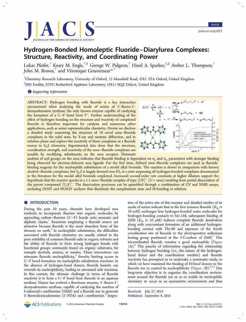
During the past 10 years, chemists have developed newmethods to incorporate fluorine into organic molecules byappending carbon−fluorine (C−F) bonds onto aromatic andaliphatic chains.1 Reactions employing a fluoride source areattractive because fluoride is the most abundant form of theelement on earth.2 In nucleophilic substitution, the difficultiesassociated with fluoride chemistry are usually related to thepoor solubility of common fluoride salts in organic solvents andthe ability of fluoride to form strong hydrogen bonds withfunctional groups commonly found on organic substrates, forexample alcohols, amines, or amides. These interactions canattenuate fluoride nucleophilicity,3 thereby limiting access toC−F bond formation via nucleophilic substitution reactions. Inthe absence of hydrogen-bond donors, fluoride basicity canoverride its nucleophilicity, leading to unwanted side reactions.In this context, the ultimate challenge in terms of fluoridereactivity is to form a C−F bond from fluoride in an aqueousmedium. Nature has evolved a fluorinase enzyme, 5′-fluoro-5′-deoxyadenosine synthase, capable of catalyzing the reaction ofS-adenosyl-L-methionine (SAM) and a fluoride ion to generate5′-fluorodeoxyadenosine (5′-FDA) and L-methionine.4 Inspec-
tion of the active site of this enzyme and detailed studies of itsmode of action indicate that in the first instance fluoride (KM ≈10 mM) exchanges four hydrogen-bonded water molecules forhydrogen-bonding contacts to Ser-158; subsequent binding ofSAM (KM ≈ 10 μM) induces complete fluoride desolvationalong with concomitant formation of an additional hydrogen-bonding contact with Thr-80 and exposure of the fourthcoordination site of fluoride to the electropositive sulfoniumleaving group positioned at the 5′C-carbon of SAM.5 Thistricoordinated fluoride remains a good nucleophile (Figure1A).6 The paucity of information regarding the relationshipbetween hydrogen bonding (i.e., the nature of the hydrogen-bond donor and the coordination number) and fluoridereactivity has prompted us to undertake a systematic study, inwhich we have examined the binding of H-bond donors to thefluoride ion to control its nucleophilicity (Figure 1B).3,7,8 Ourlong-term objective is to organize the coordination environ-ment around the fluoride ion so as to enable its nucleophilicchemistry to occur in an asymmetric environment and thus
Received: July 27, 2016Published: September 8, 2016
Article
pubs.acs.org/JACS
© 2016 American Chemical Society 13314 DOI: 10.1021/jacs.6b07501J. Am. Chem. Soc. 2016, 138, 13314−13325
function as a viable model for the enzyme fluorinase. Our firstpaper showed the variety of ways in which alcohols coordinateto fluoride and the consequences of the coordination mode onSN2 reactivity.
9 This study was largely based on a broad-rangingset of crystal structures that revealed varying coordinationnumbers for the alcohol ligand and fluctuations in the geometryof the coordination sphere. The present investigation likewiseexamines the structure and reactivity of hydrogen-bondedhomoleptic fluoride−urea complexes.1,3-Diarylureas have found widespread use in diverse areas of
chemistry as anion receptors,10 sensors,11 and gelating agents,12
as well as in the fields of molecular recognition13 andorganocatalysis.14 These studies have involved a full range ofspectroscopic tools, with structural characterization of keyhydrogen-bonding interactions by single-crystal X-ray diffrac-tion. These insights have led to the successful design ofreagents for specific binding of simple inorganic ions that aremedicinally important, including chloride and nitrate. Thestrong H-bonding ability of the urea group has also beenexplored to control the configuration of foldamers and toinduce and control helicity in polymers.15 There are someexamples of characterized urea−fluoride complexes in theliterature, where interest has largely centered on bi- andtripodal ligands that enforce specific coordination geometries.16
The species involved are of interest as colorimetric sensors.Herein, we report the synthesis of a defined set of novel 1,3-diarylurea−fluoride complexes and their characterization bysingle-crystal X-ray and neutron diffraction as well as insolution, and we demonstrate that these complexes are suitablereagents for C−F bond formation on aliphatic chains bynucleophilic substitution. Structure, reactivity, and product
selectivity can be fine-tuned through structural variation of theurea ligand.
■ RESULTS AND DISCUSSIONI. Synthesis and Characterization of Hydrogen-
Bonded Fluoride−Urea Complexes. At the commencementof this study, only one crystal structure of a simple 1,3-diarylurea−fluoride complex had been reported,17 along withothers that employ this motif within a chelating template. Thepreviously reported structure was derived from a reaction of1,3-bis(4-nitrophenyl)urea with TMAF affording a 2:1 urea/fluoride complex [U2F]
− (U = urea), a stoichiometry thatdiffers from complexes derived from chloride or acetate.The ureas selected in the present study were chosen on the
basis of varied aryl substituents to examine any influence ofsteric and electronic effects on coordination geometries in thesolid state. The complexes were prepared in good yields byadapting an established synthetic protocol (Table 1);
TBAF·3H2O was combined with the urea (2 equiv) invigorously refluxing hexane for 2 h.9 The ensuing crude solidmaterials were characterized by 1H and 13C NMR as well as IRspectroscopy, and recrystallized as appropriate to obtain single-crystals suitable for X-ray diffraction studies. In addition toTBAF·3H2O, TMAF·4H2O and TEAF·2H2O were alsoemployed for the preparation of complexes from 1,3-diphenylurea 1e. Complete synthetic procedures can befound in the Supporting Information (SI).In the 2:1 urea−fluoride complexes, the fluoride adopts a
position approximately equidistant between two urea mole-cules, acting as a doubly bifurcated hydrogen-bond acceptor.This [U2F]
− motif can be characterized by the urea−ureainterplanar angle (ϕ) and the O···F−···O angle (θ), whereby
Figure 1. Coordination diversity in hydrogen-bonded fluoridecomplexes. (A) 5′-Fluoro-5′-deoxyadenosine synthase. (B) Structureand reactivity of alcohol−fluoride complexes.
Table 1. 1,3-Diarylurea−Fluoride Complexes 2a−r AsCharacterized Using Single-Crystal X-ray Diffraction
entry urea yield complex type C.N.a
1 1a, R = 3-CH3 87% 2a A1 22 1b, R = 4-CF3 96% 2b A1 23 1c, R = 4-NO2 96% 2c A1 24 1d, R = 4-n-Pr 97% 2db A1 25 1e, R = 4-H 90% 2e A2 26 1f, R = 4-CH3 97% 2f A2 27 1g, R = 4-OCH3 91% 2g A2 28 1h, R = 4-Cl 99% 2hb A2 29 1i, R = 4-n-Bu 77% 2i A2 210 1j, R = 4-F 94% 2jb B 211 1k, R = 4-i-Pr 94% 2k B 212 1l, R = 4-I 91% 2l C 1c
13 1m, R = 4-Br 97% 2m C 1c
14 1n, R = 4-Et 95% 2n C 1c
15 1k, R = 4-i-Pr 94% 2o C 1c
16 1m, R = 4-Br 97% 2pd C 1c
17 1e, R = 4-H (NMe4+) 99% 2q D 3
18 1e, R= 4-H (NEt4+) 98% 2r D 3
aCoordination number n for (urea)nF−. bNeutron diffraction structure
also obtained. c[urea/H2O/F−] = 1:1:1. dCH2Cl2 solvate. TBAF =
tetrabutylammonium fluoride.
Journal of the American Chemical Society Article
DOI: 10.1021/jacs.6b07501J. Am. Chem. Soc. 2016, 138, 13314−13325
13315
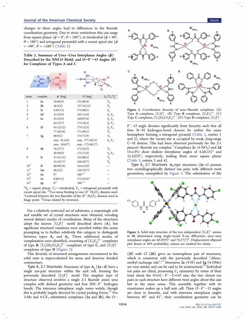
changes to these angles lead to differences in the fluoridecoordination geometry. Due to steric restrictions this can rangefrom square planar (ϕ = 0°, θ = 180°), to tetrahedral (ϕ = 90°,θ = 180°) and tetragonal pyramidal with a vacant apical site (ϕ= <90°, θ = <180°) (Table 2).
For a relatively restricted set of substrates, a surprisingly richand variable set of crystal structures were obtained, revealingseveral distinct modes of coordination. Many of the structuresadopt the known [U2F]
− motif described above (A), butsignificant structural variations were unveiled within this seriesprompting us to further subdivide this category to distinguishbetween types A1 and A2. Three additional modes ofcomplexation were identified, consisting of [U4F2]
2− complexesof type B, [U2(H2O)2F2]
2− complexes of type C, and [U3F]−
complexes of type D (Figure 2).This diversity of structural arrangements encountered in the
solid state is unprecedented for ureas and deserves detailedcommentary.Type A1 2:1 Structures. Structures of type A1 (2a−d) have a
single ion-pair structure within the unit cell, forming thepreviously described [U2F]
− motif. The simplest type ofstructure observed involves a single 2:1 fluoride anion ureacomplex with defined geometry and four NH···F− hydrogenbonds. The interurea interplanar angle varies widely, thoughthis is probably largely directed by crystal packing forces.18 For3-Me and 4-CF3 substituted complexes (2a and 2b), the O···
F−···O angle deviates significantly from linearity such that allfour N−H hydrogen-bond donors lie within the samehemisphere forming a tetragonal pyramid (Table 2, entries 1and 2), where the vacant site is occupied by weak, long-rangeC−H donors. This had been observed previously for the 2:1pinacol−fluoride ion complex.9 Complexes 2c (4-NO2) and 2d(4-n-Pr) show shallow interplanar angles of 4.26(15)° and15.52(9)°, respectively, making them more square planar(Table 2, entries 3 and 4).
Type A2 2:1 Structures. A2-type structures (2e−i) possesstwo crystallographically distinct ion pairs with different twistgeometries, exemplified by Figure 3. The substitution of Me
(2f) with Cl (2h) gives an isomorphous pair of structures,which is consistent with the previously described “chloro-methyl exchange rule”.19 Structures 2e (4-H) and 2g (4-OMe)are very similar and can be said to be isostructural.20 Individualion pairs are chiral, possessing C2 symmetry by virtue of theirtwist about the OC···F−···CO axis; the two closest ionpairs in each structure have different twist angles about this axisbut in the same sense. This assembly together with itsenantiomer makes up a half unit cell. Their O···F−···O anglesare close to linearity, and with interurea interplanar anglesbetween 40° and 81°, their coordination geometry can be
Table 2. Summary of Urea−Urea Interplanar Angles (ϕ)Described by the NNCO Motif, and O···F−···O Angles (θ)for Complexes of Types A and C
entry complex ϕ [deg] θ [deg] Sp/Td/Tpa
1 2a 59.66(9) 131.88(4) Tp
2 2b 56.3(4) 137.55(12) Tp
3 2c 4.26(15) 172.60(5) Sp4 2d 15.52(9) 163.11(4) Sp-Tp
5 2e 43.15(9) 169.87(4) Sp-Td
64.13(7) 173.54(3) Sp-Td
6 2f 43.12(12) 174.12(5) Sp-Td
77.58(10) 171.69(5) Td
7 2g 80.9(2) 174.71(9) Td
maj.: 43.5(4) maj.: 177.10(15) Sp-Td
min.: 38.8(7) min.: 172.49(17)8 2h 76.27(7) 172.23(3) Td
40.59(9) 172.71(4) Sp-Td
9 2i 27.41(15) 155.00(5) Tp
16.52(17) 168.58(7) Tp
10b 2l 60.99(18) 144.51(6)c −11b 2m 68.5(2) 138.72(7)c −12b 2n 0d 180c,d −13b 2o 6.60(15) 175.55(4)c −14b 2p 0d 180c,d −
aSp = square planar, Td = tetrahedral, Tp = tetragonal pyramidal withvacant apical site. bTwo ureas binding to one (F−·H2O)2 dianion used.cCentroid between the two fluorides of the (F−·H2O)2 dianion used ashinge point. dUreas related by inversion.
Figure 2. Coordination diversity of urea−fluoride complexes. (A)Type A complexes, [U2F]
−. (B) Type B complexes, [U4F2]2−. (C)
Type C complexes, [U2(H2O)2F2]2−. (D) Type D complexes, [U3F]
−.
Figure 3. Solid state structure of the two independent [U2F]− anions
in 2h determined using single-crystal X-ray diffraction; urea−ureainterplanar angles are 40.59(9)° and 76.27(7)°. Displacement ellipsoidplot drawn at 50% probability; cations are omitted for clarity.
Journal of the American Chemical Society Article
DOI: 10.1021/jacs.6b07501J. Am. Chem. Soc. 2016, 138, 13314−13325
13316
described either as tetrahedral or as an intermediary statebetween square planar and tetrahedral (Table 2, entries 5−8).Complex 2i (4-n-Bu) stands out, for two separate complexesare observed where the paired anionic ligands describe ashallow V-shape, forming a tetragonal pyramidal coordinationsphere with interplanar urea angles of 16.52(17)° and27.41(15)°, respectively (Table 2, entry 9). This is alsocharacterized by the O···F−···O angles’ distinct deviation fromlinearity.Type B 4:2 Structures. These involve a supramolecular 4:2
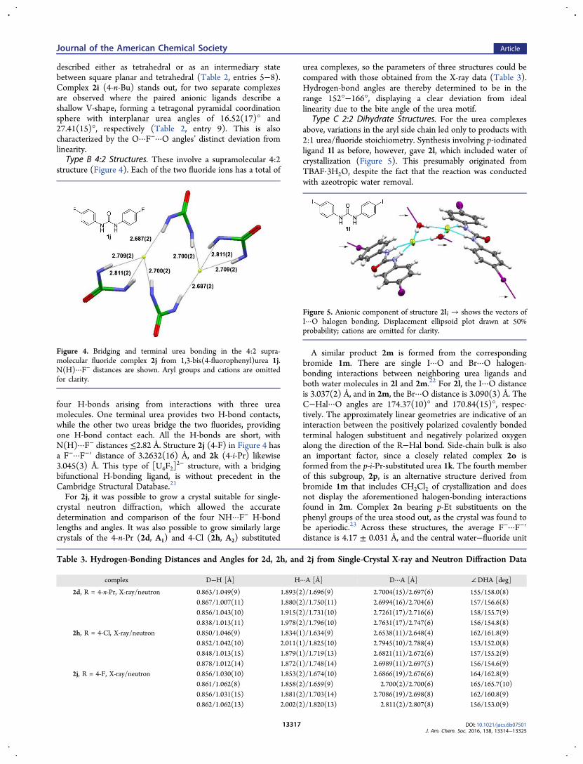
structure (Figure 4). Each of the two fluoride ions has a total of
four H-bonds arising from interactions with three ureamolecules. One terminal urea provides two H-bond contacts,while the other two ureas bridge the two fluorides, providingone H-bond contact each. All the H-bonds are short, withN(H)···F− distances ≤2.82 Å. Structure 2j (4-F) in Figure 4 hasa F−···F−′ distance of 3.2632(16) Å, and 2k (4-i-Pr) likewise3.045(3) Å. This type of [U4F2]
2− structure, with a bridgingbifunctional H-bonding ligand, is without precedent in theCambridge Structural Database.21
For 2j, it was possible to grow a crystal suitable for single-crystal neutron diffraction, which allowed the accuratedetermination and comparison of the four NH···F− H-bondlengths and angles. It was also possible to grow similarly largecrystals of the 4-n-Pr (2d, A1) and 4-Cl (2h, A2) substituted
urea complexes, so the parameters of three structures could becompared with those obtained from the X-ray data (Table 3).Hydrogen-bond angles are thereby determined to be in therange 152°−166°, displaying a clear deviation from ideallinearity due to the bite angle of the urea motif.
Type C 2:2 Dihydrate Structures. For the urea complexesabove, variations in the aryl side chain led only to products with2:1 urea/fluoride stoichiometry. Synthesis involving p-iodinatedligand 1l as before, however, gave 2l, which included water ofcrystallization (Figure 5). This presumably originated fromTBAF·3H2O, despite the fact that the reaction was conductedwith azeotropic water removal.
A similar product 2m is formed from the correspondingbromide 1m. There are single I···O and Br···O halogen-bonding interactions between neighboring urea ligands andboth water molecules in 2l and 2m.22 For 2l, the I···O distanceis 3.037(2) Å, and in 2m, the Br···O distance is 3.090(3) Å. TheC−Hal···O angles are 174.37(10)° and 170.84(15)°, respec-tively. The approximately linear geometries are indicative of aninteraction between the positively polarized covalently bondedterminal halogen substituent and negatively polarized oxygenalong the direction of the R−Hal bond. Side-chain bulk is alsoan important factor, since a closely related complex 2o isformed from the p-i-Pr-substituted urea 1k. The fourth memberof this subgroup, 2p, is an alternative structure derived frombromide 1m that includes CH2Cl2 of crystallization and doesnot display the aforementioned halogen-bonding interactionsfound in 2m. Complex 2n bearing p-Et substituents on thephenyl groups of the urea stood out, as the crystal was found tobe aperiodic.23 Across these structures, the average F−···F−′distance is 4.17 ± 0.031 Å, and the central water−fluoride unit
Figure 4. Bridging and terminal urea bonding in the 4:2 supra-molecular fluoride complex 2j from 1,3-bis(4-fluorophenyl)urea 1j.N(H)···F− distances are shown. Aryl groups and cations are omittedfor clarity.
Table 3. Hydrogen-Bonding Distances and Angles for 2d, 2h, and 2j from Single-Crystal X-ray and Neutron Diffraction Data
complex D−H [Å] H···A [Å] D···A [Å] ∠DHA [deg]
2d, R = 4-n-Pr, X-ray/neutron 0.863/1.049(9) 1.893(2)/1.696(9) 2.7004(15)/2.697(6) 155/158.0(8)0.867/1.007(11) 1.880(2)/1.750(11) 2.6994(16)/2.704(6) 157/156.6(8)0.856/1.043(10) 1.915(2)/1.731(10) 2.7261(17)/2.716(6) 158/155.7(9)0.838/1.013(11) 1.978(2)/1.796(10) 2.7631(17)/2.747(6) 156/154.8(8)
2h, R = 4-Cl, X-ray/neutron 0.850/1.046(9) 1.834(1)/1.634(9) 2.6538(11)/2.648(4) 162/161.8(9)0.852/1.042(10) 2.011(1)/1.825(10) 2.7945(10)/2.788(4) 153/152.0(8)0.848/1.013(15) 1.879(1)/1.719(13) 2.6821(11)/2.672(6) 157/155.2(9)0.878/1.012(14) 1.872(1)/1.748(14) 2.6989(11)/2.697(5) 156/154.6(9)
2j, R = 4-F, X-ray/neutron 0.856/1.030(10) 1.853(2)/1.674(10) 2.6866(19)/2.676(6) 164/162.8(9)0.861/1.062(8) 1.858(2)/1.659(9) 2.700(2)/2.700(6) 165/165.7(10)0.856/1.031(15) 1.881(2)/1.703(14) 2.7086(19)/2.698(8) 162/160.8(9)0.862/1.062(13) 2.002(2)/1.820(13) 2.811(2)/2.807(8) 156/153.0(9)
Figure 5. Anionic component of structure 2l; → shows the vectors ofI···O halogen bonding. Displacement ellipsoid plot drawn at 50%probability; cations are omitted for clarity.
Journal of the American Chemical Society Article
DOI: 10.1021/jacs.6b07501J. Am. Chem. Soc. 2016, 138, 13314−13325
13317
is approximately planar. The dihedral angles between ureaplanes in 2l and 2m are 60.99(18)° and 68.5(2)°, and togetherwith θ values of 144.51(6)° and 138.72(7)°, they describe theshallow U-shape that is formed by the hydrogen-bondedassembly (Table 2, entries 10 and 11). In contrast, the ureaplanes in structures 2n−p are parallel or almost parallel, and θshows a perfectly or almost linear relationship between the twocarbonyl oxygens and the centroid between the two fluorideanions (Table 2, entries 12−14). The Cambridge StructuralDatabase (CSD)21 reveals several examples of (F−·H2O)2dianion clusters, although their generality has not been wellrecognized, despite Emsley’s early discussion.24,25 Here, theirformation in an isolated unit is encouraged in part by thecombination of a bulky p-substituent in the 1,3-diarylurea and aspace-demanding countercation, with further interactionsavailable by halogen bonding to the bridging water.Type D 3:1 Structures. The examples discussed above all
have n-Bu4N+ as the countercation, which is itself quite spatially
demanding. This posed a question: how does the countercationinfluence the overall pattern of urea−fluoride bonding? This ledto analysis of the structures of the Et4N
+ and Me4N+ analogues
of the parent urea 1e. The resulting ion pairs, 2q and 2r, aresimilar but distinct from the 2:1 n-Bu4N
+-derived complex 2e,in that they possess a third coordinated urea ligand. Thestructures are unsymmetrical, with six different H-bond lengths,two of which are considerably longer than the norm. Overall,the three pairs of donor atoms arrange to form a distortedpaddle-wheel motif (Figure 6).
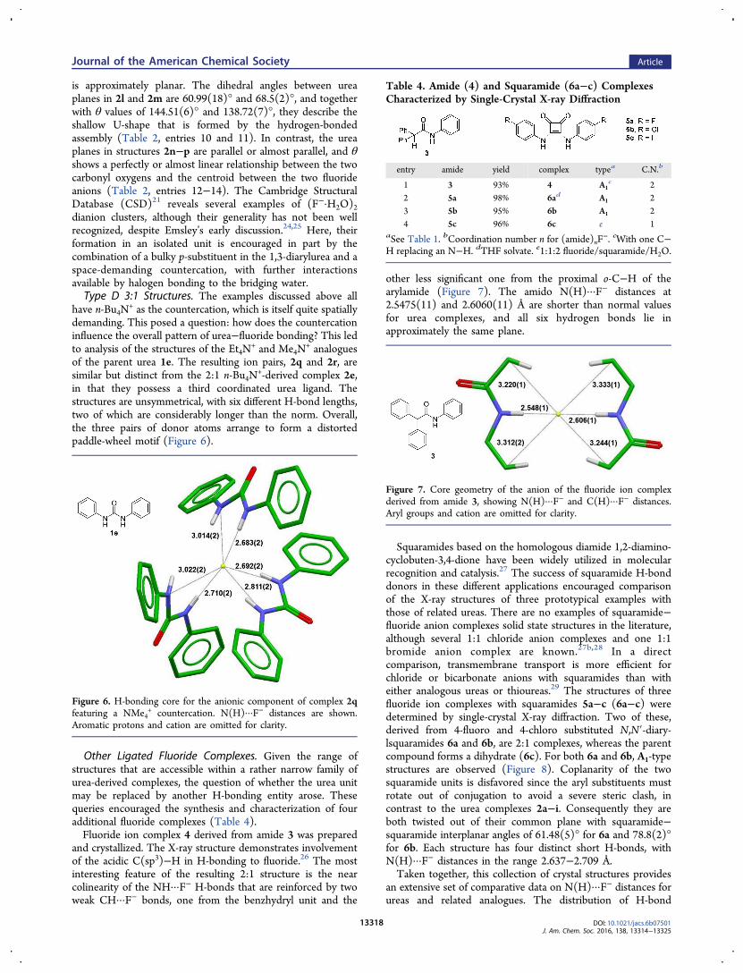
Other Ligated Fluoride Complexes. Given the range ofstructures that are accessible within a rather narrow family ofurea-derived complexes, the question of whether the urea unitmay be replaced by another H-bonding entity arose. Thesequeries encouraged the synthesis and characterization of fouradditional fluoride complexes (Table 4).Fluoride ion complex 4 derived from amide 3 was prepared
and crystallized. The X-ray structure demonstrates involvementof the acidic C(sp3)−H in H-bonding to fluoride.26 The mostinteresting feature of the resulting 2:1 structure is the nearcolinearity of the NH···F− H-bonds that are reinforced by twoweak CH···F− bonds, one from the benzhydryl unit and the
other less significant one from the proximal o-C−H of thearylamide (Figure 7). The amido N(H)···F− distances at2.5475(11) and 2.6060(11) Å are shorter than normal valuesfor urea complexes, and all six hydrogen bonds lie inapproximately the same plane.
Squaramides based on the homologous diamide 1,2-diamino-cyclobuten-3,4-dione have been widely utilized in molecularrecognition and catalysis.27 The success of squaramide H-bonddonors in these different applications encouraged comparisonof the X-ray structures of three prototypical examples withthose of related ureas. There are no examples of squaramide−fluoride anion complexes solid state structures in the literature,although several 1:1 chloride anion complexes and one 1:1bromide anion complex are known.27b,28 In a directcomparison, transmembrane transport is more efficient forchloride or bicarbonate anions with squaramides than witheither analogous ureas or thioureas.29 The structures of threefluoride ion complexes with squaramides 5a−c (6a−c) weredetermined by single-crystal X-ray diffraction. Two of these,derived from 4-fluoro and 4-chloro substituted N,N′-diary-lsquaramides 6a and 6b, are 2:1 complexes, whereas the parentcompound forms a dihydrate (6c). For both 6a and 6b, A1-typestructures are observed (Figure 8). Coplanarity of the twosquaramide units is disfavored since the aryl substituents mustrotate out of conjugation to avoid a severe steric clash, incontrast to the urea complexes 2a−i. Consequently they areboth twisted out of their common plane with squaramide−squaramide interplanar angles of 61.48(5)° for 6a and 78.8(2)°for 6b. Each structure has four distinct short H-bonds, withN(H)···F− distances in the range 2.637−2.709 Å.Taken together, this collection of crystal structures provides
an extensive set of comparative data on N(H)···F− distances forureas and related analogues. The distribution of H-bond
Figure 6. H-bonding core for the anionic component of complex 2qfeaturing a NMe4
+ countercation. N(H)···F− distances are shown.Aromatic protons and cation are omitted for clarity.
Table 4. Amide (4) and Squaramide (6a−c) ComplexesCharacterized by Single-Crystal X-ray Diffraction
entry amide yield complex typea C.N.b
1 3 93% 4 A1c 2
2 5a 98% 6ad A1 23 5b 95% 6b A1 24 5c 96% 6c e 1
aSee Table 1. bCoordination number n for (amide)nF−. cWith one C−
H replacing an N−H. dTHF solvate. e1:1:2 fluoride/squaramide/H2O.
Figure 7. Core geometry of the anion of the fluoride ion complexderived from amide 3, showing N(H)···F− and C(H)···F− distances.Aryl groups and cation are omitted for clarity.
Journal of the American Chemical Society Article
DOI: 10.1021/jacs.6b07501J. Am. Chem. Soc. 2016, 138, 13314−13325
13318
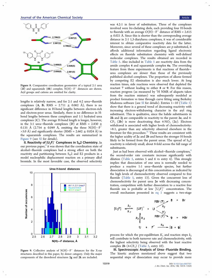
lengths is relatively narrow, and for 2:1 and 4:2 urea−fluoridecomplexes (A, B; RMS = 2.711 ± 0.062 Å), there is nosignificant difference in H-bond lengths between electron-richand electron-poor ureas. Similarly, there is no difference in H-bond lengths between these complexes and 1:1 hydrated ureacomplexes (C). The average H-bond length is longer, however,in the 3:1 urea−fluoride complexes (D) at RMS = 2.830 ±0.135 Å (2.754 ± 0.049 Å, omitting the three N(H)···F−
>3.0 Å) and significantly shorter (RMS = 2.662 ± 0.024 Å) inthe squaramide complexes. The results are summarized inFigure 9 (see SI for details).II. Reactivity of [U2F]
− Complexes in SN2 Chemistry. Inour previous paper,9 it was shown that the coordination state ofalcohol−fluoride complexes had a strong effect on both thereactivity and partitioning between SN2 and E2 products in amodel nucleophilic displacement reaction on a primary alkylbromide. In the most favorable case, the observed selectivity
was 4.2:1 in favor of substitution. Three of the complexesinvolved were bis-chelating diols, each providing four H-bondsto fluoride with an average O(H)···F− distance of RMS = 2.615± 0.021 Å. Since this is shorter than the corresponding averagedistance in 2:1 1,3-diarylurea complexes, it was of considerableinterest to obtain comparative reactivity data for the latter.Moreover, since several of these complexes are p-substituted, itaffords additional information regarding ligand electroniceffects on fluoride substitution chemistry with well-definedmolecular complexes. The results obtained are recorded inTable 5. Also included in Table 5 are reactivity data from theamide complex 4 and squaramide complex 6a. The overridingfeature from these experiments is that reactions of fluoride−urea complexes are slower than those of the previouslypublished alcohol complexes. The proportion of alkene formedby competing E2 elimination is also much lower. At longreaction times, side reactions were observed that depleted thereactant 7 without leading to either 8 or 9. For this reason,reaction progress (as measured by 1H NMR of aliquots takenfrom the reaction mixture) was subsequently modeled asproduct formation vs time through curve fitting using BerkeleyMadonna software (see SI for details). Entries 1−10 (Table 5)show that there is a general trend of decreasing reactivity withincreasing electron-withdrawing character in the aryl ringsubstituent. This is qualitative, since the halide substituents in2h and 2j are comparable in reactivity to the parent 2e, and 4-CF3 (2b) is more deactivating than 4-NO2 (2c). Electronwithdrawal is associated with higher levels of chemoselectivity:>8:1, greater than any selectivity observed elsewhere in theliterature for this procedure.3c These results are consistent withthe higher acidity of 2c and 2b and hence the stronger H-bondslikely to be formed from the parent ureas. The spread of SN2reactivity is relatively small, about 8-fold across the full range ofsubstituents.Just as had been observed with alcohol−fluoride complexes,9
the second-order rate constants increase significantly withdilution (Table 5, entries 5 and 6 vs entry 4). This stronglyimplies that dissociation of one urea is normally needed toproduce a reactive 1:1 urea−fluoride species, but furtherdissociation is discouraged at this concentration as indicated bythe high levels of chemoselectivity observed compared to freefluoride (Table 5, entry 13). Given the concurrent loss ofchemoselectivity for parent urea 2e with decreasing concen-tration, competition with further dissociation to a reactive freefluoride ion is probable at low [U2F]
− concentration. Theoverall mechanism presented in eq 1 suggests a two-stage
process for which the pre-equilibrium K1 and reaction steps k2will contribute to both turnover rate and chemoselectivity, withthe highest selectivity being observed with the least reactivecomplex 2b (4-CF3) (Table 5, entry 10).
III. Spectroscopic Analysis of Urea−Fluoride Binding.The kinetic analyses mentioned above suggest that twosequential steps of dissociation may occur to provide more
Figure 8. Comparative coordination geometries of a typical 2:1 urea(2f) and squaramide (6b) complex. N(H)···F− distances are shown.Aryl groups and cations are omitted for clarity.
Figure 9. Collective analysis of N(H)···F− distances for the X-raystructures described in this paper, by donor category. Only the majorcomponents of the disordered structures 2g and 2k are included.
Journal of the American Chemical Society Article
DOI: 10.1021/jacs.6b07501J. Am. Chem. Soc. 2016, 138, 13314−13325
13319
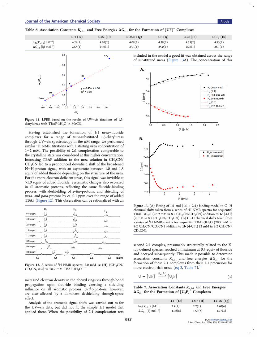
active fluoride species that react as either a nucleophile or base.We examined urea complexation of fluoride ion in solution togain further insight. It is known that the process results inchanges in the UV/visible region, and our first objective wasthus to study the effects of changes in the electronic characterof the 1,3-diarylureas. At first, UV−vis titrations of 8.0 μMsolutions of selected ureas in MeCN, with a TBAF solutionwhose exact concentration had previously been establishedusing a known method,30 were performed without changing theurea concentration over the course of the addition (Figure 10).A bathochromic shift of the band with a maximum between250−264 nm was observed, and the buildup of the absorptionat the new maximum between 259−282 nm was plotted againstthe concentration of added fluoride. Association constants,Ka,1:1, that assumed the formation of a 1:1 urea−fluoridecomplex were obtained (eq 2, Table 6) via nonlinear least-squares regression using DynaFit4 software, forming the basisof the Hammett plot of Figure 11, with a ρ-value (vs 2σp) forthe process of 0.43 ± 0.03.31 Free energies of complexationΔG1:1 ranged from 23.3−28.1 kJ/mol.
+ − −H IooooooU F [UF]K , 1:1a
(2)
In the case of electron-rich 1,3-diarylureas, assuming theformation of a 1:1 urea−fluoride complex as the only productgave good results, with the presence of two isosbestic pointsalso indicating the clean transition between two structures (seeFigure 10A). Band deconvolution analysis also showed thepresence of two species over the course of the titration (seeFigure S22). For electron-deficient 1,3-diarylureas, on the otherhand, the behavior was more complex and lacked a cleanisosbestic point (Figure 10B). Here, band deconvolutionsuggested the formation of a third species, which becomespredominant at higher F− concentrations (see Figures S38−S39). Taking the early data points in the titration (up to ca.10 equiv), a reasonable fit to the 1:1 model was obtained; athigher concentrations of F− the results deviated from this. TheUV−vis spectra of the 4-nitrophenylurea 1c in the presence ofvarious anions had been studied previously; for fluoride this was
dominated by a band at 475 nm associated with thedeprotonated species.32 This is a likely possibility in thepresent case for the more N−H acidic examples, particularly 4-CF3 substituted 1b.33
Table 5. Reactions of Urea−, Amide−, and Squaramide−Fluoride Complexes with 7 in CH3CN (Conditions: (X)2F− (0.4
mmol), 7 (0.2 mmol), CH3CN (0.8 mL), 70 °C)
entrya complex k2(SN2)b [×10−5 M−1 s−1] k′2(E2)b [×10−5 M−1 s−1] k2(SN2)/k′2(E2)c
1; 4-OMe 2g 12.7 2.45 5.22; 4-Me 2f 9.8 1.65 5.73; 3-Me 2a 5.95 1.07 5.64; H 2e 5.76 0.85 6.85; Hd 2e 18.8 4.63 4.16; He 2e 42.4 12.9 3.37; 4-F 2j 5.67 0.80 7.18; 4-Cl 2h 5.83 0.84 6.99; 4-NO2 2c 3.2 0.38 8.410; 4-CF3
f 2b 1.65 0.19 8.711g 4 178 112 1.612g,h 6a (2) (0.7) 2.813h TBAF·3H2O 375 235 1.6
aSubstituent of urea. bFrom 1H NMR analysis by curve fitting. cFor comparison, the ratio k2(SN2)/k′2(E2) for TBAF·4(t-BuOH) was 2.1 (see ref 9).d0.0625 M. e0.025 M. fIn a separate reaction under identical conditions, yields of 8 and 9 after 48 h were determined to be 45% and 4%, respectively,using 19F and 1H NMR with 1-fluoro-3-nitrobenzene as internal reference. gSee Table 4. hSlow reaction accompanied by decomposition of diamide5a.
Figure 10. (A) Series of UV−vis spectra: 8.0 μM 1e (4-H) (MeCN)vs 1.58 mM TBAF·3H2O. Inset: Titration profile, 272 nm. (B) Samefor 1b (4-CF3). Inset: Titration profile, 282 nm.
Journal of the American Chemical Society Article
DOI: 10.1021/jacs.6b07501J. Am. Chem. Soc. 2016, 138, 13314−13325
13320
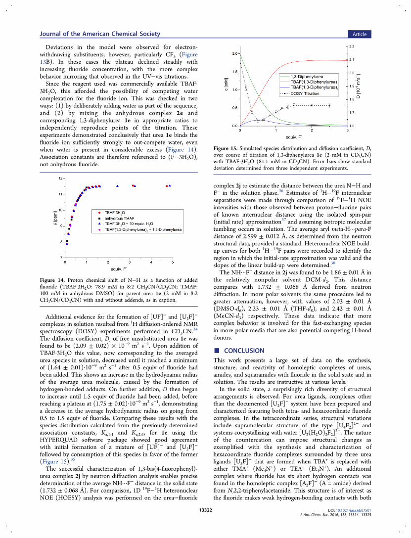
Having established the formation of 1:1 urea−fluoridecomplexes for a range of para-substituted 1,3-diarylureasthrough UV−vis spectroscopy in the μM range, we performedsimilar 1H NMR titrations with a starting urea concentration of1−2 mM. The possibility of 2:1 complexation comparable tothe crystalline state was considered at this higher concentration.Increasing TBAF addition to the urea solution in CH3CN/CD3CN led to a pronounced downfield shift of the broadenedN−H proton signal, with an asymptote between 1.0 and 1.5equiv of added fluoride depending on the structure of the urea.For the more electron-deficient ureas, this signal was invisible at>1.0 equiv of added fluoride. Systematic changes also occurredin all aromatic protons, reflecting the same fluoride-bindingprocess, with deshielding of ortho-protons, and shielding ofmeta- and para-protons by ca. 0.1 ppm over the range of addedTBAF (Figure 12). This observation can be rationalized with an
increased electron density in the phenyl rings via through-bondpropagation upon fluoride binding exerting a shieldinginfluence on all aromatic protons. Ortho-protons, however,are also affected by a dominant deshielding through-spaceeffect.Analysis of the aromatic signal shifts was carried out as for
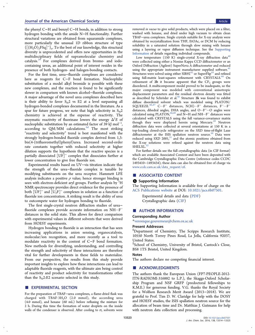
the UV−vis data, but did not fit the simple 1:1 model thatapplied there. When the possibility of 2:1 complexation was
included in the model a good fit was obtained across the rangeof substituted ureas (Figure 13A). The concentration of this
second 2:1 complex, presumably structurally related to the X-ray-defined species, reached a maximum at 0.5 equiv of fluorideand decayed subsequently. This made it possible to determineassociation constants Ka,2:1 and free energies ΔG2:1 for theformation of these 2:1 complexes from their 1:1 precursors formore electron-rich ureas (eq 3, Table 7).31
+ − −H IooooooU [UF] [U F]K , 2:1
2a
(3)
Table 6. Association Constants Ka,1:1 and Free Energies ΔG1:1 for the Formation of [UF]− Complexes
4-H (1e) 4-Me (1f) 4-OMe (1g) 4-F (1j) 4-Cl (1h) 4-CF3 (1b)
log(Ka,1:1) [M−1] 4.29(1) 4.20(2) 4.09(2) 4.38(2) 4.53(2) 4.93(1)
ΔG1:1 [kJ mol−1] 24.5(1) 24.0(1) 23.3(1) 25.0(1) 25.8(1) 28.1(1)
Figure 11. LFER based on the results of UV−vis titrations of 1,3-diarylureas with TBAF·3H2O in MeCN.
Figure 12. A series of 1H NMR spectra: 2.0 mM 1e (H) (CH3CN/CD3CN, 8:2) vs 78.9 mM TBAF·3H2O.
Figure 13. (A) Fitting of 1:1 and (1:1 + 2:1) binding model to C−Hchemical shifts taken from a series of 1H NMR spectra for sequentialTBAF·3H2O (78.9 mM in 8:2 CH3CN/CD3CN) addition to 1e (4-H)(2 mM in 8:2 CH3CN/CD3CN). (B) C−H chemical shifts taken froma series of 1H NMR spectra for sequential TBAF·3H2O (78.9 mM in8:2 CH3CN/CD3CN) addition to 1b (4-CF3) (2 mM in 8:2 CH3CN/CD3CN).
Table 7. Association Constants Ka,2:1 and Free EnergiesΔG2:1 for the Formation of [U2F]
− Complexes
4-H (1e) 4-Me (1f) 4-OMe (1g)
log(Ka,2:1) [M−1] 2.4(1) 2.7(1) 2.40(6)
ΔG2:1 [kJ mol−1] 13.6(8) 15.3(6) 13.7(3)
Journal of the American Chemical Society Article
DOI: 10.1021/jacs.6b07501J. Am. Chem. Soc. 2016, 138, 13314−13325
13321
Deviations in the model were observed for electron-withdrawing substituents, however, particularly CF3 (Figure13B). In these cases the plateau declined steadily withincreasing fluoride concentration, with the more complexbehavior mirroring that observed in the UV−vis titrations.Since the reagent used was commercially available TBAF·
3H2O, this afforded the possibility of competing watercomplexation for the fluoride ion. This was checked in twoways: (1) by deliberately adding water as part of the sequence,and (2) by mixing the anhydrous complex 2e andcorresponding 1,3-diphenylurea 1e in appropriate ratios toindependently reproduce points of the titration. Theseexperiments demonstrated conclusively that urea 1e binds thefluoride ion sufficiently strongly to out-compete water, evenwhen water is present in considerable excess (Figure 14).Association constants are therefore referenced to (F−·3H2O),not anhydrous fluoride.
Additional evidence for the formation of [UF]− and [U2F]−
complexes in solution resulted from 1H diffusion-ordered NMRspectroscopy (DOSY) experiments performed in CD3CN.
34
The diffusion coefficient, D, of free unsubstituted urea 1e wasfound to be (2.09 ± 0.02) × 10−9 m2 s−1. Upon addition ofTBAF·3H2O this value, now corresponding to the averagedurea species in solution, decreased until it reached a minimumof (1.64 ± 0.01)·10−9 m2 s−1 after 0.5 equiv of fluoride hadbeen added. This shows an increase in the hydrodynamic radiusof the average urea molecule, caused by the formation ofhydrogen-bonded adducts. On further addition, D then beganto increase until 1.5 equiv of fluoride had been added, beforereaching a plateau at (1.75 ± 0.02)·10−9 m2 s−1, demonstratinga decrease in the average hydrodynamic radius on going from0.5 to 1.5 equiv of fluoride. Comparing these results with thespecies distribution calculated from the previously determinedassociation constants, Ka,1:1 and Ka,2:1, for 1e using theHYPERQUAD software package showed good agreementwith initial formation of a mixture of [UF]− and [U2F]
−
followed by consumption of this species in favor of the former(Figure 15).35
The successful characterization of 1,3-bis(4-fluorophenyl)-urea complex 2j by neutron diffraction analysis enables precisedetermination of the average NH···F− distance in the solid state(1.732 ± 0.068 Å). For comparison, 1D 19F−1H heteronuclearNOE (HOESY) analysis was performed on the urea−fluoride
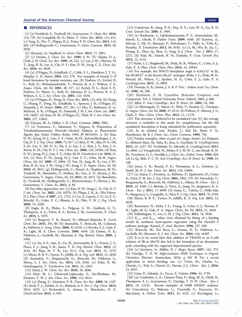
complex 2j to estimate the distance between the urea N−H andF− in the solution phase.36 Estimates of 1H−19F internuclearseparations were made through comparison of 19F−1H NOEintensities with those observed between proton−fluorine pairsof known internuclear distance using the isolated spin-pair(initial rate) approximation37 and assuming isotropic moleculartumbling occurs in solution. The average aryl meta-H···para-Fdistance of 2.599 ± 0.012 Å, as determined from the neutronstructural data, provided a standard. Heteronuclear NOE build-up curves for both 1H−19F pairs were recorded to identify theregion in which the initial-rate approximation was valid and theslopes of the linear build-up were determined.38
The NH···F− distance in 2j was found to be 1.86 ± 0.01 Å inthe relatively nonpolar solvent DCM-d2. This distancecompares with 1.732 ± 0.068 Å derived from neutrondiffraction. In more polar solvents the same procedure led togreater attenuation, however, with values of 2.03 ± 0.01 Å(DMSO-d6), 2.23 ± 0.01 Å (THF-d8), and 2.42 ± 0.01 Å(MeCN-d3) respectively. These data indicate that morecomplex behavior is involved for this fast-exchanging speciesin more polar media that are also potential competing H-bonddonors.
■ CONCLUSIONThis work presents a large set of data on the synthesis,structure, and reactivity of homoleptic complexes of ureas,amides, and squaramides with fluoride in the solid state and insolution. The results are instructive at various levels.In the solid state, a surprisingly rich diversity of structural
arrangements is observed. For urea ligands, complexes otherthan the documented [U2F]
− system have been prepared andcharacterized featuring both tetra- and hexacoordinate fluoridecomplexes. In the tetracoordinate series, structural variationsinclude supramolecular structure of the type [U4F2]
2− andsystems cocrystallizing with water [U2(H2O)2F2]
2−. The natureof the countercation can impose structural changes asexemplified with the synthesis and characterization ofhexacoordinate fluoride complexes surrounded by three urealigands [U3F]
− that are formed when TBA+ is replaced witheither TMA+ (Me4N
+) or TEA+ (Et4N+). An additional
complex where fluoride has six short hydrogen contacts wasfound in the homoleptic complex [A2F]
− (A = amide) derivedfrom N,2,2-triphenylacetamide. This structure is of interest asthe fluoride makes weak hydrogen-bonding contacts with both
Figure 14. Proton chemical shift of N−H as a function of addedfluoride (TBAF·3H2O: 78.9 mM in 8:2 CH3CN/CD3CN; TMAF:100 mM in anhydrous DMSO) for parent urea 1e (2 mM in 8:2CH3CN/CD3CN) with and without addends, as in caption.
Figure 15. Simulated species distribution and diffusion coefficient, D,over course of titration of 1,3-diphenylurea 1e (2 mM in CD3CN)with TBAF·3H2O (81.1 mM in CD3CN). Error bars show standarddeviation determined from three independent experiments.
Journal of the American Chemical Society Article
DOI: 10.1021/jacs.6b07501J. Am. Chem. Soc. 2016, 138, 13314−13325
13322
the phenyl C−H and benzyl C−H bonds, in addition to stronghydrogen bonding with the amide N−H functionality. Furtherstructural variations are obtained from squaramide complexes,more particularly the unusual 1D ribbon structure of type{[(H2O)2FSq]
−}n. To the best of our knowledge, this structuraldiversity is unprecedented and offers new opportunities in themultidisciplinary fields of supramolecular chemistry andcatalysis.39 For complexes derived from bromo- and iodo-containing ureas, an additional point of interest resides in thepresence of both hydrogen- and halogen-bonding contacts.For the first time, urea−fluoride complexes are considered
here as reagents for C−F bond formation. Nucleophilicsubstitution of a model alkyl bromide is possible with thesenew complexes, and the reaction is found to be significantlyslower in comparison with known alcohol−fluoride complexes.A major advantage of the novel urea complexes described hereis their ability to favor SN2 vs E2 at a level surpassing allhydrogen-bonded complexes documented in the literature. As aspur for future progress, we note that high selectivity in SN2chemistry is achieved at the expense of reactivity. Theenzymatic reactivity of fluorinase lowers the energy Δ‡E ofnucleophilic substitution by an estimated factor of 39 kJ mol−1,according to QM/MM calculations.5d The most striking“reactivity and selectivity” trend is best manifested with thestrongly hydrogen-bonded fluoride complex derived from 1,3-bis(4-(trifluoromethyl)phenyl)urea. Increased second-orderrate constants together with reduced selectivity at higherdilution supports the hypothesis that the reactive species is apartially dissociated [UF]− complex that dissociates further atlower concentration to give free fluoride ion.Experimental results based on UV−vis titration indicate that
the strength of the urea−fluoride complex is tunable bymodifying substituents on the urea receptor. Hammett LFEanalysis indicates a positive ρ value; hence stronger binding isseen with electron-deficient aryl groups. Further analysis by 1HNMR spectroscopy provides direct evidence for the presence ofboth [UF]− and [U2F]
− complexes in solution as a function offluoride ion concentration. A striking result is the ability of ureato outcompete water for hydrogen bonding to fluoride.The first single-crystal neutron diffraction studies of urea−
fluoride complexes provide accurate information on NH···F−
distances in the solid state. This allows for direct comparisonwith experimental values in different solvents that were derivedfrom HOESY experiments.Hydrogen bonding to fluoride is an interaction that has seen
increasing applications in anion sensing, organocatalysis,molecular/ion recognition, and more recently as a tool tomodulate reactivity in the context of C−F bond formation.New methods for diversifying, understanding, and controllingthe strength and selectivity of these interactions are thereforevital for further developments in these fields to materialize.From our perspective, the results from this study provideimportant insights to explore how these interactions can lead toadaptable fluoride reagents, with the ultimate aim being controlof reactivity and product selectivity for transformations otherthan the SN2:E2 scenario selected in this study.
■ EXPERIMENTAL SECTIONFor the preparation of TBAF−urea complexes, a flame-dried flask wascharged with TBAF·3H2O (2.0 mmol), the according urea(4.0 mmol), and hexane (60 mL) before refluxing the mixture for2 h. During this time the formation of water droplets on the insidewalls of the condenser is observed. After cooling to rt, solvents were
removed in vacuo to give solid products, which were placed on a filter,washed with hexane, and dried under high vacuum to obtain cleanTBAF−urea complexes. Single crystals suitable for X-ray analysis wereobtained by recrystallization from THF, EtOAc, or DCM by reducingsolubility in a saturated solution through slow mixing with hexaneusing a layering or vapor diffusion technique. See the SupportingInformation of details regarding individual compounds.
Low temperature (150 K) single-crystal X-ray diffraction data40
were collected using either a Nonius Kappa CCD diffractometer or anOxford Diffraction (Agilent) SuperNova A diffractometer and reducedusing the appropriate instrument manufacturer supplied software.41
Structures were solved using either SIR9242 or SuperFlip43 and refinedusing full-matrix least-squares refinement with CRYSTALS.44 Onrefinement of 2b it became apparent that the CF3 groups weredisordered. A multicomponent model proved to be inadequate, so themajor component was modeled with conventional anisotropicdisplacement parameters and the residual electron density was fittedas described by Schroder et al.45 Structure 2k was found to exhibitdiffuse disordered solvent which was modeled using PLATON/SQUEEZE.46,47 C···F− distances, N(H)···F− distances, F−···F−
distances, dihedral angles, DHA angles, and O···F−···O angles werecalculated using PLATON,46,48 and N−H and NH···F− distances werecalculated with CRYSTALS using the full variance-covariance matrixthough they were displayed herein using Mercury.49 Neutrondiffraction data were collected at several orientations at 150 K in atop-loading closed-cycle refrigerator on the SXD time-of-flight Lauediffractometer at the ISIS spallation neutron source.50 Data werereduced using SXD 2001,51 and the atomic positions obtained fromthe X-ray solutions were refined against the neutron data usingSHELXL.52
For further details see the full crystallographic data (in CIF format)which are available Associated Content and have been deposited withthe Cambridge Crystallographic Data Centre (reference codes CCDC1493410−1493434); these data can also be obtained free of charge viawww.ccdc.cam.ac.uk/data_request/cif.
■ ASSOCIATED CONTENT*S Supporting InformationThe Supporting Information is available free of charge on theACS Publications website at DOI: 10.1021/jacs.6b07501.
Experimental details and data (PDF)Crystallographic data (CIF)
■ AUTHOR INFORMATIONCorresponding Author*[email protected] Addresses§Department of Chemistry, The Scripps Research Institute,10550 North Torrey Pines Road, La Jolla, California 92037,United States.∥School of Chemistry, University of Bristol, Cantock’s Close,BS8 1TS Bristol, United Kingdom.NotesThe authors declare no competing financial interest.
■ ACKNOWLEDGMENTSThe authors thank the European Union (FP7-PEOPLE-2012-ITN-RADIOMI-316882 to L.P.), the Skaggs-Oxford Scholar-ship Program and NSF GRFP (predoctoral fellowships toK.M.E.) for generous funding. V.G. thanks the Royal Societyfor a Wolfson Research Merit Award (2013−2018). We aregrateful to Prof. Tim D. W. Claridge for help with the DOSYand HOESY studies, the ISIS spallation neutron source for theallocation of beam time and Dr. Matthias J. Gutmann for helpwith neutron data collection and processing.
Journal of the American Chemical Society Article
DOI: 10.1021/jacs.6b07501J. Am. Chem. Soc. 2016, 138, 13314−13325
13323
■ REFERENCES(1) (a) Preshlock, S.; Tredwell, M.; Gouverneur, V. Chem. Rev. 2016,116, 719. (b) Campbell, M. G.; Ritter, T. Chem. Rev. 2015, 115, 612.(c) Yang, X.; Wu, T.; Phipps, R. J.; Toste, F. D. Chem. Rev. 2015, 115,826. (d) Hollingworth, C.; Gouverneur, V. Chem. Commun. 2012, 48,2929.(2) Harsanyi, A.; Sandford, G. Green Chem. 2015, 17, 2081.(3) (a) Emsley, J. Chem. Soc. Rev. 1980, 9, 91. (b) Adams, D. J.;Clark, J. H. Chem. Soc. Rev. 1999, 28, 225. (c) Lee, J.-W.; Oliveira, M.T.; Jang, H. B.; Lee, S.; Chi, D. Y.; Kim, D. W.; Song, C. E. Chem. Soc.Rev. 2016, 45, 4638.(4) (a) O’Hagan, D.; Schaffrath, C.; Cobb, S. L.; Hamilton, J. T. G.;Murphy, C. D. Nature 2002, 416, 279. For examples of formal C−Fbond formation by mutant enzymes, see: (b) Nashiru, O.; Zechel, D.L.; Stoll, D.; Mohammadzadeh, T.; Warren, R. A. J.; Withers, S. G.Angew. Chem., Int. Ed. 2001, 40, 417. (c) Zechel, D. L.; Reid, S. P.;Nashiru, O.; Mayer, C.; Stoll, D.; Jakeman, D. L.; Warren, R. A. J.;Withers, S. G. J. Am. Chem. Soc. 2001, 123, 4350.(5) (a) O’Hagan, D.; Deng, H. Chem. Rev. 2015, 115, 634. (b) Dong,C.; Huang, F.; Deng, H.; Schaffrath, C.; Spencer, J. B.; O’Hagan, D.;Naismith, J. H. Nature 2004, 427, 561. (c) Zhu, X.; Robinson, D. A.;McEwan, A. R.; O’Hagan, D.; Naismith, J. H. J. Am. Chem. Soc. 2007,129, 14597. (d) Senn, H. M.; O’Hagan, D.; Thiel, W. J. Am. Chem. Soc.2005, 127, 13643.(6) Vincent, M. A.; Hillier, I. H. Chem. Commun. 2005, 5902.(7) (a) Yonezawa, T.; Sakamoto, Y.; Nogawa, K. Preparation ofTetrabutylammonium Fluoride−Alcohol Adducts as FluorinationAgents. Jpn. Kokai Tokkyo Koho, 1994, JP 06316551 A. (b) Kim,D. W.; Jeong, H.-J.; Lim, S. T.; Sohn, M.-H.; Katzenellenbogen, J. A.;Chi, D. Y. J. Org. Chem. 2008, 73, 957. (c) Kim, D. W.; Ahn, D.-S.; Oh,Y.-H.; Lee, S.; Kil, H. S.; Oh, S. J.; Lee, S. J.; Kim, J. S.; Ryu, J. S.;Moon, D. H.; Chi, D. Y. J. Am. Chem. Soc. 2006, 128, 16394. (d) Kim,D. W.; Jeong, H.-J.; Lim, S. T.; Sohn, M.-H. Tetrahedron Lett. 2010, 51,432. (e) Kim, D. W.; Jeong, H.-J.; Lim, S. T.; Sohn, M.-H. Angew.Chem., Int. Ed. 2008, 47, 8404. (f) Yan, H.; Jang, H. B.; Lee, J.-W.;Kim, H. K.; Lee, S. W.; Yang, J. W.; Song, C. E. Angew. Chem., Int. Ed.2010, 49, 8915. (g) Hollingworth, C.; Hazari, A.; Hopkinson, M. N.;Tredwell, M.; Benedetto, E.; Huiban, M.; Gee, A. D.; Brown, J. M.;Gouverneur, V. Angew. Chem., Int. Ed. 2011, 50, 2613. (h) Benedetto,E.; Tredwell, M.; Hollingworth, C.; Khotavivattana, T.; Brown, J. M.;Gouverneur, V. Chem. Sci. 2013, 4, 89.(8) For other approaches, see: (a) Kim, D. W.; Song, C. E.; Chi, D. Y.J. Am. Chem. Soc. 2002, 124, 10278. (b) Pliego, J. R., Jr.; Pilo-Veloso,D. J. Phys. Chem. B 2007, 111, 1752. (c) Lu, S.; Lepore, S. D.; Li, S. Y.;Mondal, D.; Cohn, P. C.; Bhunia, A. K.; Pike, V. W. J. Org. Chem.2009, 74, 5290.(9) Engle, K. E.; Pfeifer, L.; Pidgeon, G. W.; Giuffredi, G. T.;Thompson, A. L.; Paton, R. S.; Brown, J. M.; Gouverneur, V. Chem.Sci. 2015, 6, 5293.(10) (a) Bregovic, V. B.; Basaric, N.; Mlinaric-Majerski, K. Coord.Chem. Rev. 2015, 295, 80. (b) Amendola, V.; Boiocchi, M.; Colasson,B.; Fabbrizzi, L. Inorg. Chem. 2006, 45, 6138. (c) Brooks, S. J.; Gale, P.A.; Light, M. E. Chem. Commun. 2006, 4344. (d) Gomez, D. E.;Fabbrizzi, L.; Licchelli, M.; Monzani, E. Org. Biomol. Chem. 2005, 3,1495.(11) (a) Xu, S.-Y.; Sun, X.; Ge, H.; Arrowsmith, R. L.; Fossey, J. S.;Pascu, S. I.; Jiang, Y.-B.; James, T. D. Org. Biomol. Chem. 2015, 13,4143. (b) Raju, M. V. R.; Lin, H.-C. Org. Lett. 2013, 15, 1274.(c) Elmes, R. B. P.; Turner, P.; Jolliffe, K. A. Org. Lett. 2013, 15, 5638.(d) Amendola, V.; Bergamaschi, G.; Boiocchi, M.; Fabbrizzi, L.;Mosca, L. J. Am. Chem. Soc. 2013, 135, 6345. (e) Duke, R. M.;Gunnlaugsson, T. Tetrahedron Lett. 2011, 52, 1503.(12) Steed, J. W. Chem. Soc. Rev. 2010, 39, 3686.(13) Etter, M. C.; Urbanczyk-Lipkowska, Z.; Zia-Ebrahimi, M.;Panunto, T. W. J. Am. Chem. Soc. 1990, 112, 8415.(14) (a) Zhang, Z.; Schreiner, P. R. Chem. Soc. Rev. 2009, 38, 1187.(b) Auvil, T. J.; Schafer, A. G.; Mattson, A. E. Eur. J. Org. Chem. 2014,2014, 2633. (c) Beckendorf, S.; Asmus, S.; Mancheno, O. G.ChemCatChem 2012, 4, 926.
(15) Custelcean, R.; Jiang, D.-E.; Hay, B. P.; Luo, W. S.; Gu, B. H.Cryst. Growth Des. 2008, 8, 1909.(16) (a) Ravikumar, I.; Lakshminarayanan, P. S.; Arunachalam, M.;Suresh, E.; Ghosh, P. Dalton Trans. 2009, 4160. (b) Kormos, A.;Moczar, I.; Pal, D.; Baranyai, P.; Holczbauer, T.; Pallo, A.; Toth, K.;Huszthy, P. Tetrahedron 2013, 69, 8142. (c) Li, M.; Wu, B.; Jia, C.;Huang, X.; Zhao, Q.; Shao, S.; Yang, X.-J. Chem. - Eur. J. 2011, 17,2272. (d) Paul, M.; Adarsh, N. N.; Dastidar, P. Cryst. Growth Des.2012, 12, 4135.(17) Kirby, I. L.; Brightwell, M.; Pitak, M. B.; Wilson, C.; Coles, S. J.;Gale, P. A. Phys. Chem. Chem. Phys. 2014, 16, 10943.(18) For example, the NNCO interplanar angle is 4.26(15)° in 2c,but 40.30(3)° in the known Me4N
+ analogue: Kirby, I. L.; Pitak, M. B.;Wenzel, M.; Wilson, C.; Sparkes, H. A.; Coles, S. J.; Gale, P. A.CrystEngComm 2013, 15, 9003.(19) Desiraju, G. R.; Sarma, J. A. R. P. Proc. - Indian Acad. Sci., Chem.Sci. 1986, 96, 599.(20) Herbstein, F. H. Crystalline Molecular Complexes andCompounds, Vol. I; Oxford University Press: Oxford, 2005; p 606.(21) Allen, F. Acta Crystallogr., Sect. B: Struct. Sci. 2002, 58, 380.(22) (a) Metrangolo, P.; Meyer, F.; Pilati, T.; Resnati, G.; Terraneo,G. Angew. Chem., Int. Ed. 2008, 47, 6114. (b) Politzer, P.; Murray, J. S.;Clark, T. Phys. Chem. Chem. Phys. 2013, 15, 11178.(23) This structure is believed to be modulated (see SI); the averagestructure is included in this study for completeness, but the fullmodulated description is being submitted for publication separately.(24) As an isolated unit: Emsley, J.; Arif, M.; Bates, P. A.;Hursthouse, M. B. J. Chem. Soc., Chem. Commun. 1989, 738.(25) Further examples, often as part of a larger cluster: (a) Dalapati,S.; Akhtarul Alam, M.; Saha, R.; Jana, S.; Guchhait, N. CrystEngComm2012, 14, 1527. (b) Trzybin ski, D.; Sikorski, A. CrystEngComm 2013,15, 6808. (c) Vreugdenhil, W.; Birker, P. J. M. W. L.; ten Hoedt, R. W.M.; Verschoor, G. C.; Reedijk, J. J. Chem. Soc., Dalton Trans. 1984, 429.(d) Li, Q.; Mak, T. C. W. Acta Crystallogr., Sect. B: Struct. Sci. 1998, 54,180.(26) Jones, C. R.; Baruah, P. K.; Thompson, A. L.; Scheiner, S.;Smith, M. D. J. Am. Chem. Soc. 2012, 134, 12064.(27) (a) Garau, C.; Frontera, A.; Ballester, P.; Quinonero, D.; Costa,A.; Deya, P. M. Eur. J. Org. Chem. 2005, 2005, 179. (b) Amendola, V.;Bergamaschi, G.; Boiocchi, M.; Fabbrizzi, L.; Milani, M. Chem. - Eur. J.2010, 16, 4368. (c) Aleman, J.; Parra, A.; Jiang, H.; Jørgensen, K. A.Chem. - Eur. J. 2011, 17, 6890. (d) Gaeta, C.; Talotta, C.; Della Sala,P.; Margarucci, L.; Casapullo, A.; Neri, P. J. Org. Chem. 2014, 79, 3704.(28) Elmes, R. B. P.; Turner, P.; Jolliffe, K. A. Org. Lett. 2013, 15,5638.(29) Busschaert, N.; Kirby, I. L.; Young, S.; Coles, S. J.; Horton, P.N.; Light, M. E.; Gale, P. A. Angew. Chem., Int. Ed. 2012, 51, 4426.(30) Sokkalingam, P.; Lee, C.-H. J. Org. Chem. 2011, 76, 3820.(31) Ka,1:1 and Ka,2:1 values were obtained by fitting of a bindingmodel by nonlinear least-squares regression using the DynaFit 4software package: Kuzmic, P. Anal. Biochem. 1996, 237, 260.(32) Boiocchi, M.; Del Boca, L.; Gomez, D. E.; Fabbrizzi, L.;Licchelli, M.; Monzani, E. J. Am. Chem. Soc. 2004, 126, 16507.(33) It is to be noted here that addition of TBAOH to an 8 μMsolution of 1b in MeCN also led to the formation of an absorptionpeak coinciding with the supposed deprotonated species.(34) (a) Jerschow, A.; Muller, N. J. Magn. Reson. 1997, 125, 372.(b) Claridge, T. D. W. High-resolution NMR Techniques in OrganicChemistry; Elsevier: Amsterdam, 2016; p 381 ff. For a recentapplication to anion binding, see: (c) Toure, M.; Charles, L.;Chendo, C.; Viel, S.; Chuzel, O.; Parrain, J.-L. Chem. - Eur. J. 2016,22, 8937.(35) Gans, P.; Sabatini, A.; Vacca, A. Talanta 1996, 43, 1739.(36) (a) Combettes, L. E.; Clausen-Thue, P.; King, M. A.; Odell, B.;Thompson, A. L.; Gouverneur, V.; Claridge, T. D. W. Chem. - Eur. J.2012, 18, 13133. Recent examples of NMR HOESY analyses:(b) Ciancaleoni, G.; Belpassi, L.; Tarantelli, F.; Zuccaccia, D.;Macchioni, A. Dalton Trans. 2013, 42, 4122. (c) Rocchigiani, L.;
Journal of the American Chemical Society Article
DOI: 10.1021/jacs.6b07501J. Am. Chem. Soc. 2016, 138, 13314−13325
13324
Ciancaleoni, G.; Zuccaccia, C.; Macchioni, A. J. Am. Chem. Soc. 2014,136, 112.(37) (a) Neuhaus, D.; Williamson, M. P. The Nuclear OverhauserEffect in Structural and Conformational Analysis, 2nd ed.; Wiley: NewYork, 2000. (b) Butts, C. P.; Jones, C. R.; Towers, E. C.; Flynn, J. L.;Appleby, L.; Barron, N. J. Org. Biomol. Chem. 2011, 9, 177.(38) Distances were then calculated as rNH...F = rmH...pF[sNH...F/smH...pF]
−1/6 in which rNH...F is the distance to be determined, rmH...pFthe reference distance, and sNH...F and smH...pF the corresponding slopesof the linear build-up curves.(39) Upon addition of 20 mol% of 1b to a reaction of 7 with2.0 equiv of CsF the yield of 8 improved from 7% to 46%, asdetermined by 19F and 1H NMR using 1-fluoro-3-nitrobenzene as aninternal reference (see SI).(40) Cosier, J.; Glazer, A. M. J. Appl. Crystallogr. 1986, 19, 105.(41) Otwinowski, Z.; Minor, W. Processing of X-ray Diffraction DataCollected in Oscillation Mode. In Methods in Enzymology; Carter, C.W., Jr., Sweet, R. M., Eds.; Academic Press: New York, 1997; Vol. 276,pp 307−326.(42) Altomare, A.; Cascarano, G.; Giacovazzo, C.; Guagliardi, A.;Burla, M. C.; Polidori, G.; Camalli, M. J. Appl. Crystallogr. 1994, 27,435.(43) Palatinus, L.; Chapuis, G. J. Appl. Crystallogr. 2007, 40, 786.(44) (a) Betteridge, P. W.; Carruthers, J. R.; Cooper, R. I.; Prout, K.;Watkin, D. J. J. Appl. Crystallogr. 2003, 36, 1487. (b) Cooper, R. I.;Thompson, A. L.; Watkin, D. J. J. Appl. Crystallogr. 2010, 43, 1100.(c) Parois, P.; Cooper, R. I.; Thompson, A. L. Chem. Cent. J. 2015, 9,30. (d) Thompson, A. L.; Watkin, D. J. J. Appl. Crystallogr. 2011, 44,1017.(45) Schroder, L.; Watkin, D. J.; Cousson, A.; Cooper, R. I.; Paulus,W. J. Appl. Crystallogr. 2004, 37, 545.(46) Spek, A. L. J. Appl. Crystallogr. 2003, 36, 7.(47) van der Sluis, P.; Spek, A. L. Acta Crystallogr., Sect. A: Found.Crystallogr. 1990, 46, 194.(48) Spek, A. L. PLATON, A Multipurpose Crystallographic Tool;Utrecht University: Utrecht, The Netherlands, 1998.(49) Macrae, C. F.; Bruno, I. J.; Chisholm, J. A.; Edgington, P. R.;McCabe, P.; Pidcock, E.; Rodriguez-Monge, L.; Taylor, R.; van deStreek, J.; Wood, P. A. J. Appl. Crystallogr. 2008, 41, 466.(50) Keen, D. A.; Gutmann, M. J.; Wilson, C. C. J. Appl. Crystallogr.2006, 39, 714.(51) Gutmann, M. J. Acta Crystallogr., Sect. A: Found. Crystallogr.2005, 61, C164.(52) Sheldrick, G. M. Acta Crystallogr., Sect. A: Found. Crystallogr.2008, 64, 112.
Journal of the American Chemical Society Article
DOI: 10.1021/jacs.6b07501J. Am. Chem. Soc. 2016, 138, 13314−13325
13325