Embed Size (px)
Citation preview
Full Terms & Conditions of access and use can be found athttps://www.tandfonline.com/action/journalInformation?journalCode=bfsn20
Critical Reviews in Food Science and Nutrition
ISSN: 1040-8398 (Print) 1549-7852 (Online) Journal homepage: https://www.tandfonline.com/loi/bfsn20
Hydrocolloid Gel Particles: Formation,Characterization, and Application
P. Burey , B. R. Bhandari , T. Howes & M. J. Gidley
To cite this article: P. Burey , B. R. Bhandari , T. Howes & M. J. Gidley (2008) Hydrocolloid GelParticles: Formation, Characterization, and Application, Critical Reviews in Food Science andNutrition, 48:5, 361-377, DOI: 10.1080/10408390701347801
To link to this article: https://doi.org/10.1080/10408390701347801
Published online: 02 May 2008.
Submit your article to this journal
Article views: 3463
View related articles
Citing articles: 188 View citing articles

Critical Reviews in Food Science and Nutrition, 48:361–377 (2008)Copyright C©© Taylor and Francis Group, LLCISSN: 1040-8398DOI: 10.1080/10408390701347801
Hydrocolloid Gel Particles:Formation, Characterization,and Application
P. BUREY,1 B. R. BHANDARI,2 T. HOWES,3 and M. J. GIDLEY1
1Centre for Nutrition and Food Sciences, The University of Queensland, Brisbane, QLD 4072, Australia2School of Land, Crop, and Food Sciences, The University of Queensland, Brisbane, QLD 4072, Australia3School of Engineering, The University of Queensland, Brisbane, QLD 4072, Australia
Hydrocolloid gel particles of micron and sub-micron size are particularly attractive for use in many applications in the food,agricultural, pharmaceutical, and chemical industries, due to their biocompatibility, perception as “natural” materials,and soft-solid texture. Industrial applications for such particles include uses as texturizers in confectionery and cosmeticproducts, slow-release encapsulation agents for flavors, nutrients, and pharmaceutical products, and thickeners in soupsand sauces. Properties such as particle size, hardness, shape, texture, and molecular release rates can be important forindividual applications. In addition, product formats will determine specific needs for physical form (e.g. dry or wet) andcompatibility with other components. The diverse range of potential applications for hydrocolloid gel particles provide adriver for understanding-led tailoring of raw material and process conditions. This review introduces some of the materialsthat are used to form hydrocolloid gel particles and the corresponding gel formation mechanisms. One issue of importancein the production of hydrocolloid gel particles is the control of particle properties, such as release profiles, strength, anddetectability within products. An alternative technique to traditional methods of hydrocolloid gel particle production isevaluated and a model for control of particle size, and subsequently other particle properties, is proposed. Key properties ofhydrocolloid gel particles are identified and characterization methods for evaluating these properties are described.
Keywords hydrocolloid, polysaccharide, gel particle, gelation, spray drying
INTRODUCTION
Hydrocolloids are hydrophilic polymers which generallycontain many hydroxyl groups and may be polyelectrolytes.They are derived from vegetable, animal, microbial, or syntheticorigins and are naturally present in foodstuffs or added to controlthe functional properties of such materials (Glicksman, 1983a;Hoefler, 2004). In most practical applications of hydrocolloids,they are primarily polysaccharides, although some proteins maybe used. Hydrocolloids are used to modify many food propertiesincluding rheology (in the form of thickening and gelling) andwater binding as well as emulsion stabilization, prevention ofice recryztallization and enhancement of organoleptic properties(Hoefler, 2004; Nussinovitch, 1997). Additional applications in-clude adhesion, suspension, flocculation, foam stabilization, andfilm formation.
Address correspondence to Paulomi (Polly) Burey, Centre for Nutrition andFood Sciences, Hartley Teakle Building 83, University of Queensland, Brisbane,QLD 4072, Australia, E-mail: [email protected]; [email protected]
One particular use of hydrocolloid materials, which is ofgrowing interest, is in the form of gel particles for encapsulationor texture control within food, pharmaceutical, probiotic, med-ical, and cosmetic products (Gidley and Hedges, 1994; Hunikand Tramper, 1993; Juang et al., 2002; King, 1995; Klokk andMelvik, 2002; Krasaekoopt et al., 2003; Lapitsky and Kaler,2004; Malone and Appelqvist, 2003; Mukai-Correa et al., 2004;Ocio et al., 1997; Sriamornsak and Nuthanid, 1998; Sugiuraet al., 2005; Vogelsang et al., 2000; Wandrey et al., 2003; Wonget al., 2002; Zimmerman et al., 2005). Particulate forms of gelledsystems are prized for their “short” texture, their ability to beprocessed in a flowable form, and the opportunity to tailor molec-ular release properties based on particle size, shape, and mate-rial characteristics. Hydrocolloids are particularly attractive forthese applications as they are biocompatible and often from nat-ural sources.
This paper reviews current work related to the formation andapplication of hydrocolloid gel particles. The need for studyingthese systems further is addressed. Network formation mech-anisms of some gelling hydrocolloids are introduced and then
361
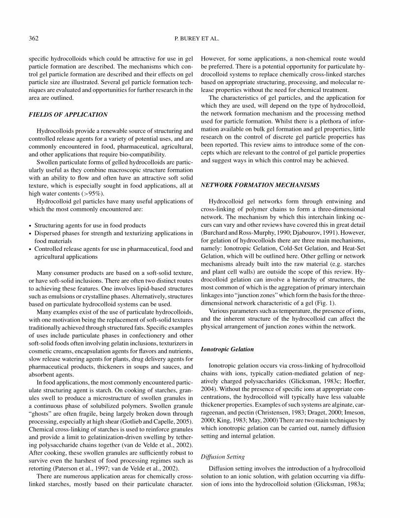
362 P. BUREY ET AL.
specific hydrocolloids which could be attractive for use in gelparticle formation are described. The mechanisms which con-trol gel particle formation are described and their effects on gelparticle size are illustrated. Several gel particle formation tech-niques are evaluated and opportunities for further research in thearea are outlined.
FIELDS OF APPLICATION
Hydrocolloids provide a renewable source of structuring andcontrolled release agents for a variety of potential uses, and arecommonly encountered in food, pharmaceutical, agricultural,and other applications that require bio-compatibility.
Swollen particulate forms of gelled hydrocolloids are partic-ularly useful as they combine macroscopic structure formationwith an ability to flow and often have an attractive soft solidtexture, which is especially sought in food applications, all athigh water contents (>95%).
Hydrocolloid gel particles have many useful applications ofwhich the most commonly encountered are:
• Structuring agents for use in food products• Dispersed phases for strength and texturizing applications in
food materials• Controlled release agents for use in pharmaceutical, food and
agricultural applications
Many consumer products are based on a soft-solid texture,or have soft-solid inclusions. There are often two distinct routesto achieving these features. One involves lipid-based structuressuch as emulsions or crystalline phases. Alternatively, structuresbased on particulate hydrocolloid systems can be used.
Many examples exist of the use of particulate hydrocolloids,with one motivation being the replacement of soft-solid texturestraditionally achieved through structured fats. Specific examplesof uses include particulate phases in confectionery and othersoft-solid foods often involving gelatin inclusions, texturizers incosmetic creams, encapsulation agents for flavors and nutrients,slow release watering agents for plants, drug delivery agents forpharmaceutical products, thickeners in soups and sauces, andabsorbent agents.
In food applications, the most commonly encountered partic-ulate structuring agent is starch. On cooking of starches, gran-ules swell to produce a microstructure of swollen granules ina continuous phase of solubilized polymers. Swollen granule“ghosts” are often fragile, being largely broken down throughprocessing, especially at high shear (Gotlieb and Capelle, 2005).Chemical cross-linking of starches is used to reinforce granulesand provide a limit to gelatinization-driven swelling by tether-ing polysaccharide chains together (van de Velde et al., 2002).After cooking, these swollen granules are sufficiently robust tosurvive even the harshest of food processing regimes such asretorting (Paterson et al., 1997; van de Velde et al., 2002).
There are numerous application areas for chemically cross-linked starches, mostly based on their particulate character.
However, for some applications, a non-chemical route wouldbe preferred. There is a potential opportunity for particulate hy-drocolloid systems to replace chemically cross-linked starchesbased on appropriate structuring, processing, and molecular re-lease properties without the need for chemical treatment.
The characteristics of gel particles, and the application forwhich they are used, will depend on the type of hydrocolloid,the network formation mechanism and the processing methodused for particle formation. Whilst there is a plethora of infor-mation available on bulk gel formation and gel properties, littleresearch on the control of discrete gel particle properties hasbeen reported. This review aims to introduce some of the con-cepts which are relevant to the control of gel particle propertiesand suggest ways in which this control may be achieved.
NETWORK FORMATION MECHANISMS
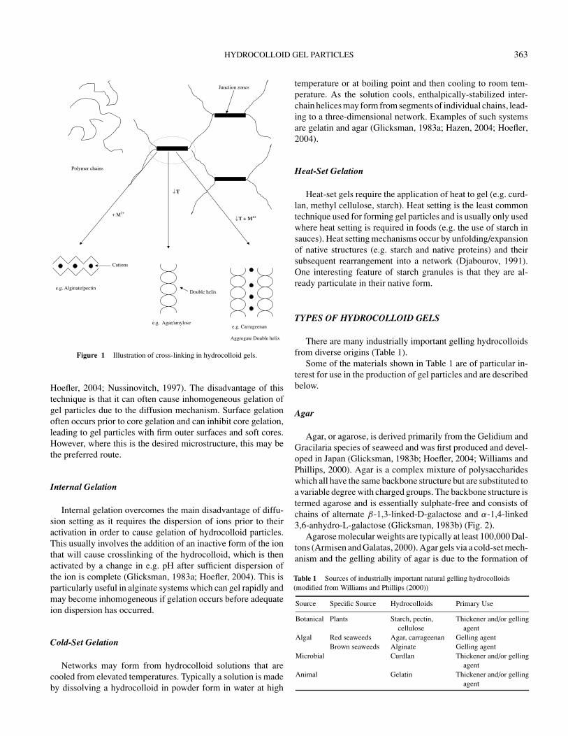
Hydrocolloid gel networks form through entwining andcross-linking of polymer chains to form a three-dimensionalnetwork. The mechanism by which this interchain linking oc-curs can vary and other reviews have covered this in great detail(Burchard and Ross-Murphy, 1990; Djabourov, 1991). However,for gelation of hydrocolloids there are three main mechanisms,namely: Ionotropic Gelation, Cold-Set Gelation, and Heat-SetGelation, which will be outlined here. Other gelling or networkmechanisms already built into the raw material (e.g. starchesand plant cell walls) are outside the scope of this review. Hy-drocolloid gelation can involve a hierarchy of structures, themost common of which is the aggregation of primary interchainlinkages into “junction zones” which form the basis for the three-dimensional network characteristic of a gel (Fig. 1).
Various parameters such as temperature, the presence of ions,and the inherent structure of the hydrocolloid can affect thephysical arrangement of junction zones within the network.
Ionotropic Gelation
Ionotropic gelation occurs via cross-linking of hydrocolloidchains with ions, typically cation-mediated gelation of neg-atively charged polysaccharides (Glicksman, 1983c; Hoefler,2004). Without the presence of specific ions at appropriate con-centrations, the hydrocolloid will typically have less valuablethickener properties. Examples of such systems are alginate, car-rageenan, and pectin (Christensen, 1983; Draget, 2000; Imeson,2000; King, 1983; May, 2000) There are two main techniques bywhich ionotropic gelation can be carried out, namely diffusionsetting and internal gelation.
Diffusion Setting
Diffusion setting involves the introduction of a hydrocolloidsolution to an ionic solution, with gelation occurring via diffu-sion of ions into the hydrocolloid solution (Glicksman, 1983a;
HYDROCOLLOID GEL PARTICLES 363
+ M2+
T
T + Mx+
e.g. Alginate/pectin
e.g. Agar/amylose e.g. Carrageenan
Double helix
Cations
Polymer chains
Junction zones
Aggregate Double helix
Figure 1 Illustration of cross-linking in hydrocolloid gels.
Hoefler, 2004; Nussinovitch, 1997). The disadvantage of thistechnique is that it can often cause inhomogeneous gelation ofgel particles due to the diffusion mechanism. Surface gelationoften occurs prior to core gelation and can inhibit core gelation,leading to gel particles with firm outer surfaces and soft cores.However, where this is the desired microstructure, this may bethe preferred route.
Internal Gelation
Internal gelation overcomes the main disadvantage of diffu-sion setting as it requires the dispersion of ions prior to theiractivation in order to cause gelation of hydrocolloid particles.This usually involves the addition of an inactive form of the ionthat will cause crosslinking of the hydrocolloid, which is thenactivated by a change in e.g. pH after sufficient dispersion ofthe ion is complete (Glicksman, 1983a; Hoefler, 2004). This isparticularly useful in alginate systems which can gel rapidly andmay become inhomogeneous if gelation occurs before adequateion dispersion has occurred.
Cold-Set Gelation
Networks may form from hydrocolloid solutions that arecooled from elevated temperatures. Typically a solution is madeby dissolving a hydrocolloid in powder form in water at high
temperature or at boiling point and then cooling to room tem-perature. As the solution cools, enthalpically-stabilized inter-chain helices may form from segments of individual chains, lead-ing to a three-dimensional network. Examples of such systemsare gelatin and agar (Glicksman, 1983a; Hazen, 2004; Hoefler,2004).
Heat-Set Gelation
Heat-set gels require the application of heat to gel (e.g. curd-lan, methyl cellulose, starch). Heat setting is the least commontechnique used for forming gel particles and is usually only usedwhere heat setting is required in foods (e.g. the use of starch insauces). Heat setting mechanisms occur by unfolding/expansionof native structures (e.g. starch and native proteins) and theirsubsequent rearrangement into a network (Djabourov, 1991).One interesting feature of starch granules is that they are al-ready particulate in their native form.
TYPES OF HYDROCOLLOID GELS
There are many industrially important gelling hydrocolloidsfrom diverse origins (Table 1).
Some of the materials shown in Table 1 are of particular in-terest for use in the production of gel particles and are describedbelow.
Agar
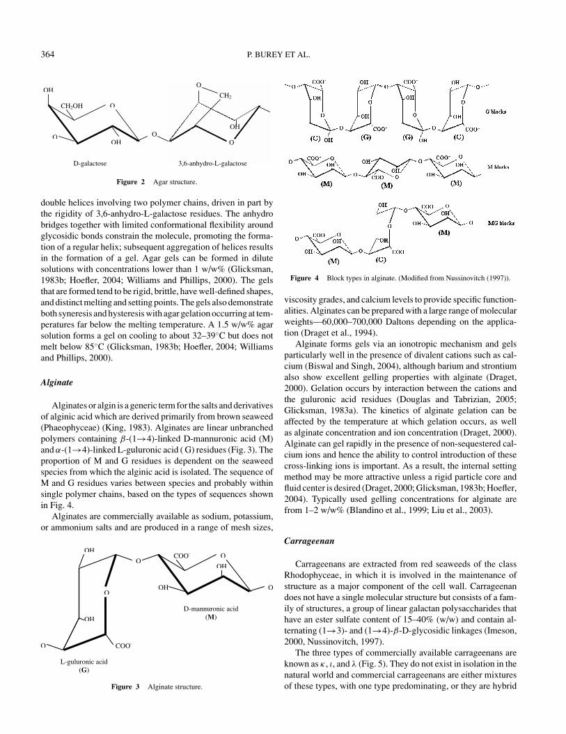
Agar, or agarose, is derived primarily from the Gelidium andGracilaria species of seaweed and was first produced and devel-oped in Japan (Glicksman, 1983b; Hoefler, 2004; Williams andPhillips, 2000). Agar is a complex mixture of polysaccharideswhich all have the same backbone structure but are substituted toa variable degree with charged groups. The backbone structure istermed agarose and is essentially sulphate-free and consists ofchains of alternate β-1,3-linked-D-galactose and α-1,4-linked3,6-anhydro-L-galactose (Glicksman, 1983b) (Fig. 2).
Agarose molecular weights are typically at least 100,000 Dal-tons (Armisen and Galatas, 2000). Agar gels via a cold-set mech-anism and the gelling ability of agar is due to the formation of
Table 1 Sources of industrially important natural gelling hydrocolloids(modified from Williams and Phillips (2000))
Source Specific Source Hydrocolloids Primary Use
Botanical Plants Starch, pectin,cellulose
Thickener and/or gellingagent
Algal Red seaweeds Agar, carrageenan Gelling agentBrown seaweeds Alginate Gelling agent
Microbial Curdlan Thickener and/or gellingagent
Animal Gelatin Thickener and/or gellingagent
364 P. BUREY ET AL.
OCH2OH
OOH
OH
O
O
OH
O
CH2
a-6,3 esotcalag-D nhydro-L-galactose
Figure 2 Agar structure.
double helices involving two polymer chains, driven in part bythe rigidity of 3,6-anhydro-L-galactose residues. The anhydrobridges together with limited conformational flexibility aroundglycosidic bonds constrain the molecule, promoting the forma-tion of a regular helix; subsequent aggregation of helices resultsin the formation of a gel. Agar gels can be formed in dilutesolutions with concentrations lower than 1 w/w% (Glicksman,1983b; Hoefler, 2004; Williams and Phillips, 2000). The gelsthat are formed tend to be rigid, brittle, have well-defined shapes,and distinct melting and setting points. The gels also demonstrateboth syneresis and hysteresis with agar gelation occurring at tem-peratures far below the melting temperature. A 1.5 w/w% agarsolution forms a gel on cooling to about 32–39◦C but does notmelt below 85◦C (Glicksman, 1983b; Hoefler, 2004; Williamsand Phillips, 2000).
Alginate
Alginates or algin is a generic term for the salts and derivativesof alginic acid which are derived primarily from brown seaweed(Phaeophyceae) (King, 1983). Alginates are linear unbranchedpolymers containing β-(1→4)-linked D-mannuronic acid (M)and α-(1→4)-linked L-guluronic acid ( G) residues (Fig. 3). Theproportion of M and G residues is dependent on the seaweedspecies from which the alginic acid is isolated. The sequence ofM and G residues varies between species and probably withinsingle polymer chains, based on the types of sequences shownin Fig. 4.
Alginates are commercially available as sodium, potassium,or ammonium salts and are produced in a range of mesh sizes,
OCOO-
OH
OH
D-mannuronic acid (M)
L-guluronic acid (G)
O
O
O
OH
OH
O COO-
Figure 3 Alginate structure.
Figure 4 Block types in alginate. (Modified from Nussinovitch (1997)).
viscosity grades, and calcium levels to provide specific function-alities. Alginates can be prepared with a large range of molecularweights—60,000–700,000 Daltons depending on the applica-tion (Draget et al., 1994).
Alginate forms gels via an ionotropic mechanism and gelsparticularly well in the presence of divalent cations such as cal-cium (Biswal and Singh, 2004), although barium and strontiumalso show excellent gelling properties with alginate (Draget,2000). Gelation occurs by interaction between the cations andthe guluronic acid residues (Douglas and Tabrizian, 2005;Glicksman, 1983a). The kinetics of alginate gelation can beaffected by the temperature at which gelation occurs, as wellas alginate concentration and ion concentration (Draget, 2000).Alginate can gel rapidly in the presence of non-sequestered cal-cium ions and hence the ability to control introduction of thesecross-linking ions is important. As a result, the internal settingmethod may be more attractive unless a rigid particle core andfluid center is desired (Draget, 2000; Glicksman, 1983b; Hoefler,2004). Typically used gelling concentrations for alginate arefrom 1–2 w/w% (Blandino et al., 1999; Liu et al., 2003).
Carrageenan
Carrageenans are extracted from red seaweeds of the classRhodophyceae, in which it is involved in the maintenance ofstructure as a major component of the cell wall. Carrageenandoes not have a single molecular structure but consists of a fam-ily of structures, a group of linear galactan polysaccharides thathave an ester sulfate content of 15–40% (w/w) and contain al-ternating (1→3)- and (1→4)-β-D-glycosidic linkages (Imeson,2000, Nussinovitch, 1997).
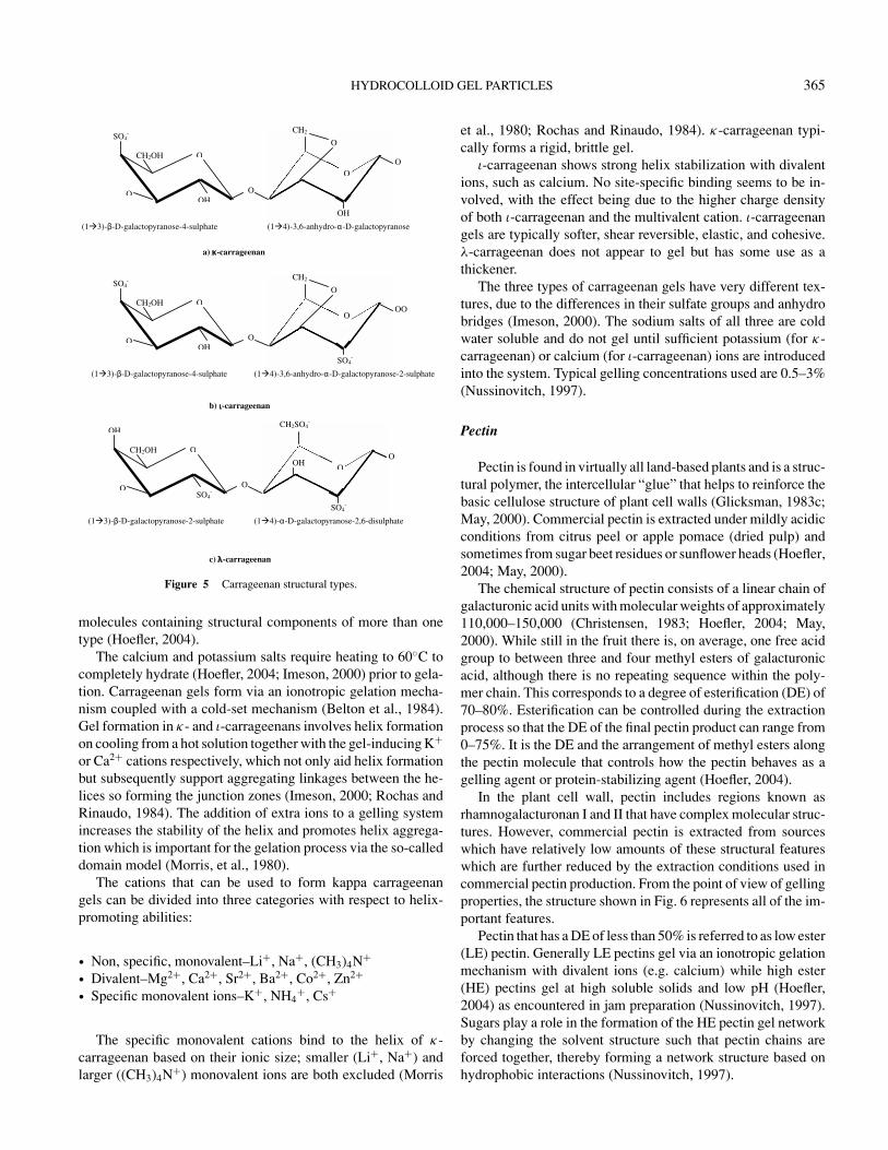
The three types of commercially available carrageenans areknown as κ , ι, and λ (Fig. 5). They do not exist in isolation in thenatural world and commercial carrageenans are either mixturesof these types, with one type predominating, or they are hybrid
HYDROCOLLOID GEL PARTICLES 365
O
O
CH2
O
OH
OCH2OH
OOH
O
(1 3)- -D-galactopyranose-4-sulphate (1 4)-3,6-anhydro- -D-galactopyranose
SO4-
O
CH2SO4-
O
SO4-
OCH2OH
O
OH
O
OH
(1 3)- -D-galactopyranose-2-sulphate (1 4)- -D-galactopyranose-2,6-disulphate
SO4-
(1 3)- -D-galactopyranose-4-sulphate
OCH2OH
OOH
SO4-
O
O
O
CH2
(1 4)-3,6-anhydro- -D-galactopyranose-2-sulphate
SO4-
OO
a) -carrageenan
c) -carrageenan
b) -carrageenan
Figure 5 Carrageenan structural types.
molecules containing structural components of more than onetype (Hoefler, 2004).
The calcium and potassium salts require heating to 60◦C tocompletely hydrate (Hoefler, 2004; Imeson, 2000) prior to gela-tion. Carrageenan gels form via an ionotropic gelation mecha-nism coupled with a cold-set mechanism (Belton et al., 1984).Gel formation in κ- and ι-carrageenans involves helix formationon cooling from a hot solution together with the gel-inducing K+
or Ca2+ cations respectively, which not only aid helix formationbut subsequently support aggregating linkages between the he-lices so forming the junction zones (Imeson, 2000; Rochas andRinaudo, 1984). The addition of extra ions to a gelling systemincreases the stability of the helix and promotes helix aggrega-tion which is important for the gelation process via the so-calleddomain model (Morris, et al., 1980).
The cations that can be used to form kappa carrageenangels can be divided into three categories with respect to helix-promoting abilities:
• Non, specific, monovalent–Li+, Na+, (CH3)4N+• Divalent–Mg2+, Ca2+, Sr2+, Ba2+, Co2+, Zn2+• Specific monovalent ions–K+, NH4
+, Cs+
The specific monovalent cations bind to the helix of κ-carrageenan based on their ionic size; smaller (Li+, Na+) andlarger ((CH3)4N+) monovalent ions are both excluded (Morris
et al., 1980; Rochas and Rinaudo, 1984). κ-carrageenan typi-cally forms a rigid, brittle gel.
ι-carrageenan shows strong helix stabilization with divalentions, such as calcium. No site-specific binding seems to be in-volved, with the effect being due to the higher charge densityof both ι-carrageenan and the multivalent cation. ι-carrageenangels are typically softer, shear reversible, elastic, and cohesive.λ-carrageenan does not appear to gel but has some use as athickener.
The three types of carrageenan gels have very different tex-tures, due to the differences in their sulfate groups and anhydrobridges (Imeson, 2000). The sodium salts of all three are coldwater soluble and do not gel until sufficient potassium (for κ-carrageenan) or calcium (for ι-carrageenan) ions are introducedinto the system. Typical gelling concentrations used are 0.5–3%(Nussinovitch, 1997).
Pectin
Pectin is found in virtually all land-based plants and is a struc-tural polymer, the intercellular “glue” that helps to reinforce thebasic cellulose structure of plant cell walls (Glicksman, 1983c;May, 2000). Commercial pectin is extracted under mildly acidicconditions from citrus peel or apple pomace (dried pulp) andsometimes from sugar beet residues or sunflower heads (Hoefler,2004; May, 2000).
The chemical structure of pectin consists of a linear chain ofgalacturonic acid units with molecular weights of approximately110,000–150,000 (Christensen, 1983; Hoefler, 2004; May,2000). While still in the fruit there is, on average, one free acidgroup to between three and four methyl esters of galacturonicacid, although there is no repeating sequence within the poly-mer chain. This corresponds to a degree of esterification (DE) of70–80%. Esterification can be controlled during the extractionprocess so that the DE of the final pectin product can range from0–75%. It is the DE and the arrangement of methyl esters alongthe pectin molecule that controls how the pectin behaves as agelling agent or protein-stabilizing agent (Hoefler, 2004).
In the plant cell wall, pectin includes regions known asrhamnogalacturonan I and II that have complex molecular struc-tures. However, commercial pectin is extracted from sourceswhich have relatively low amounts of these structural featureswhich are further reduced by the extraction conditions used incommercial pectin production. From the point of view of gellingproperties, the structure shown in Fig. 6 represents all of the im-portant features.
Pectin that has a DE of less than 50% is referred to as low ester(LE) pectin. Generally LE pectins gel via an ionotropic gelationmechanism with divalent ions (e.g. calcium) while high ester(HE) pectins gel at high soluble solids and low pH (Hoefler,2004) as encountered in jam preparation (Nussinovitch, 1997).Sugars play a role in the formation of the HE pectin gel networkby changing the solvent structure such that pectin chains areforced together, thereby forming a network structure based onhydrophobic interactions (Nussinovitch, 1997).

366 P. BUREY ET AL.
O
OH
OH
O
COOCH3
(1 4)- galacturonic acid with carboxyl acid group
(1 4)- galacturonic acid partially methylated with methyl ester
Figure 6 Pectin structure.
For low-ester pectins an “egg-box” model of network forma-tion has been suggested for the primary junction zones in the gelnetwork (Rees, 1982) which is similar to the network structureformed in alginate gels. The basis of the network is pectin chainscross-linked by calcium ions through chelation by carboxyl andhydroxyl groups (Rees, 1982).
Pectin can be in acid or metal salt form. It is water-soluble inall forms and can achieve high solubility in water at room tem-perature with sufficient shear, although for complete hydrationit is necessary to heat to about 60◦C (Hoefler, 2004). Typical gelconcentrations range from 2–4 w/w% for HE pectins and 0.1–4w/w% for LE pectins (Glicksman, 1983c; May, 2000). Similarto alginate, the gel properties of LE pectin gels are affected byhydrocolloid concentration, ion concentration, and the methodof preparation.
Gelatin
Gelatin differs from the hydrocolloids discussed above in thatit is derived from an animal protein, collagen, via controlled acidor alkaline hydrolysis. Collagen may come from hide, bone,or other collagenous material (Belitz and Grosch, 1999). Com-monly the collagen used is of bovine (cow), porcine (pig), orpiscine (fish) origins (Johnston-Banks, 1990). The properties ofgelatins are affected by the source, age, and type of collagen(Rix, 1990).
A typical gelatin consists of 14% moisture, 84% protein,and 2% ash (Rix, 1990). The protein consists of a mixture ofamino acids of which glycine, proline, and hydroxyproline arepresent in the most abundance (Cuppo et al., 2001; Johnston-Banks, 1990). Gelatin molecules contain repeating sequences ofglycine-X-Y triplets, where X and Y are frequently proline andhydroxyproline amino acids (Fig. 7).
These repeating sequences occur over extended regions of thepolymer chain. This regularity is necessary in order to form thecharacteristic triple helical structure in gelatin and other collagenfamily proteins and the subsequent ability to form gels in whichtriple helical segments form the basis for cross-linking and three-dimensional network formation (Clark and Ross-Murphy, 1990).
Figure 7 Repeating structure in gelatin responsible for triple helix structure.
Gelatin forms a thermoreversible gel through a cold-settingmechanism in aqueous solvents (Djabourov, 1991). Above 40◦Cgelatin in solution behaves like a typical synthetic polymer withthe individual macromolecules each assuming random-coil con-figurations (Finer et al., 1975) with typical molecular weights of2 × 105 Daltons (Bohidar and Jena, 1993). These random coilsconsist of single polypeptide chains, termed α-chains that maybe entangled. Upon cooling these coils undergo a coil-to helixtransition, leading to gelation (Fig. 8) (Finer et al., 1975).
Gelatin gels are typically made from higher concentrationsthan gels formed with agar, alginate, carrageenan, and pectin.Typical gelatin gel concentrations used in food products are 1–5w/w% (Tosh et al., 2003; Williams, 1964). Gel properties aretypically affected by gelatin concentration, cooling rate usedduring gelation, pH, and temperature at which the particularsource of gelatin will gel. Gelation temperatures for gelatinare typically just above room temperature; this is one of thefactors leading to the relatively slow gel setting kinetics forgelatin.
HYDROCOLLOID GEL PARTICLES–IN-USEPROPERTIES
Hydrocolloid gel particles can be characterized in manydifferent ways in relation to their use in structured materials.
Figure 8 Schematic of gelatin gelation. Two steps are involved A) formationof a loose network and B) renaturation to a collagen assembly (modified fromDjabourov 1991).
HYDROCOLLOID GEL PARTICLES 367
Two aspects will be discussed here—size/shape, and in-mouthdetectability.
Size and Shape Definition
As a broad dictionary definition, a particle is defined as “aminute fragment or quantity of matter or the smallest percepti-ble or discernible part of an aggregation or mass.” Using mod-ern analytical techniques, particles can now be detected on thenanometer scale; however, particles may not be discernible byhuman senses (sight in visible light, touch/mouthfeel) when in-corporated into materials until the micron or millimeter scale.Techniques for assessing the size and shape of the microgel par-ticles will be addressed in a subsequent section.
In-Mouth Detectability and Factors AffectingSensorial Detection
Acceptance of hydrocolloid gel particles in food productsmay be based on (lack of) detectability in the mouth. Particledetectability in foodstuffs can depend on the particle character-istics as well as features of the medium in which they reside,including such characteristics as water absorption, sedimenta-tion rate of particles, size, shape, and deformation resistance(Imai et al., 1999).
Visual acceptability of a product containing particles may bebased on whether it is desirable to observe particles in visiblelight. In some applications, the use of suspended particles willgive food products a cloudy appearance which may not be idealfor consumer acceptance. Visibility of gel particles will dependon size; very small particles may be minute enough that theycannot be observed in visible light, whilst very large expandedgel particles may also be invisible due to their refractive indexapproaching that of their surrounding medium.
When particles are detected sensorially by the mouth, thematerials are often described as “grainy” or “gritty.” Harderparticles have been shown to be detected at smaller sizesthrough detection of a gritty texture (Utz, 1986). (Engelen et al.,2005) found that large, hard, sharp particles in a low viscositymedium produce a more rough, gritty, and unpleasant sensa-tion than small, soft, and smooth particles in a higher viscositymedium.
An example of size of particles combined with their mediumhaving an effect on detectability comes from the fact that sugarparticles ≥25 µm in chocolate can be detected by the roof of themouth (Rostagno, 1969), although they cannot be detected in asofter product such as fondant (Woodruff and Gilder, 1931). Insmoother, creamier products such as margarine and ice cream,particle detection can vary, with 22 µm particles the minimumsize detected in margarine by the mouth, and 55 µm the min-imum size detectable in ice cream (Imai et al., 1999). Hardparticles, such as alumina, can be detected at smaller sizes of10 µm.
Size
Hardness
Undetectable
MediumViscosity
Detectable
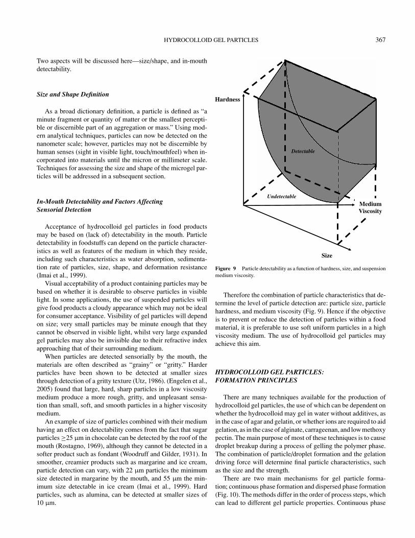
Figure 9 Particle detectability as a function of hardness, size, and suspensionmedium viscosity.
Therefore the combination of particle characteristics that de-termine the level of particle detection are: particle size, particlehardness, and medium viscosity (Fig. 9). Hence if the objectiveis to prevent or reduce the detection of particles within a foodmaterial, it is preferable to use soft uniform particles in a highviscosity medium. The use of hydrocolloid gel particles mayachieve this aim.
HYDROCOLLOID GEL PARTICLES:FORMATION PRINCIPLES
There are many techniques available for the production ofhydrocolloid gel particles, the use of which can be dependent onwhether the hydrocolloid may gel in water without additives, asin the case of agar and gelatin, or whether ions are required to aidgelation, as in the case of alginate, carrageenan, and low methoxypectin. The main purpose of most of these techniques is to causedroplet breakup during a process of gelling the polymer phase.The combination of particle/droplet formation and the gelationdriving force will determine final particle characteristics, suchas the size and the strength.
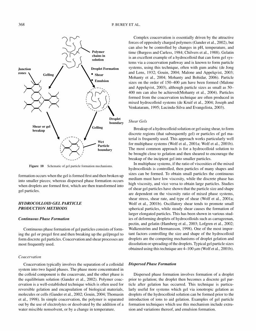
There are two main mechanisms for gel particle forma-tion; continuous phase formation and dispersed phase formation(Fig. 10). The methods differ in the order of process steps, whichcan lead to different gel particle properties. Continuous phase
368 P. BUREY ET AL.
Droplet Formation
• Shear
• Emulsion
Gelling
GellingShear or gel breakup
Polymerchains in solution
Junctionzones
WetParticle boundary
Dropletboundary
Figure 10 Schematic of gel particle formation mechanisms.
formation occurs when the gel is formed first and then broken upinto smaller pieces; whereas dispersed phase formation occurswhen droplets are formed first, which are then transformed intogel particles.
HYDROCOLLOID GEL PARTICLEPRODUCTION METHODS
Continuous Phase Formation
Continuous phase formation of gel particles consists of form-ing the gel or pregel first and then breaking up the gel/pregel toform discrete gel particles. Coacervation and shear processes aremost frequently used.
Coacervation
Coacervation typically involves the separation of a colloidalsystem into two liquid phases. The phase more concentrated inthe colloid component is the coacervate, and the other phase isthe equilibrium solution (Gander et al., 2002). Polymer coac-ervation is a well-established technique which is often used forreversible gelation and encapsulation of biological materials,molecules or cells (Gander et al., 2002; Gouin, 2004; Thomasinet al., 1998). In simple coacervation, the polymer is separatedout by the use of electrolytes or desolvated by the addition of awater miscible nonsolvent, or by a change in temperature.
Complex coacervation is essentially driven by the attractiveforces of oppositely charged polymers (Gander et al., 2002), butcan also be be controlled by changes in pH, temperature, andtime (Burgess and Carless, 1984; Chilvers et al., 1988). Gelatinis an excellent example of a hydrocolloid that can form gel sys-tems via a coacervation pathway and is known to form particlesystems, using this technique, often with gum arabic (de Jongand Lens, 1932; Gouin, 2004; Malone and Appelqvist, 2003;Mohanty et al., 2004; Mohanty and Bohidar, 2006). Particlesizes on the order of 150–400 µm have been formed (Maloneand Appelqvist, 2003), although particle sizes as small as 50–400 nm can also be achieved(Mohanty et al., 2004). Particlesformed from the coacervation technique are often produced inmixed hydrocolloid systems (de Kruif et al., 2004; Joseph andVenkataram, 1995; Lucinda-Silva and Evangelista, 2003).
Shear Gels
Breakup of a hydrocolloid solution or gel using shear, to formdiscrete regions (that subsequently gel) or particles of gel ma-terial is frequently used. This approach works particularly wellfor multiphase systems (Wolf et al., 2001a; Wolf et al., 2001b).The most common approach is for a hydrocolloid solution tobe brought close to gelation and then sheared to encourage thebreakup of the incipient gel into smaller particles.
In multiphase systems, if the ratio of viscosities of the mixedhydrocolloids is controlled, then particles of many shapes andsizes can be formed. To obtain small particles the continuousmedium must have low viscosity, while the discrete phase hashigh viscosity, and vice versa to obtain large particles. Studiesof shear gel particles have shown that the particle size and shapeare dependent on the viscosity ratio of mixed phase systems,shear stress, shear rate, and type of shear (Wolf et al., 2001a;Wolf et al., 2001b). Oscillatory shear tends to promote smallspherical particles, while steady shear causes the formation oflarger elongated particles. This has been shown in various stud-ies of deforming droplets of hydrocolloids such as carrageenan,pectin, and gelatin (Hamberg et al., 2003; Lofgren et al., 2002;Walkenstrom and Hermansson, 1998). One of the most impor-tant factors controlling the size and shape of the hydrocolloiddroplets are the competing mechanisms of droplet gelation anddissolution or spreading of the droplets. Typical gel particle sizesobtained using this technique are 4–100 µm (Wolf et al., 2001b).
Dispersed Phase Formation
Dispersed phase formation involves formation of a dropletprior to gelation; the droplet then becomes a discrete gel par-ticle after gelation has occurred. This technique is particu-larly useful for systems which gel via ionotropic gelation asdroplets of the hydrocolloid solution can be formed prior to theintroduction of ions to aid gelation. Examples of gel particleformation techniques which use this mechanism include extru-sion and variations thereof, and emulsion formation.

HYDROCOLLOID GEL PARTICLES 369
Syringe
Polymer solution
Gellingsolution
Gelparticle
Polymer solutiondroplet
Crosslink
Figure 11 Extrusion formation of gel particles.
Extrusion
Extrusion is a commonly used technique for the formationof gel particles. On a small scale this may involve the use ofa syringe needle (Blandino et al., 1999; Cheng and Lim, 2004;Hills et al., 2000; Hunik and Tramper, 1993). A hydrocolloidsolution is loaded in a syringe and then extruded through aneedle, to form solution droplets which then gel based on theconditions that the solution is extruded into (Fig. 11). The sizeof the droplets, and thus the subsequent gel particles, dependsupon the diameter of the needle, the flow rate of the solution,and the viscosity of the solution.
This technique may be applicable to fast gelling hydrocol-loids that do not require ions for gelation, but can also be usedto extrude droplets into a hardening bath containing ions to pro-mote gelation of the solution. If the gel is ionotropic, then theconcentration of the ionic solution in the hardening bath willalso affect final gel particle size. Typical gel particle sizes usingthis technique are 0.5–6 mm using conventional dropping meth-ods (Blandino et al., 1999; Murakata et al., 2001; Ouwerx et al.,1998) and on the scale of hundreds of microns if modified tech-niques are used to atomize the hydrocolloid solution (Murakataet al., 2001). On an industrial scale commercial extruders andscraped-surface heat exchangers may be used to form gel parti-cles (Brown et al., 2004; Peng et al., 2006).
Electrostatic
The electrostatic technique is a modified version of the small-scale extrusion technique where the syringe and the solution haveopposite charges, which can affect gel particle size (Goosen,2003; Klokk and Melvik, 2002; Zvitov and Nussinovitch, 2001;Zvitov and Nussinovitch, 2003). The rate at which the dropletsform, and droplet size, is needle diameter, charge arrangement(electrode geometry and spacing), and strength of electric field
(Bugarski et al., 1994), although hydrocolloid solution and hard-ening solution (if present) properties can also have an effect.The most effective electrode and charge arrangement for pro-ducing small droplets is a positively charged needle and agrounded plate. Two other arrangements are also possible; posi-tively charged plate attached to needle, and a positively chargedcollecting solution (Goosen, 2003). If a positive charge is al-ways on the needle, this ensures that the smallest gel particlesize is produced at the lowest applied potential (Goosen, 2003).Particle sizes ranging from 40–2500 µm can be achieved usingthis method (Bugarski et al., 1994; Goosen, 2003; Zvitov andNussinovitch, 2003).
Ultrasonic
This technique is also a modified version of the extrusionmethod in which ultrasonic breakup of a polymer solution streamis used. Liquid is pushed from a reservoir to a nozzle, whichis forced to vibrate at ultrasonic frequencies. The liquid isbroken into homogeneous droplets that gel when dropped into ahardening bath (Cellesi et al., 2004; Hunik and Tramper, 1993).Droplet, and subsequently gel particle size, is controlled by theflow rate of solution and the ultrasonic frequency at which thedroplets are vibrated. Gel particles can range in size from hun-dreds of microns (Cellesi et al., 2004) up to 1–5 mm (Hunik andTramper, 1993) using this technique.
Emulsion
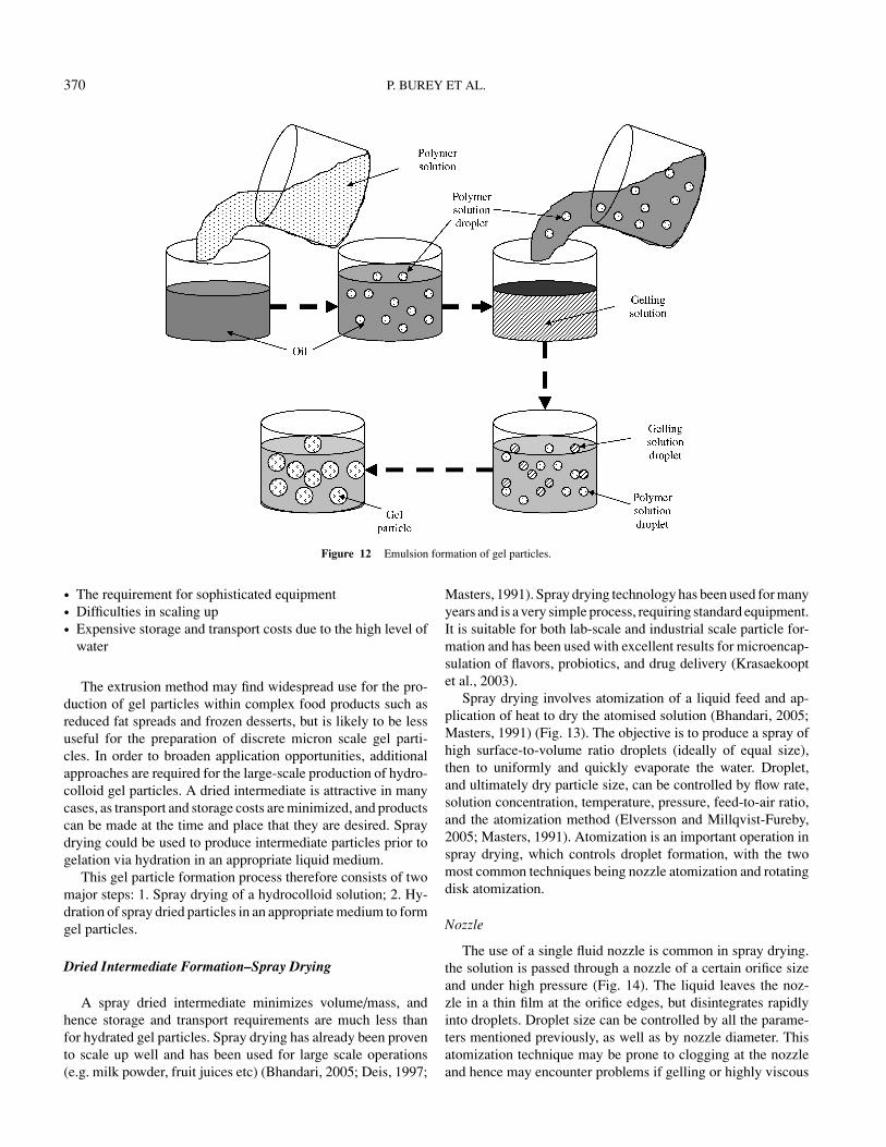
The emulsion technique uses media other than water to aidgel particle formation. In food applications, the hydrocolloidsolution is often suspended in vegetable oil and then introducedto the appropriate ionic solution to promote gelation (Lamprechtet al., 2000, Sugiura et al., 2005). The hydrocolloid and ionicsolution droplets collide with each other in the stream of oil,and the reaction between the hydrocolloid and ions proceedswhen successful coalescence of the droplets takes place (Fig. 12)(Campbell et al., 2004; Liu et al., 2003; Malone and Appelqvist,2003).
The size of the gel particles formed using this method isdependent on the viscosity of the suspending oil, ratio of oilto hydrocolloid solution, emulsifier type, and amount of energyused to create the oil-hydrocolloid emulsion (Reis et al., 2006).Hydrocolloid solution droplet sizes can range from 0.2–80 µm(Huang et al., 2001), although they can be as large as 5000 µm(Malone and Appelqvist, 2003). Gel particle sizes can rangefrom as low as 10 µm up to 3 mm (Liu et al., 2003; Malone andAppelqvist, 2003). There is a difficulty with this technique ifaqueous gel particles are required as adequate removal of the oilphase from the droplets can prove difficult or messy, and addsanother separation step to the process.
AN ALTERNATIVE APPROACH: FORMATION OF GELPARTICLES FROM A DRIED INTERMEDIATE
There are several limitations of the techniques describedabove. These may include:
370 P. BUREY ET AL.
Figure 12 Emulsion formation of gel particles.
• The requirement for sophisticated equipment• Difficulties in scaling up• Expensive storage and transport costs due to the high level of
water
The extrusion method may find widespread use for the pro-duction of gel particles within complex food products such asreduced fat spreads and frozen desserts, but is likely to be lessuseful for the preparation of discrete micron scale gel parti-cles. In order to broaden application opportunities, additionalapproaches are required for the large-scale production of hydro-colloid gel particles. A dried intermediate is attractive in manycases, as transport and storage costs are minimized, and productscan be made at the time and place that they are desired. Spraydrying could be used to produce intermediate particles prior togelation via hydration in an appropriate liquid medium.
This gel particle formation process therefore consists of twomajor steps: 1. Spray drying of a hydrocolloid solution; 2. Hy-dration of spray dried particles in an appropriate medium to formgel particles.
Dried Intermediate Formation–Spray Drying
A spray dried intermediate minimizes volume/mass, andhence storage and transport requirements are much less thanfor hydrated gel particles. Spray drying has already been provento scale up well and has been used for large scale operations(e.g. milk powder, fruit juices etc) (Bhandari, 2005; Deis, 1997;
Masters, 1991). Spray drying technology has been used for manyyears and is a very simple process, requiring standard equipment.It is suitable for both lab-scale and industrial scale particle for-mation and has been used with excellent results for microencap-sulation of flavors, probiotics, and drug delivery (Krasaekooptet al., 2003).
Spray drying involves atomization of a liquid feed and ap-plication of heat to dry the atomised solution (Bhandari, 2005;Masters, 1991) (Fig. 13). The objective is to produce a spray ofhigh surface-to-volume ratio droplets (ideally of equal size),then to uniformly and quickly evaporate the water. Droplet,and ultimately dry particle size, can be controlled by flow rate,solution concentration, temperature, pressure, feed-to-air ratio,and the atomization method (Elversson and Millqvist-Fureby,2005; Masters, 1991). Atomization is an important operation inspray drying, which controls droplet formation, with the twomost common techniques being nozzle atomization and rotatingdisk atomization.
Nozzle
The use of a single fluid nozzle is common in spray drying.the solution is passed through a nozzle of a certain orifice sizeand under high pressure (Fig. 14). The liquid leaves the noz-zle in a thin film at the orifice edges, but disintegrates rapidlyinto droplets. Droplet size can be controlled by all the parame-ters mentioned previously, as well as by nozzle diameter. Thisatomization technique may be prone to clogging at the nozzleand hence may encounter problems if gelling or highly viscous

HYDROCOLLOID GEL PARTICLES 371
Solution
Pump reird yarpS deeF
Atomiser
Spray
Cyclone Filter for fines
Particlecollection
Finescollection
Heat Air
Figure 13 Schematic of the spray drying process.
solutions are used. The size of droplets formed using a nozzle at-omizer range from 10–600 µm (Bhandari, 2005; Masters, 1991;Oakley, 2004). Particle size distributions are typically at thelarger end of this scale, but have a narrow range of diameters(Bhandari, 2005). The mean particle size of spray dried particlescan range from 30–80 µm, although particles as small as 1–2 µmcan be formed, if there is a large difference between initial andfinal solids content.
Rotating Disk
This is the most commonly used atomizer in the food industry(Masters, 1991). The solution is dropped onto the rotating disk
Figure 14 Schematic of a) nozzle atomization; b) disk atomization.
which then spreads into a thin film at the disk edge (Fig. 14). Therotation of the disk and friction with the surrounding air causesthe film to disintegrate into droplets. A wide range of particlesizes can be formed and can be controlled by manipulating diskrotational speed, as well as other parameters mentioned previ-ously. Clogging rarely occurs as there are no small orifices toget blocked. Usage of a rotating disk atomizer causes formationof droplets ranging in size from 10–600 µm (Bhandari, 2005;Masters, 1991; Oakley, 2004). Particle size distributions aretypically very broad, with particles of diameters as small asa few microns, ranging up to hundreds of microns. The meanparticle size of spray dried particles can range from 30–80 µmand size distribution is dependent on the initial solids contentand desired final solids content.
Gel Particle Formation–Hydration
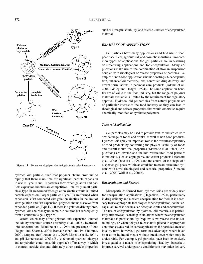
The use of a spray-dried intermediate allows ease of controlof particle properties via manipulation of particle gelation andexpansion kinetics (Fig. 15). By controlling gelation and expan-sion in tandem, it is possible to create a range of particle sizesfrom a spray dried intermediate ranging from 2–30 times the sizeof the dried particle, typically 5–400 µm (Gidley and Hedges,1994).
Figure 15 depicts various hydration regimes, which can leadto several different gel products, ranging from low-swelling par-ticles (Type I), through to medium (Type II) and large swellingparticles (Type III) to dissolved polymer chains (Type IV), tocontinuous gel networks (Type V).
Type I particles are formed when gelation kinetics are veryfast compared with hydration-driven expansion of the dried
372 P. BUREY ET AL.
Figure 15 Formation of gel particles and gels from a dried intermediate.
hydrocolloid particle, such that polymer chains crosslink sorapidly that there is no time for significant particle expansionto occur. Type II and III particles form when gelation and par-ticle expansion kinetics are competitive. Relatively small parti-cles (Type II) are formed when gelation kinetics result in limitedparticle expansion. Larger particles (Type III) are formed whenexpansion is fast compared with gelation kinetics. In the limit ofslow gelation and fast expansion, polymer chains dissolve fromexpanded particles (Type IV). If there is a gelation driving force,hydrocolloid chains may not remain in solution but subsequentlyform a continuous gel (Type V).
Factors which may affect gelation and expansion kineticsinclude hydrocolloid source (Wandrey et al., 2003), hydrocol-loid concentration (Blandino et al., 1999), the presence of ions(Bajpai and Sharma, 2004; Ramakrishnan and Prud’homme,2000), temperature (Lootens et al., 2003; Marcotte et al., 2001),and pH (Lootens et al., 2003). By selection of hydrocolloid typeand rehydration conditions, this approach offers a way in whichto control particle size and ultimately other particle properties
such as strength, solubility, and release kinetics of encapsulatedmaterial.
EXAMPLES OF APPLICATIONS
Gel particles have many applications and find use in food,pharmaceutical, agricultural, and cosmetic industries. Two com-mon types of applications for gel particles are in texturingor structuring applications and for encapsulation. Many ap-plications make use of the combination of flow in suspensioncoupled with rheological or release properties of particles. Ex-amples of non-food applications include coatings, bioencapsula-tion, enhanced oil recovery, inks, controlled drug delivery, andcream formulations in personal care products (Adams et al.,2004; Gidley and Hedges, 1994). The same application bene-fits are of value to the food industry, but the range of polymermaterials available is limited by the requirement for regulatoryapproval. Hydrocolloid gel particles from natural polymers areof particular interest to the food industry as they can lead torheological and release properties that would otherwise requirechemically-modified or synthetic polymers.
Textural Applications
Gel particles may be used to provide texture and structure toa wide range of foods and drinks, as well as non-food products.Hydrocolloids play an important role in the overall acceptabilityof food products by controlling the physical stability of foodsand overall mouth-feel properties (Marcotte et al., 2001). Ap-plications are diverse and include restructured food particlesin materials such as apple puree and carrot products (Marcotteet al., 2000; Ocio et al., 1997) and the control of the shape of adispersed gel phase within an emulsion to create structured sys-tems with novel rheological and sensorial properties (Simeoneet al., 2005; Wolf et al., 2001b).
Encapsulation and Release
Microparticles formed from hydrocolloids are widely usedfor encapsulation applications (Hegenbart, 1993), particularlyin drug delivery and nutrient encapsulation for food. It is neces-sary to use appropriate techologies for encapsulation, so that en-capsulant release occurs at an acceptable rate and concentration.The use of encapsulation by hydrocolloid materials is particu-larly attractive as it can help in situations where the encapsulatedmaterial has poor solubility, requires slow release into its sur-roundings, or when delayed release until placed in appropriateconditions is desired. In some applications the particles are usedin a dry form; however, a gel form has advantages where it canbe used in hydrated media without breakdown which may beundesirable. For example, gel particles have been extensivelyinvestigated as a means of encapsulating “healthy” bacteria toimprove survival under gastric conditions to maximize delivery

HYDROCOLLOID GEL PARTICLES 373
to the colon (Nussinovitch et al., 1997; Zvitov and Nussinovitch,2003). Other uses for such particles are the encapsulation of in-sulin or drugs in pectin particles (Cheng and Lim, 2004; Sri-amornsak and Nuthanid, 1998). Some examples of food appli-cations include the use of alginate gel particles to encapsulatevitamin C (Desai et al., 2005) and hydrocolloid particles for theencapsulation of flavors (Malone and Appelqvist, 2003).
GEL PARTICLE CHARACTERISATION TECHNIQUES
Many gel particle characteristics are directly related to endproduct perception by consumers, and hence success in appli-cation. Some of the major properties of gel particles and thetechniques used for characterizing them are described below.
Particle Size, Shape, and Structure
Particle morphology is important as it can directly impact onconsumer perception (Engelen et al., 2005; Imai et al., 1999) aswell as influence other properties. Therefore it is important tounderstand particle morphology and use appropriate techniquesfor studying particle properties. Two commonly used techniquesfor evaluating particle size and morphology are laser diffractionanalysis and microscopy (Gavini et al., 2005; Klokk and Melvik,2002; Kortesuo et al., 2000; Rosinski et al., 2002). Nuclear Mag-netic Resonance (NMR) can also be used for examining porosityof gel particles as well as observing the gelation process (Dobieset al., 2005; Duez et al., 2000; Hills et al., 2000).
Laser Diffraction
Particle size distributions can be determined by the use oflaser diffraction analysis, which is based on the diffraction oflight at the surface of particles. Two factors are important foreffectively obtaining particle size distributions, namely the laserwavelength coupled with machine optics and the presence ofsufficient contrast between a particle surface and the suspend-ing fluid to define the surface location. Standard measurementdevices such as the Malvern Mastersizer use different opticalattachments to detect particles in different size ranges, whichcan be as wide as 0.02–2000 µm.
Particle size distributions of hard particles have been deter-mined using laser diffraction for many years (Pike, 1979) andnow gel particle size distributions can also be obtained. Hy-drocolloid gel particle systems have recently been studied usinglaser diffraction analysis with alginate being the most commonlystudied material (Gavini et al., 2005; Klokk and Melvik, 2002;Liu et al., 2003; Rosinski et al., 2002). Particle sizes on thescale of µm are typically described. Light scattering may alsoprovide information about structure on a mesoscopic scale, typ-ically 100–1000 nm (Poon and Haw, 1997). It is important torealize that some difficulties may arise in using laser diffractionfor highly swollen particles, where the concentration of hydro-
colloid is low, resulting in limited optical contrast between themedium and the gel particles.
Microscopy
Particle size can also be determined via microscopy tech-niques, such as light microscopy (LM), scanning electron mi-croscopy (SEM), transmission electron microscopy (TEM), andatomic force microscopy (AFM); in addition, microscopy tech-niques can also provide information about the shape and thetopological features of particles.
Bright field and fluorescence LM are frequently used becausethey allow selective staining of different chemical components.LM may be useful in observing microscopic gel particles, butit does not have the resolution of SEM or TEM. It is however,a quick and simple method to characterize particle preparationsbefore more complex methods are utilized. Provided contrastbetween particles and a suspending fluid can be achieved (e.g.through the use of selective stains), image analysis software canbe used to provide descriptions of particle population propertiessuch as size distributions and shape factors.
SEM is useful for examining the surface of microstruc-tural components (Egerton, 2005). Samples are typically frozen,fractured, and coated with a metal compound in order to ex-amine macromolecular organization within particles (Bhatnagarand Hanna, 1997). Environmental scanning electron microscopy(ESEM) allows viewing of the surface of a hydrated samplewithout the need for coating. This is possible by observing thesamples under a partial vacuum (Egerton, 2005). Although res-olution is not as high as for SEM, the milder preparation processprovides more confidence that artefactual structure modificationis avoided.
TEM is a method that may be used to view the microstructureof a thin sample by passing electrons through it. In the case offood gels, the section must be fixed and stained with a heavymetal compound to provide contrast between the various com-ponents (Kalab et al., 1995).
There are some difficulties with sample preparation tech-niques for both SEM and TEM. Cryo-sectioning involves freez-ing and ice crystals may damage the structure. Plastic embed-ding involves dehydration and can cause shrinkage. Confocalscanning laser microscopy (CSLM) offers some advantages inthat there is minimal sample processing, and three-dimensionalimages can be obtained, however, resolution is similar to LM,so electron microscopy is still required to investigate fine details(Autio and Laurikainen, 1997).
Previous studies of hydrocolloid gel particles have mostlyused LM to observe the particles and have provided useful infor-mation on particle shape and size (Cellesi et al., 2004; Lamprechtet al., 2000; Liu et al., 2003; Sriamornsak and Nuthanid, 1998;Sriamornsak et al., 2004; Wong et al., 2002; Zvitov and Nussi-novitch, 2003). The limitation of LM is that it mostly producesonly two-dimensional images and does not have enough resolu-tion to observe sub-micron structural features on or within theparticles.
374 P. BUREY ET AL.
Several recent studies have used SEM to observe gel particlefeatures more closely. The use of SEM has allowed observationof fiber and pore structures, as well as surface topology (Ouw-erx et al., 1998; Sriamornsak et al., 2004; Velings and Mestdagh,1995; Zvitov and Nussinovitch, 2003). Conventional SEM re-quires drying of samples so there is some loss of structure dueto the removal of water, but it has proved useful for observingthe internal structure of the hydrocolloid network within beadparticles. TEM has also been used to observe network structure(Adams et al., 2004), but again would have the same difficultiesassociated with dehydration.
Traditionally AFM has been used to observe surface struc-tures of materials, and is an attractive technique to utilize for thispurpose as it has the ability of performing three-dimensionalmeasurements with a resolution on the order of nanometres(Bonell, 1993). Another advantage of this technique is the verysimple sample preparation, hence avoiding damaging or alteringthe sample prior to measurement (Starostina and West, 2006),and also the ability to observe samples in air or under solution.The use of AFM for observing nanoscale particles has devel-oped only in the last few years and hence the capabilities arestill being explored. Interest in the use of AFM for observingsoft particles has grown in recent times, particularly in the med-ical field (Starostina and West, 2006), and has great potential foruse in observing hydrocolloid gel particles. AFM also has thecapability of carrying out mechanical measurements, providingfurther information on sample characteristics.
Image analysis is used to determine properties of microscopicfeatures from micrographs and can be used in conjunctionwith images from any of the microscopy techniques mentionedabove. Characterization of both particle shape and size can becarried out, providing detailed information about particle sizedistributions as well as individual particle properties (Langtonand Hermansson, 1993; Russ, 2005). Simple measurementsof particle characteristics (e.g diameter, shape factor) can bequickly measured. It has been used to analyse such systemsas biopolymer particle suspensions (Wolf et al., 2001b) andshear gel systems (Adams et al., 2004), providing quantitativemeasures of images.
NMR techniques have more recently been used to study in-ternal gel particle structure as well as gelation of gel particles(Dobies et al., 2005; Duez et al., 2000; Grant et al., 2005; Hillset al., 2000). In these studies, water proton relaxation time dis-tributions are obtained, to complement the information on thehydrocolloid architecture observable from SEM or TEM.
Mechanical Characteristics–Rheology/Texture/Mouthfeel
There are many techniques available for evaluating the me-chanical behavior of macroscopic particles; however, there canbe difficulties associated with analyzing the sub-micron andmicron-sized particles of interest in food applications, requiringmore specialized techniques. Some techniques that have beenused include nano-indentation and AFM for the study of indi-vidual particles, and rheometry and texture analysis for bulk
dispersions of particles. These analysis techniques are morecommonly used for the study of hard particles, and so whilstpotentially useful for the study of soft gel particles, there maybe practical difficulties that need to be addressed.
Nanoindentation
Nanoindentation testing is used to determine elastic modulusand hardness data of samples from load-displacement measure-ments. In a typical test, a measure of the residual impressionleft by an indenter is carried out (Fischer-Cripps, 2004). Theimpression is measured as a function of indenter load. As theimpression that is left is so small, it is not possible to view theimpression by simple optical means (LM), hence the depth ofpenetration is measured, and the impression size is determinedfrom known indenter geometry and the measured penetrationdepth (Fischer-Cripps, 2004). There have been very few stud-ies of nanoindentation of soft gel particles, as the technique istypically used for hard particles or surfaces.
Rheometry
Rheometry is traditionally an extremely useful technique formeasuring deformation and flow of both synthetic polymer andbiopolymer materials. Shear rheometry is commonly used onliquid or semi-solid materials, while extensional or compressiverheometry may be used on semi-solid or solid materials.
Rheometry can be useful for evaluating the deformation andflow behavior of particle suspensions and has been used suc-cessfully to characterize agar gel microparticles (Adams et al.,2004) and alginate particles encapsulating biocatalysts (Vogel-sang et al., 2000). Information about the rheological behavior ofgel particles can be useful in determining both static (small de-formation) and yield/flow behavior of particles in different me-dia which can then be related to sensorial properties in productssuch as food materials or cosmetic products. The measurementsmay also relate back to in-mouth particle detectability, a keyquality determinant in food applications.
Texture Analysis
Texture profile analysis involves large deformation testing ofsamples so it is often limited to measurements on macroscalesamples. It was developed in the early 1960s to imitate defor-mation of food samples by the jaw, by observing the behaviorthat would correspond to the first two bites of a sample (Bordeet al., 2002; Friedman et al., 1963). The test involves a two-cyclepenetration test into the food sample, with the force developedobserved over time. The output obtained shows a two peak forcecurve, and gives an indication of recovery behavior after defor-mation. Calculations are carried out to determine the magnitudeof textural parameters (Bourne, 2002). These parameters are de-pendent on testing geometries, so it is important to be consistentwith the sample size when testing materials. The technique couldbe useful for testing bulk samples of hydrocolloid gel particlesor for testing products that contain such particles.
HYDROCOLLOID GEL PARTICLES 375
Molecular Release–Food/Pharmaceuticals
Hydrocolloid gel particles have potential applications in en-capsulation of materials such as flavors, nutrients, probiotics,and drugs. The gel particles can release the encapsulated ma-terial slowly or the hydrocolloid “shell” can break down underappropriate conditions to facilitate release of the material. Therate at which material is released is one of the most impor-tant factors for application success and can be influenced bycharacteristics such as particle size and particle membrane dif-fusivity. The characterization of release profiles together withparticle morphology allows the development of understandingof mechanisms involved in determining release rates. The abil-ity to withstand different environments of pH, temperature, andionic strength may also be an issue depending upon the applica-tion (Nussinovitch et al., 1997; Velings and Mestdagh, 1995).
Studies of pectin as an encapsulation material for drug deliv-ery have shown that the dissolution medium affects drug releasefrom pectin microspheres by modifying the drug solubility andintegrity of the pectin matrix (Sriamornsak and Nuthanid, 1998).A comprehensive study of various hydrocolloids for use in en-capsulation of flavors was carried out by Malone and Appelqvist(2003), where it was found that particle composition and struc-ture both had an effect on the rate of release. The particles weretested for release under various conditions of mechanical fail-ure, temperature, and alpha-amylase susceptibility in order tomimic the breakdown conditions within the human body. It wasconcluded that the control of these parameters could control therelease profile of the encapsulated flavor.
OPPORTUNITIES FOR FURTHER RESEARCH
The application opportunities for hydrocolloid gel particlesin foods are widespread and also linked to current trends in foodinnovation such as the controlled delivery of functional activesand the development of lower calorie equivalents of traditionalfoods. These drivers ensure that gel particles will be further in-vestigated and applied in the food sector. Three areas of specificopportunity for future R&D are:
• Translation of findings related to drug delivery to bioactivedelivery from foods
• Understanding the role of mechanical and thermal processingoperations on the formation and stability of gel particles infinished foods.
• Exploitation of dried intermediates as a convenient route togel particles of controllable size and properties produced whenneeded. This area is currently under investigation by the au-thors.
REFERENCES
Adams, S., Frith, W. J., and Stokes, J. R. (2004). Influence of particle modulus onthe rheological properties of agar microgel suspensions. J Rheol., 48:1195–1213.
Armisen, R., and Galatas, F. (2000). Agar. In: Handbook of Hydrocolloids. pp.21–40. Phillips, G. O. and Williams, P. A., Eds., CRC Press, Boca Raton.
Autio, K., and Laurikainen, T. (1997). Relationships between flour/dough mi-crostructure and dough handling and baking properties. Trends Food Sci.Technol., 8:181–185.
Bajpai, S. K., and Sharma, S. (2004). Investigation of swelling/degradation be-haviour of alginate beads crosslinked with Ca2+ and Ba2+ ions. React FunctPolym., 59:129–140.
Belitz, H.-D. and Grosch, W. (1999). Food Chemistry. Springer Verlag, Berlin.Belton, P. S., Chilvers, G. R., Morris, V. J., and Tanner, S. F. (1984). Effects
of group I cations on the gelation of iota carrageenan. Int J Biol Macromol.,6:303–308.
Bhandari, B. (2005). Spray Drying and Powder Properties. In:Handbook of FoodScience, Technology and Engineering. In press. Hui, Y. H. and Sherkat, F.,Eds., Taylor and Francis, Boca Raton.
Bhatnagar, S., and Hanna, M. A. (1997). Modification of microstructure of starchextruded with selected lipids. Starch., 49:12–20.
Biswal, D. R., and Singh, R. P. (2004). The flocculation and rheological charac-teristics of hydrolyzed and unhydrolyzed grafted sodium alginate in aqueoussolutions. J Appl Polym Sci., 94:1480–1488.
Blandino, A., Macias, M., and Cantero, D. (1999). Formation of calcium algi-nate gel capsules: Influence of sodium alginate and CaCl2 concentration ongelation kinetics. J Biosci Bioeng., 88:686–689.
Bohidar, H. B., and Jena, S. S. (1993). Kinetics of sol-gel transition in thermore-versible gelation of gelatin. J Chem Phys., 98:8970–8977.
Borde, B., Bizot, H., Vigier, G., and Buleon, A. (2002). Calorimetric analysisof the structural relaxation in partially hydrated amorphous polysaccharides.Carbohyd Polym., 48:111–123.
Bourne, M. C. (2002). Food Texture and Viscosity: Concept and Measurement.Academic Press Inc., New York.
Brown, C. R. T., Fairley, P., and Lam, S. (2004). Hair treatment compositions.US Patent no. 6719967.
Bugarski, B., Li, Q. L., Goosen, M. F. A., Poncelet, D., Neufeld, R. J., and Vun-jakg (1994). Electrostatic droplet generation–mechanism of polymer dropletformation. AICHE J., 40:1026–1031.
Burchard, W., and Ross-Murphy, S. B. (1990). Physical Networks–Polymersand Gels. Elsevier Science, Essex.
Burgess, D. J., and Carless, J. E. (1984). Microelectrophoretic studies of gelatinand acacia for the prediction of complex coacervation. J Colloid Interf Sci.,98:1–8.
Campbell, A., Taylor, P., Cayre, O. J., and Paunov, V. N. (2004). Preparation ofaqueous gel beads coated by lipid bilayers. Chem Commun., 21:2378–2379.
Cellesi, F., Weber, W., Fussenegger, M., Hubbell, J. A., and Tirelli, N. (2004).Towards a fully synthetic substitute of alginate: optimization of a thermal gela-tion/chemical cross-linking scheme (“tandem” gelation) for the production ofbeads and liquid-core capsules. Biotechnol Bioeng., 88:740–749.
Cheng, K., and Lim, L.-Y. (2004). Insulin-loaded calcium pectinate nanopar-ticles: Effects of pectin molecular weight and formula pH. Drug Dev IndPharm., 30:359–367.
Chilvers, G. R., Gunning, A. P., and Morris, V. J. (1988). Coacervation of gelatin-xm6 mixtures and their use in microencapsulation. Carbohyd Polym., 8:55–61.
Christensen, S. H. (1983). Pectins. In:Food Hydrocolloids. pp. 205–229. Glicks-man, M., Eds., CRC Press Inc., Boca Raton
Clark, A. H., and Ross-Murphy, S. B. (1990). Shear Modulus–ConcentrationRelationships for Biopolymer Gels. Comparison of Independent and Coop-erative Crosslink Descriptions. In:Physical Networks–Polymers and Gels.pp. 209–230. Burchard, W., and Ross-Murphy, S. B., Eds., Elsevier Science,London.
Cuppo, F., Venuti, M., and Cesaro, A. (2001). Kinetics of gelatin transitions withphase separation: T-jump and step-wise DSC study. Int J Biol Macromol.,28:331–341.
de Jong, H. G. B., and Lens, J. (1932). Coacervation (separation) in mixturesconcentrated gum arabic and gelatine sol. Kolloid Z Z Polym., 58:209–214.
de Kruif, C. G., Weinbreck, F., and de Vries, R. (2004). Complex coacervationof proteins and anionic polysaccharides. Curr Opin Colloid In., 9:340–349.

376 P. BUREY ET AL.
Deis, R. C. (1997). Spray Drying–Innovative Use of an Old Process.Food Product Design. Available from: http://www.foodproductdesign.com/archive/1997/0597DE.html [Accessed 12 Dec 2005]
Desai, K. G. H., Liu, C., and Park, H. J. (2005). Characteristics of vitaminC immobilized particles and sodium alginate beads containing immobilizedparticles. J Microencapsul., 22:363–376.
Djabourov, M. (1991). Gelation–A Review. Polym Int., 25:135–143.Dobies, M., Kusmia, S., and Jurga, S. (2005). 1H NMR and rheological studies
of the calcium induced gelation process in aqueous low methoxyl pectinsolutions. Acta Phys Pol A., 108:33–46.
Douglas, K. L., and Tabrizian, M. (2005). Effect of experimental parameterson the formation of alginate-chitosan nanoparticles and evaluation for theirpotential application as DNA carrier. J Biomat Sci-Polym E., 16:43–56.
Draget, K. I. (2000). Alginates. In: Handbook of Hydrocolloids. pp. 379–395.Phillips, G. O., and Williams, P. A., Eds., CRC Press, Boca Raton.
Draget, K. I., Skjak Braek, G., and Smidsrod, O. (1994). Alginic acid gels:the effect of alginate chemical composition and molecular weight. CarbohydPolym., 25:31–38.
Duez, J.-M., Mestdagh, M., Demeure, R., Goudemant, J.-F., Hills, B. P., andGodward, J. (2000). NMR studies of calcium-induced alginate gelation. PartI–MRI tests of gelation models. Magn Reson Chem., 38:324–330.
Egerton, R. F. (2005). Physical principles of electron microscopy: An introduc-tion to TEM, SEM, and AEM Springer, New York.
Elversson, J., and Millqvist-Fureby, A. (2005). Particle size and density in spraydrying–effects of carbohydrate properties J. Pharm. Sci., 94:2049–2060.
Engelen, L., van der Bilt, A., Schipper, M., and Bosman, F. (2005). Oral sizeperception of particles: effect of size, type, viscosity and method. J TextureStud., 36:373–386.
Finer, E. G., Franks, F., Phillips, M. C., and Sugget, A. (1975). Gel formationfrom solutions of single chain gelatin. Biopolymers., 14:1995–2005.
Fischer-Cripps, A. C. (2004). Nanoindentation. Springer-Verlag, New York.Friedman, H. H., Whitney, J. E., and Sczczesniak, A. S. (1963). The
texturometer–a new instrument for objective texture measurement. J FoodSci., 28:390–396.
Gander, B., Blanco-Prıeto, M. J., Thomasin, C., Wandrey, C., and Hunkeler, D.(2002). Coacervation of Phase Separation. In: Encyclopedia of Pharmaceu-tical Technology. pp. 481–496. Swarbrick, J., Eds.
Gavini, E., Rassu, G., Sanna, V., Cossu, M., and Giunchedi, P. (2005). Mucoad-hesive microspheres for nasal administration of an antiemetic drug, metoclo-pramide: in-vitro/ex-vivo studies. J Pharm Pharmacol., 57:287–294.
Gidley, M. J., and Hedges, N. D. (1994). Suspensions of gelled biopolymers.US Patent No. 5738897.
Glicksman, M. (1983a). Food Hydrocolloids–Volume 1. CRC Press Inc, BocaRaton.
Glicksman, M. (1983b). Food Hydrocolloids–Volume 2. CRC Press Inc., BocaRaton.
Glicksman, M. (1983c). Food Hydrocolloids–Volume 3. CRC Press Inc., BocaRaton.
Goosen, M. F. A. (2003). Experimental and modeling studies of mass transferin microencapsulated cell systems. Trop J Pharm Res., 1:3–14.
Gotlieb, K. F., and Capelle, A. (2005). Starch derivatization : fascinat-ing and unique industrial opportunities Wageningen Academic Publishers,Wageningen.
Gouin, S. (2004). Microencapsulation: industrial appraisal of existing technolo-gies and trends. Trends Food Sci Tech., 15:330–347.
Grant, S. C., Celper, S., Gauffin-Holmberg, I., Simpson, N. E., Blackband, S.J., and Constantinidis, I. (2005). Alginate assessment by NMR microscopy. JMater Sci-Mater M., 16:511–514.
Hamberg, L., Wohlwend, M., Walkenstrom, P., and Hermansson, A.-M. (2003).Shapes and shaping of biopolymer drops in hyperbolic flow. Food Hydrocol-loid., 17:641–652.
Hazen, C. (2004). Hydrocolloid Handbook. Food Product Design. Availablefrom: http://www.foodproductdesign.com/archive/2004/1004CS.html [Ac-cessed 06 May 2005]
Hegenbart, S. (1993). Encapsulated Ingredients: Keep Problems Covered.Food Product Design. Available from: http://www.foodproductdesign.com/archive/1993/0493CS.html [Accessed 28 Jun 2005]
Hills, B. P., Godward, J., Debatty, M., Barras, L., Saturio, C. P., and Ouwerx,C. (2000). NMR studies of calcium induced alginate gelation. Part II. Theinternal bead structure. Magn Reson Chem., 38:719–728.
Hoefler, A. C. (2004). Hydrocolloids. Eagen Press, Minnesota.Huang, X., Kakuda, Y., and Cui, W. (2001). Hydrocolloids in emulsions: particle
size distribution and interfacial activity. Food Hydrocolloid., 15:533–542.Hunik, J. H., and Tramper, J. (1993). Large-scale production of k-carrageenan
droplets for gel-bead production: theoretical and practical limitations of sizeand production rate, Biotechnol Progr., 9:186–192.
Imai, E., Saito, K., Hatakeyama, A., Hatae, K., and Shimada, A. (1999). Effectof physical properties of food particles on the degree of graininess perceivedin the mouth. J Texture Stud., 30:59–88.
Imeson, A. P. (2000). Carrageenan. In: Handbook of Hydrocolloids. pp. 87–102.Phillips, G. O., and Williams, P. A., Eds., CRC Press, Boca Raton.
Johnston-Banks, F. A. (1990). Gelatine. In: Food Gels. pp. 233–269. Harris, P.,Elsevier Science Publishers Ltd., New York.
Joseph, I., and Venkataram, S. (1995). Indomethacin sustained release fromalginate-gelatin or pectin-gelatin coacervates. Int J Pharm., 126:161–168.
Juang, R.-S., Wu, F.-C., and Tseng, R.-L. (2002). Use of chemically modifiedchitosan beads for sorption and enzyme immobilisation. Adv Environ Res.,6:171–177.
Kalab, M., Allan-Wojtas, P., and Miller, S. S. (1995). Microscopy and otherimaging techniques in food structure analysis. Trends Food Sci. Technol.,6:177–186.
King, A. H. (1983). Brown Seaweed Extracts (Alginates). In: Food Hydrocollids.pp. 115–182. Glicksman, M., Eds., CRC Press, Boca Raton.
King, A. H. (1995). Encapsulation of Food Ingredients: A Review of AvailableTechnology, Focusing on Hydrocolloids. In: Encapsulation and ControlledRelease of Food Ingredients. pp. 26–39. Risch, S. J., and Reineccius, G. A.,Eds., American Chemical Society, Washington.
Klokk, T. E., and Melvik, J. E. (2002). Controlling the size of alginate gel beadsby use of high electrostatic potential. J Microencapsul., 19:415–424.
Kortesuo, P., Ahola, M., Kangas, M., Kangasniemi, I., Yli-Urpo, A., and Kies-vaara, J. (2000). In vitro evaluation of sol-gel processed spray dried silica gelmicrospheres as carrier in controlled drug delivery. Int J Pharm., 200:223–229.
Krasaekoopt, W., Bhandari, B., and Deeth, H. (2003). Evaluation of encapsula-tion techniques of probiotics for yoghurt. Int Dairy J., 13:3–13.
Lamprecht, A., Schafer, U., and Lehr, C.-M. (2000). Structural Analysis of Mi-croparticles by Confocal Laser Scanning Microscopy. AAPS PharmSciTech.1. Available from: http://aapspharmscitech.org/articles/pt0103/pt010317/pt010317.pdf [Accessed 28 Jun 2005]
Langton, M., and Hermansson, A. M. (1993). Image-analysis determination ofparticle-size distribution. Food Hydrocolloid., 7:11–22.
Lapitsky, Y., and Kaler, E. W. (2004). Formation of surfactant and polyelectrolytegel particles in aqueous solutions. Colloid Surface A., 250:179–187.
Liu, X. D., Bao, D. C., Xue, W. M., Xiong, Y., Yu, W. T., Yu, X. J., Ma, X. J.,and Yuan, Q. (2003). Preparation of uniform calcium alginate gel beads bymembrane emulsification coupled with internal gelation. J App Polym Sci.,87:848–852.
Lofgren, C., Walkenstrom, P., and Hermansson, A.-M. (2002). Microstructureand rheological behavior of pure and mixed pectin gels. Biomacromolecules.,3:1144–1153.
Lootens, D., Capel, F., Durand, D., Nicolai, T., Boulenguer, P., and Langendorff,V. (2003). Influence of pH, Ca concentration, temperature and amidation onthe gelation of low methoxyl pectin. Food Hydrocolloid., 17:237–244.
Lucinda-Silva, R. M., and Evangelista, R. C. (2003). Microspheres of alginate-chitosan containing isoniazid. J Microencapsul., 20:145–152.
Malone, M., and Appelqvist, I. A. M. (2003). Gelled emulsion particles for thecontrolled release of lipophilic volatiles during eating. J Control Release.,90:227–241.
Marcotte, M., Hoshahili, A. R. T., and Ramaswamy, H. S. (2001). Rheologi-cal properties of selected hydrocolloids as a function of concentration andtemperature. Food Res Int., 34:695–703.
Marcotte, M., Taherian, A. R., and Ramaswamy, H. S. (2000). Physical proper-ties of reconstituted carrot/alginate particles stable for aseptic processing. JFood Process Eng., 23:463–480.

HYDROCOLLOID GEL PARTICLES 377
Masters, K. (1991). Spray Drying Handbook. Longman and Scientific Technical,New York.
May, C. D. (2000). Pectins. In: Handbook of Hydrocolloids. pp. 169–188.Phillips, G. O., and Williams, P. A., Eds., CRC Press, Boca Raton.
Mohanty, B., Aswal, V. K., Goyal, P. S., and Bohidar, H. B. (2004). Small-angleneutron and dynamic light scattering study of gelatin coacervates. Pramana-JPhys., 63:271–276.
Mohanty, B., and Bohidar, H. B. (2006). Microscopic structure of gelatin coac-ervates. Int. J. Biol. Macromol., 3:39–46.
Morris, E. R., Rees, D. A., and Robinson, G. (1980). Cation-specific aggregationof carrageenan helices: domain model of polymer gel structure. J. Mol. Biol.,138:349–362.
Mukai-Correa, R., Prata, A. S., Alvim, I. D., and Grosso, C. R. F. (2004). Con-trolled release of protein from hydrocolloid gel microbeads before and afterdrying. Curr Drug Del., 1:265–273.
Murakata, T., Honma, H., Nakazato, S., Kuroda, C., and Sato, S. (2001). Controlof particle size of calcium alginate gel bead by application of electric fieldto interface between aqueous and organic phases. J Chem Eng Jpn., 34:299–305.
Nussinovitch, A. (1997). Hydrocolloid Applications. Chapman and Hall, Lon-don.
Nussinovitch, A., Gershon, Z., and Nussinovitch, M. (1997). Temperature stableliquid core hydrocolloid capsules. Food Hydrocolloid., 11:209–215.
Oakley, D. E. (2004). Spray dryer modeling in theory and practice. Dry. Technol.,22:1371–1402.
Ocio, M. J., Fiszman, S. M., Gasque, F., Rodrigo, M., and Martinez, A.(1997). Development of a restructured alginate food particle suitable forhigh temperature-short time process validation. Food Hydrocolloid., 11:423–427.
Ouwerx, C., Velings, N., Mestdagh, M. M., and Axelos, M. A. V. (1998).Physico-chemical properties and rheology of alginate gel beads formed withvarious divalent cations. Polym Gels Netw., 6:393–408.
Paterson, L. A., Hill, S. E., Mitchell, J. R., and Blanshard, J. M. V. (1997).Sulphite and oxidative-reductive depolymerization reactions. Food Chem.,60:143–147.
Peng, H., Martineau, L., and Shek, P. (2006). Method of producing layeredpolymeric articles. US Patent No. 20060061005.
Pike, E. R. (1979). The Malvern correlator. Phys Technol., 10:104–109.Poon, W. C. K., and Haw, M. D. (1997). Mesoscopic structure formation in
colloidal aggregation and gelation. Adv Colloid Interfac., 73:71–126.Ramakrishnan, S., and Prud’homme, R. K. (2000). Effect of solvent quality and
ions on the rheology and gelation of kappa-carrageenan. J Rheol., 44:885–896.
Rees, D. A. (1982). Polysaccharide conformation in solutions and gels–recentresults on pectins. Carbohyd Polym., 2:254–263.
Reis, C. P., Neufeld, R. J., Vilela, S., Ribeiro, A. J., and Veiga, F. (2006). Re-view and current status of emulsion/dispersion technology using an internalgelation process for the design of alginate particles. J Microencapsul., 23:245–257.
Rix, A. (1990). Gelling and Whipping Agents. In: Sugar Confectionery Manu-facture. pp. 57–76. Jackson, E. B., Eds., Van Nostrand Reinhold, Glasgow.
Rochas, C., and Rinaudo, M. (1984). Mechanism of gel formation in κ-carrageenan. Biopolymers., 23:735–745.
Rosinski, S., Grigorescu, G., Lewinska, D., Ritzen, L. G., Viernstein, H., Teunou,E., Poncelet, D., Zhang, Z., Fan, Z., D., S., Marison, I., and Hunkeler, D.(2002). Characterisation of microcapsules: Recommended methods based onround-robin testing. J Microencapsul., 19:641–659.
Rostagno, W. (1969). Chocolate particle size and its organoleptic influence.Manuf Conf., 49:81–85.
Russ, J. C. (2005). Image analysis of food microstructures. CRC Press, BocaRaton.
Simeone, M., Tassieri, M., Sibillo, V., and Guido, S. (2005). Effect of sol-geltransition on shear-induced drop deformation in aqueous mixtures of gellanand κ-carrageenan. J Colloid Interf Sci., 281:488–494.
Sriamornsak, P., and Nuthanid, J. (1998). Calcium pectinate gel beads for con-trolled release drug delivery: I. Preparation and in vitro release studies. Int JPharm., 160:207–212.
Sriamornsak, P., Thirawong, N., and Puttipipatkhachorn, S. (2004). Morphol-ogy and Buoyancy of Oil-entrapped Calcium Pectinate Gel Beads. TheAmerican Association of Pharmaceutical Scientists Journal. 6. Availablefrom: http://www.aapsj.org/articles/aapsj0603/aapsj060324/aapsj060324.pdf[Accessed 09 Aug 2005]
Sugiura, S., Odo, T., Izumida, Y., Aoyagi, Y., Satake, M., Ochiai, A., Ohkohchi,N., and Nakajima, M. (2005). Size control of calcium alginate beads contain-ing living cells using micro-nozzle array. Biomaterials., 26:3327–3331.
Thomasin, C., Merkle, H. P., and Gander, B. (1998). Drug microencapsulationby PLA/PLGA coacervation in the light of thermodynamics. 2. Parametersdetermining microsphere formation. J Pharm Sci., 87:269–275.
Tosh, S., Marangoni, A., Hallet, F., and Britt, I. (2003). Aging dynamics ingelatin gel microstructure. Food Hydrocolloid., 17:503–513.
Utz, K.-H. (1986). The interocclusal tactile fine sensitivity of the natural teethas studied by aluminium oxide particles. Deut Zahnaerztl Z., 41:313–315.
van de Velde, F., van Riel, J., and Tromp, R. H. (2002). Visualisation of starchgranule morphologies using confocal scanning laser microscopy (CSLM). JFood Sci Agr., 82:1528–1536.
Velings, N. M., and Mestdagh, M. M. (1995). Physico-Chemical Properties ofAlginate Gel Beads. Polym Gels Netw., 3:311–330.
Vogelsang, C., Wijffels, R. H., and Ostgaard, K. (2000). Rheological propertiesand mechanical stability of new gel-entrapment systems applied in bioreac-tors. Biotechnol Bioeng., 70:247–253.
Walkenstrom, P., and Hermansson, A.-M. (1998). Effects of shear on pureand mixed gels of gelatin and particulate whey protein. Food Hydrocolloid.,12:77–87.
Wandrey, C., Espinosa, D., Rehor, A., and Hunkeler, D. (2003). Influence ofalginate characteristics on the properties of multi-component microcapsules.J Microencapsul., 20:591–611.
Williams, C. T. (1964). Chocolate and Confectionery. Leonard Hill, London.Williams, P. A., and Phillips, G. O. (2000). Introduction to Food Hydrocolloids.
In: Handbook of Hydrocolloids. pp. 1–19. Phillips, G. O., and Williams, P.A., Eds., CRC Press, Boca Raton.
Wolf, B., Frith, W. J., and Norton, I. T. (2001a). Influence of gelation on particleshape in sheared biopolymer blends. J Rheol., 45:1141–1157.
Wolf, B., Frith, W. J., Singleton, S., Tassieri, M., and Norton, I. T. (2001b).Shear behaviour of biopolymer suspensions with spheroidal and cylindricalparticles. Rheol Acta., 40:238–247.
Wong, T. W., Lee, H. Y., Chan, L. W., and Heng, P. W. S. (2002). Drug releaseproperties of pectinate microspheres prepared by emulsification method. IntJ Pharm., 242:233–237.
Woodruff, S., and Gilder, H. V. (1931). Photomicrographic studies of sucrosecrystals. J Phys Chem., 35:1355–1367.
Zimmerman, H., Zimmerman, D., Reuss, R., Feilen, P. J., Manz, B., Katsen, A.,Weber, M., Ihmig, F. R., Ehrhart, F., Gebner, P., Behringer, M., Steinbach,A., Wegner, L. H., Sukhorukov, V. L., Vasquez, J. A., Schneider, S., Weber,M. M., Volke, F., Wolf, R., and Zimmerman, U. (2005). Towards a medicallyapproved technology for alginate-based microcapsules allowing long-termimmunoisolated transplantation. J Mater Sci-Mater M., 16:491–501.
Zvitov, R., and Nussinovitch, A. (2001). Weight, mechanical and structuralchanges induced in alginate beads by DC electrical field. Food Hydrocol-loid., 15:33–42.
Zvitov, R., and Nussinovitch, A. (2003). Changes induced by DC electrical fieldin agar, agarose, alginate and gellan gel beads. Food Hydrocolloid., 17:255–263.