Embed Size (px)
Citation preview

Human Papillomavirus (HPV)
and Management of Abnormal
Pap Tests
Christopher P. DeSimone, M.D.
Assistant professor
Division of Gynecologic Oncology
Department of Obstetrics and Gynecology
University of Kentucky Medical Center

Papanicolaou and Traut:
Diagnosis of uterine cancer by the
vaginal smear, 1943
The Papanicolaou Test
Developed by Dr’s. Papanicolaou and
Traut in the 1940’s
Consists of collecting cervical cytology
from the cervix and depositing them
onto a slide for microscopic evaluation
Easy to collect and objectively interpret
results
Initially used to detect cervical cancer

Parkin et al. Int J Cancer, 2005.
The Papanicolaou Test
The pap has dramatically decreased the
the incidence and mortality rate of
cervical cancer in the United States
U.S. 10,000 new cases each year
3700 deaths annually from cervical cancer
Worldwide 493,000 new cases
293,000 deaths annually worldwide

The Papanicolaou Test
Estimated 50 million paps
performed in the U.S.
5% will be diagnosed as
abnormal
2-3 million ASC-US paps
1 million LSIL paps
600,000 HSIL paps
Economics behind these
numbers are staggering
ASCUS
LGSIL
HGSIL
Cancer

Who Develops Cervical
Cancer?
50% of women diagnosed with cervical
cancer have not had a pap test in 5 years
25% of all cervical cancers are diagnosed in
women older than 65
In women older than 65, it is estimated that
over 50% have not had a pap test in the past
10 years
Bottom Line – the majority of women with
cervical cancer fail to get annual pap tests

Overview
American Cancer Society guidelines for Pap testing
Bethesda 2001 Pap nomenclature
Human papillomavirus (HPV)
Types of HPV
Incidence of HPV
Incidence of HPV in cervical cancer and preinvasive disease
Mechanism for oncogenesis
Associated risk of cervical cancer with smoking
Management of abnormal Pap tests
Types of treatment
Screening intervals after treatment
HPV vaccines

Saslow et al. Cancer J Clin, 2002.
American Cancer Society screening
guidelines for Pap testing
Begin testing 3 years after starting
vaginal intercourse or no later than 21
Interval is every year for a conventional
pap test or every other year with liquid
cytology

Saslow et al. Cancer J Clin, 2002.
American Cancer Society screening
guidelines for Pap testing
At 30 years of age: IF > 3
normal/negative, satisfactory,
consecutive pap tests then every 3
years IF no high-risk factors
High-risk factors include: any abnormal
pap tests, HPV infections, other STD’s –
syphilis, HIV, gonorrhea and chlamydia

Saslow et al. Cancer J Clin, 2002
American Cancer Society screening
guidelines for Pap testing
Cease screening IN
Women > 70 with normal pap tests in the
past 10 years
Women status post total hysterectomy for
non-neoplastic diseases

Abnormal Pap Smears
(Old Bethesda Nomenclature)
HPV (Human
Papilloma Virus)
LGSIL (Low Grade
Squamous
Intraepithelial
Lesion)
ASCUS (Atypical
Squamous Cells of
Unknown
Significance)
HGSIL (High Grade
Squamous
Intraepithelial Lesion)
AGUS (Atypical
Glandular Cells of
Unknown Significance)
AIS (Adenocarcinoma
In Situ)
CIS or Carcinoma

Solomon et al. JAMA, 2002.
2001 Bethesda Nomenclature,
squamous cells
Mild dysplasia
HPV effect (Human
Papilloma Virus)
ASC-US (Atypical
Squamous Cells of
Unknown
Significance)
LSIL (Low Grade
Squamous
Intraepithelial
Lesion)
Severe dysplasia
ASC-H (Cannot rule
out HGSIL)
HSIL (High Grade
Squamous
Intraepithelial
Lesion)
CIS (Squamous
Carcinoma In-Situ)

Solomon et al. JAMA, 2002.
2001 Bethesda Nomenclature,
glandular cells
Glandular abnormalities
Endometrial cells in a woman 40 years of age
or older
AGC (Atypical Glandular Cells of Unknown
Significance)
AIS (Adenocarcinoma In-Situ)
Adenocarcinoma
AGC and AIS should be handled with utmost
caution

Cytology versus Histology
Cytology pertains to a sample of cells
Pap test
Histology pertains to a tissue sample
Colposcopic biopsy
Cervical intraepithelial neoplasia (CIN) is the
nomenclature used for colposcopic/cervical
biopsies
Cervical intraepithelial neoplasia (CIN) and
dysplasia are synonymous

Dysplasia
Dysplasia is the medical term for preinvasive
disease
Dysplasia represents the POTENTIAL for
abnormal cells to progress to invasive cancer
This potential is subdivided:
CIN 1 = mild dysplasia
CIN 2 = moderate dysplasia
CIN 3 = severe dysplasia
Carcinoma in situ and CIN 3 are the same

Anatomy of Dysplasia

Human Papillomavirus
Member of the
Papovaviridae family
Double Stranded DNA
tumor virus
45-55 nm icosohedral
capsid
More than 100 types
Specific for target
epithelium
Epitheliotrophic and causes
proliferation

HPV and types of infection
Mucocutaneous
Verruca plantaris
1,2,4
Verruca vulgaris
2,4,29,38
Verruca plana
3,10,28
Anogenital
Condyloma 6,11
SIL and Cancer
16,18,31,33,35,45,51
,56

Ho et al. NEJM 1998
Incidence of HPV
608 college aged women studied from
1992-1994
Followed 3 years at 6 month intervals
Incidence of infection 43%
Median duration of any HPV infection, 8
months
70% cleared in one year, 90% in two
years
Ho et al. NEJM 1998
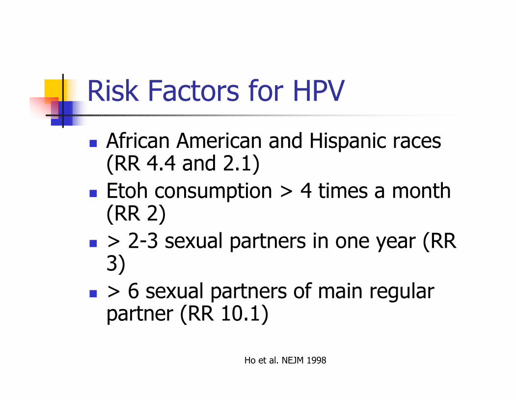
Risk Factors for HPV
African American and Hispanic races
(RR 4.4 and 2.1)
Etoh consumption > 4 times a month
(RR 2)
> 2-3 sexual partners in one year (RR
3)
> 6 sexual partners of main regular
partner (RR 10.1)

Ho et al. NEJM 1998
Incidence of HPV Types
Most common types are high risk types
51,66,16,18
Type 16 found in 7% of 514 women
Type 18 found in 4% of 525 women

Ho et al. NEJM 1998
HPV and SIL
Persistent HPV more likely to progress
to SIL
High risk types take longer to clear
(Median of 12 month)
Women infected with high risk types
documented at two 6 month visits were
38 times more likely to develop SIL

Winer et al. Am J Epidemiol,
2003.
Incidence of HPV
Winer et al. also studied 603 college women
from 1990-2000
Followed for 4 month intervals
Incidence of HPV infection 32%
Incidence among virginal women and sexually
active women was the same
Report of a new partner (5-8 months before a
visit) was the greatest risk factor for
acquiring HPV infection, RR 2.1

Richardson et al. Cancer
Epidemiol Biomark Prev, 2003.
Incidence of HPV
Richardson et al. studied 621 college
students from 1996 to 2001
Followed at six month intervals for 2
years
Incidence was 36%
Median time to regression was 13
months for high risk HPV

Incidence of HPV types
HPV 16 most common
Ho et al. 7%
Kuhn et al. 6%
Winer et al. 10%
Richardson et al. 8%
HPV 18
Roughly 3-4%
HPV 33, 39
Roughly 3-4%

Bosch et al. J Natl Cancer Inst,
1995
HPV and cervical cancer
Bosch et al in 1995, accrued 932 cases of
cervical cancer from around the world
Using polymerase chain reactions (PCR), his
group amplified HPV DNA from the tumor and
recorded their findings
93% of cervical carcinoma had HPV DNA
Common types included 16, 18, 31, 33, 35,
39, 45, 51 (high risk HPV subtypes)

Walboomers et al. J Pathol, 1999
HPV and cervical cancer
Walboomers et al. repeated Bosch’s
experiment using new PCR primers
Those cancers that failed to test
positive for HPV DNA were retested
with these new primers
Results showed that 99.7% of Bosch’s
original cases tested positive for HPV
DNA

Solomon et al. J Natl Cancer Inst,
2001
HPV and HSIL/CIN 3
Severe cervical dysplasia can be described as
the step before invasive carcinoma
HPV is closely tied to the development of
CIN 3
ALTS group examined 136 women with
histology proven CIN 3
126/136 or 93% tested positive for high risk
HPV

Sherman et al. Am J Clin Pathol,
2001.
HPV and LSIL, ASC-H, ASC-US
ALTS group found that a significant number
of patients were positive for high risk HPV
with an LSIL Pap test (83%)
ASC-H also tests positive for high risk HPV in
85% of liquid cytology Pap tests or 70% of
conventional Pap test
ASC-US has a 50% rate of testing positive for
high risk HPV

HPV and oncogenesis
Viral DNA E6 and E7 believed to be crucial in
stimulating cellular proliferation
E6 acts by inhibiting p53 which is a crucial cell protein
involved in programmed cell death (apoptosis)
E7 acts by binding the retinoblastoma (Rb) protein
Once bound, Rb releases E2F transcription factor which
causes cellular proliferation
Combined they inhibit the regulatory mechanism for
apoptosis while stimulating the cell to proliferate

HPV and oncogenesis

HPV and Smoking

Smoking and HPV
Prior to understanding the role of HPV
in cervical cancer, studies which
focused on smoking as a risk factor
were often contradictory
Once stratified for HPV status, many
recent studies have shown that smokers
with HPV are more likely to develop
cervical cancer and CIN 3

Smoking and Oncogenesis
Two probable causes for oncogenesis
Accumulation of carcinogens from
tobacco smoke in cervical mucous
Decreased host immune system
Decreased T cells more likely to lead to
uncontrolled cell growth

Plummer et al. Cancer Causes
Control, 2003
Smoking and cervical cancer
Plummer et al. and the IARC performed a
case-control study to determine if smoking
was a cofactor for progression of HPV to
cancer
Included:
1463 squamous cell carcinomas
124 adenocarcinomas
211 CIN 3 cases
254 control women
Only women positive for HPV DNA were
included

Smoking and cervical cancer
Results:
ever smoking and HPV had an OR 2.17
(95% CI 1.46-3.22)
Stronger risk for squamous cell carcinomas
OR 2.3 (95% CI 1.31-4.04)
Ex-smokers also had an increased risk for
developing squamous cell carcinoma, OR
1.8 (95% CI 0.95-3.44)
No increased risk for smoking and
adenocarcinoma

Lacey et al. Cancer Causes
Control, 2001
Smoking and cervical cancer
Lacey et al. performed a smaller case-control
study in the US aimed at examining smoking
and adenocarcinomas and squamous cell
carcinomas of the cervix
Included:
124 adenocarcinomas
139 squamous carcinomas
309 control women
All women were positive for HPV DNA

Smoking and cervical cancer
Results:
Women who smoked 1 pack per day were
at an increased risk to develop squamous
cell carcinoma OR 1.8 (95% CI 1.0-3.3)
Women who smoked 1 pack per day had a
decreased risk of developing
adenocarcinomas OR 0.7 (95% CI 0.4-1.3)

McIntyre-Seltman et al. Cancer
Epidemiol Biomarkers Prev, 2005
Smoking and CIN 3
The ALTS group examined smoking as a
risk factor for developing CIN 3 or
cervical cancer
Included:
Originally 5,060 women with ASCUS or
LSIL pap tests
3,133 women with high risk HPV
506 women with CIN 3 or cancer

Smoking and CIN 3
Concluded:
Current smokers (OR 1.7) and ex-smokers (1.7)
had a mildly increased risk for developing >CIN 3
Women who smoked more cigarettes and who
smoked for a longer duration were at a higher risk
for developing > CIN3
Smoked more than 2 packs per day OR 3.3 (95%
CI 1.5-7.5)
Smoked greater than 11 years OR 2.1 (95% CI
1.5-2.9)
Both the smoking duration and smoking intensity
trended towards significance (P
trend
< 0.0005)

Coker et al. Cancer Detect Prev,
2002
Passive smoking and CIN 3
Coker et al evaluated passive cigarette
smoke exposure as a risk factor for
developing CIN 3
Small case-control study
Women exposed to passive tobacco
smoke were more likely to have CIN 3
(p <0.05)

Wright et al. JAMA, 2002.
Atypical Squamous Cells of
Unknown Significance (ASC-US)
COMMON
5% of all pap smears
2 million a year
20% - 30% have CIN
(mild – severe) on any
one ASC-US Pap
5% - 17% have CIN II
and III
Fortunately, invasive
cancer is low 0.1% to
0.2%

ASC-US
Changes in management have come
from two sources: technology and a
NIH study
Liquid cytology
ThinPrep (Cytyc Corporation)
SurePath/PrepStain (TriPath Corporation)
ASC-US/LSIL triage study (ALTS) data

Liquid Cytology
Wooden spatula
replaced by
cytobrush
Cells collected in
liquid medium
instead of slide
End result: fewer
cells plated per
slide, thus easier to
interpret

Liquid Cytology

Vassilakos et al. Acta Cytol 1998
Liquid Cytology
Several studies published documenting
decreased rates of ASC-US and
increased rates of SIL
Vassilakos studied 15,000 women by
conventional pap smear and 32000
women by liquid cytology
Concluded 130% reduction in ASC-US
275% increase in LSIL

Minge et al. J Rep Med 2000
Liquid Cytology
Minge took 2156 patients and performed both
conventional paps and liquid cytology paps
Same-patient conventional and liquid cytology
were given to separate cytopathologists
Results: 78% of SIL discovered by thin prep
versus 59% by conventional pap (p < 0.01)
Discordant cases: thin prep found 88% more
LGSIL lesions than conventional (p < 0.05)

Liquid Cytology
Proven to be highly
effective in reducing
ASCUS and
increasing SIL
Should replace
conventional pap
smears

Solomon et al. JNCI 2001
ALTS Trial
Multicenter, prospective, randomized
controlled study
Took 3488 ASCUS referrals
Each patient had thin prep and HPV typing
prior to randomly being assigned a study arm
Placed into three arms: immediate
colposcopy; HPV screen, if positive then
colposcopy; and conservative management
with colpo only for HSIL

Kuhn et al, J Natl Cancer Inst
2000
Testing for HPV
HPV is obtained with a cytobrush
Hybrid capture II
®
(Digene) is the
commercial test
Detects 13 high risk strains (16, 18, 31,
33, 35, 39, 45, 51, 52, 56, 58, 59, 68)
Positive test > 1pg/ml of DNA content

Solomon et al. JNCI 2001
ALTS
All referral paps brought before pathology
review board for quality control
3389 pap smears analyzed by this board
55% concurred ASC-US
45% changed !!!
31% NORMAL
14% LSIL

Solomon et al. JNCI 2001
ALTS
Immediate Colposcopy
Normal 539 (62.9%)
CIN I 167 (19.5%)
CIN II 72 (7.4%)
CIN III 59 (6.9%)
N 857
35% are CIN
HPV screening
Normal 237 (48%)
CIN I 111 (22.5%)
CIN II 59 (11.9%)
CIN III 77 (15.6%)
N 494
50% are CIN

Solomon et al. JNCI 2001
ALTS
136 CIN III patients in both arms
125 of these were HPV positive
Sensitivity 96.3%
PPV 10%
NPV 99.5%

ASC-US
Benefit is NPV 99.5%
Clinical Implications: If
a patient is negative for
high risk HPV then it is
highly unlikely she will
have CIN III
Therefore: colposcopy
and biopsies are
unlikely to yield CIN3

ASC-US
1) Repeat Pap test in 6 months
If ASCUS or greater - Colposcopy
If normal repeat in 6 months; continue until two
normal pap tests are achieved then place patient
on yearly Pap test
2) Reflex HPV testing
If HPV positive – Colposcopy
If HPV negative – repeat Pap test in one year
Reflex HPV testing should only include high risk
strains
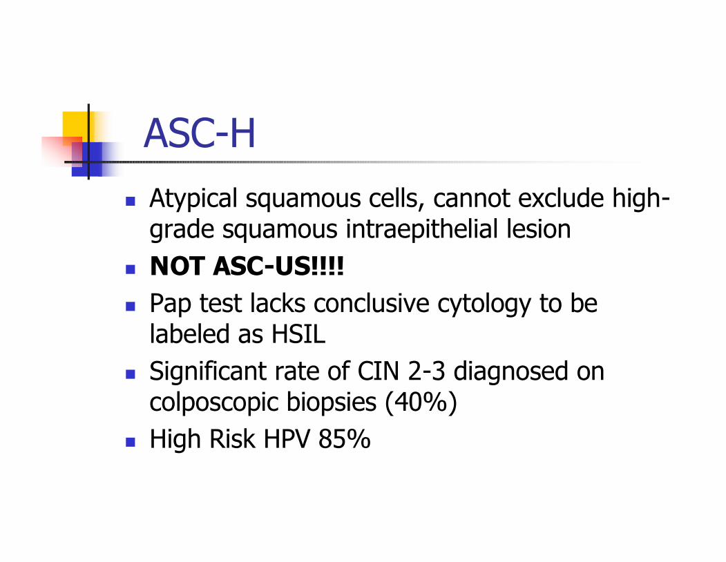
ASC-H
Atypical squamous cells, cannot exclude high-
grade squamous intraepithelial lesion
NOT ASC-US!!!!
Pap test lacks conclusive cytology to be
labeled as HSIL
Significant rate of CIN 2-3 diagnosed on
colposcopic biopsies (40%)
High Risk HPV 85%

ASC-H
COLPOSCOPY
If colposcopy and
biopsies are normal
AND discussion with
pathologist upholds
ASC-H…
Repeat Pap test in 6
months or HPV
testing in 12 months

Low Grade Squamous
Intraepithelial Lesion (LSIL)
1 million reported a
year
Usually histology
confirms CIN 1
High risk HPV
associated with LSIL
Pap tests 83% of time
Colposcopy is the
initial management

LSIL
Common mistake made by practitioners is to
equate LSIL with CIN 1
LSIL is a cytologic specimen
CIN 1 is a histologic specimen
LSIL does not equal CIN 1
30% of LSIL paps actually harbor worse
disease: CIN 2 or CIN 3
COLPOSCOPY NEEDED

Colposcopic Findings
Acetyl white plaques
Bright white
Clearly demarcated
Fine punctations
Acetic Acid - more is
better

LSIL Pap and Colposcopy

LSIL Pap with Colposcopy

Holowaty et al. JNCI 1999
Natural Progression of CIN 1
Ontario Cancer Registry conducted a
study of Pap tests in a single cytologic
laboratory
17,000 women identified between 1970
and 1980 with dysplasia
Conservative treatment

Holowaty et al. NJCI 1999
Regression of CIN 1
Mild dysplasia regressed to normal
44% in 2 years
74% in 5 years
88% in 10 years

Wright. ACOG Colposcopic Course
2001
Regression of CIN 1 in One
Year
16%Progress to CIS
22%Persist
62%Regress
4500No. Patients
17No. Studies
Mild DysplasiaVariable

Holowaty et al. JNCI 1999
Progression of CIN 1
Mild dysplasia progressed to invasive
cervical carcinoma 0.1% in 2 years
0.3% in 5 years
1.6% in 10 years

Management of CIN 1
Most cases of CIN 1 spontaneously resolve in one to
two years
Resolution similar to regression of HPV infection
Follow with serial Pap tests every 4 to 6 months
Persistence of LSIL for more than one year warrants
repeat colposcopy
Persistence of CIN 1 beyond 1.5 to 2 years should be
treated
Pap test progression to HSIL merits colposcopy and
biopsy

High Grade Squamous
Intraepithelial Lesion (HSIL)
The most aggressive
type of squamous
cell dysplasia before
invasive cancer
Needs prompt
evaluation and
treatment (within 4
weeks of diagnosis)
Easiest to manage:
colpo, biopsy, treat

Colposcopic Findings
Dull acetyl white plaques
Cobblestoning
Coarse punctations
Atypical vessels
Mosaicism

Colposcopy

Colposcopy

Holowaty et al. JNCI 1999
Progression of CIN III
Most CIN III lesions progress to carcinoma if
left untreated
This risk grows the longer it is left untreated
CIN III has a RR 22 for progression to
cervical cancer OR
12% of patients will develop cancer in 2
years
70% will develop cancer in 8 years

Derchain et al. Gyn Oncol, 2004.
Atypical Glandular Cells (AGC)
Old Bethesda system AGC was known as
AGUS
AGC was created to clear confusion between
ASC-US and AGUS
THE TWO ARE NOT THE SAME !!!
Incidence is 0.1 to 1.5% of all Pap tests
High risk HPV correlated with 38% of AGC
Pap tests.
HPV testing not recommended

AGC
AGC is worrisome for several pathologies: CIN, adenocarcinoma
in situ, cervical adenocarcinoma and endometrial
adenocarcinoma
Many studies have documented the incidence of these disease
processes:
AGUS system
Eddy et al. 36%, 1997 Am J Obstet Gynecol
Duska et al. 34%, 1998 Obstet Gynecol
Veljovich et al. 32%, 1998 Am J Obstet Gynecol
Manetta et al. 45%, 1999 Gynecol Oncol
Tam et al. 31%, 2003 Gynecol Oncol
AGC system
DeSimone et al. 38%, 2005 Obstet Gynecol

Management of AGC
American Society for Colposcopy and
Cervical Pathology (ASCCP)
recommends:
Colposcopy with or without biopsies
An endocervical curettage (ECC)
Endometrial biopsy in women with
menorrhagia or age greater than 35
OR referral to GYN or GYN oncologist

Management of AGC
Does every patient need an endometrial
biopsy?
Age is important
Premenopausal women more likely to have
HSIL vs. postmenopausal women (30.4% vs.
7.4%) p = 0.04 Duska et al. Obstet Gynecol, 1998.
Women over the age of 40 were more likely
to have adenocarcinoma than dysplasia
(31% vs. 6%) p=0.002 DeSimone et al. Obstet Gynecol, 2005

Lee et al. Diagn Cytopathol, 1995.
Adenocarcinoma in-situ (AIS)
Aggressive form of dysplasia for
columnar cells
AIS cytology associated with:
AIS histology (48-69%)
Cervical adenocarcinoma (38%)

AIS
AIS cytology mandates colposcopic biopsies and an
ECC
AIS histology is managed with a cold knife cone
(CKC)
Numerous studies support CKC over loop
electricosurgical excision procedure (LEEP) because
of margin status
CKC has fewer positive margins than LEEP
Women with positive margins have 40-70% risk of
residual AIS
Women with negative margins have a 20 to 40% risk
of residual AIS

AIS
Recommend referral to GYN or GYN
oncologist
Nulliparous women are difficult to
manage secondary to a high risk of
residual disease
Don’t underestimate the risk of invasive
adenocarcinoma with an AIS Pap test

Treatment Modalities
Cryosugery
Loop Electricosurgical Excision
Procedure (LEEP)
Laser Ablation
Cold Knife Conization
Hysterectomy

Cryosurgery
Inexpensive, easy to perform, tolerated well
by patients
Cells are destroyed by (cold) thermal damage
3 minute freeze/ 1 minute thaw/ 3 minute
freeze well documented technique
Does cause 2 -3 weeks of malodorous
discharge
Does hinder repeat colposcopy (SCJ often
obscured)

LEEP
Procedure of choice for most OB/GYN’s
Easy to perform, well tolerated and
provides specimen for pathologic
evaluation (Margins)
Concern that multiple excisions or one
large excision will increase rate of
preterm labor/incompetent cervix

Sadler, JAMA 2004 and Tan, J
Obstet Gynecol 2004
Preterm Delivery and LEEP
NO conclusive evidence that LEEP increases
preterm labor or incompetent cervix
2 retrospective studies from 2004 show no
difference between control and study patients
However, most studies are small and
retrospective
Good rule to keep specimens less than 4 cm wide
and 2 cm tall

Dietrich, Obstet Gynecol 2002
Margins and LEEP
Margin status helpful in predicting
recurrence of cervical dysplasia
Negative margins ~15%
Positive margins ~ 30-60%
Re-excision not needed. Follow patient
with serial pap tests and treat
accordingly if patient recurs

Laser
CO2 laser works by vaporizing cervical
cells
Very precise method; only need 5-7 mm
of vaporization for treatment
Heals great, spares cervical excisions
COST major problem
No pathology specimen

Cold Knife Conization (CKC)
Used to be the treatment of choice
before LEEP
Surgically excises dysplasia with
scalpel/scissors
Large cost to patient from physician,
anesthesia, and hospital charges
Incompetent cervix an issue
Indications to perform are few

Hysterectomy
The final treatment for cervical dysplasia
Comes with significant morbidity/ mortality
and lengthy recovery (6 weeks)
Complications include: hemorrhage,
infections, bowel & bladder injuries, MI,
pulmonary embolus, stroke, death
10-20% of patients will continue to have
abnormal pap tests: vaginal dysplasia

Mitchell, Obstet Gynecol 1998.
Efficacy of treatment
Randomized controlled trial between
cryosurgery, LEEP and laser showed no
statistical difference in efficacy
Recurrences were measure from 6-37
months
Cryosurgery 19%
LEEP 13%
Laser 13%

Which method to chose?
Several factors to consider: age, desire for
fertility, size of lesion, size of the cervix,
severity of dysplasia, and prior therapies
Generalizations:
Cryosurgery- best for young women with few
finances and CIN 1 or 2.
LEEP- the majority of women with CIN 2 or 3.
Women with endocervical lesions also suited for
LEEP

Which method to chose?
Laser- women who have had multiple recurrences
of CIN 2 or 3 and who want to retain fertility.
Example: a 19 year old G
0
who has CIN 3, prior
LEEP, and a small cervix.
CKC- glandular abnormalities (AIS) or early
invasive cancer
Hysterectomy- women finished with childbearing
and who have persistent CIN. Often best utilized
with other gynecologic problems like pelvic pain or
abnormal uterine bleeding

Guido et al. Am J Obstet Gynecol,
2003.
Post Procedure Surveillance
CIN 1
Repeat Pap testing at 6 and 12 months
ASC-US or greater = colposcopy (referral 63%)
2 normal Pap tests = annual cytology
screening
High risk HPV testing at 12 months
Positive test = colposcopy
HPV 92% sensitive for detecting CIN 2-3
(referral 55%)

Wright et al. Am J Obstet
Gynecol, 2003
Post Procedure Surveillance
CIN 2-3
Repeat Pap testing at 4 to 6 month
intervals
If 3 consecutive normal Pap tests = annual
cytology screening
Colposcopy if ASC-US or greater
High risk HPV testing at 6 or 12 months
If positive = colposcopy
If negative =annual cytology screening

HPV vaccines
3 published, double
blinded randomized
control trials
2 trials with Merck
1 trial with
GlaxoSmithKline
All trials target the L1
protein of the HPV virus
L1 is the major capsid
protein

Koutsky et al. N Engl J Med, 2002.
HPV Vaccines
First trial (Merck) studied HPV type 16
HPV 16 chosen since it accounts for 50% of CIN 2-3
and carcinoma
2392 women randomized between Placebo or HPV 16
virus-like-particle (VLP) vaccine [40µg] given on day
0, month 2 and month 6
Women had to be HPV 16 negative on enrollment
Genital samples were obtained on enrollment, month
7 and then every 6 months to check for HPV 16 by
PCR
Primary endpoint was persistent HPV 16 infection
defined as HPV 16 diagnosed on two or more visits

Koutsky et al. N Engl J Med, 2002.
HPV Vaccines
Results
Median follow up 17 months
Incidence of 3.8 per 100 woman-years,
placebo
Incidence of 0 per 100 woman-years,
vaccine
P=0.001
Nine cases of CIN, HPV 16 induced
occurred in placebo group

Harper et al. Lancet, 2004.
HPV Vaccines
2
nd
study was funded by GlaxoSmithKline
It evaluated a bivalent vaccine for HPV 16 and 18
Together HPV 16 and 18 account for 70% of CIN 2-3 and
carcinoma
1113 women were randomized to receive placebo or vaccine
(0.5ml) at 0, 1 and 6 months
Women were HPV 16 and 18 negative on enrollment
Genital samples were obtained on enrollment and every 3
months to check for HPV 16 and 18 by PCR
Primary endpoint was prevention of HPV 16 and 18 infection,
secondary endpoint was prevention of cytologic (LSIL, HSIL)
and histologic abnormalities (CIN 1-3)

Harper et al. Lancet, 2004.
HPV Vaccines
Results
Vaccine efficacy 91.6% against incident infections
Efficacy 100% against persistent infection
Vaccine arm two cytologic abnormalities (ASC-US,
LSIL)
Placebo arm 30 cytologic abnormalities (15 ASC-
US, 14 LSIL, 1 HSIL
Vaccine efficacy 93.5% (95% CI 3-99.1; p=0.002)

Villa et al. Lancet, 2005.
HPV Vaccines
3
rd
study (Merck) evaluated a
quadrivalent vaccine HPV 16, 18, 6, 11
HPV 6, 11 account for 90% of
condyloma (genital warts)
552 women randomized to placebo or
vaccine at 0, 2 and 6 months
Women were HPV 6,11,16,18 negative
on enrollment

Villa et al. Lancet, 2005.
HPV Vaccines
Results
Vaccine 4/235 cases of infection or clinical disease
Placebo 36/233 cases of infection or clinical
disease
Efficacy difference 90% (95% CI 71-97;
p=0.0001)
No cases of condyloma or CIN in vaccine arm
3 cases of CIN and 3 cases of condyloma in
placebo arm

HPV Vaccines
HPV vaccines are effective in preventing HPV
infection and clinical infections such as
condyloma and CIN
Large study by Merck presented at the
meeting for Infectious Diseases Society of
America confirmed these three studies with
5,000 women per arm
Vaccine will be a boon for 3
rd
world countries
with high rates of cervical cancer

HPV Vaccines
Limits
Vaccines not widely available yet. Projected for 8/2006
Estimated cost $600 for three shots
Who do we start immunizing?
FDA recommend girls
What about boys?
At what age do we start immunizations?
Age 9-21
Best to immunize before onset of coitarche (sex)
Length of duration?
Latest studies suggest activity at 36 months
What about cross over immunization between other HPV strains?
Latest reports suggest there is cross over immunization
Will Pap testing cease?
Not likely in the next 40 years
We will need to see wide spread acceptance of the vaccine and
immunizing men and women before Pap testing can be discontinued

Summary
HPV is THE cause of cervical
cancer and dysplasia
Majority of men and women
will have been exposed to
HPV before the age of 50
Smoking is an important co-
factor in oncogenesis
Upcoming vaccinations are
effective in preventing HPV
infections and histologic
abnormalities

Summary of Squamous
Abnormalities
ASC-US Pap tests should either be
repeated in 6 months or a reflex HPV
testing should be performed
On repeat cytology, women with ASC-US or
greater or women with positive HPV testing
should undergo colposcopy
LSIL /ASC-H /HSIL Pap tests should
undergo colposcopy

Summary of Glandular
Abnormalities
AGC Pap tests should undergo
colposcopy, ECC and endometrial
biopsy (women >35 or menorrhagia)
AIS Pap tests should be referred to a
GYN or GYN oncologist

Summary of CIN
CIN 1 should be followed with serial cytology
every 6 months or HPV testing at 12 months
Women with 2 normal Pap tests can resume
annual cytology screening
Women with HPV negative testing can resume
annual cytology screening
CIN 2-3 should be treated with an excision
procedure
Post procedure cytology should be evaluated
every 4 to 6 months OR
HPV testing at 6 to 12 months