Embed Size (px)
Citation preview

Human MBL Immunoassay
Quantikine® ELISA
This package insert must be read in its entirety before using this product. For research use only. Not for use in diagnostic procedures.
Catalog Number DMBL00
For the quantitative determination of human Mannan-Binding Lectin (MBL) concentrations in cell culture supernates, serum, and plasma.

TABLE OF CONTENTS
SECTION PAGE
INTRODUCTION ....................................................................................................................................................................1PRINCIPLE OF THE ASSAY ..................................................................................................................................................2LIMITATIONS OF THE PROCEDURE ................................................................................................................................2TECHNICAL HINTS ................................................................................................................................................................2MATERIALS PROVIDED & STORAGE CONDITIONS ..................................................................................................3OTHER SUPPLIES REQUIRED ............................................................................................................................................3PRECAUTIONS ........................................................................................................................................................................4SAMPLE COLLECTION & STORAGE ................................................................................................................................4SAMPLE PREPARATION.......................................................................................................................................................4REAGENT PREPARATION ....................................................................................................................................................5ASSAY PROCEDURE ............................................................................................................................................................6CALCULATION OF RESULTS ..............................................................................................................................................7TYPICAL DATA ........................................................................................................................................................................7PRECISION ...............................................................................................................................................................................8RECOVERY................................................................................................................................................................................8LINEARITY ................................................................................................................................................................................8SENSITIVITY ............................................................................................................................................................................9CALIBRATION .........................................................................................................................................................................9SAMPLE VALUES ....................................................................................................................................................................9SPECIFICITY .............................................................................................................................................................................9REFERENCES ........................................................................................................................................................................ 10
Manufactured and Distributed by:
USA R&D Systems, Inc. 614 McKinley Place NE, Minneapolis, MN 55413TEL: 800 343 7475 612 379 2956FAX: 612 656 4400E-MAIL: [email protected]
Distributed by:
Europe | Middle East | Africa Bio-Techne Ltd.19 Barton Lane, Abingdon Science ParkAbingdon OX14 3NB, UKTEL: +44 (0)1235 529449FAX: +44 (0)1235 533420E-MAIL: [email protected]
China Bio-Techne China Co., Ltd.Unit 1901, Tower 3, Raffles City Changning Office,1193 Changning Road, Shanghai PRC 200051TEL: +86 (21) 52380373 (400) 821-3475FAX: +86 (21) 52371001E-MAIL: [email protected]

www.RnDSystems.com 1
INTRODUCTIONMannan- or mannose-Binding Lectin (MBL) belongs to the collectin family of innate immune defense proteins (1-3). Collectin family members share common structural features: a cysteine rich amino-terminal domain, a collagen-like region, an α-helical coiled-coil neck domain, and a carboxy terminal C-type (Ca++-dependent) lectin or carbohydrate recognition domain (CRD). MBL forms coiled-coil mediated homotrimers as a structural unit. The units are joined by N-terminal disulfide bridges into oligomers of up to 6 trimers, with higher-order structures allowing stronger ligand binding (4-6). Three- and four-trimer complexes (230 and 305 kDa) predominate in circulating human MBL (6). Whereas two forms of MBL proteins (MBL-1 and MBL-2) exist in rodents and other animals, only one functional MBL protein is present in humans. Mature human MBL shares 58% and 61% amino acid sequence identity with mouse MBL-1 and MBL-2, respectively (4).
Of human collectins, MBL and ficolins can activate complement via the lectin complement pathway (1). The complex of oligomeric MBL with MBL-associated serine protease (MASP) proenzymes in plasma recognizes horizontal hydroxyl groups at the 3' and 4' position of pyranose rings, such as mannose, glucose, L-fucose, or N-acetyl-glucosamine, that are present at a terminal non-reducing position on the cell surface (2, 3, 6). Various pathogens, certain tumor cells, and atypically glycosylated immunoglobulins are recognized while normal self-components are not, due to sialic acid or galactose at the terminus of the carbohydrate chain (2, 7, 8). Binding to carbohydrate ligands induces a conformational change in MBL that allows pro-enzyme activation (9, 10). The enzyme may then cleave complement components C4 and C2 to activate the complement cascade, resulting in opsonization and pathogen removal (1). MBL is also thought to cooperate with toll-like receptors TLR2 and TLR6 after opsonized bacteria are taken into phagosomes (11). MBL and complement component C1q can both bind membrane receptors such as calreticulin, a non-transmembrane protein present on the cell surface in complex with CD91 (12, 13). Such complexes are thought to mediate MBL activities in clearance of apoptotic cells.
MBL is primarily produced in the liver, with minor expression in the human small intestine and testis (14). MBL is mainly secreted into the bloodstream, although an intracellular form, thought to mediate Golgi trafficking of glycosylated proteins, has been described (15). Plasma concentrations in normal humans can vary 1000-fold due to differential expression of genes with polymorphisms (16, 17). Polymorphisms can also alter the MBL oligomeric structure and/or ability to activate MASPs and complement, thus altering its effectiveness (18-21). Expression of these polymorphisms is quite common and varies by ethnicity (16-21). Chemotherapy and other immunocompromised conditions reveal greater susceptibility to infectious disease in individuals expressing genotypes with low expression or function (21-23). Although studies vary, these genotypes may also be over-represented in autoimmune diseases such as systemic lupus erythematosus (SLE) (24). This may be related to MBL-mediated clearance of apoptotic cells, which removes potential autoantigens from circulation (22). On the other hand, low expression of MBL appears to lower the risk of ischemia-reperfusion injury and diabetic nephropathy (25, 26).
The Quantikine® Human MBL Immunoassay is a 4.5 hour solid phase ELISA designed to measure human MBL in cell culture supernates, serum, and plasma. It contains recombinant human MBL and has been shown to accurately quantitate the recombinant factor. Results obtained using natural human MBL showed dose-response curves that were parallel to the standard curves obtained using the Quantikine® kit standards. These results indicate that this kit can be used to determine relative mass values of natural human MBL.

For research use only. Not for use in diagnostic procedures.2
PRINCIPLE OF THE ASSAYThis assay employs the quantitative sandwich enzyme immunoassay technique. A monoclonal antibody specific for human MBL has been pre-coated onto a microplate. Standards and samples are pipetted into the wells and any MBL present is bound by the immobilized antibody. After washing away any unbound substances, an enzyme-linked monoclonal antibody specific for human MBL is added to the wells. Following a wash to remove any unbound antibody-enzyme reagent, a substrate solution is added to the wells and color develops in proportion to the amount of MBL bound in the initial step. The color development is stopped and the intensity of the color is measured.
LIMITATIONS OF THE PROCEDURE• FOR RESEARCH USE ONLY. NOT FOR USE IN DIAGNOSTIC PROCEDURES.
• The kit should not be used beyond the expiration date on the kit label.
• Do not mix or substitute reagents with those from other lots or sources.
• If samples generate values higher than the highest standard, dilute the samples with calibrator diluent and repeat the assay.
• Any variation in diluent, operator, pipetting technique, washing technique, incubation time or temperature, and kit age can cause variation in binding.
• Variations in sample collection, processing, and storage may cause sample value differences.
• This assay is designed to eliminate interference by other factors present in biological samples. Until all factors have been tested in the Quantikine® Immunoassay, the possibility of interference cannot be excluded.
TECHNICAL HINTS• When mixing or reconstituting protein solutions, always avoid foaming.
• To avoid cross-contamination, change pipette tips between additions of each standard level, between sample additions, and between reagent additions. Also, use separate reservoirs for each reagent.
• To ensure accurate results, proper adhesion of plate sealers during incubation steps is necessary.
• When using an automated plate washer, adding a 30 second soak period following the addition of Wash Buffer, and/or rotating the plate 180 degrees between wash steps may improve assay precision.
• Substrate Solution should remain colorless until added to the plate. Keep Substrate Solution protected from light. Substrate Solution should change from colorless to gradations of blue.
• Stop Solution should be added to the plate in the same order as the Substrate Solution. The color developed in the wells will turn from blue to yellow upon addition of the Stop Solution.

www.RnDSystems.com 3
MATERIALS PROVIDED & STORAGE CONDITIONSStore the unopened kit at 2-8 °C. Do not use past kit expiration date.
PART PART # DESCRIPTIONSTORAGE OF OPENED/ RECONSTITUTED MATERIAL
Human MBL Microplate
893711 96 well polystyrene microplate (12 strips of 8 wells) coated with a monoclonal antibody specific for human MBL.
Return unused wells to the foil pouch containing the desiccant pack. Reseal along entire edge of the zip-seal. May be stored for up to 1 month at 2-8 °C.*
Human MBL Conjugate
893712 12 mL of a monoclonal antibody specific for human MBL conjugated to horseradish peroxidase with preservatives.
May be stored for up to 1 month at 2-8 °C.*
Human MBL Standard
893713 2 vials of recombinant human MBL in a buffered protein solution with preservatives; lyophilized. Refer to the vial label for reconstitution volume.
Assay Diluent RD1-21
895215 12 mL of a buffered protein solution with preservatives.
Calibrator Diluent RD5-26 Concentrate
895525 21 mL of a concentrated buffered protein solution with preservatives. Use diluted 1:4 in this assay.
Wash Buffer Concentrate
895003 21 mL of a 25-fold concentrated solution of buffered surfactant with preservative. May turn yellow over time.
Color Reagent A 895000 12 mL of stabilized hydrogen peroxide.
Color Reagent B 895001 12 mL of stabilized chromogen (tetramethylbenzidine).
Stop Solution 895174 23 mL of a diluted hydrochloric aid solution.
Plate Sealers N/A 4 adhesive strips.
* Provided this is within the expiration date of the kit.
OTHER SUPPLIES REQUIRED• Microplate reader capable of measuring absorbance at 450 nm, with the correction
wavelength set at 540 nm or 570 nm.
• Pipettes and pipette tips.
• Deionized or distilled water.
• Squirt bottle, manifold dispenser, or automated microplate washer.
• 100 mL and 500 mL graduated cylinders.
• Horizontal orbital microplate shaker (0.12" orbit) capable of maintaining a speed of 500 ± 50 rpm.
• Test tubes for dilution of standards and samples.
• Human MBL Controls (optional; R&D Systems®, Catalog # QC118).

For research use only. Not for use in diagnostic procedures.4
PRECAUTIONSThe Stop Solution provided with this kit is an acid solution.
Some components in this kit contain a preservative which may cause an allergic skin reaction. Avoid breathing mist.
Color Reagent B may cause skin, eye, and respiratory irritation. Avoid breathing fumes.
Wear protective gloves, clothing, eye, and face protection. Wash hands thoroughly after handling. Refer to the SDS on our website prior to use.
SAMPLE COLLECTION & STORAGEThe sample collection and storage conditions listed below are intended as general guidelines. Sample stability has not been evaluated.
Cell Culture Supernates - Remove particulates by centrifugation and assay immediately or aliquot and store samples at ≤ -20 °C. Avoid repeated freeze-thaw cycles.
Serum - Use a serum separator tube (SST) and allow samples to clot for 30 minutes at room temperature before centrifugation for 15 minutes at 1000 x g. Remove serum and assay immediately or aliquot and store samples at ≤ -20 °C. Avoid repeated freeze-thaw cycles.
Plasma - Collect plasma using EDTA or heparin as an anticoagulant. Centrifuge for 15 minutes at 1000 x g within 30 minutes of collection. Assay immediately or aliquot and store samples at ≤ -20 °C. Avoid repeated freeze-thaw cycles.
Note: Citrate plasma has not been validated for use in this assay.
SAMPLE PREPARATIONSerum and plasma samples require a 400-fold dilution. A suggested 400-fold dilution can be achieved by adding 10 μL of sample to 190 μL of Calibrator Diluent RD5-26 (diluted 1:4).* Complete the 400-fold dilution by adding 10 μL of the diluted sample to 190 μL of Calibrator Diluent RD5-26 (diluted 1:4)*.
*See Reagent Preparation section.

www.RnDSystems.com 5
REAGENT PREPARATIONBring all reagents to room temperature before use.
Wash Buff er - If crystals have formed in the concentrate, warm to room temperature and mix gently until the crystals have completely dissolved. Add 20 mL of Wash Buff er Concentrate to 480 mL of deionized or distilled water to prepare 500 mL of Wash Buff er.
Substrate Solution - Color Reagents A and B should be mixed together in equal volumes within 15 minutes of use. Protect from light. 100 μL of the resultant mixture is required per well.
Calibrator Diluent RD5-26 (diluted 1:4) - Add 20 mL of Calibrator Diluent RD5-26 Concentrate to 60 mL of deionized or distilled water to prepare 80 mL of Calibrator Diluent RD5-26 (diluted 1:4).
Human MBL Standard - Refer to the vial label for reconstitution volume. Reconstitute the Human MBL Standard with Calibrator Diluent RD5-26 (diluted 1:4). This reconstitution produces a stock solution of 100 ng/mL. Mix the standard to ensure complete reconstitution and allow the standard to sit for a minimum of 5 minutes with gentle agitation prior to making dilutions.
Pipette 900 μL of Calibrator Diluent RD5-26 (diluted 1:4) into the 10 ng/mL tube. Pipette 500 μL into the remaining tubes. Use the stock solution to produce a dilution series (below). Mix each tube thoroughly before the next transfer. The 10 ng/mL standard serves as the high standard. Calibrator Diluent RD5-26 (diluted 1:4) serves as the zero standard (0 ng/mL).
100 µL Std.
100 ng/mL 10 ng/mL 5 ng/mL 2.5 ng/mL 1.25 ng/mL 0.625 ng/mL 0.313 ng/mL 0.156 ng/mL
500 µL 500 µL 500 µL 500 µL 500 µL 500 µL

For research use only. Not for use in diagnostic procedures.6
ASSAY PROCEDURE Bring all reagents and samples to room temperature before use. It is recommended that all standards, controls, and samples be assayed in duplicate.
1. Prepare all reagents, working standards, and samples as directed in the previous sections.
2. Remove excess microplate strips from the plate frame, return them to the foil pouch containing the desiccant pack, and reseal.
3. Add 50 μL of Assay Diluent RD1-21 to each well.
4. Add 50 μL of standard, control, or sample* per well. Cover with the adhesive strip provided. Incubate for 2 hours at room temperature on a horizontal orbital microplate shaker (0.12" orbit) set at 500 ± 50 rpm.
5. Aspirate each well and wash, repeating the process three times for a total of four washes. Wash by filling each well with Wash Buffer (400 μL) using a squirt bottle, manifold dispenser, or autowasher. Complete removal of liquid at each step is essential to good performance. After the last wash, remove any remaining Wash Buffer by aspirating or decanting. Invert the plate and blot it against clean paper towels.
6. Add 100 μL of the Human MBL Conjugate to each well. Cover with a new adhesive strip. Incubate for 2 hours at room temperature on the shaker.
7. Repeat the aspiration/wash as in step 5.
8. Add 100 μL of Substrate Solution to each well. Incubate for 30 minutes at room temperature on the benchtop. Protect from light.
9. Add 100 μL of Stop Solution to each well. Gently tap the plate to ensure thorough mixing.
10. Determine the optical density of each well within 30 minutes, using a microplate reader set to 450 nm. If wavelength correction is available, set to 540 nm or 570 nm. If wavelength correction is not available, subtract readings at 540 nm or 570 nm from the readings at 450 nm. This subtraction will correct for optical imperfections in the plate. Readings made directly at 450 nm without correction may be higher and less accurate.
*Samples may require dilution. See Sample Preparation section.

www.RnDSystems.com 7
CALCULATION OF RESULTSAverage the duplicate readings for each standard, control, and sample and subtract the average zero standard optical density (O.D.).
Create a standard curve by reducing the data using computer software capable of generating a four parameter logistic (4-PL) curve-fit. As an alternative, construct a standard curve by plotting the mean absorbance for each standard on the y-axis against the concentration on the x-axis and draw a best fit curve through the points on the graph. The data may be linearized by plotting the log of the human MBL concentrations versus the log of the O.D. and the best fit line can be determined by regression analysis. This procedure will produce an adequate but less precise fit of the data.
If samples have been diluted, the concentration read from the standard curve must be multiplied by the dilution factor.
TYPICAL DATAThis standard curve is provided for demonstration only. A standard curve should be generated for each set of samples assayed.
(ng/mL) O.D. Average Corrected0 0.011 0.011 —
0.0110.156 0.075 0.076 0.065
0.0760.313 0.146 0.147 0.136
0.1480.625 0.279 0.282 0.271
0.2841.25 0.513 0.523 0.512
0.5332.5 0.960 0.962 0.951
0.9635 1.675 1.678 1.667
1.68010 2.558 2.574 2.563
2.590
For research use only. Not for use in diagnostic procedures.8
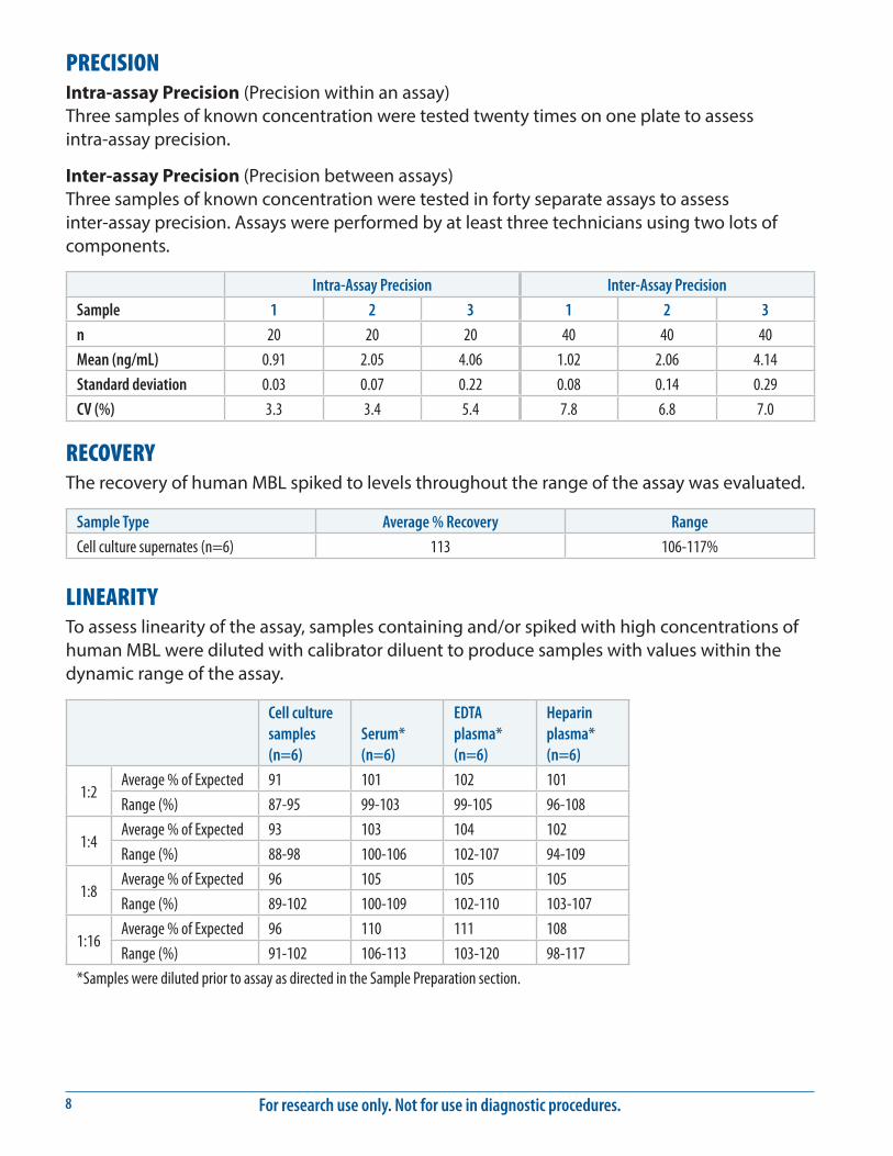
PRECISIONIntra-assay Precision (Precision within an assay) Three samples of known concentration were tested twenty times on one plate to assess intra-assay precision.
Inter-assay Precision (Precision between assays) Three samples of known concentration were tested in forty separate assays to assess inter-assay precision. Assays were performed by at least three technicians using two lots of components.
Intra-Assay Precision Inter-Assay Precision
Sample 1 2 3 1 2 3
n 20 20 20 40 40 40
Mean (ng/mL) 0.91 2.05 4.06 1.02 2.06 4.14
Standard deviation 0.03 0.07 0.22 0.08 0.14 0.29
CV (%) 3.3 3.4 5.4 7.8 6.8 7.0
RECOVERYThe recovery of human MBL spiked to levels throughout the range of the assay was evaluated.
Sample Type Average % Recovery Range
Cell culture supernates (n=6) 113 106-117%
LINEARITYTo assess linearity of the assay, samples containing and/or spiked with high concentrations of human MBL were diluted with calibrator diluent to produce samples with values within the dynamic range of the assay.
Cell culture samples (n=6)
Serum* (n=6)
EDTA plasma* (n=6)
Heparin plasma* (n=6)
1:2Average % of Expected 91 101 102 101
Range (%) 87-95 99-103 99-105 96-108
1:4Average % of Expected 93 103 104 102
Range (%) 88-98 100-106 102-107 94-109
1:8Average % of Expected 96 105 105 105
Range (%) 89-102 100-109 102-110 103-107
1:16Average % of Expected 96 110 111 108
Range (%) 91-102 106-113 103-120 98-117
*Samples were diluted prior to assay as directed in the Sample Preparation section.

www.RnDSystems.com 9
SENSITIVITYSeventy two assays were evaluated and the minimum detectable dose (MDD) of human MBL ranged from 0.002-0.029 ng/mL. The mean MDD was 0.008 ng/mL.
The MDD was determined by adding two standard deviations to the mean O.D. value of twenty zero standard replicates and calculating the corresponding concentration.
CALIBRATIONThis immunoassay is calibrated against a highly purified recombinant human MBL produced at R&D Systems®.
SAMPLE VALUESSerum/Plasma - Samples from apparently healthy volunteers were evaluated for the presence of human MBL in this assay. No medical histories were available for the donors used in this study.
Sample Type Mean (ng/mL) Range (ng/mL) Standard Deviation (ng/mL)
Serum (n=36) 1135 103-3308 874
EDTA plasma (n=36) 1097 85-3077 845
Heparin plasma (n=36) 1099 91-3347 867
Cell Culture Supernates - HepG2 human hepatocellular carcinoma cells (1 x 10⁶ cells/mL) were cultured in MEM supplemented with 10% fetal bovine serum, 2 mM L-glutamine, 100 U/mL penicillin, and 100 μg/mL of streptomycin sulfate for 5 days. An aliquot of the cell culture supernate was removed, assayed for human MBL, and measured 0.211 ng/mL.
SPECIFICITYThis assay recognizes oligomeric forms of natural and recombinant human MBL.
The factors listed below were prepared at 100 ng/mL in calibrator diluent and assayed for cross-reactivity. Preparations of the following factors at 100 ng/mL in a mid-range recombinant human MBL control were assayed for interference. No significant cross-reactivity or interference was observed.
Recombinant human:CL-K1CL-P1DC-SIGNDC-SIGN RLangerinMASP-3 (catalytic domain)MMRSP-D
Recombinant mouse:MBL-1MBL-2
Other:human IgA

For research use only. Not for use in diagnostic procedures.10
REFERENCES1. Thiel, S. (2007) Mol. Immunol. 44:3875.2. Takahashi, K. et al. (2006) Curr. Opin. Immunol. 18:16.3. Bohlson, S.S. et al. (2007) Mol. Immunol. 44:33.4. Sastry, K. et al. (1989) J. Exp. Med. 170:1175.5. Sheriff, S. et al. (1994) Struct. Biol. 1:789.6. Teillet, F. et al. (2005) J. Immunol. 174:2870.7. Saevarsdottir, S. et al. (2004) Scand. J. Immnunol. 60:23.8. Arnold, J.N. et al. (2006) Immunol. Lett. 106:103.9. Dong, M. et al. (2007) J. Immunol. 178:3016.
10. Chen, C.B. et al. (2004) J. Biol. Chem. 279:26058.11. Ip, W.K.E. et al. (2008) J. Exp. Med. 205:169.12. Pagh, R. et al. (2008) FEBS J. 275:515.13. Ogden, C.A. et al. (2001) J. Exp. Med. 194:781.14. Seyfarth, J. et al. (2006) Mol. Immunol. 43:962.15. Nonaka, M. et al. (2007) J. Biol. Chem. 282:17908.16. Garred, P. et al. (2006) Genes Immun. 7:85.17. Bernig, T. et al. (2004) Genes Immun. 5:461. 18. Terai, I. et al. (2003) Eur. J. Immunol. 33:2755.19. Wallis, R. et al. (2005) J. Immunol. 175:6846.20. Super, M. et al. (1992) Nat. Genet. 2:50.21. Larsen, F. et al. (2004) J. Biol. Chem. 279:21302.22. Thiel, S. et al. (2006) Mol. Immunol. 43:86.23. Mullighan, C.G. et al. (2008) Blood 112:2120.24. Monticielo, O.A. et al. (2008) Clin. Rheumatol. 27:413.25. Bilgin, Y.M. et al. (2008) Transfusion 48:601.26. Busche, M.N. et al. (2008) Diabetologia 51:1544.
04.09 752005.4 6/18
©2018 R&D Systems®, Inc.
All trademarks and registered trademarks are the property of their respective owners.