Embed Size (px)
Citation preview
JOURNAL OF VIROLOGY, Feb. 2003, p. 2124–2133 Vol. 77, No. 30022-538X/03/$08.00�0 DOI: 10.1128/JVI.77.3.2124–2133.2003Copyright © 2003, American Society for Microbiology. All Rights Reserved.
Human Immunodeficiency Virus Type 1 Nef-Mediated Downregulationof CD4 Correlates with Nef Enhancement of Viral Pathogenesis
Cheryl A. Stoddart,1 Romas Geleziunas,1 Sharon Ferrell,1 Valerie Linquist-Stepps,1 Mary E. Moreno,1Christopher Bare,1 Weiduan Xu,1 Wes Yonemoto,1 Patricia A. Bresnahan,1
Joseph M. McCune,1,2,3 and Warner C. Greene1,2,3*Gladstone Institute of Virology and Immunology1 and Departments of Medicine2 and Microbiology
and Immunology,3 University of California, San Francisco, California 94141
Received 28 May 2002/Accepted 25 October 2002
The nef gene products encoded by human immunodeficiency virus type 1 (HIV-1) and simian immunodefi-ciency virus type 1 (SIV-1) increase viral loads in infected hosts and accelerate clinical progression to AIDS.Nef exhibits a spectrum of biological activities, including the ability to downregulate surface expression of CD4and major histocompatibility complex (MHC) class I antigens, to alter the state of T-cell activation, and toenhance the infectivity of viral particles. To determine which of these in vitro functions most closely correlateswith the pathogenic effects of Nef in vivo, we constructed recombinant HIV-1 NL4-3 viruses carrying mutationswithin the nef gene that selectively impair these functions. These mutant viruses were evaluated for pathogenicpotential in severe combined immunodeficiency (SCID) mice implanted with human fetal thymus and liver(SCID-hu Thy/Liv mice), in which virus-mediated depletion of thymocytes is known to be Nef dependent.Disruption of the polyproline type II helix (Pxx)4 within Nef (required for binding of Hck and p21-activatedkinase-like kinases, downregulation of MHC class I, and enhancement of HIV-1 infectivity in vitro butdispensable for CD4 downregulation) did not impair thymocyte depletion in virus-infected Thy/Liv humanthymus implants. Conversely, three separate point mutations in Nef that compromised its ability to down-regulate CD4 attenuated thymocyte depletion while not diminishing viral replication. These findings indicatethat the functional ability of Nef to downregulate CD4 and not MHC class I downregulation, Hck or PAKbinding, or (Pxx)4-associated enhancement of infectivity most closely correlates with Nef-mediated enhance-ment of HIV-1 pathogenicity in vivo. Nef-mediated CD4 downregulation merits consideration as a new targetfor the development of small-molecule inhibitors.
Nef, a 25- to 27-kDa regulatory protein encoded by humanand simian immunodeficiency viruses, accelerates clinical pro-gression to AIDS in humans infected with either human im-munodeficiency virus type 1 (HIV-1) or HIV-2 and in rhesusmacaques experimentally infected with simian immunode-ficiency virus (SIV) (6, 15, 27, 33, 35, 38, 46, 58, 62, 71).Similarly, Nef enhances the pathogenicity of HIV-1 in severecombined immunodeficiency (SCID) mice implanted withfragments of human fetal thymus and liver (SCID-hu Thy/Livmice) (3, 4, 16, 31). The molecular mechanisms underlyingthese effects of Nef remain unknown. However, in newbornrhesus macaques, Nef is dispensable for disease progression (5,6), indicating that it is not an obligate virulence factor.
Among its numerous properties, Nef can downregulate cellsurface expression of CD4 (19) and many major histocompat-ibility complex (MHC) class I (13, 39, 67) antigens, enhancethe infectivity of viral particles (12, 48), and stimulate thereplication of HIV-1 in peripheral blood mononuclear cells(PBMC) (48, 69). Nef also perturbs various intracellular signaltransduction pathways (14, 20, 47, 53, 65, 72) and impairs Fas-and tumor necrosis factor receptor-mediated apoptosis (21).
Downregulation of CD4 by Nef involves endocytosis andlysosomal degradation of CD4, likely the result of Nef-medi-
ated recruitment of cellular adaptors to the cytoplasmic tail ofCD4 (9, 25, 39, 41, 42, 44, 54). These interactions seem torequire distinct residues within Nef, situated at both the aminoand carboxy termini of this viral regulatory protein. At theamino terminus, residues W57 and L58 of Nef have beenproposed to interact with the cytoplasmic tail of CD4 (28, 42,45). Two pairs of residues situated at the carboxy terminus ofNef participate in CD4 downregulation as well (9, 25, 41). Adileucine motif, L164-L165, is required for the interaction ofNef with clathrin-associated adaptor molecules such as AP-1and AP-2. The role of the second pair of residues (D174 andD175) is unclear. These residues may either interact with theCD4 cytoplasmic tail or bind to a vacuolar ATPase involved inboth adaptor binding and acidification of endosomes (42, 45).
Nef-mediated downregulation of CD4 was first postulated asa mechanism to prevent viral superinfection (7). Additionally,it has been proposed that Nef-mediated removal of CD4 fromthe cell surface could lead to liberation of the CD4-associatedprotein tyrosine kinase Lck, in turn promoting enhanced phos-phorylation of various substrates such as the T-cell receptor �chain. Such events could modify the overall state of T-cellactivation, perhaps creating an intracellular milieu more con-ducive to HIV replication (14, 20). This mechanism might alsocontribute to the finding that thymocytes from Nef-transgenicmice display surface markers indicative of cellular activation(29, 40, 68).
To increase HIV-1 virion infectivity, Nef must be present
* Corresponding author. Mailing address: Gladstone Institute ofVirology and Immunology, P.O. Box 419100, San Francisco, CA94141-9100. Phone: (415) 695-3800. Fax: (415) 826-1817. E-mail:[email protected].
2124
Dow
nloa
ded
from
http
s://j
ourn
als.
asm
.org
/jour
nal/j
vi o
n 26
Nov
embe
r 20
21 b
y 45
.224
.161
.118
.

during viral particle assembly in the host producer cell (2, 49,52). The effect of Nef, however, is only manifested in theinfected target cell. In terms of enhancement of virion infec-tivity, both CD4-dependent and CD4-independent compo-nents are demonstrable. HIV-1 virion infectivity enhancementoccurring in the absence of CD4 was believed to be attribut-able to Nef’s facilitation of proviral DNA synthesis, occurringduring the afferent portion of the viral life cycle (2, 11, 66).However, more recent studies also suggest that Nef enhancesvirion entry into the cytosol (64), perhaps reflecting changes inthe lipid composition of the virion membrane (74). Consistentwith this notion, when HIV-1 particles are forced to enter cellsvia acidified endosomes by vesicular stomatitis virus G pseu-dotyping, Nef no longer enhances infectivity (1). When CD4 ispresent on the producer cell, Nef may enhance infectivitythrough CD4 downregulation, leading to either more efficientrelease of virions or more effective envelope engagement ofCD4 receptors present on the subsequent target cells (36, 60).
Nef-mediated downregulation of MHC class I may enableHIV-1-infected cells to more effectively evade cytotoxic T-lymphocyte-mediated recognition and lysis (13). MHC class Idownregulation involves different determinants in Nef thanCD4 downregulation. Specifically, MHC class I downregula-tion is dependent on an acidic region (E64EEE) and a polypro-line stretch [(P69xx)4] situated within the central portion of theNef polypeptide (26, 45). This acidic region within Nef appearsto bind the cellular protein PACS-1, which participates inrelocalization of MHC class I from the cell surface to thetrans-Golgi network (55). Downregulation of MHC class I byNef may also occur through delayed transit from the Golgicomplex to the cell surface (70). The polyproline-rich region isalso necessary to confer enhanced, CD4-independent infec-tious potential upon HIV-1 particles (24, 61) and represents ahigh-affinity binding site for the SH3 domain of the Src familyprotein tyrosine kinase Hck (61). Additionally, this region isrequired for Nef interaction with a complex of cellular pro-teins, including PIX-p95, Vav, and p21-activated kinase-2(PAK 2) (10, 17, 18, 34, 51, 57, 63, 73). However, thus far,neither Hck nor PAK 2 has been clearly implicated in eitherNef-mediated MHC class I downregulation or enhancement ofHIV-1 infectivity.
Since Nef accelerates clinical progression to AIDS in hostsinfected with primate immunodeficiency viruses, a thoroughunderstanding of Nef function within both the infected hostcell and the infected host is needed to delineate the molecularbasis for its acceleration of viral pathogenesis in vivo. BecauseNef is capable of numerous effector functions, it is importantto first determine which function or combination of functionsis linked to its pathogenic effects in vivo. We now describe aseries of studies that define the molecular determinants in Nefthat are required for HIV-1-associated cytopathicity in vivo inthe SCID-hu Thy/Liv mouse model.
MATERIALS AND METHODS
Mammalian expression vector preparation. Nef coding sequences were am-plified by PCR from the pNL4-3N proviral plasmid (61) (kindly provided byKalle Saksela, University of Tempere, Tempere, Finland) with the amplimers5�-GAGAGAAGCTTGACAGGGCTTGGAAAGG-3�, which is situated 30 bpupstream of the start codon of nef and introduces a HindIII site at the 5� end, and5�-CTAGTCTAGATTCACAATGATGCATAATCAGGCACGTCGTACGG
ATAGCAGTCTTTGTAGTACTCCG-3�, which introduces an influenza virushemagglutinin (HA) epitope tag and an XbaI site at the 3� end of nef. TheseNef-encoding DNA sequences were next subcloned into the mammalian expres-sion vector pCMV4neo (24) as HindIII-XbaI fragments to create pNL43Nef-HA.Mutations were introduced into nef with the Muta-Gene Phagemid in vitromutagenesis kit (Bio-Rad, Richmond, Calif.) and confirmed by DNA sequenc-ing. Wild-type and mutated nef genes were then subcloned into the pCI mam-malian expression vector (Promega, Madison, Wis.) to create pCI-NL43Nef-HA.Inserts were removed from pNL43Nef-HA as 5� HindIII and 3� XbaI fragments.The 5� HindIII site was blunt ended with Klenow polymerase, and the insertswere introduced into pCI at a 5� blunt-ended EcoRI site and a 3� XbaI site.
Proviral plasmid preparation. To create viruses containing select Nef muta-tions, internal 5� XhoI–3� PmlI fragments of nef-bearing targeted point mutationswere subcloned from pNL43Nef-HA into the pNL4-3N plasmid. The pNL4-3NXho construct, which fails to express full-length Nef (only 35 N-terminal aminoacids of Nef are translated), was produced by digesting pNL4-3N DNA withXhoI, filling in the overhanging ends with Klenow polymerase, and religating theplasmid. This mutation introduces a frameshift mutation leading to early termi-nation of the nef open reading frame. The presence of each nef mutation withinthe HIV-1 molecular clones was confirmed by DNA sequencing.
Hck-Nef interaction assay. 293T cells transfected with a Hck tyrosine kinaseexpression vector (59) served as the source of recombinant Hck. Lysates fromHck-transfected 293 cells were incubated with glutathione S-transferase (GST)-Nef fusion proteins conjugated to glutathione-agarose beads for 4 h at 4°C. Thebeads were extensively washed and boiled in sodium dodecyl sulfate-polyacryl-amide gel electrophoresis (SDS-PAGE) sample buffer. Proteins retained on theGST or GST-Nef matrices were analyzed by SDS-PAGE and subsequently im-munoblotted with an anti-Hck antibody (Transduction Laboratories, San Diego,Calif.).
CD4 downregulation assays. 293T cells were grown in Dulbecco’s modifiedEagle’s medium (Cellgro, Herndon, Va.) supplemented with 10% heat-inacti-vated fetal bovine serum and antibiotics. Cells were transfected in duplicate bythe calcium phosphate precipitation method with 200 ng of a human CD4expression vector (pCD4-neo) together with 2 �g of a human CD25 expressionvector (pCMV4-IL-2R�) (24) as an internal control and 1 �g of either wild-typeor mutant Nef expression plasmid DNA (pCI-NL43Nef-HA). The transfectionmixture was added directly to the cells (1 � 106 to 2 � 106) at the time of platingin six-well (35-mm diameter) plates in 2 ml of medium. Eighteen hours later, thetransfection mixture was replaced with fresh medium. The cells were harvested24 h later by scraping in the presence of phosphate-buffered saline–5% heat-inactivated fetal bovine serum (staining buffer). Three-quarters of the harvestedcells were stained and analyzed by flow cytometry, while the remainder werelysed in SDS-PAGE sample buffer and analyzed by immunoblotting.
For the flow cytometry studies, the cells were stained with anti-CD4 antibodyconjugated to tricolor and an anti-CD25 antibody conjugated to fluoresceinisothiocyanate (Caltag, Burlingame, Calif.), followed by analysis on a FACSCali-bur flow cytometer (BD Immunocytometry Systems, San Jose, Calif.). To verifyNef expression levels, a portion of the lysates was immunoblotted with an an-ti-HA epitope tag polyclonal antibody (BabCo, Richmond, Calif.).
MHC class I downregulation assays. Jurkat cells expressing high levels of CD4(JJK cells) were generously provided by Dan Littman (Skirball Institute, NewYork, N.Y.). JJK cells were maintained in RPMI 1640 supplemented with 10%heat-inactivated fetal bovine serum and antibiotics. These cells were transfectedwith pCI-NL43Nef-HA or the Nef mutants by electroporation (250 V and 950�F) with a Gene Pulser (Bio-Rad). Eighteen hours later, the cells were stainedwith anti-MHC class I (HLA-ABC) antibodies conjugated to phycoerythrin(Dako, Carpinteria, Calif.) prior to flow cytometric analysis. Equal amounts oftotal cell lysate (30 �g of protein/lane) were used for immunoblotting withanti-HA epitope antibodies (BabCo) to assess the level of wild-type and mutantNef protein expression.
Production of HIV-1 viral stocks. Viral stocks were produced by transfecting293T cells with pNL4-3N plasmids by the calcium phosphate precipitationmethod. 293T cells were maintained as described in the previous section. Toproduce 2 ml of viral stock, 1 � 106 to 2 � 106 293T cells were transfected with1 �g of pNL4-3N proviral DNA carrying either wild-type or mutated nef genesand 3 �g of pCMV4neo vector DNA in six-well plates. Mock viral stocks wereprepared by transfecting only pCMV4neo vector. Eighteen hours later, themedium was changed to remove the calcium phosphate precipitate. Virus-con-taining supernatants were collected 24 h later and centrifuged to remove cellulardebris. Large viral stocks were made by pooling multiple individual transfections,and 1-ml aliquots of clarified culture supernatants were frozen in liquid nitrogen.Viral stocks were quantified by the p24 enzyme-linked immunosorbent assay(ELISA) (NEN Life Sciences, Boston, Mass.).
VOL. 77, 2003 Nef-MEDIATED CD4 DOWNREGULATION AND PATHOGENESIS 2125
Dow
nloa
ded
from
http
s://j
ourn
als.
asm
.org
/jour
nal/j
vi o
n 26
Nov
embe
r 20
21 b
y 45
.224
.161
.118
.
Titration of viral stocks by TCID50 determination. Cryopreserved phytohem-agglutinin-stimulated peripheral blood mononuclear cells (PBMC) pooled fromat least seven donors were thawed and cultured for 2 days in RPMI 1640(Cellgro) supplemented with 10% heat-inactivated fetal bovine serum and 10 Uof human interleukin-2 (Boehringer Mannheim, Indianapolis, Ind.) per ml (com-plete RPMI). Phytohemagglutinin-stimulated PBMC (105) were plated in eachwell of a 96-well plate. Quadruplicate infections were performed with half-logserial dilutions. The infections were maintained for 7 days. The plates werecentrifuged, and the supernatant was removed, diluted twofold, and subjected toa p24 ELISA (NEN Life Sciences). Wells with more than 50 pg of p24 per mlwere scored as positive, and the 50% tissue culture infectious dose (TCID50)value was determined by Reed-Muench calculations (32).
Spreading PBMC infections. Cryopreserved, pooled, phytohemagglutinin-stimulated PBMC from at least seven donors were thawed and cultured for 2days in complete RPMI. Triplicate infections were performed with 4 � 106
phytohemagglutinin-stimulated PBMC in 2.5 ml of medium in six-well plates.Culture supernatants containing virus (25 ng of p24) or culture supernatantsfrom mock transfections were added to each well, and the cells were cultured for22 days. One-half of the medium was replaced twice weekly, and the removedsupernatant was frozen for subsequent p24 ELISAs (NEN Life Sciences).
Construction and infection of SCID-hu Thy/Liv mice. Homozygous C.B-17scid/scid (SCID) mice were bred and implanted with human fetal liver andhuman fetal thymus to create SCID-hu Thy/Liv mice as previously described(50). The human thymus implants of these mice were inoculated with HIV-1 asdescribed before (56). Each experiment was performed in a single cohort madewith tissue from the same donor.
Thy/Liv implant processing. Thy/Liv implants were collected from SCID-humice as previously described (23). For the p24 ELISA, pellets of 2.5 � 106 cellswere resuspended in 400 �l of p24 lysis buffer (1% Triton X-100, 0.5% sodiumdeoxycholate, 5 mM EDTA, 25 mM Tris-Cl, 250 mM NaCl, and 1% aprotinin),rotated overnight at 4°C, and stored at �20°C. Thawed samples were transferredinto HIV p24 antibody-coated microplates (Dupont-NEN) for quantitativeELISA. Flow cytometry analysis was performed to assess thymocyte depletion.Specifically, pellets containing 106 cells were resuspended in 50 �l of a mono-clonal antibody mixture containing phycoerythrin-conjugated anti-CD4 (BectonDickinson, San Jose, Calif.) and tricolor-conjugated anti-CD8 (Caltag, Burlin-game, Calif.). Cells from one implant were also stained with conjugated, isotype-matched antibodies to control for nonspecific antibody binding. Cells were in-cubated for 30 min in the dark, washed twice with phosphate-buffered saline–2%fetal bovine serum, resuspended in 200 �l of phosphate-buffered saline–2% fetalbovine serum containing 1% paraformaldehyde in 1.5-ml flow cytometry tubes,and analyzed on a FACScan (Becton Dickinson, San Jose, Calif.). After collect-ing 10,000 events, percentages of marker-positive (CD4�, CD8�, and CD4�
CD8�) thymocytes in the implant samples were determined by gating on alymphoid cell population identified by forward- and side-scatter properties.
RESULTS
Confirmation of functional phenotypes of HIV-1 nef mu-tants. To identify regions within the NL4-3 nef allele that areimportant for Nef-mediated enhancement of HIV-1-inducedpathogenesis in SCID-hu Thy/Liv mice, we created a panel ofviruses containing specific point mutations in nef that compro-mised various in vitro functions of this regulatory protein.Mutations disrupting the ability of Nef to downregulate cellsurface CD4 included WL57/58AA, LL164/165AA, andDD174/175AA (9, 25, 41, 42, 45). Mutations interrupting thepolyproline type II helix within Nef, which serves as an HckSH3-binding domain (61) and is important for MHC class Idownregulation (26, 45) and CD4-independent enhancementof viral infectivity in vitro (24, 61), were also prepared. Thismutant contains alanine substitutions at P69, P72, and P75(P1P2P3AAA). Finally, an acidic region within Nef correspond-ing to four contiguous glutamic acid residues at positions 64 to67 was replaced with four alanines (E4A). This acidic regionhas been implicated in MHC class I downregulation (26, 55).
To verify the phenotype of these Nef mutants with respect toCD4 downregulation, we tested each for the ability to down-
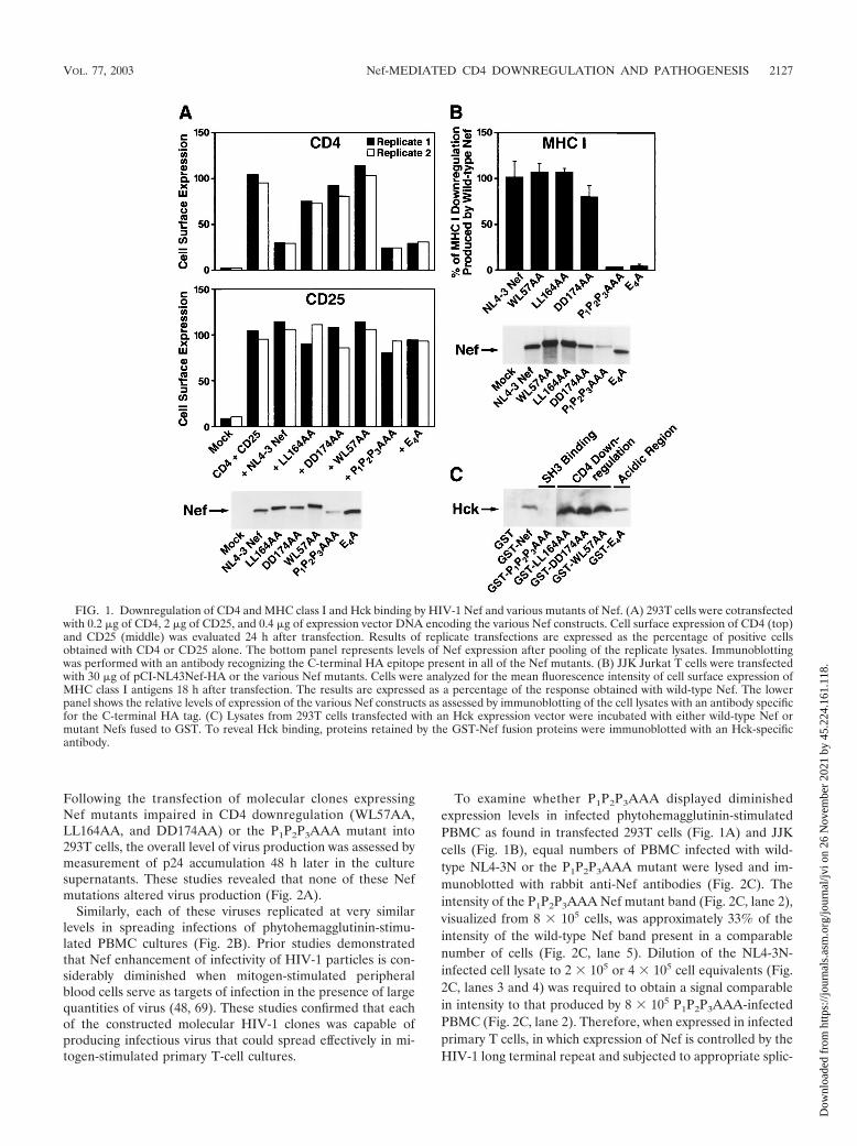
regulate ectopically expressed CD4 in 293T human embryonickidney cells. These cells lack expression of the Src family pro-tein tyrosine kinase Lck, which can associate with the cytoplas-mic tail of CD4 in T cells (reviewed in reference 8). To ensurethat the Nef-mediated effects were specific for CD4, a controlexpression vector encoding the � chain of the human interleu-kin-2 receptor complex (CD25) was cotransfected togetherwith CD4 and the various Nef mutants. The Nef mutantsLL164AA, DD174AA, and WL57AA each displayed impaireddownregulation of surface CD4 (Fig. 1A). In contrast, disrup-tion of the polyproline type II helix within Nef (P1P2P3AAA)or mutation of the acidic region (E4A) within Nef did notimpair CD4 downregulation in 293T cells. None of the Nefmutants altered surface expression of CD25 in these cultures(Fig. 1A, middle panel).
To assess the expression levels of these Nef mutants, analiquot of the cell lysates from these cultures was immunoblot-ted with an antibody reacting with the C-terminal HA epitopetag present on the proteins (Fig. 1A, lower panel). Thesestudies revealed that the mutant Nefs defective for CD4 down-regulation were expressed at levels comparable to wild-typeNef. Of note, we consistently observed that the P1P2P3AAANef was expressed at significantly lower levels than the otherNef constructs. Additionally, this mutant and the E4A mutantdisplayed slightly more rapid electrophoretic migration, whileWL57AA was slightly retarded.
To examine the ability of these Nef mutants to downregulatecell surface expression of MHC class I, the human JJK JurkatT-cell line, which expresses high levels of MHC class I andCD4, was electroporated with the various Nef constructs. Mu-tation of either the polyproline stretch (P1P2P3AAA) or theacidic region (E4A) within Nef significantly impaired the abil-ity of Nef to downregulate surface MHC class I (Fig. 1B).Conversely, mutations of Nef that compromised CD4 down-regulation (WL57AA, LL164AA, and DD174AA) did not af-fect MHC class I downregulation. Expression levels of the Nefmutants in lysates of the transfected Jurkat T cells were as-sessed (Fig. 1B, lower panel). Again, the P1P2P3AAA Nef wasexpressed at lower levels than the other Nef mutants. Thus, theMHC class I downregulation results with this mutant must beinterpreted with caution.
Finally, the Nef mutants expressed as GST fusion proteinswere tested for their ability to interact with the Src familykinase Hck. Consistent with the prior finding that Nef inter-action with Hck requires an intact polyproline stretch withinNef (61), the GST-P1P2P3AAA mutant failed to bind Hck (Fig.1C). Conversely, each of the mutants that impaired CD4 down-regulation and the E4A mutant retained the capacity to inter-act with Hck in vitro.
Together, these results confirm the in vitro phenotypes ofthe Nef mutants and are consistent with the previous findingthat Nef-mediated CD4 downregulation versus class I MHCdownregulation and Hck binding involve different moleculardeterminants (26, 45, 61). These studies also highlight theoverall instability of the P1P2P3AAA Nef mutant in two dif-ferent cellular environments.
HIV-1 containing mutant nef alleles that impair CD4 down-regulation are less infectious than wild-type HIV-1. Each ofthe various Nef mutations was introduced into NL4-3N, aCXCR4-utilizing infectious molecular clone of HIV-1 (61).
2126 STODDART ET AL. J. VIROL.
Dow
nloa
ded
from
http
s://j
ourn
als.
asm
.org
/jour
nal/j
vi o
n 26
Nov
embe
r 20
21 b
y 45
.224
.161
.118
.
Following the transfection of molecular clones expressingNef mutants impaired in CD4 downregulation (WL57AA,LL164AA, and DD174AA) or the P1P2P3AAA mutant into293T cells, the overall level of virus production was assessed bymeasurement of p24 accumulation 48 h later in the culturesupernatants. These studies revealed that none of these Nefmutations altered virus production (Fig. 2A).
Similarly, each of these viruses replicated at very similarlevels in spreading infections of phytohemagglutinin-stimu-lated PBMC cultures (Fig. 2B). Prior studies demonstratedthat Nef enhancement of infectivity of HIV-1 particles is con-siderably diminished when mitogen-stimulated peripheralblood cells serve as targets of infection in the presence of largequantities of virus (48, 69). These studies confirmed that eachof the constructed molecular HIV-1 clones was capable ofproducing infectious virus that could spread effectively in mi-togen-stimulated primary T-cell cultures.
To examine whether P1P2P3AAA displayed diminishedexpression levels in infected phytohemagglutinin-stimulatedPBMC as found in transfected 293T cells (Fig. 1A) and JJKcells (Fig. 1B), equal numbers of PBMC infected with wild-type NL4-3N or the P1P2P3AAA mutant were lysed and im-munoblotted with rabbit anti-Nef antibodies (Fig. 2C). Theintensity of the P1P2P3AAA Nef mutant band (Fig. 2C, lane 2),visualized from 8 � 105 cells, was approximately 33% of theintensity of the wild-type Nef band present in a comparablenumber of cells (Fig. 2C, lane 5). Dilution of the NL4-3N-infected cell lysate to 2 � 105 or 4 � 105 cell equivalents (Fig.2C, lanes 3 and 4) was required to obtain a signal comparablein intensity to that produced by 8 � 105 P1P2P3AAA-infectedPBMC (Fig. 2C, lane 2). Therefore, when expressed in infectedprimary T cells, in which expression of Nef is controlled by theHIV-1 long terminal repeat and subjected to appropriate splic-
FIG. 1. Downregulation of CD4 and MHC class I and Hck binding by HIV-1 Nef and various mutants of Nef. (A) 293T cells were cotransfectedwith 0.2 �g of CD4, 2 �g of CD25, and 0.4 �g of expression vector DNA encoding the various Nef constructs. Cell surface expression of CD4 (top)and CD25 (middle) was evaluated 24 h after transfection. Results of replicate transfections are expressed as the percentage of positive cellsobtained with CD4 or CD25 alone. The bottom panel represents levels of Nef expression after pooling of the replicate lysates. Immunoblottingwas performed with an antibody recognizing the C-terminal HA epitope present in all of the Nef mutants. (B) JJK Jurkat T cells were transfectedwith 30 �g of pCI-NL43Nef-HA or the various Nef mutants. Cells were analyzed for the mean fluorescence intensity of cell surface expression ofMHC class I antigens 18 h after transfection. The results are expressed as a percentage of the response obtained with wild-type Nef. The lowerpanel shows the relative levels of expression of the various Nef constructs as assessed by immunoblotting of the cell lysates with an antibody specificfor the C-terminal HA tag. (C) Lysates from 293T cells transfected with an Hck expression vector were incubated with either wild-type Nef ormutant Nefs fused to GST. To reveal Hck binding, proteins retained by the GST-Nef fusion proteins were immunoblotted with an Hck-specificantibody.
VOL. 77, 2003 Nef-MEDIATED CD4 DOWNREGULATION AND PATHOGENESIS 2127
Dow
nloa
ded
from
http
s://j
ourn
als.
asm
.org
/jour
nal/j
vi o
n 26
Nov
embe
r 20
21 b
y 45
.224
.161
.118
.

ing, the P1P2P3AAA Nef is consistently expressed at levelsbetween 25 and 50% of that of wild-type Nef.
We next tested the infectivity of the recombinant viruses car-rying the various Nef mutations. The infectivity titer (TCID50)was determined for each virus by limiting dilution in phyto-hemagglutinin-stimulated PBMC (Fig. 2D). NL4-3N carryingNef mutations making it defective for CD4 downregulation(LL164AA, DD174AA, or WL57AA) displayed diminishedinfectivity in PBMC; this reduction was comparable to thatobserved with NL4-3N lacking Nef expression (Xho) and morepronounced than that observed with the NL4-3N clone ex-pressing the P1P2P3AAA Nef mutant (24, 61) (Fig. 2D). Incontrast, the E4A Nef mutant, which is impaired in MHC classI downregulation, displayed a level of infectivity similar to thatdetected with the wild-type virus. These results confirm the
correlation between Nef-mediated CD4 downregulation andNef-mediated enhancement of HIV-1 infectivity in primary Tcells expressing CD4 and serving as virus producers (36, 43,60).
Determination of minimal inoculum of HIV-1 NL4-3N re-quired to initiate a productive infection in the implants ofSCID-hu Thy/Liv mice. To compare the replication kineticsand pathogenic properties of the nef mutants in vivo, we em-ployed the SCID-hu Thy/Liv mouse model of HIV-1 infection(50). Prior studies have shown that while HIV-1 Nef is re-quired for efficient in vivo viral replication and pathogenicity,the infectivity and pathogenicity of HIV-1 lacking Nef areattenuated but not abolished in this model. Viruses lacking nefretain the ability to induce thymocyte depletion if implantsare inoculated with larger amounts of virus (1,000 infectious
FIG. 2. Virus production, spreading infection, and infectivity of HIV-1 expressing wild-type Nef or the various Nef mutants. (A) Culturesupernatants from 293T cells transfected with a molecular clone of HIV-1 carrying the various Nef mutations were analyzed for virus productionby p24 antigen-capture ELISA. (B) Phytohemagglutinin-stimulated PBMC were infected (25 ng of p24 per 4 � 106 cells in 2.5 ml of medium), andthe culture supernatants were monitored by p24 antigen-capture ELISA. Results show that viral infections peaked on day 8 after virus inoculation.(C) Lysates from NL4-3N- and P1P2P3AAA-infected PBMC were immunoblotted with an anti-Nef rabbit serum. (D) The TCID50 for each viruswas determined by limiting dilution with phytohemagglutinin-stimulated PBMC as cellular targets. The data are expressed as a percentage of theinfectivity of wild-type NL4-3N.
2128 STODDART ET AL. J. VIROL.
Dow
nloa
ded
from
http
s://j
ourn
als.
asm
.org
/jour
nal/j
vi o
n 26
Nov
embe
r 20
21 b
y 45
.224
.161
.118
.

units) and observed for longer periods (6 to 9 weeks) (3, 4,16, 31).
To establish the minimal inoculum of wild-type NL4-3Nrequired for productive infection, 4 � 103 to 4 � 107 TCID50
(in 50 �l) were inoculated into each implant of SCID-hu Thy/Liv mice by direct injection. Productive infection of the im-plants was assessed by p24 ELISA on lysates of dispersedthymocytes from implants collected 4 weeks after inoculation.Only one of four implants inoculated with 4 � 103 TCID50
contained detectable p24 (410 pg/106 cells), while all implantsinoculated with higher doses became productively infected(510 to 2,900 pg of p24/106 cells). (Data are expressed aspicograms of p24 per 106 cells rather than p24 per implant fora measure of infectivity on a per-cell basis because virus-me-diated thymocyte depletion reduces overall implant viralloads.) For subsequent studies, we therefore used 20,000TCID50 per implant of NL4-3N and NL4-3N containing thevarious Nef mutations (all generated by transfection of 293Tcells) and collected the implants 6 weeks after inoculation.
HIV-1 expressing nef mutations defective for CD4 down-regulation are less pathogenic than wild-type HIV-1 inSCID-hu Thy/Liv mice. To determine if the ability of Nef todownregulate CD4 plays a role in the pathogenic events ob-served in the Thy/Liv mouse model, we analyzed viral replica-tion (Fig. 3A) and thymocyte depletion (Fig. 3B and C) in micewhose implants were inoculated with wild-type NL4-3N,
NL4-N (Xho), or the three Nef mutants defective for CD4downregulation, LL164AA [defective in clathrin adaptor bind-ing (9, 25, 41)], DD174AA [defective in assembly with a cel-lular vacuolar ATPase responsible for endocytic acidification(42) or in interaction with the cytoplasmic tail of CD4 (45)],and WL57AA [defective in interaction with the cytoplasmictail of CD4 (42, 45)]. Implants were also inoculated with theP1P2P3AAA mutant, which is capable of CD4 downregulationdespite 50 to 75% lower levels of Nef expression.
At 6 weeks after virus inoculation, no statistically significantdifferences in implant p24 production were observed betweenwild-type NL4-3N and each of the Nef mutants (Fig. 3A), andmost inoculated implants had high levels of p24 (�500 pg per106 cells) regardless of nef genotype. Despite similar levels ofp24 production, which indicate comparable infectivity, two ofthe three Nef mutants defective for CD4 downregulation(LL164AA and WL57AA) were attenuated for thymocyte de-pletion, as assessed by the percentage of viable cells (events inthe live lymphocyte gate as a percentage of total events) basedon forward- and side-scatter properties (Fig. 3B), and by thepercentage of immature cortical CD4� CD8� thymocytes (Fig.3C). Unlike wild-type NL4-3N, which reduced viable thymo-cytes from 86% to 23% and CD4� CD8� thymocytes from89% to 17%, LL164AA and WL57AA produced only smalldeclines in these percentages by 6 weeks after inoculation (Fig.3B and C).
FIG. 3. HIV-1 containing nef mutations that impair CD4 downregulation produces significantly less depletion of thymocytes in SCID-huThy/Liv mice. (A) Cell-associated p24 accumulation was measured to assess the relative level of viral replication at 6 weeks after inoculation ofthe Thy/Liv implant mice. (B) At 6 weeks after inoculation of Thy/Liv implant mice with either wild-type NL4-3N or NL4-3N expressing the variousNef mutants, the percentage of live cells was determined by flow cytometric forward- and side-scatter analysis. (C) In cells derived from the sameinfected implants, the percentage of immature double-positive (CD4� CD8�) thymocytes present was determined. The height of each bar indicatesthe mean of the experimental values shown by the solid circles. Error bars indicate standard errors of the mean. P values from the Mann-WhitneyU test were calculated for each mutant relative to wild-type NL4-3N with StatView 5.0 (SAS Institute Inc., Cary, N.C.). NS, not significant. Wedid not exclude the Xho-infected implant that had no detectable p24 because it was successfully inoculated with 20,000 TCID50 and appeared tobe of good quality.
VOL. 77, 2003 Nef-MEDIATED CD4 DOWNREGULATION AND PATHOGENESIS 2129
Dow
nloa
ded
from
http
s://j
ourn
als.
asm
.org
/jour
nal/j
vi o
n 26
Nov
embe
r 20
21 b
y 45
.224
.161
.118
.
The diminished cytopathic properties of these two mutationsdefective for CD4 downregulation resembled that of NL4-3N(Xho), which encodes only the 35 N-terminal amino acids ofNef. In contrast, and despite its lower level of Nef expression,P1P2P3AAA depleted human thymocytes in the SCID-hu Thy/Liv mice as effectively as wild-type NL4-3N did (Fig. 3B andC). This result agrees with previous work in the SCID-huThy/Liv mouse model reported by Aldrovandi et al. (3) andsuggests that polyproline-dependent enhancement of infectiv-ity (Fig. 2D), MHC class I downregulation (Fig. 1B), andinteraction with the cellular kinases PAK and Hck (Fig. 1C)(73) are dispensable for Nef-mediated HIV-1 pathogenesis inSCID-hu Thy/Liv mice. These results thus reveal a correlationbetween CD4 downregulation and virus-induced depletion ofthymocytes in the SCID-hu Thy/Liv mouse model.
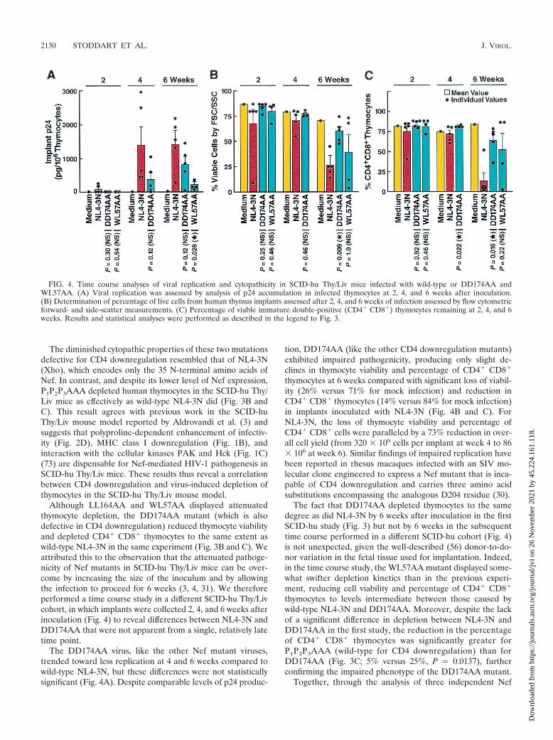
Although LL164AA and WL57AA displayed attenuatedthymocyte depletion, the DD174AA mutant (which is alsodefective in CD4 downregulation) reduced thymocyte viabilityand depleted CD4� CD8� thymocytes to the same extent aswild-type NL4-3N in the same experiment (Fig. 3B and C). Weattributed this to the observation that the attenuated pathoge-nicity of Nef mutants in SCID-hu Thy/Liv mice can be over-come by increasing the size of the inoculum and by allowingthe infection to proceed for 6 weeks (3, 4, 31). We thereforeperformed a time course study in a different SCID-hu Thy/Livcohort, in which implants were collected 2, 4, and 6 weeks afterinoculation (Fig. 4) to reveal differences between NL4-3N andDD174AA that were not apparent from a single, relatively latetime point.
The DD174AA virus, like the other Nef mutant viruses,trended toward less replication at 4 and 6 weeks compared towild-type NL4-3N, but these differences were not statisticallysignificant (Fig. 4A). Despite comparable levels of p24 produc-
tion, DD174AA (like the other CD4 downregulation mutants)exhibited impaired pathogenicity, producing only slight de-clines in thymocyte viability and percentage of CD4� CD8�
thymocytes at 6 weeks compared with significant loss of viabil-ity (26% versus 71% for mock infection) and reduction inCD4� CD8� thymocytes (14% versus 84% for mock infection)in implants inoculated with NL4-3N (Fig. 4B and C). ForNL4-3N, the loss of thymocyte viability and percentage ofCD4� CD8� cells were paralleled by a 73% reduction in over-all cell yield (from 320 � 106 cells per implant at week 4 to 86� 106 at week 6). Similar findings of impaired replication havebeen reported in rhesus macaques infected with an SIV mo-lecular clone engineered to express a Nef mutant that is inca-pable of CD4 downregulation and carries three amino acidsubstitutions encompassing the analogous D204 residue (30).
The fact that DD174AA depleted thymocytes to the samedegree as did NL4-3N by 6 weeks after inoculation in the firstSCID-hu study (Fig. 3) but not by 6 weeks in the subsequenttime course performed in a different SCID-hu cohort (Fig. 4)is not unexpected, given the well-described (56) donor-to-do-nor variation in the fetal tissue used for implantation. Indeed,in the time course study, the WL57AA mutant displayed some-what swifter depletion kinetics than in the previous experi-ment, reducing cell viability and percentage of CD4� CD8�
thymocytes to levels intermediate between those caused bywild-type NL4-3N and DD174AA. Moreover, despite the lackof a significant difference in depletion between NL4-3N andDD174AA in the first study, the reduction in the percentageof CD4� CD8� thymocytes was significantly greater forP1P2P3AAA (wild-type for CD4 downregulation) than forDD174AA (Fig. 3C; 5% versus 25%, P � 0.0137), furtherconfirming the impaired phenotype of the DD174AA mutant.
Together, through the analysis of three independent Nef
FIG. 4. Time course analyses of viral replication and cytopathicity in SCID-hu Thy/Liv mice infected with wild-type or DD174AA andWL57AA. (A) Viral replication was assessed by analysis of p24 accumulation in infected thymocytes at 2, 4, and 6 weeks after inoculation.(B) Determination of percentage of live cells from human thymus implants assessed after 2, 4, and 6 weeks of infection assessed by flow cytometricforward- and side-scatter measurements. (C) Percentage of viable immature double-positive (CD4� CD8�) thymocytes remaining at 2, 4, and 6weeks. Results and statistical analyses were performed as described in the legend to Fig. 3.
2130 STODDART ET AL. J. VIROL.
Dow
nloa
ded
from
http
s://j
ourn
als.
asm
.org
/jour
nal/j
vi o
n 26
Nov
embe
r 20
21 b
y 45
.224
.161
.118
.

mutants, our findings reveal a correlation between the inabilityof Nef to downregulate cell surface expression of CD4 andattenuated HIV-1 pathogenicity in SCID-hu Thy/Liv mice, asevidenced by slower kinetics of thymocyte depletion.
DISCUSSION
In this series of studies, we analyzed which of the various invitro functions ascribed to HIV-1 Nef are most closely associ-ated with in vivo pathogenesis in the SCID-hu Thy/Liv mousemodel. We found that introduction of three different Nef mu-tations that compromised CD4 downregulation (WL57AA,LL164AA, and DD174AA) into the NL4-3N molecular cloneleads to viruses that display attenuated pathogenicity in thehuman thymus implants of these mice. These changes in cyto-pathic effect did not significantly correlate with changes inreplication of the mutant viruses, although each of these mu-tant viruses tended to produce lower levels of p24 in the im-plants. The viruses containing Nef alleles defective for CD4downregulation did display significantly lower infectivity inphytohemagglutinin-stimulated PBMC, based on limiting-dilu-tion determination of TCID50.
More study is required to determine whether cytopathicityand viral replication are indeed separable phenomena in thethymus. In this regard, Glushakova and colleagues recentlyreported that CD4 downregulation by HIV-1 Nef also corre-lates with depletion of mature T cells in human lymphoid cellhistocultures (22). However, in this system, viral replicationwas clearly linked to cytopathicity. In contrast to the Nefmutants compromised for CD4 downregulation, NL4-3Nthat carried a polyproline (P1P2P3AAA) mutation and exhib-ited diminished infectivity and failed to downregulate MHCclass I antigens displayed pathogenic potential comparable tothat of wild-type virus.
Three recent reports have highlighted the importance of cellsurface CD4 downregulation by Nef in augmenting viral pro-duction and infectivity (36, 43, 60). The first report indicatesthat Nef is required to prevent the incorporation of CD4 intonascent HIV-1 particles (36). Viral particles produced in theabsence of Nef incorporate fewer envelope proteins and moreCD4 molecules. The resulting viral particles are less infectious.A second report shows that in the presence of high levels ofCD4, HIV-1 structural proteins accumulate in the host cell,causing a reduction in the release of viral particles (60). Nefreverses this reduction in viral particle release. Finally, Lund-quist and colleagues, with a battery of Nef point mutants,reported a strong genetic correlation between replication effi-ciency and Nef-induced CD4 downregulation (but not Nef-induced enhancement of virion infectivity) in activated primaryCD4� T cells (43). These molecular mechanisms may contrib-ute to our observation that HIV-1 recombinants carrying Nefmutations defective for CD4 downregulation are less patho-genic in SCID-hu Thy/Liv mice.
Our finding that HIV-1 carrying a Nef mutant in which thefirst three prolines of the (Pxx)4 polyproline stretch were mu-tated to alanines (P1P2P3AAA) displays the same pathogenicpotential as wild-type HIV-1 is consistent with a previous studyin SCID-hu Thy/Liv mice (3). This result further argues thatMHC class I downregulation and Pxx-dependent enhancementof HIV-1 infectivity in phytohemagglutinin-stimulated PBMC
are not required for optimal HIV-1 pathogenic potential inhuman thymus. The dispensability of MHC class I downregu-lation for pathogenesis in SCID-hu Thy/Liv mice is perhapsnot surprising because no immune response is mounted againstHIV-1-infected cells in this model.
The (Pxx)4 region within Nef was also suggested to be im-portant for activation of certain cellular signal transductionpathways (72). This function of Nef may also be dispensable ininfected thymocytes, as these rapidly dividing and differentiat-ing cells may be in an elevated state of activation compared toquiescent nondividing PBMC. However, it is intriguing thatdisruption of a similar region within SIV Nef (Pxx)2 did notdisrupt pathogenesis in rhesus macaques in one study (37).However, in a second study, this region within SIV Nef ap-peared to be important, as mutations in this region rapidlyreverted to wild-type sequence in infected animals (34). Sincethe P1P2P3AAA Nef mutant was expressed very poorly com-pared to wild-type Nef yet was capable of comparable induc-tion of pathogenic effects, it seems plausible that a thresholdlevel of Nef (well below that found in cells infected with wild-type HIV-1) is sufficient to promote pathogenesis. However,since this Nef mutant retained the ability to downregulateCD4, it is perhaps this function of Nef that is most crucial foracceleration of pathogenesis.
In summary, our studies demonstrate a correlation betweenNef-mediated CD4 downregulation and HIV-1-associated thy-mocyte depletion in SCID-hu Thy/Liv mice. These studiessuggest that the development of small-molecule inhibitors ofNef-mediated downregulation of CD4 might impair HIV-1pathogenicity.
ACKNOWLEDGMENTS
C. A. Stoddart, R. Geleziunas, and S. Ferrell contributed equally tothis work.
This work was supported in part by NIH grant R0ICA86814 anda grant from the James B. Pendleton Charitable Trust (awardedto W.C.G.), the UCSF-GIVI Center for AIDS Research (P30-MH59037), and NIH contracts AI05418 (awarded to C.A.S.) andAI65309 (awarded to J.M.M.).
We thank John Carroll, Stephen Gonzales, and Chris Goodfellowfor preparation of the figures. We thank Laura Napolitano and EricWeider for technical assistance, Robert Grant for statistical advice,and Robin Givens, Sue Cammack, Stephen Ordway, and Gary Howardfor assistance with manuscript preparation and editing.
REFERENCES
1. Aiken, C. 1997. Pseudotyping human immunodeficiency virus type 1 (HIV-1)by the glycoprotein of vesicular stomatitis virus targets HIV-1 entry to anendocytic pathway and suppresses both the requirement for Nef and thesensitivity to cyclosporin A. J. Virol. 71:5871–5877.
2. Aiken, C., and D. Trono. 1995. Nef stimulates human immunodeficiencyvirus type 1 proviral DNA synthesis. J. Virol. 69:5048–5056.
3. Aldrovandi, G. M., L. Gao, G. Bristol, and J. A. Zack. 1998. Regions ofhuman immunodeficiency virus type 1 nef required for function in vivo.J. Virol. 72:7032–7039.
4. Aldrovandi, G. M., and J. A. Zack. 1996. Replication and pathogenicity ofhuman immunodeficiency virus type 1 accessory gene mutants in SCID-humice. J. Virol. 70:1505–1511.
5. Baba, T. W., Y. S. Jeong, D. Penninck, R. Bronson, M. F. Greene, and R. M.Ruprecht. 1995. Pathogenicity of live, attenuated SIV after mucosal infectionof neonatal macaques. Science 267:1820–1825.
6. Baba, T. W., V. Liska, A. H. Khimani, N. B. Ray, P. J. Dailey, D. Penninck,R. Bronson, M. F. Greene, H. M. McClure, L. N. Martin, and R. M. Ru-precht. 1999. Live attenuated, multiply deleted simian immunodeficiencyvirus causes AIDS in infant and adult macaques. Nat. Med. 5:194–203.
7. Benson, R. E., A. Sanfridson, J. S. Ottinger, C. Doyle, and B. R. Cullen. 1993.Downregulation of cell surface CD4 expression by SIV Nef prevents viralsuperinfection. J. Exp. Med. 177:1561–1566.
VOL. 77, 2003 Nef-MEDIATED CD4 DOWNREGULATION AND PATHOGENESIS 2131
Dow
nloa
ded
from
http
s://j
ourn
als.
asm
.org
/jour
nal/j
vi o
n 26
Nov
embe
r 20
21 b
y 45
.224
.161
.118
.

8. Bolen, J. B., and A. Veillette. 1989. A function for the lck proto-oncogene.Trends Biochem. Sci. 14:404–407.
9. Bresnahan, P. A., W. Yonemoto, S. Ferrell, D. Williams-Herman, R. Geleziu-nas, and W. C. Greene. 1998. A dileucine motif in HIV-1 Nef acts as aninternalization signal for CD4 downregulation and binds the AP-1 clathrinadaptor. Curr. Biol. 8:1235–1238.
10. Brown, A., X. Wang, E. Sawai, and C. Cheng-Mayer. 1999. Activation of thePAK-related kinase by human immunodeficiency virus type 1 Nef in primaryhuman peripheral blood lymphocytes and macrophages leads to phosphor-ylation of a PIX-p95 complex. J. Virol. 73:9899–9907.
11. Chowers, M. Y., M. W. Pandori, C. A. Spina, D. D. Richman, and J. C.Guatelli. 1995. The growth advantage conferred by HIV-1 Nef is determinedat the level of viral DNA formation and is independent of CD4 downregu-lation. Virology 212:451–457.
12. Chowers, M. Y., C. A. Spina, T. J. Kwoh, N. J. S. Fitch, D. D. Richman, andJ. C. Guatelli. 1994. Optimal infectivity in vitro of human immunodeficiencyvirus type 1 requires an intact nef gene. J. Virol. 68:2906–2914.
13. Collins, K. L., B. K. Chen, S. A. Kalams, B. D. Walker, and D. Baltimore.1998. HIV-1 Nef protein protects infected primary cells against killing bycytotoxic T lymphocytes. Nature 391:397–401.
14. Cullen, B. R. 1998. HIV-1 auxilliary proteins: making connections in a dyingcell. Cell 93:685–692.
15. Deacon, N. J., A. Tsykin, A. Solomon, K. Smith, M. Ludford-Menting,A. Ellett, D. J. Hooker, D. A. McPhee, A. L. Greenway, C. Chatfield, V. A.Lawson, S. Crowe, A. Maerz, S. Sonza, J. Learmont, J. S. Sullivan, A.Cunningham, D. Dwyer, D. Dowton, and J. Mills. 1995. Genomic structureof an attenuated quasi species of HIV-1 from a blood transfusion donor andrecipients. Science 270:988–991.
16. Duus, K. M., E. D. Miller, J. A. Smith, G. I. Kovalev, and L. Su. 2001.Separation of human immunodeficiency virus type 1 replication from Nef-mediated pathogenesis in the human thymus. J. Virol. 8:3916–3924.
17. Fackler, O. T., X. Lu, J. A. Frost, M. Geyer, B. Jiang, W. Luo, A. Abo, A. S.Alberts, and B. M. Peterlin. 2000. p21-activated kinase 1 plays a critical rolein cellular activation by Nef. Mol. Cell. Biol. 20:2619–2627.
18. Fackler, O. T., W. Luo, M. Geyer, A. S. Alberts, and B. M. Peterlin. 1999.Activation of Vav by Nef induces cytoskeletal rearrangements and down-stream effector functions. Mol. Cell 3:729–739.
19. Garcia, J. V., and A. D. Miller. 1991. Serine phosphorylation-independentdownregulation of cell surface CD4 by Nef. Nature 350:508–511.
20. Geleziunas, R., M. D. Miller, and W. C. Greene. 1996. Unraveling thefunction of HIV type 1 Nef. AIDS Res. Hum. Retrovir. 12:1579–1582.
21. Geleziunas, R., W. Xu, K. Takeda, H. Ichijo, and W. C. Greene. 2001. HIV-1Nef inhibits ASK-1-dependent death signalling providing a potential mech-anism for protecting the infected host cell. Nature 410:834–838.
22. Glushakova, S., J. Munch, S. Carl, T. C. Greenough, J. L. Sullivan, L.Margolis, and F. Kirchhoff. 2001. CD4 down-modulation by human immu-nodeficiency virus type 1 Nef correlates with the efficiency of viral replicationand with CD4� T-cell depletion in human lymphoid tissue ex vivo. J. Virol.75:10113–10117.
23. Gobbi, A., C. A. Stoddart, M. S. Malnati, G. Locatelli, F. Santoro, N. W.Abbey, C. Bare, V. Linquist-Stepps, M. B. Moreno, B. G. Herndier, P. Lusso,and J. M. McCune. 1999. Human herpesvirus 6 (HHV-6) causes severethymocyte depletion in SCID-hu Thy/Liv mice. J. Exp. Med. 189:1953–1960.
24. Goldsmith, M. A., M. T. Warmerdam, R. E. Atchison, M. D. Miller, andW. C. Greene. 1995. Dissociation of the CD4 downregulation and viralinfectivity enhancement functions of human immunodeficiency virus type 1Nef. J. Virol. 69:4112–4121.
25. Greenberg, M., L. DeTulleo, I. Rapoport, J. Skowronski, and T. Kirch-hausen. 1998. A dileucine motif in HIV-1 Nef is essential for sorting intoclathrin-coated pits and for downregulation of CD4. Curr. Biol. 8:1239–1242.
26. Greenberg, M. E., A. J. Iafrate, and J. Skowronski. 1998. The SH3 domain-binding surface and an acidic motif in HIV-1 Nef regulate trafficking of classI MHC complexes. EMBO J. 17:2777–2789.
27. Greenough, T. C., J. L. Sullivan, and R. C. Desrosiers. 1999. Declining CD4T-cell counts in a person infected with Nef-deleted HIV-1. N. Engl. J. Med.340:236–237.
28. Grzesiek, S., S. J. Stahl, P. T. Wingfield, and A. Bax. 1996. The CD4determinant for downregulation by HIV-1 Nef directly binds to Nef: map-ping of the Nef binding surface by NMR. Biochemistry 35:10256–10261.
29. Hanna, Z., D. G. Kay, N. Rebai, A. Guimond, S. Jothy, and P. Jolicoeur.1998. Nef harbors a major determinant of pathogenicity for an AIDS-likedisease induced by HIV-1 in transgenic mice. Cell 95:163–175.
30. Iafrate, A. J., S. Carl, S. Bronson, C. Stahl-Hennig, T. Swigut, J. Skowronski,and F. Kirchhoff. 2000. Disrupting surfaces of Nef required for downregu-lation of CD4 and for enhancement of virion infectivity attenuates simianimmunodeficiency virus replication in vivo. J. Virol. 74:9836–9844.
31. Jamieson, B. D., G. M. Aldrovandi, V. Planelles, J. B. Jowett, L. Gao, L. M.Bloch, I. S. Chen, and J. A. Zack. 1994. Requirement of human immuno-deficiency virus type 1 Nef for in vivo replication and pathogenicity. J. Virol.68:3478–3485.
32. Johnson, V. A., and R. E. Byington. 1990. Quantitative assays for virus
infectivity, p. 71. In A. Aldovini and B. D. Walker (ed.), Techniques in HIVresearch. Stockton Press, London, United Kingdom.
33. Kestler, H. W. d., D. J. Ringler, K. Mori, D. L. Panicali, P. K. Sehgal, M. D.Daniel, and R. C. Desrosiers. 1991. Importance of the nef gene for mainte-nance of high virus loads and for development of AIDS. Cell 65:651–662.
34. Khan, I. H., E. T. Sawai, E. Antonio, C. J. Weber, C. P. Mandell, P. Mont-briand, and P. A. Luciw. 1998. Role of the SH3-ligand domain of simianimmunodeficiency virus Nef in interaction with Nef-associated kinase andsimian AIDS in rhesus macaques. J. Virol. 72:5820–5830.
35. Kirchhoff, F., T. C. Greenough, D. B. Brettler, J. L. Sullivan, and R. C.Desrosiers. 1995. Absence of intact nef sequences in a long-term survivorwith nonprogressive HIV-1 infection. N. Engl. J. Med. 332:228–232.
36. Lama, J., A. Mangasarian, and D. Trono. 1999. Cell surface expression ofCD4 reduces HIV-1 infectivity by blocking Env incorporation in a Nef- andVpu-inhibitable manner. Curr. Biol. 9:622–631.
37. Lang, S. M., A. J. Iafrate, H. C. Stahl, E. M. Kuhn, T. Nisslein, F. J. Kaup,M. Haupt, G. Hunsmann, J. Skowronski, and F. Kirchhoff. 1997. Associationof simian immunodeficiency virus Nef with cellular serine/threonine kinasesis dispensable for the development of AIDS in rhesus macaques. Nat. Med.3:860–865.
38. Learmont, J. C., A. F. Geczy, J. Mills, L. J. Ashton, C. H. Raynes-Greenow,R. J. Garsia, W. B. Dyer, L. McIntyre, R. B. Oelrichs, D. I. Rhodes, N. J.Deacon, and J. S. Sullivan. 1999. Immunologic and virologic status after 14to 18 years of infection with an attenuated strain of HIV-1: a report from theSydney Blood Bank Cohort. N. Engl. J. Med. 340:1715–1722.
39. Le Gall, S., L. Erdtmann, S. Benichou, C. Berlioz-Torrent, L. Liu, R. Ben-arous, J. M. Heard, and O. Schwartz. 1998. Nef interacts with the mu subunitof clathrin adaptor complexes and reveals a cryptic sorting signal in MHC Imolecules. Immunity 8:483–495.
40. Lindemann, D., R. Wilhelm, P. Renard, A. Althage, R. Zinkernagel, and J.Mous. 1994. Severe immunodeficiency associated with an HIV-1 Nef/3� longterminal repeat transgene. J. Exp. Med. 179:797–807.
41. Lock, M., M. E. Greenberg, A. J. Iafrate, T. Swigut, J. Muench, F. Kirchhoff,N. Shohdy, and J. Skowronski. 1999. Two elements target SIV Nef to theAP-2 clathrin adaptor complex, but only one is required for the induction ofCD4 endocytosis. EMBO J. 18:2722–2733.
42. Lu, X., H. Yu, S. H. Liu, F. M. Brodsky, and B. M. Peterlin. 1998. Interac-tions between HIV-1 Nef and vacuolar ATPase facilitate the internalizationof CD4. Immunity 8:647–656.
43. Lundquist, C. A., M. Tobiume, J. Zhou, D. Unutmaz, and C. Aiken. 2002.Nef-mediated downregulation of CD4 enhances human immunodeficiencyvirus type 1 replication in primary T lymphocytes. J. Virol. 9:4625–4633.
44. Mangasarian, A., M. Foti, C. Aiken, D. Chin, J. L. Carpentier, and D. Trono.1997. The HIV-1 Nef protein acts as a connector with sorting pathways in theGolgi and at the plasma membrane. Immunity 6:67–77.
45. Mangasarian, A., V. Piguet, J. K. Wang, Y. L. Chen, and D. Trono. 1999.Nef-induced CD4 and major histocompatibility complex class I (MHC classI) down-regulation are governed by distinct determinants: N-terminal alphahelix and proline repeat of Nef selectively regulate MHC class I trafficking.J. Virol. 73:1964–1973.
46. Mariani, R., F. Kirchhoff, T. C. Greenough, J. L. Sullivan, R. C. Desrosiers,and J. Skowronski. 1996. High frequency of defective nef alleles in a long-term survivor with nonprogressive human immunodeficiency virus type 1infection. J. Virol. 70:7752–7764.
47. Marsh, J. W. 1999. The numerous effector functions of Nef. Arch. Biochem.Biophys. 365:192–198.
48. Miller, M. D., M. T. Warmerdam, I. Gaston, W. C. Greene, and M. B.Feinberg. 1994. The HIV-1 Nef gene product: a positive factor for viralinfection and replication in primary lymphocytes and macrophages. J. Exp.Med. 179:101–113.
49. Miller, M. D., M. T. Warmerdam, K. A. Page, M. B. Feinberg, and W. C.Greene. 1995. Expression of the human immunodeficiency virus type 1(HIV-1) nef gene during HIV-1 production increases progeny particle infec-tivity independently of gp160 or viral entry. J. Virol. 69:570–584.
50. Namikawa, R., K. N. Weilbaecher, H. Kaneshima, E. J. Yee, and J. M.McCune. 1990. Long-term human hematopoiesis in the SCID-hu mouse. J.Exp. Med. 172:1055–1063.
51. Nunn, M. F., and J. W. Marsh. 1996. Human immunodeficiency virus type 1Nef associates with a member of the p21-activated kinase family. J. Virol.70:6157–6161.
52. Pandori, M. W., N. J. S. Fitch, H. M. Craig, D. D. Richman, C. A. Spina, andJ. C. Guatelli. 1996. Producer-cell modification of human immunodeficiencyvirus type 1: Nef is a virion protein. J. Virol. 70:4283–4290.
53. Peter, F. 1998. HIV Nef: the mother of all evil? Immunity 9:433–437.54. Piguet, V., Y. L. Chen, A. Mangasarian, M. Foti, J. L. Carpentier, and D.
Trono. 1998. Mechanism of Nef-induced CD4 endocytosis: Nef connectsCD4 with the mu chain of adaptor complexes. EMBO J. 17:2472–2481.
55. Piguet, V., L. Wan, C. Borel, A. Mangasarian, N. Demaurex, G. Thomas, andD. Trono. 2000. HIV-1 Nef protein binds to the cellular protein PACS-1 todownregulate class I major histocompatibility complexes. Nat. Cell Biol.2:163–167.
56. Rabin, L., M. Hincenbergs, M. B. Moreno, S. Warren, V. Linquist, R.
2132 STODDART ET AL. J. VIROL.
Dow
nloa
ded
from
http
s://j
ourn
als.
asm
.org
/jour
nal/j
vi o
n 26
Nov
embe
r 20
21 b
y 45
.224
.161
.118
.

Datema, B. Charpiot, J. Seifert, H. Kaneshima, and J. M. McCune. 1996.Use of standardized SCID-hu Thy/Liv mouse model for preclinical efficacytesting of anti-human immunodeficiency virus type 1 compounds. Antimi-crob. Agents Chemother. 40:755–762.
57. Renkema, G. H., A. Manninen, D. A. Mann, M. Harris, and K. Saksela. 1999.Identification of the Nef-associated kinase as p21-activated kinase 2. Curr.Biol. 9:1407–1410.
58. Rhodes, D., A. Solomon, W. Bolton, J. Wood, J. Sullivan, J. Learmont, andN. Deacon. 1999. Identification of a new recipient in the Sydney Blood BankCohort: a long-term HIV type 1-infected seroindeterminate individual.AIDS Res. Hum. Retrovir. 15:1433–1439.
59. Robbins, S. M., N. A. Quintrell, and J. M. Bishop. 1995. Myristoylation anddifferential palmitoylation of the HCK protein-tyrosine kinases govern theirattachment to membranes and association with caveolae. Mol. Cell. Biol.15:3507–3515.
60. Ross, T. M., A. E. Oran, and B. R. Cullen. 1999. Inhibition of HIV-1 progenyvirion release by cell surface CD4 is relieved by expression of the viral Nefprotein. Curr. Biol. 9:613–621.
61. Saksela, K., G. Cheng, and D. Baltimore. 1995. Proline-rich (PxxP) motifs inHIV-1 Nef bind to SH3 domains of a subset of Src kinases and are requiredfor the enhanced growth of Nef� viruses but not for down-regulation ofCD4. EMBO J. 14:484–491.
62. Salvi, R., A. R. Garbuglia, A. Di Caro, S. Pulciani, F. Montella, and A.Benedetto. 1998. Grossly defective nef gene sequences in a human immuno-deficiency virus type 1-seropositive long-term nonprogressor. J. Virol. 72:3646–3657.
63. Sawai, E. T., I. H. Khan, P. M. Montbriand, B. M. Peterlin, M. C. Cheng,and P. A. Luciw. 1996. Activation of PAK by HIV and SIV Nef: importancefor AIDS in rhesus macaques. Curr. Biol. 6:1519–1527.
64. Schaeffer, E., R. Geleziunas, and W. C. Greene. 2001. Human immunodefi-
ciency virus type 1 Nef functions at the level of virus entry by enhancingcytoplasmic delivery of virions. J. Virol. 75:2993–3000.
65. Schrager, J. A., and J. W. Marsh. 1999. HIV-1 Nef increases T-cell activationin a stimulus-dependent manner. Proc. Natl. Acad. Sci. USA 96:8167–8172.
66. Schwartz, O., V. Marechal, O. Danos, and J. M. Heard. 1995. Humanimmunodeficiency virus type 1 Nef increases the efficiency of reverse tran-scription in the infected cell. J. Virol. 69:93–100.
67. Schwartz, O., V. Marechal, G. S. Le, F. Lemonnier, and J. M. Heard. 1996.Endocytosis of major histocompatibility complex class I molecules is inducedby the HIV-1 Nef protein. Nat. Med. 2:338–342.
68. Skowronski, J., D. Parks, and R. Mariani. 1993. Altered T-cell activationand development in transgenic mice expressing the HIV-1 nef gene. EMBOJ. 12:703–713.
69. Spina, C. A., T. J. Kwoh, M. Y. Chowers, J. C. Guatelli, and D. D. Richman.1994. The importance of Nef in the induction of HIV-1 replication fromprimary quiescent CD4 lymphocytes. J. Exp. Med. 179:115–123.
70. Swann, S. A., M. Williams, C. M. Story, K. R. Bobbitt, R. Fleis, and K. L.Collins. 2001. HIV-1 Nef blocks transport of MHC class I molecules to thecell surface via a PI 3-kinase-dependent pathway. Virology 282:267–277.
71. Switzer, W. M., S. Wiktor, V. Soriano, A. Silva-Graca, K. Mansinho, I. M.Coulibaly, E. Ekpini, A. E. Greenberg, T. M. Folks, and W. Heneine. 1998.Evidence of Nef truncation in human immunodeficiency virus type 2 infec-tion. J. Infect. Dis. 177:65–71.
72. Wang, J. K., E. Kiyokawa, E. Verdin, and D. Trono. 2000. The Nef proteinof HIV-1 associates with rafts and primes T cells for activation. Proc. Natl.Acad. Sci. USA 97:394–399.
73. Wiskerchen, M., and M. C. Cheng. 1996. HIV-1 Nef association with cellularserine kinase correlates with enhanced virion infectivity and efficient proviralDNA synthesis. Virology 224:292–301.
74. Zheng, Y., A. Plemenitas, T. Linnemann, O. T. Fackler, and B. M. Peterlin.2001. Nef increases infectivity of HIV via lipid rafts. Curr. Biol. 11:875–879.
VOL. 77, 2003 Nef-MEDIATED CD4 DOWNREGULATION AND PATHOGENESIS 2133
Dow
nloa
ded
from
http
s://j
ourn
als.
asm
.org
/jour
nal/j
vi o
n 26
Nov
embe
r 20
21 b
y 45
.224
.161
.118
.