Embed Size (px)
Citation preview
Human GeneticsConcepts and Applications
Tenth Edition
RICKI LEWIS
Copyright ©The McGraw-Hill Companies, Inc. Permission required for reproduction or display
PowerPoint® Lecture Outlines Prepared by Johnny El-Rady, University of South Florida
2 Cells

2
Introducing Cells
Cellular activities and abnormalities underlie our inherited traits, quirks, and illnesses
Understanding genetic diseases can suggest ways to treat the condition
Lack of
dystrophin
Figure 2.1

3
Introducing Cells
Our bodies include more than 260 cell types
Somatic (body) cells have two copies of the genome and are said to be diploid
Sperm and egg cells have one copy of the genome and are haploid
Stem cells can both replicate themselves and give rise to differentiated cells

4
Types of Cells
All cells can be divided into two main types
Prokaryotic cells
- Lack a nucleus
Eukaryotic cells
- Possess a nucleus and other organelles
Figure 2.2
5
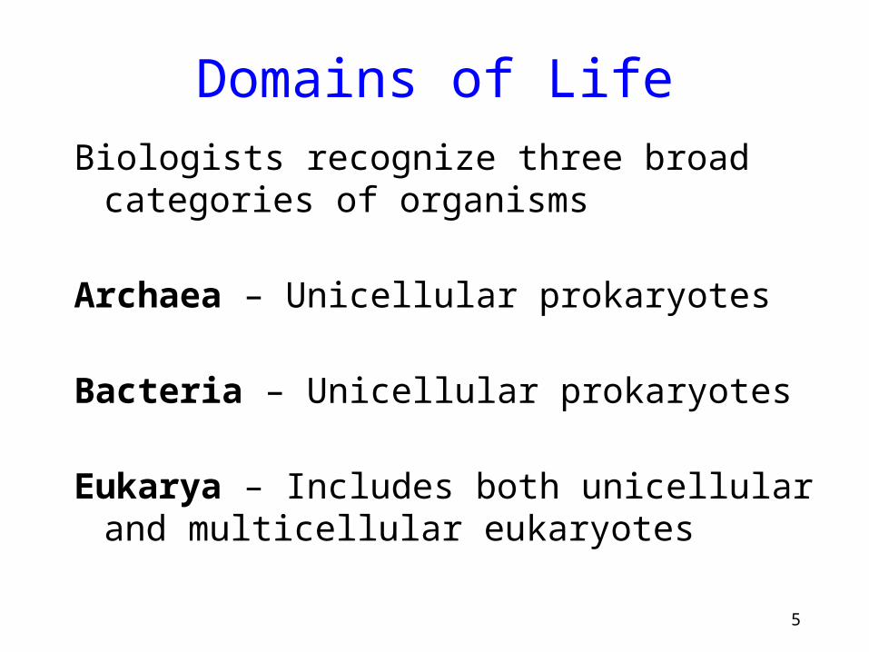
Domains of Life
Biologists recognize three broad categories of organisms
Archaea – Unicellular prokaryotes
Bacteria – Unicellular prokaryotes
Eukarya – Includes both unicellular and multicellular eukaryotes

6
Chemical ConstituentsCells contain four types of macromolecules
Type Examples Functions
Carbohydrates Sugars, starches Energy, structure
Lipids Fats, oils Membranes, hormones
Proteins Myosin, collagen Enzymes, structure
Nucleic Acids DNA, RNA Genetic information
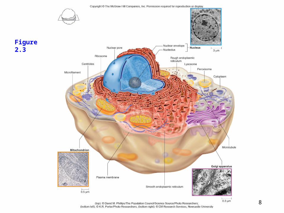
7Figure 2.3
An Animal Cell
Surrounded by the plasma membrane
Contains:
- Cytoplasm
- Organelles
- Divide labor by partitioning certain areas or serving specific functions
8
An Animal Cell
Figure 2.3
Figure 2.3
9Figure 2.3
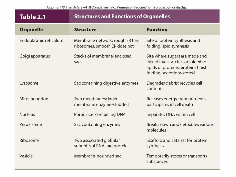
The Nucleus
The largest structure in a cell
Surrounded by a double-layered nuclear envelope
Contains:
- Nuclear pores that allow movement of some molecules in and out
- Nucleolus, which is the site of ribosome production
- Chromosomes composed of DNA and proteins

10
Figure 2.3
Figure 2.4
The Nucleus
Figure 2.4

11
Secretion illustrates how organelles function together to coordinate the basic functions of life
Figure 2.5

12Figure 2.3
Endoplasmic Reticulum (ER)
Interconnected membranous tubules & sacs
Winds from the nuclear envelope to the plasma membrane
Rough ER contains ribosomes and is involved in protein synthesis
Smooth ER does not contain ribosomes and is important in lipid synthesis

13Figure 2.3
Golgi Apparatus
Stack of flat membrane-enclosed sacs
Processing center that adds sugars forming glycoproteins and glycolipids
Site of final protein folding
Products are released into vesicles that bud off to the plasma membrane

14Figure 2.3
LysosomesMembrane-bound sacs
containing > 40 types of digestive enzymes
Break down bacteria, cellular debris, and nutrients
Tay-Sachs is an inherited lysosomal storage disorder Figure 2.6

15Figure 2.3
Peroxisomes
Sacs with outer membranes studded with several types of enzymes
Break down lipids, rare biochemicals
Synthesize bile acids
Detoxify compounds from free radicals
Abundant in liver and kidney cells

16Figure 2.3
MitochondriaSurrounded by two
membranes
Site of ATP (energy) production
Contain their own circular DNA
Human mitochondrial DNA is inherited only from the mother
Figure 2.7
17
Structures and Functions of Organelles
Table 2.1

18Figure 2.3
Plasma Membrane
Forms a selective barrier
A phospholipid bilayer
- Phosphate end (hydrophilic)
- Fatty acid chains (hydrophobic)
Figure 2.8

19Figure 2.3
Plasma Membrane
Contains proteins, glycoproteins, and glycolipids
- Important to cell function and interactions
- May be receptors
- Form channels for ions
Figure 2.9

20Figure 2.3
Faulty Ion Channels Cause Inherited Diseases
Sodium channels
- Mutations lead to absence or extreme pain
Potassium channels
- Mutations lead to impaired heart function and deafness
Chloride channels
- Mutations lead to cystic fibrosis

21Figure 2.3
Cytoskeleton
A meshwork of protein rods and tubules
Includes three major types of proteins
- Microtubules
- Microfilaments
- Intermediate filaments
Figure 2.10

22Figure 2.3
Cytoskeleton Functions
Maintain cell shape
Connect cells to each other
Transport organelles and small molecules
Provide cell motility (some cell types)
Move chromosomes in cell division
Compose cilia

23Figure 2.11

24Figure 2.12

25Figure 2.3
Cell Division and Death
Normal growth and development require an intricate interplay between the rates of two processes
Mitosis – Cell division
- Produces two somatic cells from one
Apoptosis – Cell death
- Precise genetically-programmed sequence

26Figure 2.12
Figure 2.13
27Figure 2.3
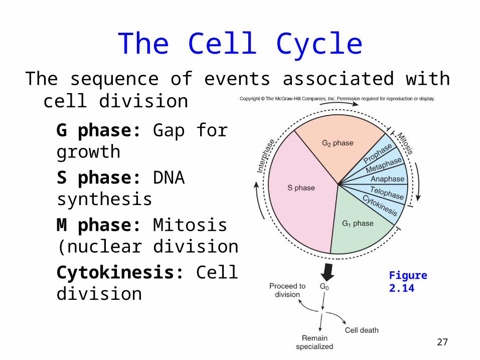
The Cell CycleThe sequence of events associated with cell division
G phase: Gap for growth
S phase: DNA synthesis
M phase: Mitosis (nuclear division)
Cytokinesis: Cell division
Figure 2.14

28Figure 2.3
Stages of the Cell Cycle
Interphase
- Prepares for cell division
- Replicates DNA and subcellular structures
- Composed of G1, S, and G2
- Cells may exit the cell cycle at G1 or enter G0, a quiescent phase
Mitosis – Division of the nucleus
Cytokinesis – Division of the cytoplasm

29Figure 2.3
Replication of Chromosomes
Chromosomes are replicated during S phase prior to mitosis
The result is two sister chromatids held together at the centromere
Figure 2.15
30Figure 2.3
MitosisUsed for growth, repair, and replacement
Consists of a single division that produces two identical daughter cells
A continuous process divided into 4 phases
- Prophase
- Metaphase
- Anaphase
- Telophase

31
Figure 2.15
Figure 2.16
Mitosis in a Human Cell
32Figure 2.3
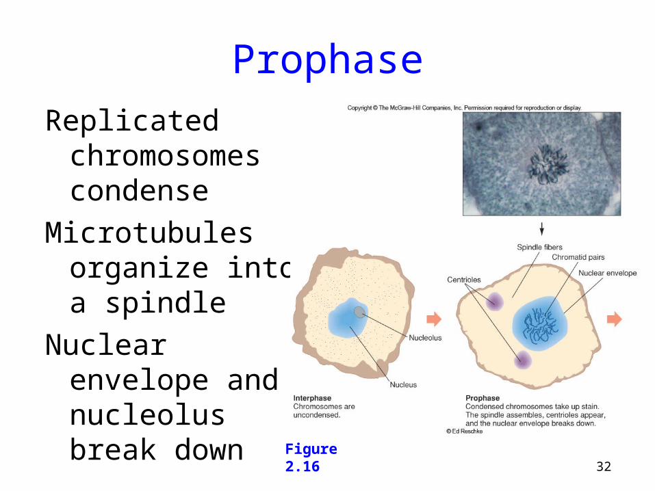
Prophase
Replicated chromosomes condense
Microtubules organize into a spindle
Nuclear envelope and nucleolus break down
Figure 2.16
33Figure 2.3
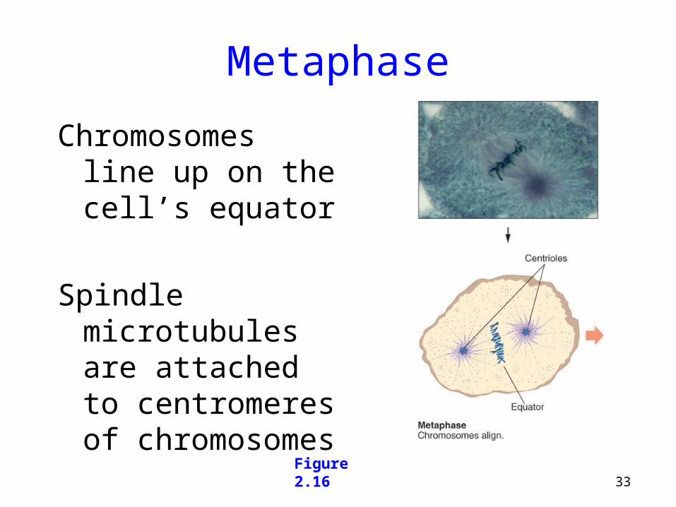
Metaphase
Chromosomes line up on the cell’s equator
Spindle microtubules are attached to centromeres of chromosomes
Figure 2.16

34Figure 2.3
Anaphase
Centromeres divide
Chromatids separate and become independent chromosomes
- They move to opposite ends of the cell
Figure 2.16
35Figure 2.3
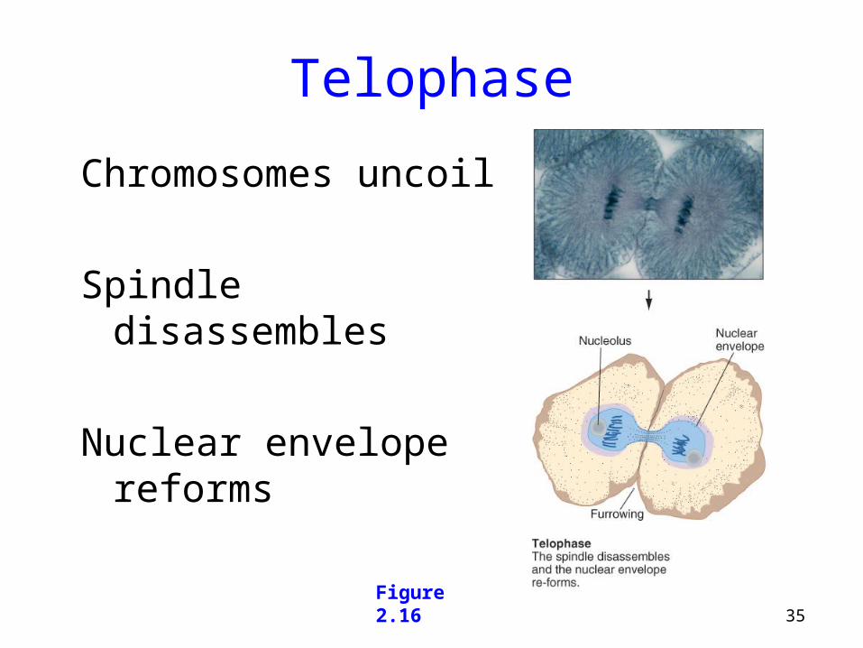
Telophase
Chromosomes uncoil
Spindle disassembles
Nuclear envelope reforms
Figure 2.16

36Figure 2.3
Cytokinesis
Cytoplasmic division occurs after nuclear division is complete
Organelles and macromolecules are distributed between the two daughter cells
Microfilament band contracts, separating the two cells

37Figure 2.3
Cell Cycle Control
Checkpoints ensure that mitotic events occur in the correct sequence
Internal and external factors are involved
Many types of cancer result from faulty checkpoints

38
Figure 2.16
Cell Cycle Control
Figure 2.17
39Figure 2.3
TelomeresLocated at the ends of the chromosomes
Contain hundreds to thousands of repeats of a 6-base DNA sequence
Most cells lose 50-200 endmost bases after each cell division
After about 50 divisions, shortened telomeres signal the cell to stop dividing
Sperm, eggs, bone marrow, and cancer cells produce telomerase that prevent shortening of telomeres

40Figure 2.18
41Figure 2.3
Apoptosis
Begins when a cell receives a “death signal”
Killer enzymes called caspases are activated
-Destroy cellular components
Phagocytes digest the remains
Dying cell forms bulges called blebs

42
Programmed cell death is part of normal development
Figure 2.18
Mitosis and apotosis work together to form functional body
Cancer can result from too much mitosis, too little apotosis
Figure 2.19

43Figure 2.3
Cell-to-Cell Interactions
Make multicellular life possible
Two broad types
1) Signal transduction
2) Cellular adhesion
Defects cause certain inherited disorders

44Figure 2.3
Signal Transduction
The process of transmitting a signal from the environment to a cell
- Receptor binds to “first messenger”
- Interacts with regulator
- Causes enzyme to produce “second messenger”
- Elicits cellular response, which is typically enzyme activation
- Amplification due to cascade

45
Figure 2.19
Signal Transduction
Figure 2.20

46Figure 2.3
Cellular Adhesion
A precise sequence of interactions among proteins that connect cells
Example = Inflammation
- Three types of cellular adhesion molecules (CAMs) help guide WBCs to the injured area
- Secretins, integrins, and adhesion receptor proteins

47Figure 2.20
Cellular Adhesion
Figure 2.21
48Figure 2.3
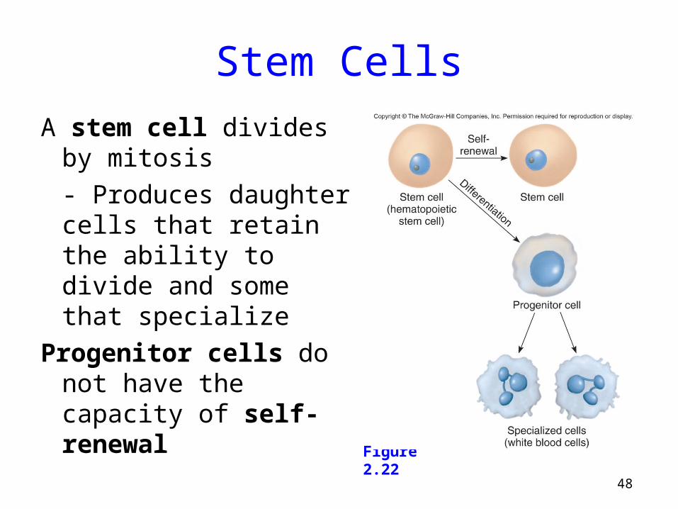
Stem Cells
A stem cell divides by mitosis
- Produces daughter cells that retain the ability to divide and some that specialize
Progenitor cells do not have the capacity of self-renewal
Figure 2.22

49Figure 2.3
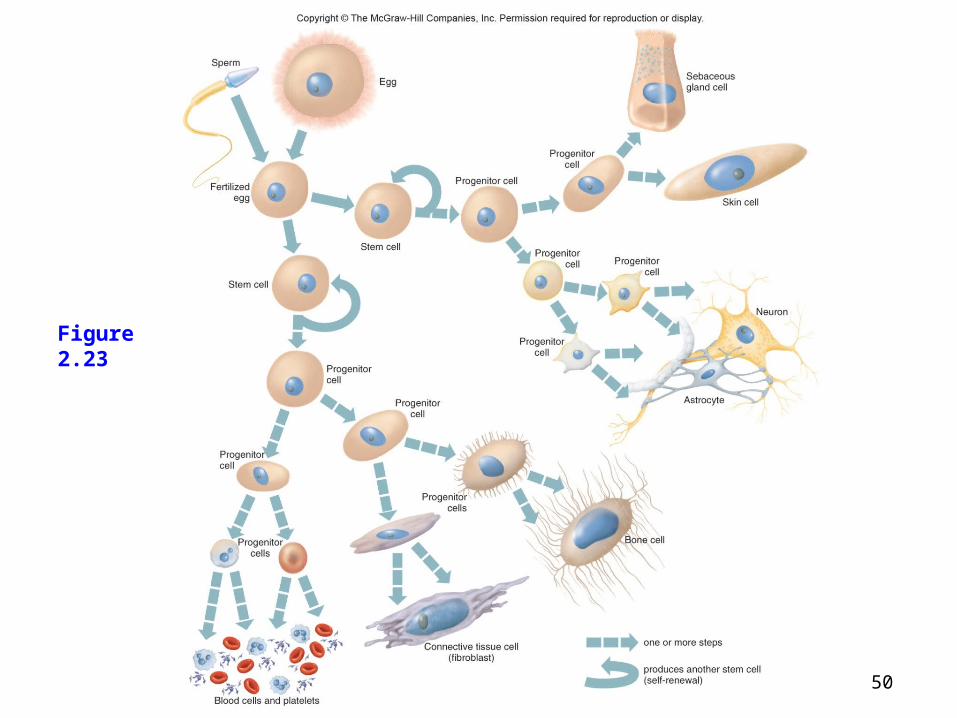
Stem Cells
All cells in the human body descend from stem cells via mitosis and differentiation
Cells differentiate down cell lineages by differential gene expression
Stem cells are present throughout life and provide growth and repair
50Figure 2.3
Figure 2.23

51Figure 2.3
Stem Cells
Stem cells and progenitor cells are described in terms of their developmental potential
Totipotent – Can give rise to every cell type
Pluripotent – Have fewer possible fates
Multipotent – Have only a few fates

52Figure 2.3
Stem Cells in Health Care
There are 3 general sources of human stem cells
1) Embryonic stem cells – Created in a lab dish using the inner cell mass (ICM) of an embryo
2) Induced pluripotent stem (iPS) cells – Somatic cells reprogrammed to differentiate into any of several cell types
3) Adult stem cells – Tissue-specific or somatic stem cells

53Figure 2.24
Stem Cells in Health Care
Figure 2.24
54Figure 2.3
Stem Cell Applications
Stem cells are being used in four basic ways
1) Discovery and development of drugs
2) Observing the earliest sign of disease
3) Treatment of disease via implants and transplants
4) Stimulating stem cells in the body via the introduction of reprogramming proteins

55Figure 2.3
Stem Cell Applications
Figure 2.25