Embed Size (px)
Citation preview

Human Cytomegalovirus Induces TGF-b1 Activation inRenal Tubular Epithelial Cells after Epithelial-to-Mesenchymal TransitionMasako Shimamura1*, Joanne E. Murphy-Ullrich2, William J. Britt1,3
1 Department of Pediatrics, University of Alabama at Birmingham, Birmingham, Alabama, United States of America, 2 Department of Pathology, University of Alabama at
Birmingham, Birmingham, Alabama, United States of America, 3 Department of Microbiology, University of Alabama at Birmingham, Birmingham, Alabama, United States
of America
Abstract
Human cytomegalovirus (HCMV) infection is associated epidemiologically with poor outcome of renal allografts due tomechanisms which remain largely undefined. Transforming growth factor-b1 (TGF-b1), a potent fibrogenic cytokine, is moreabundant in rejecting renal allografts that are infected with either HCMV or rat CMV as compared to uninfected, rejectinggrafts. TGF-b1 induces renal fibrosis via epithelial-to-mesenchymal transition (EMT) of renal epithelial cells, a process bywhich epithelial cells acquire mesenchymal characteristics and a migratory phenotype, and secrete molecules associatedwith extracellular matrix deposition and remodeling. We report that human renal tubular epithelial cells infected in vitrowith HCMV and exposed to TGF-b1 underwent morphologic and transcriptional changes of EMT, similar to uninfected cells.HCMV infected cells after EMT also activated extracellular latent TGF-b1 via induction of MMP-2. Renal epithelial cellstransiently transfected with only the HCMV IE1 or IE2 open reading frames and stimulated to undergo EMT also inducedTGF-b1 activation associated with MMP-2 production, suggesting a role for these viral gene products in MMP-2 production.Consistent with the function of these immediate early gene products, the antiviral agents ganciclovir and foscarnet did notinhibit TGF-b1 production after EMT by HCMV infected cells. These results indicate that HCMV infected renal tubularepithelial cells can undergo EMT after exposure to TGF-b1, similar to uninfected renal epithelial cells, but that HCMVinfection by inducing active TGF-b1 may potentiate renal fibrosis. Our findings provide in vitro evidence for a pathogenicmechanism that could explain the clinical association between HCMV infection, TGF-b1, and adverse renal allograftoutcome.
Citation: Shimamura M, Murphy-Ullrich JE, Britt WJ (2010) Human Cytomegalovirus Induces TGF-b1 Activation in Renal Tubular Epithelial Cells after Epithelial-to-Mesenchymal Transition. PLoS Pathog 6(11): e1001170. doi:10.1371/journal.ppat.1001170
Editor: Jay A. Nelson, Oregon Health and Science University, United States of America
Received December 16, 2009; Accepted September 29, 2010; Published November 4, 2010
Copyright: � 2010 Shimamura et al. This is an open-access article distributed under the terms of the Creative Commons Attribution License, which permitsunrestricted use, distribution, and reproduction in any medium, provided the original author and source are credited.
Funding: This work was supported by NIH 5K08AI059428 (M.S.), the Children’s Center for Research and Innovation of the Alabama Children’s HospitalFoundation (M.S.), and the Kaul Pediatric Research Initiative of The Children’s Hospital of Alabama (M.S.). The funders had no role in study design, data collectionand analysis, decision to publish, or preparation of the manuscript.
Competing Interests: The authors have declared that no competing interests exist.
* E-mail: [email protected]
Introduction
Human cytomegalovirus (HCMV) has been associated with
poor renal allograft outcome in numerous seroepidemiologic
studies [1,2,3,4]. Evidence of active CMV infection (DNAemia,
antigenemia) or CMV disease in renal transplant recipients is also
associated with poor graft outcome [5]. In a rat renal allograft
model, infection with rat CMV accelerates and intensifies rejection
in infected allografts as compared to uninfected allografts [6,7,8].
These studies support an association between HCMV and adverse
renal allograft outcome, but the mechanisms by which HCMV
contributes to renal allograft loss remain cryptic.
The fibrogenic cytokine transforming growth factor-b1 (TGF-
b1) is present in biopsy specimens of human renal allografts
undergoing rejection [9,10,11]. TGF-b1 is produced by infiltrating
leukocytes during rejection and may also be produced by renal
tubular epithelium [12,13,14]. TGF-b1 is expressed at higher
levels in HCMV infected renal allografts compared to uninfected
allografts [15]. In a rat renal transplantation model, allografts from
rat CMV infected animals also contain greater quantities of TGF-b1
as compared to uninfected allografts [6,16,17]. TGF-b1 contributes
to renal fibrosis in numerous animal models and in human fibrotic
renal disease, by inducing epithelial-to-mesenchymal transition
(EMT) of renal tubular epithelial cells [18,19,20,21]. During
EMT, renal tubular cells demonstrate loss of epithelial characteristics
and cellular adhesions, develop changes in the actin cytoskeleton,
induce expression of fibrogenic molecules, and acquire a migratory
phenotype [22]. These fibroblastoid renal tubular cells are key
contributors to renal fibrosis, as inhibition of TGF-b1 mediated
EMT prevents and reverses experimentally induced renal fibrosis in
animal models [22,23,24]. The association between CMV and
TGF-b1 in renal allografts raises the possibility that CMV might
accelerate renal allograft loss via viral induction of TGF-b1 with
resultant fibrosis within the allograft.
Studies performed in vitro have shown that CMV induces
secretion of TGF-b1 from infected fibroblasts, astrocytes, and
osteosarcoma cells [25,26,27]. TGF-b1 production can also be
induced by transient transfection of expression plasmids contain-
ing the HCMV immediate early 1 and 2 (IE1, IE2) genes into
fibroblasts and astrocytoma cells [25,28]. In those studies,
PLoS Pathogens | www.plospathogens.org 1 November 2010 | Volume 6 | Issue 11 | e1001170

increases in TGF-b1 were associated with induction of TGF-b1
mRNA. However, the local effects of TGF-b1 are often controlled
in vivo by activation of the extracellular latent form [29]. Known
activators of latent TGF-b1 include proteases (plasmin), matrix
metalloproteases (MMPs), thrombospondin-1 (TSP-1), and the
avb6 and avb8 integrins [30]. In the HCMV infected placenta,
HCMV infected endothelial cells have been shown to induce
production of TGF-b1 and collagen IV via induction of avb6
integrin [31]. Thus, precedent exists for the possibility that
HCMV infected renal cells might induce TGF-b1 production or
activation in pathological settings.
HCMV can infect renal tubular epithelial cells. HCMV
antigens and DNA are found in renal epithelial cells in kidneys
of trauma victims examined during autopsy as well as in biopsies of
renal allografts, indicating that these cells can harbor HCMV in
both healthy persons and allograft recipients [32,33]. HCMV
antigens have also been detected in tubular cells of biopsies from
HCMV seropositive patients with rejection [34]. Furthermore,
HCMV has been detected more often in renal tubular epithelium
of allograft biopsies with rejection compared to those without
rejection using both immunohistochemistry and in situ hybridiza-
tion [35,36].
Based on the epidemiologic data associating HCMV infection
with long-term allograft loss, histologic evidence that TGF-b1
production is increased in HCMV infected renal allografts, and in
vitro data supporting HCMV induction of TGF-b1 production by
fibroblasts and other cells, we hypothesized that HCMV infected
renal tubular epithelium undergoing EMT might develop a
fibroblast-like phenotype with secretion of TGF-b1. In the
following studies, we demonstrate that HCMV infected renal
tubular epithelial cells undergo EMT and thereafter induce active
TGF-b1 production. In this system, MMP-2 mediates the
extracellular activation of TGF-b1 in HCMV infected cells, and
can be recapitulated by transient transfection of the HCMV IE1
or IE2 open reading frames into renal epithelial cells stimulated to
undergo EMT. These data provide supportive evidence that
HCMV infected renal epithelium may contribute to long-term
renal allograft loss by exacerbating fibrosis via TGF-b1, and
provides a potential in vitro mechanism for the association of
HCMV infection with greater TGF-b1 production in renal
allografts and long-term allograft loss.
Results
HCMV infects renal tubular epitheliumPrimary human foreskin fibroblasts and the immortalized
human proximal renal tubular epithelial cell line, HK-2, were
infected in parallel with HCMV strain TR at an MOI of 1. Some
HK-2 cultures were treated with recombinant active (ra) TGF-b1
at 15 ng/ml, starting 1 hour post-infection. Culture media and
cells were harvested separately on days 1-6 post-infection and viral
titers determined by the detection of early antigen fluorescent foci
(DEAFF) assay. Separate cell cultures were infected and harvested
at day 5 post-infection, and cellular lysates subjected to western
blotting using a high titered human CMV immune globulin
(Cytogam).
Fibroblasts supported productive infection with logarithmic
increases in viral progeny observed in both media and cells
(Figure 1A). HK-2 cells also supported productive infection
(Figure 1B) but the kinetic was linear and the virions remained
cell-associated, with very few virions detectable in media. By day 3
post-infection, virus was not detectable in the media. No difference
in viral growth kinetics was observed in the HK-2 cultures treated
with TGF-b1 (Figure 1C), compared to HK-2 cultures without
TGF-b1. Viral titers in HK-2 cells and media were similar at each
time point post infection, in the presence and absence of TGF-b1.
This result differs from TGF-b1 effects upon HFFs, where HCMV
replication is induced by exposure to TGF-b1 [37]. Western
blotting of the infected cell lysates (Figure 1, insets) indicates a
similar pattern of infected cell viral proteins in both HFFs and
HK-2 cells, consistent with productive infection in both cell types.
Viral replication studies were repeated in primary renal epithelial
cells and results similar to those in HK-2 cells were observed, with
the majority of the replicating virus remaining cell-associated
(Figure S1).
HCMV infected primary renal tubular epithelial cells andHK-2 cells undergo TGF-b1 induced EMT in vitro
We next investigated whether HCMV infected renal tubular
cells could undergo EMT similar to uninfected cells, or whether
infection caused cells to remain epithelioid in the presence of
TGF-b1. HK-2 cells were infected with HCMV strain TR,
followed by incubation with raTGF-b1 for 48 hours as an inducer
of EMT. Cells were fixed, permeabilized, and stained using an
anti-HCMV IE1 antibody (p63-27) and species-specific Alexa-
Fluor 488 conjugated secondary antibody, and the AlexaFluor 594
conjugated phalloidin (Figure 2A). Imaging by confocal micros-
copy demonstrated that HCMV infected renal tubular epithelial
cells exhibited cuboidal structural actin cytoskeleton, similar to
uninfected cells (Figure 2A, left column). The HCMV infected
renal epithelial cells appeared morphologically indistinguishable
from uninfected cells and could only be distinguished by
immunostaining for HCMV IE1. After raTGF-b1 stimulation,
HCMV infected epithelial cells showed changes in actin
cytoskeleton, developing an elongated mesenchymal phenotype
associated with parallel actin stress fiber formation and loss of
cuboidal epithelial morphology. The raTGF-b1 induced cytoskel-
etal changes appeared similar in both HCMV infected and
uninfected renal tubular cells (Figure 2A, right column). Imaging
results were similar using primary renal tubular epithelial cells
Author Summary
Human cytomegalovirus (HCMV) is a common virus thatestablishes lifelong persistence in the host. Althoughasymptomatic in healthy people, HCMV can reactivateand cause disease in immunosuppressed patients, such asthose undergoing kidney transplantation. HCMV infectionis associated with inferior renal allograft survival comparedto transplants without HCMV infection. HCMV infectedallografts also contain higher levels of the fibrogeniccytokine, transforming growth factor-b1 (TGF-b1), com-pared to uninfected allografts. TGF-b1 is a potent inducerof renal fibrosis and causes epithelial-to-mesenchymaltransition (EMT), whereby epithelial cells acquire charac-teristics of cells of mesenchymal origin and expressmolecules associated with fibrosis. Our work shows thatrenal epithelial cells infected in vitro with HCMV canundergo EMT, but that HCMV infected cells producegreater amounts of the fibrogenic molecule TGF-b1,compared to uninfected cells after EMT. We have shownthat this effect is likely due to specific HCMV genes (IE1,IE2), and cannot be prevented by administration ofantivirals such as ganciclovir or foscarnet. These datasuggest that HCMV may contribute to adverse renalallograft outcome by exacerbating TGF-b1 induced renalfibrosis. Understanding such mechanisms will permit thedevelopment of treatments that could improve long-termrenal allograft survival in HCMV infected patients.
CMV Infected Renal Cells Induce TGF-b1 Activation
PLoS Pathogens | www.plospathogens.org 2 November 2010 | Volume 6 | Issue 11 | e1001170

(data not shown [dns]). These results showed that HCMV infected
renal tubular cells can undergo morphologic changes consistent
with EMT, similar to those described for uninfected cells [22].
In the next series of experiments, primary renal tubular cells
were left untreated, or were infected with HCMV strain TR,
followed by incubation with or without raTGF-b1 for 48 hours.
This permitted analysis of effects of HCMV alone, TGF-b1 alone,
or the combination of HCMV and TGF-b1 upon renal tubular
cells. Cells were lysed and equivalent protein lysates were assayed
for expression of HCMV IE1, E-cadherin, vimentin, or actin by
immuno-blotting (Figure 2B). At baseline, both HCMV uninfected
and infected cells expressed E-cadherin but not vimentin,
consistent with an epithelial phenotype. Following raTGF-b1
stimulation, E-cadherin was no longer present, and vimentin was
induced by both HCMV uninfected and infected cells, consistent
with a mesenchymal phenotype. Immunoblotting for phosphory-
lated SMAD2 (p-SMAD2) in nuclear extracts revealed the
presence of p-SMAD2 in nuclei of raTGF-b1 treated cells, both
in the presence and absence of HCMV infection, whereas cells
that were not treated with raTGF-b1 did not contain nuclear p-
SMAD2. Again, this result was consistent with the induction of
TGF-b1 dependent SMAD signaling in HCMV infected cells.
HCMV infected cells expressed IE1 as expected, and actin
staining showed equivalent protein loading for all samples. Taken
together, these results were consistent with findings from imaging
studies (Figure 2A) and indicated that HCMV infected renal
tubular epithelial cells could undergo loss of E-cadherin expres-
sion, acquisition of vimentin expression, and SMAD2 phosphor-
ylation in the presence of raTGF-b1. The phenotypic changes
were consistent with changes indicative of EMT in uninfected
renal tubular epithelium [22,38].
To characterize the phenotype of HCMV infected HK-2 cells
before and after EMT, HK-2 cells were examined at baseline, or
after HCMV infection, in the presence or absence of raTGF-b1.
Thus, effects of HCMV alone, TGF-b1 alone, or HCMV and
TGF-b1 together could be compared to HK-2 cells at baseline.
RNA was extracted from cell lysates, and RT-PCR performed
using a commercial PCR array to detect transcripts associated
with extracellular matrix molecules. Transcripts from cells under
various experimental conditions were compared to those produced
at baseline by uninfected, unstimulated HK-2 cells. Compared
to uninfected cells, cells infected with HCMV showed less than
10-fold induction of many mRNA transcripts encoding fibrogenic
matrix proteins represented in this array (Figure 2C, grey bars).
This is consistent with the light microscopic appearance of HK-2
cells infected with HCMV, which appeared epithelioid and
morphologically indistinguishable from adjacent uninfected HK-
2 cells (Figure 2A), and confirmed that, in the absence of raTGF-
b1 stimulation, HCMV infected HK-2 cells did not induce global
transcriptional changes suggestive of EMT.
In contrast, after exposure to raTGF-b1, transcriptional up-
regulation of a number of fibrogenic molecules was observed,
indicating induction of EMT in both HCMV uninfected and
infected cells (Figure 2C). Some transcripts, such as for fibronectin,
MMP-9, and the av integrin, were highly upregulated in both
uninfected (hatched bars) and HCMV infected (black bars) cells
stimulated with raTGF-b1. A few transcripts, such as for TIMP-2,
were induced to higher levels in HCMV uninfected cells compared
to HCMV infected cells after raTGF-b1 stimulation. Transcripts
encoding many fibrogenic molecules (ADAMTS1, TGF-b1, b-
catenin, collagens, MMPs, TIMP-1, thrombospondins) were
induced to greater levels in HCMV infected cells as compared
to uninfected cells after EMT. These results suggested that HCMV
infected renal tubular cells expressed mRNA transcripts of
numerous fibrogenic proteins at similar or higher levels than
uninfected renal tubular cells after induction of EMT with raTGF-
b1. These results indicated that HCMV infected cells were capable
of exhibiting the fibrogenic phenotype of EMT, and that HCMV
infection does not reduce or prevent the EMT phenotype in these
cells. To confirm the results from those studies using PCR array,
individual RT-PCR assays were repeated in separate experiments
using both HK-2 cells and primary renal tubular epithelial cells,
utilizing commercial primer-probe sets for several of the mRNAs
(Figure 2C, asterisks) that were upregulated in HCMV infected
HK-2 cells after EMT (fibronectin, MMP-9, ADAMTS1, TGF-
b1, collagen 5A1, MMP-2, and thrombospondin-1). These
individual PCR assays validated the upregulation of these mRNA
transcripts observed in the PCR array (Figure S2). The mRNAs
for MMP-9 and ADAMTS1 showed a lesser log induction in the
individual assays compared to the PCR array (103–104 fold
induction vs. 106–07 fold induction); however, the individual assays
did confirm that these mRNA transcripts were highly upregulated
after TGF-b1 stimulation, consistent with the array results, in both
HK-2 cells and primary PTECs (Figure S2).
Figure 1. HCMV replication in HFFs and HK-2 cells. HFF (A) and HK-2 cells (B) were infected with HCMV strain TR at MOI of 1, cells and mediaharvested daily, and viral titers determined by DEAFF assay. A parallel set of HK-2 cells were infected with HCMV and exposed to recombinant activeTGF-b1 (C), and cells and media analyzed as for HK-2 cells. A separate set of cells were uninfected or infected with HCMV strain TR at MOI of 1, cellpellets harvested at day 5 post-infection, and western blotting for viral proteins performed using HCMV hyperimmune globulin (insets). HFFssupported logarithmic viral replication with viral progeny in both cells and media, whereas HK-2 supported linear productive infection in cells only.Upright triangles, cells; inverted triangles, media.doi:10.1371/journal.ppat.1001170.g001
CMV Infected Renal Cells Induce TGF-b1 Activation
PLoS Pathogens | www.plospathogens.org 3 November 2010 | Volume 6 | Issue 11 | e1001170

Taken together, these results demonstrate by morphologic and
phenotypic assays that HCMV infected cells can undergo EMT
similar to uninfected cells. HCMV infection does not prevent or
diminish the EMT phenotype as compared to uninfected cells.
HCMV infected renal tubular epithelial cells induce activeTGF-b1 production after EMT
Because of the association of TGF-b1 with HCMV infection in
renal allografts, we next explored whether HCMV infected HK-2
cells could produce TGF-b1. HK-2 cells were untreated, or
infected with HCMV strain TR at an MOI of 1 and/or stimulated
with raTGF-b1 for 48 hours to induce EMT. Cells were washed
extensively to remove exogenous TGF-b1, fresh media applied
and samples harvested at 24 hours after washing. Samples were
assayed for active or total (active + latent) TGF-b1 activity using a
published luciferase reporter bioassay (Figure 3A, B) and a
commercial human TGF-b1 ELISA (Figure 3E). Because all
exogenous raTGF-b1 was washed from the cultures and only fresh
media not containing exogenous TGF-b1 was assayed, the TGF-
b1 observed in samples was presumed to be derived from the
cultured cells. No TGF-b1 was detectable in the serum-free media
used for HK-2 cell culture by luciferase bioassay or ELISA (dns).
Figure 2. HCMV infected renal tubular epithelial cells undergo epithelial-to-mesenchymal transition (EMT) after TGF-b1 exposure.(A) Primary human renal tubular epithelial cells were uninfected (top row) or infected with HCMV strain TR (bottom row), without (left column) orwith recombinant human active TGF-b1 (raTGF-b1) to induce EMT (right column). Cells were stained using a monoclonal antibody against HCMV IE1(mab63-27) and an isotype-specific AlexaFluor 488-conjugated secondary antibody (green nuclei), and co-stained with AlexaFluor 594-conjugatedphalloidin (red) and Topro3 (blue nuclei) nuclear stain. Images were collected by confocal microscopy using similar exposure time and identical gain.Cells at baseline had epithelioid morphology with concentric structural actin cytoskeleton, both without (top left) and with (bottom left) HCMVinfection, whereas cells after raTGF-b1 stimulation showed elongated mesenchymal morphology indicative of EMT in both HCMV uninfected (topright) and infected (bottom right) cells. (B) Primary human renal tubular epithelial cells were untreated, or were infected with HCMV strain TR at MOIof 1 and/or treated with raTGF-b1, lysed, and subjected to western blotting using antibodies against HCMV IE1, e-cadherin, vimentin, or actin. Nuclearextracts from cellular lysates were also subjected to western blotting for phospho-SMAD2. Both uninfected (IE1 negative) and HCMV infected (IE1positive) cells expressed E-cadherin but not vimentin at baseline, but after raTGF-b1 stimulation, both uninfected and infected cells lost E-cadherinexpression and demonstrated both vimentin expression and SMAD2 phosphorylation, indicative of EMT in both. (C) HK-2 cells were untreated, orwere infected with HCMV strain TR at MOI of 1 and/or stimulated with raTGF-b1 to induce EMT, lysed, total RNA reverse transcribed to cDNA, andcDNA analyzed for presence of extracellular matrix associated mRNAs using the SuperArray extracellular matrix PCR array. Results were normalized toGAPDH expression and quantitated as fold-change compared to mRNA levels in uninfected, unstimulated HK-2 cells. HCMV infected cells (grey bars)induced some mRNA transcripts of fibrogenic molecules, but only at less than 10-fold induction. Both uninfected cells stimulated with raTGF-b1(hatched bars) and HCMV infected cells stimulated with raTGF-b1 (black bars) demonstrated induction of many fibrogenic molecules represented inthis array, consistent with induction of EMT in both uninfected and HCMV infected cells. Transcripts upregulated in HCMV infected HK-2 cells afterraTGF-b1 stimulation (denoted with asterisks) were confirmed by individual RT-PCR assays using both HK-2 cells and primary renal tubular epithelialcells (Figure S2).doi:10.1371/journal.ppat.1001170.g002
CMV Infected Renal Cells Induce TGF-b1 Activation
PLoS Pathogens | www.plospathogens.org 4 November 2010 | Volume 6 | Issue 11 | e1001170

At baseline, non-infected HK-2 cells produced some detectable
active TGF-b1 (HCMV TR-/raTGF-b1-) (Figure 3A). HCMV
infected HK-2 cells (HCMV TR+/raTGF-b1-) also produced
similar amounts of active TGF-b1 compared to uninfected cells,
indicating that HCMV infection alone did not induce de novo TGF-
b1 production in these epithelial cells (Figure 3A). This result
differed from results shown by others for HCMV infected
fibroblasts and astrocytes, where HCMV infection induced
TGF-b1 production, and may reflect biological differences
between infection of those cell types as compared to renal
epithelial cells and possibly the strain of virus utilized in these
previous studies [25,26]. Uninfected HK-2 cells stimulated with
raTGF-b1 (HCMV TR-/raTGF-b1+) also produced similar levels
of active TGF-b1 compared to both infected and uninfected cells
Figure 3. HCMV infected renal tubular epithelial cells induce active TGF-b1 production after EMT. Immortalized renal tubular cells, HK-2(A, B), or primary human renal tubular epithelial cells (C, D) were untreated, or were infected with HCMV strain TR (HCMV TR 2/+) and/or treated withraTGF-b1 (raTGF-b1 2/+) at 15 ng/ml (0.6 nM) to induce EMT. Cells were washed 3 times and re-incubated in fresh media not containing raTGF-b1.Supernatants were assayed for de novo active and total TGF-b1 production using a TGF-b1 responsive luciferase bioassay. Only the HCMV infectedcells stimulated with raTGF-b1 induced production of active TGF-b1 in both HK-2 and primary cells. (E) HK-2 cells were infected with HCMV strain TRat MOI of 1 and/or stimulated with raTGF-b1 at 15 ng/ml as described, and supernatants were assayed using the Quantikine ELISA. Only the HCMVinfected, raTGF-b1 stimulated cells produced detectable active TGF-b1 in this assay. (F) HK-2 cells were infected with HCMV strain TR at MOI of 1 and/or stimulated with raTGF-b1 at 15 ng/ml as described, and a blocking antibody against TGF-b1 was added to cell cultures simultaneously with raTGF-b1. After 48 hours, cells were washed and re-incubated in fresh media, and luciferase assay for active TGF-b1 performed. The blocking antibodyreduced TGF-b1 activation in a dose-dependent manner. Legend: (*) p.0.05, ns; (***) p,0.01.doi:10.1371/journal.ppat.1001170.g003
CMV Infected Renal Cells Induce TGF-b1 Activation
PLoS Pathogens | www.plospathogens.org 5 November 2010 | Volume 6 | Issue 11 | e1001170
not exposed to raTGF-b1, indicating that EMT alone did not
induce HK-2 cells to produce additional amounts of active TGF-
b1 (Figure 3A). This result also confirmed that the exogenous
raTGF-b1, used to induce EMT, did not carry over in detectable
quantities to our assay for active TGF-b1. In contrast, HCMV
infected HK-2 cells stimulated with raTGF-b1 (HCMV TR+/
raTGF-b1+) produced significantly more active TGF-b1 (p#0.01)
than did cells in other conditions (Figure 3A). The quantity of total
TGF-b1 was similar in all conditions (Figure 3B), indicating that
changes in active TGF-b1 did not derive from an increase in latent
TGF-b1. A commercial ELISA for TGF-b1 indicated that the
TGF-b detected in the luciferase bioassay was TGF-b1 and not
other TGF-b isoforms, and confirmed the induction of TGF-b1 in
HCMV infected, raTGF-b1 infected cells (Figure 3E). The
quantity of active TGF-b1 protein measured in the ELISA was
approximately 3 log (1000-fold) lower than the quantity of TGF-
b1 activity detected by luciferase assay. These studies were
performed with equivalent volume samples obtained from the
same experimental cultures, confirming that the picomolar
measurements indeed differed between the two assays; however,
significant induction of active TGF-b1 was measured in the
HCMV infected, raTGF-b1 treated cells by both methods. These
studies have not been performed in parallel by other investigators,
and the differing results obtained from assays of the same samples
suggests that the luciferase bioassay may detect the downstream
function of a given quantity of protein more sensitively than the
measurement of protein by antibody binding in the ELISA.
Together, these results suggested that HCMV infection of HK-2
cells, without EMT, did not induce de novo production of active
TGF-b1, whereas the HCMV infected epithelial cells after EMT
appeared to acquire the capacity to produce de novo active TGF-
b1, similar to the phenotype demonstrated by HCMV infected
fibroblasts [25]. Uninfected HK-2 cells, before or after EMT, did
not acquire the capacity to produce de novo active TGF-b1.
These studies were repeated using primary renal proximal
tubular epithelial cells (Figure 3C, D). These cells produced
undetectable active TGF-b1 at baseline (HCMV TR-/raTGF-b1-),
with HCMV infection alone (HCMV TR+/raTGF-b1-), and
produced only a small amount of detectable active TGF-b1 after
raTGF-b1 stimulation (HCMV TR-/raTGF-b1+), consistent with
data derived from studies of HK-2 cells (Figure 3A). Similar to the
HK-2 cells, HCMV infected, raTGF-b1 stimulated primary renal
epithelial cells (HCMV TR+/raTGF-b1+) produced active TGF-
b1 (p,0.01). The total TGF-b1 was similar in all treatment
conditions, consistent with results from HK-2 cells (Figure 3D).
These results indicated that the induction of active TGF-b1
production observed in HCMV infected HK-2 cells after EMT
was not unique to immortalized cells such as HK-2 but was likely a
general phenotype observed for both immortalized and primary
renal tubular epithelial cells after HCMV infection.
Next, HK-2 cells were infected with HCMV at an MOI of 1,
stimulated with raTGF-b1 at 15 ng/ml, and incubated with a
function blocking antibody against TGF-b1 in increasing concen-
trations from 0.3 mg/ml to 3 mg/ml (Figure 3F). Increasing
amounts of anti-TGF-b1 resulted in progressively decreasing
quantities of active TGF-b1 produced. This effect was not due to
the presence of anti-TGF-b1 in the luciferase assay because the
blocking antibody was removed during the final washing, prior to
incubation with fresh media for the luciferase assay. These results
show that blockade of the stimulating dose of raTGF-b1 abrogated
active TGF-b1 production.
To determine whether the TGF-b1 blocking antibody might
block effects of the TGF-b1 produced by HCMV infected cells,
EMT was first induced in uninfected HK-2 cells using raTGF-b1
for 48 hours, after which cells were washed to remove raTGF-b1,
infected with HCMV at MOI of 1, and finally incubated with
either media alone or media containing the TGF-b1 blocking
antibody at 3 mg/ml. After 24 hours, cells were lysed, RNA
extracted and RT-PCR performed for mRNAs induced in the
PCR array. Results from cells incubated with the blocking
antibody are shown as percent reduction compared to cells
incubated with media alone (Figure S3). These results show that
cells, after undergoing EMT and subsequently infected with
HCMV, demonstrate 50–95% reduction of mRNA transcripts of
molecules associated with EMT in the presence of the TGF-b1
blocking antibody, compared to similar cells in the absence of the
TGF-b1 blocking antibody. This result suggests that the TGF-b1
produced by HCMV infected cells after EMT may have activity
upon the producing cells (true autocrine activity), which is
inhibited by the TGF-b1 blocking antibody.
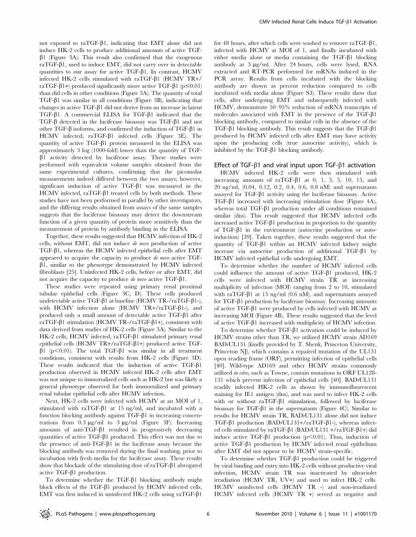
Effect of TGF-b1 and viral input upon TGF-b1 activationHCMV infected HK-2 cells were then stimulated with
increasing amounts of raTGF-b1 at 0, 1, 3, 5, 10, 15, and
20 ng/ml, (0.04, 0.12, 0.2, 0.4, 0.6, 0.8 nM) and supernatants
assayed for TGF-b1 activity using the luciferase bioassay. Active
TGF-b1 increased with increasing stimulation dose (Figure 4A),
whereas total TGF-b1 production under all conditions remained
similar (dns). This result suggested that HCMV infected cells
increased active TGF-b1 production in proportion to the quantity
of TGF-b1 in the environment (autocrine production or auto-
induction) [39]. Taken together, these results suggested that the
quantity of TGF-b1 within an HCMV infected kidney might
increase via autocrine production of additional TGF-b1 by
HCMV infected epithelial cells undergoing EMT.
To determine whether the number of HCMV infected cells
could influence the amount of active TGF-b1 produced, HK-2
cells were infected with HCMV strain TR at increasing
multiplicity of infection (MOI) ranging from 2 to 10, stimulated
with raTGF-b1 at 15 ng/ml (0.6 nM), and supernatants assayed
for TGF-b1 production by luciferase bioassay. Increasing amounts
of active TGF-b1 were produced by cells infected with HCMV at
increasing MOI (Figure 4B). These results suggested that the level
of active TGF-b1 increased with multiplicity of HCMV infection.
To determine whether TGF-b1 activation could be induced by
HCMV strains other than TR, we utilized HCMV strain AD169
BADrUL131 (kindly provided by T. Shenk, Princeton University,
Princeton NJ), which contains a repaired mutation of the UL131
open reading frame (ORF), permitting infection of epithelial cells
[40]. Wild-type AD169 and other HCMV strains commonly
utilized in vitro, such as Towne, contain mutations in ORF UL128-
131 which prevent infection of epithelial cells [40]. BADrUL131
readily infected HK-2 cells as shown by immunofluorescent
staining for IE1 antigen (dns), and was used to infect HK-2 cells
with or without raTGF-b1 stimulation, followed by luciferase
bioassay for TGF-b1 in the supernatants (Figure 4C). Similar to
results for HCMV strain TR, BADrUL131 alone did not induce
TGF-b1 production (BADrUL131+/raTGF-b1-), whereas infect-
ed cells stimulated by raTGF-b1 (BADrUL131 +/raTGF-b1+) did
induce active TGF-b1 production (p,0.01). Thus, induction of
active TGF-b1 production by HCMV infected renal epithelium
after EMT did not appear to be HCMV strain-specific.
To determine whether TGF-b1 production could be triggered
by viral binding and entry into HK-2 cells without productive viral
infection, HCMV strain TR was inactivated by ultraviolet
irradiation (HCMV TR, UV+) and used to infect HK-2 cells.
HCMV uninfected cells (HCMV TR -) and non-irradiated
HCMV infected cells (HCMV TR +) served as negative and
CMV Infected Renal Cells Induce TGF-b1 Activation
PLoS Pathogens | www.plospathogens.org 6 November 2010 | Volume 6 | Issue 11 | e1001170

positive controls, respectively. Under these conditions, irradiated
HCMV did not induce active TGF-b1 production after EMT
(p.0.05), and did not affect total TGF-b1 levels either before or
after EMT (Figure 4D). These results indicated that active TGF-
b1 could not be induced solely by binding and entry of viral
particles into cells undergoing EMT.
A matrix metalloprotease complex is associated withTGF-b1 induction by HCMV infected cells after EMT
To characterize the mechanism by which HCMV infection
might induce active TGF-b1 production, we added known
inhibitors of TGF-b1 activation - aprotinin (serine protease
inhibitor against plasmin), GM6001 (matrix metalloprotease
inhibitor), anti-thrombospondin-1, anti-avb6 integrin - to HCMV
infected HK-2 cells prior to raTGF-b1 stimulation and again after
washing, and the TGF-b1 luciferase bioassay was performed
(Figure 5A). Active TGF-b1 production was reduced by
approximately 60% (p,0.01) in the presence of GM6001. In the
presence of aprotinin, active TGF-b1 production was decreased to
a lesser but still statistically significant amount (19%, p,0.05).
Active TGF-b1 was not significantly decreased in the presence of
the other inhibitors, the blocking antibodies against thrombos-
pondin 1 and the avb6 integrin. This result suggested that MMPs
and serine proteases might be involved in TGF-b1 activation by
HCMV infected epithelial cells after EMT.
Because TGF-b1 is activated by MMP-2, we next analyzed
MMP-2 production in our experimental conditions. Lysates from
HK-2 cells with or without HCMV infection and/or raTGF-b1
stimulation were separated under non-reducing conditions by
gelatin-containing SDS-PAGE and developed for presence of
gelatinase activity. In Figure 5B, a representative zymogram
showed bands at 72 and 62 kDa, consistent with pro- and active
MMP-2, only in lysates from HCMV infected, raTGF-b1
stimulated cells (HCMV TR +/raTGF-b1 +). Identity of these
bands as MMP-2 was confirmed by immunoblotting using anti-
MMP-2 (Figure 5B). This result indicated that only HCMV
infected, raTGF-b1 stimulated cells had cell-associated pro-MMP-2
and active MMP-2 as detected in this assay of functional MMP-2.
Figure 4. Effect of TGF-b1 and viral input upon TGF-b1 activation. (A) HK-2 cells were infected with HCMV strain TR at MOI of 1 andstimulated with increasing doses of raTGF-b1 from 1–20 ng/ml (0.04 nM–0.8 nM), washed, and assayed for de novo TGF-b1 production as described.HCMV infected HK-2 cells induced active TGF-b1 production in proportion to the amount of stimulating raTGF-b1. (B) HK-2 cells were infected withHCMV strain TR at increasing MOI (2-8) with raTGF-b1 stimulation at 15 ng/ml (0.6 nM), and luciferase bioassay performed to quantitate active TGF-b1production. Active TGF-b1 production increased with increasing HCMV MOI. (C) HCMV AD169 strain BADrUL131 at MOI of 1 was used to infect HK-2cells prior to raTGF-b1 stimulation, and TGF-b1 luciferase bioassay performed. Similar to results for HCMV strain TR, HCMV AD169 strain BADrUL131induced active TGF-b1 production after EMT. (D) HCMV strain TR at MOI of 1 was inactivated by UV irradiation (HCMV TR UV+) and used to infect HK-2cells prior to raTGF-b1 stimulation, and active TGF-b1 measured by luciferase bioassay. Irradiated virus failed to induce active TGF-b1 production afterEMT. Legend: (*) p.0.05, ns; (***) p,0.01.doi:10.1371/journal.ppat.1001170.g004
CMV Infected Renal Cells Induce TGF-b1 Activation
PLoS Pathogens | www.plospathogens.org 7 November 2010 | Volume 6 | Issue 11 | e1001170

Since pro-MMP-2 is known to undergo activation in complex
with other MMPs on the cell surface, we next performed
immunoprecipitation experiments using lysates of HK-2 cells with
or without HCMV infection and/or raTGF-b1 stimulation.
Lysates were subjected to western blotting for TIMP-2, MT3-
MMP, and MT1-MMP directly (Figure 5C), or were incubated
with mouse anti-MMP-2, immune complexes collected with
protein A-agarose, and Western blotting of immunoprecipitated
proteins performed using a rabbit anti-MMP-2 antibody, or
antibodies against TIMP-2, MT3-MMP, and MT1-MMP
(Figure 5D). Immunoblotting for MMP-2 in the immunoprecip-
itates showed that MMP-2 was detectable in all samples after
immunoprecipitation (Figure 5D). Immunoblotting of immuno-
precipitated proteins demonstrated the presence of TIMP-2 and
MT3-MMP only in the HCMV infected, raTGF-b1 treated cells
(HCMV TR+/raTGF-b1+) (Figure 5D). MT1-MMP did not
immunoprecipitate with anti-MMP-2 in any samples, and was not
detectable even with membrane overexposure (Figure 5D). These
results suggested that MMP-2 activation by HCMV infected,
raTGF-b1 stimulated cells could occur via formation of a
membrane-associated complex of MT3-MMP, TIMP-2, and
MMP-2 [41].
Figure 5. MMPs are expressed and a MMP complex forms in HCMV infected cells after EMT. (A) HK-2 cells were infected with HCMV strainTR at MOI of 1 and incubated with inhibitors, GM6001, aprotinin, anti-thrombospondin 1(a-TSP), or anti-avb6 integrin (a- avb6), prior to stimulationwith raTGF-b1 at 15 ng/ml (0.6 nM), washed, and TGF-b1 luciferase bioassay performed for active TGF-b1. Results were compared to those fromuninfected, unstimulated HK-2 cells (HCMV TR-/raTGF-b1-) as well as HK-2 cells infected with HCMV and stimulated with raTGF-b1 (HCMV TR+/raTGF-b1+). Both GM6001 and aprotinin significantly inhibited active TGF-b1 production. Legend: (**) p,0.05; (***) p,0.01. (B) HK-2 cells were untreated, orwere infected with HCMV at MOI of 1 and/or treated with raTGF-b1. Cell lysates were subjected to gelatin zymography (zymogram) and westernblotting using anti-MMP-2 (anti-MMP-2). Pro- and active MMP-2 could be detected only in HCMV infected, raTGF-b1 stimulated cells. (C, D) HK-2 cellswere treated as in (A), but lysates were either subjected directly to western blotting for TIMP-2, MT3-MMP, MT1-MMP, or actin (C) or incubated withmouse anti-MMP-2 followed by protein A-agarose, and immunoprecipitated proteins subjected to western blotting using rabbit anti-MMP-2, anti-TIMP-2, anti-MT3-MMP, and anti-MT1-MMP. TIMP-2 and MT3-MMP immunoprecipitated with MMP-2 only in HCMV infected, raTGF-b1 stimulated cells.(E) HK-2 cells were transfected with MMP-2 shRNA plasmid (MMP-2), or a control scrambled plasmid (Ctrl). Cells were infected with HCMV strain TR atMOI of 1 and/or stimulated with raTGF-b1 at 15 ng/ml. Supernatants were subjected to luciferase assay for active TGF-b1 (top panel). A portion of thecell pellets were subjected to western blotting for MMP-2, GFP, and actin (middle panel). RNA was extracted from the remainder of the cell pelletsand RT-PCR performed for MMP-2 (bottom panel), with results depicted as fold change between raTGF-b1 exposed and non-exposed transfections.These assays showed that MMP-2 shRNA transfection reduced active TGF-b1, MMP-2 protein and mRNA; the control transfections stimulated withraTGF-b1 did induce active TGF-b1, MMP-2 protein and mRNA.doi:10.1371/journal.ppat.1001170.g005
CMV Infected Renal Cells Induce TGF-b1 Activation
PLoS Pathogens | www.plospathogens.org 8 November 2010 | Volume 6 | Issue 11 | e1001170

To determine whether reduction of MMP-2 production could
inhibit TGF-b1 activation, we next transfected a GFP-expressing
shRNA construct against MMP-2 mRNA into HK-2 cells prior to
HCMV infection and raTGF-b1 stimulation. A control plasmid,
consisting of scrambled RNA serving to control for off-target
effects, was transfected in parallel cultures. GFP expression was
confirmed by fluorescence microscopy daily during the assay and
confirmed similar transfection efficiencies. Active TGF-b1 in
supernatants was evaluated by luciferase assay, and cell lysates
were divided into equal portions and either assessed by western
blotting for MMP-2, GFP, and actin, or RNA extracted for RT-
PCR analysis of MMP-2. Active TGF-b1 production was inhibited
by the MMP-2 shRNA construct but not the control irrelevant
shRNA (Figure 5E, top panel). MMP-2 protein was not detectable
in the samples transfected with the MMP-2 shRNA, but was
detectable in the samples transfected with the control irrelevant
shRNA treated with raTGF-b1 (Figure 5E, middle panel). GFP
and actin expression were similar in all samples (Figure 5E, middle
panel). RT-PCR analysis, depicted as a fold change between
samples with and without raTGF-b1 exposure for either MMP-2
shRNA or the control shRNA, demonstrated detectable MMP-2
mRNA only in the control shRNA (Figure 5E, bottom panel).
Taken together, these results indicate that reduction of MMP-2
production results in inhibition of TGF-b1 activation in this
experimental system.
HCMV IE1 and IE2 genes independently mediate TGF-b1auto-induction
To characterize the viral gene product requirements for TGF-
b1 activation by HCMV infected epithelial cells after EMT, viral
polymerase inhibitors ganciclovir (GCV) and foscarnet (PFA) were
added at a range of concentrations to inhibit viral replication, one
hour after HCMV TR infection but before raTGF-b1 stimulation,
and again after washing, and the luciferase bioassay for TGF-b1
performed (Figure 6A, left panel). For each condition, DNA was
extracted from cell lysates and quantitative DNA PCR for the
HCMV UL55 ORF (gB) was performed (Figure 6A, right panel).
Production of active and total TGF-b1 was not affected by either
of the viral inhibitors, suggesting that the viral effect resulting in
TGF-b1 production preceded viral replication as represented by
viral polymerase inhibition. Together with results from experi-
ments using irradiated viruses, this result suggested that TGF-b1
production after EMT and HCMV infection might involve the
function of immediate early or early gene products.
To investigate the possible function of the major HCMV
immediate early genes IE1 or IE2 in TGF-b1 activation, HK-2
cells were transiently transfected with expression plasmids
containing the open reading frame for either IE1 or IE2,
stimulated with raTGF-b1, and luciferase bioassay performed. A
plasmid containing ORF UL55, encoding gB, was also transfected
separately into HK-2 cells as a representative HCMV late gene
product. Expression of transfected constructs was confirmed by
immunofluorescent analysis for HCMV IE1, IE2, or gB antigens
by cells grown on coverslips and by western blotting for IE1, IE2,
or gB proteins in transfected cell lysates (dns). A lacZ-expressing
plasmid was transfected separately or co-transfected with the other
plasmids, and transfection efficiency was determined by quanti-
tation of b-galactosidase activity in cell lysates. Results of the
bioassay for active TGF-b1 were normalized to transfection
efficiency. Plasmids containing either IE1 or IE2 did not induce
TGF-b1 production before EMT, but did induce active TGF-b1
production after EMT, whereas control plasmids containing
HCMV UL55 or lacZ did not induce TGF-b1 production either
before or after EMT (Figure 6B). This result suggested that IE1
and IE2 might have a common transactivating function permitting
TGF-b1 production by HCMV infected HK-2 cells after EMT.
Because MMP-2 was implicated in the TGF-b1 activating
phenotype in HCMV infected cells (Figure 5), lysates from cells
transfected with either IE1, IE2, or gB were divided equally and
subjected to western blotting for MMP-2 or RT-PCR for MMP-2
mRNA. MMP-2 protein and mRNA were detectable in IE1 and
IE2 transfections treated with raTGF-b1, but not in gB
tranfections (Figure 6C, D). Immunoblotting for actin was
performed as a loading control. These results are consistent with
those from HCMV infected cells and suggest that the HCMV
IE1/2 gene products may promote TGF-b1 activation via MMP-2
induction.
Discussion
Epithelial-to-mesenchymal transition (EMT) is a well-charac-
terized phenotypic change manifested by renal tubular epithelial
cells after exposure to TGF-b1 and has been associated with
various forms of renal fibrosis. TGF-b1 is present in renal
allografts, including both human renal biopsies after transplanta-
tion, and in animal models of renal transplantation. TGF-b1 has
been described to be more abundant in CMV infected renal
allografts in both patient biopsies and animal transplantation
models. However, the phenotype of CMV infected renal epithelial
cells undergoing EMT has not previously been characterized.
In this study we have shown that, in vitro, HCMV infected renal
tubular epithelial cells can undergo EMT after exposure to TGF-b1.
After EMT, HCMV infected cells can induce extracellular
activation of TGF-b1 via induction of MMP-2. This autocrine
production of TGF-b1 could contribute to the observation in
human and animal renal transplantation associating HCMV
infection with increased expression of intra-renal TGF-b1. This
autocrine process is thought to contribute to pathologic fibrosis in
vivo [39]. Our in vitro findings are consistent with findings in vivo by
Helantera et al., showing increased TGF-b1 in HCMV infected
human renal biopsies as well as increased urinary TGF-b1 from
patients with HCMV infected allografts [15,42]. MMPs also
participate in degradation of basement membrane, enhancement
of cellular motility, activation of growth factors, and modulation of
cell adhesion molecules, and have been implicated in various forms
of renal fibrotic disease [43,44]. Elevation of urinary MMPs has
been described during renal allograft rejection but has not been
explored in the context of HCMV infected renal allografts [45,46].
Our finding that the complex of MMP-2, MT3-MMP, and TIMP-2
may contribute to TGF-b1 activation in HCMV infected HK-2
cells demonstrates that this ternary complex, which has been well
described in vitro, may serve as a mechanism for TGF-b1 activation
by HCMV infected renal tubular epithelial cells [41,47,48].
The concept that other factors (in this case, HCMV infection)
might amplify the fibrogenic phenotype of HK-2 cells undergoing
TGF-b1 induced EMT has been validated by others, who have
shown that epidermal growth factor enhances the migratory
phenotype of HK-2 cells and synergistically increases MMP-9
production after TGF-b1 induced EMT [49]. Interestingly, the
fibrogenic phenotype enhanced by HCMV occurs only after
EMT, as HCMV infected HK-2 cells in epithelial form did not
manifest significant induction of TGF-b1. This suggests that
HCMV infected renal epithelial cells, at baseline, do not induce
fibrogenic renal changes, which would be consistent with the
absence of primary renal pathology in asymptomatic humans
infected with HCMV.
CMV effects upon extracellular matrix and fibrosis have been
previously characterized in the context of rat CMV infection of
CMV Infected Renal Cells Induce TGF-b1 Activation
PLoS Pathogens | www.plospathogens.org 9 November 2010 | Volume 6 | Issue 11 | e1001170

Figure 6. Effects of viral inhibitors and HCMV IE1 or IE2 ORFs upon TGF-b1 production. (A) HK-2 cells were infected with HCMV strain TRin the presence of increasing concentrations of ganciclovir or foscarnet prior to raTGF-b1 stimulation, and active TGF-b1 measured by luciferasebioassay (left panel). These inhibitors did not affect active TGF-b1 production by HCMV infected cells after EMT. DNA was extracted from cell pellets
CMV Infected Renal Cells Induce TGF-b1 Activation
PLoS Pathogens | www.plospathogens.org 10 November 2010 | Volume 6 | Issue 11 | e1001170

renal and cardiac allografts. In those models, rat CMV intensified
production of fibrogenic molecules such as TGF-b1, PDGF, and
collagens in renal allografts, and was associated with transcrip-
tional upregulation of numerous fibrogenic and angiogenic
molecules in the cardiac allograft [7,17,50]. HCMV infection
has also been shown to induce TGF-b1 activation by endothelial
cells via an integrin-mediated mechanism, suggesting that
placental infection by HCMV may alter extracellular matrix and
permit HCMV translocation across the placenta, contributing to
congenital infection of the fetus [31]. Our work, the first to
investigate HCMV infection of renal tubular cells in vitro, supports
the findings by others that HCMV may modify the extracellular
matrix during inflammatory conditions such as solid organ
transplantation or chorioamnionitis.
We have also shown that, similar to fibroblasts and astrocytes,
transient transfection of plasmids encoding the HCMV IE1 or IE2
gene products can induce TGF-b1 activation via MMP-2 by renal
epithelial cells after EMT. The inability of viral DNA polymerase
inhibitors, ganciclovir and foscarnet, to affect TGF-b1 activation
after EMT in HCMV infected epithelial cells supports the role of
gene products from IE1 and/or IE2 in this phenotype, and
suggests potential limited utility of these antivirals in preventing
HCMV associated fibrosis in the clinical setting. Interestingly, the
promoters for IE1, IE2, and numerous MMPs all contain AP-1
binding sites, and in the case of MMPs, this transcription factor is
thought to contribute to control of transcriptional upregulation
[51].
Our finding that either the IE1 or IE2 gene products may be
sufficient to induce TGF-b1 production suggests that HCMV
reactivation within the transplanted kidney may be associated with
the pathogenesis of HCMV associated renal allograft damage.
These conditions would be present uniquely in renal allografts,
with host T cell immunosuppression and local allograft inflam-
mation permitting HCMV reactivation within the allograft, and
presence of local TGF-b1 in the allograft inducing EMT
[34,52,53]. These conditions would all exist simultaneously after
renal transplantation, but not necessarily in other forms of fibrotic
renal disease in which TGF-b1 may be present.
In summary, this in vitro model shows that HCMV infected renal
tubular epithelial cells undergo EMT after exposure to TGF-b1.
HCMV infection may amplify autocrine TGF-b1 production via
an MMP cascade. The HCMV IE1/IE2 transcription factors are
each capable of inducing active TGF-b1 production, suggesting
that clinically utilized antivirals might not prevent this HCMV
effect within the kidney. These in vitro studies provide a potential
pathogenic mechanism for the observed association between
HCMV infection, TGF-b1 production, and poor clinical allograft
outcome in human renal transplant recipients.
Materials and Methods
Cells and virusesThe immortalized human renal proximal tubular epithelial cell
line, HK-2, was purchased from American Type Culture
Collection (Manassas, VA) and maintained in keratinocyte
serum-free media (Invitrogen, Carlsbad CA). Primary human
renal proximal tubular epithelial cells (Lonza, Walkersville MD)
were maintained in renal epithelial growth media (Lonza). Mink
lung epithelial cells stably expressing the TGF-b response element
of the plasminogen activator inhibitor-1 promoter fused to the
firefly luciferase reporter gene (gift of D. Rifkin, New York
University, New York NY) were maintained as described [54].
Primary human foreskin fibroblasts (HFFs) were maintained as
described [55].
HCMV strain TR (gift of J. Nelson, Oregon Health and
Sciences University, Portland OR) and BADrUL131 (gift of T.
Shenk, Princeton University, Princeton NJ) were propagated in
HFFs. Viruses were concentrated by centrifugation at 16,0006g
for 2 hours at 4uC, resuspended in keratinocyte serum-free media,
and frozen at 280uC until use. For ultraviolet inactivation,
HCMV was exposed to ultraviolet radiation at 150 mJ in a cross-
linking chamber (Bio-Rad, Hercules CA) [56]. Virus titers were
determined using a standard assay for detection of early antigen
fluorescent foci (DEAFF) in fibroblasts [57,58].
Chemicals, antibodies, and reagentsThe following reagents were purchased from commercial
vendors: recombinant human active TGF-b1 (raTGF-b1), Quan-
tikine human TGF-b1 ELISA, TGF-b1 blocking antibody (clone
9016) (R&D Systems, Minneapolis MN); luciferase assay reagent,
b-galactosidase assay kit (Promega Corp., Madison WI); RNeasy
kit (Qiagen, Valencia CA); RT2 First Strand Kit, SuperArray
Human Extracellular Matrix PCR Array, human MMP-2 shRNA
kit (SABiosciences, Frederick MD), Cells-to-CT kit (Applied
Biosystems, Foster City CA); GM6001, rabbit anti-MMP-2
antibody (AB19167); rabbit anti- MT3-MMP antibody (AB853),
mouse anti-avb6 blocking antibody (MAB2077Z) (Millipore,
Billerica MA); pEF1/myc-his/lacZ plasmid, anti-GFP antibody,
AlexaFluor conjugated phalloidin and secondary antibodies, and
SuperScript III kit (Invitrogen); Cytogam (CSL Behring, King of
Prussia PA); Nucleofector device and transfection kit V (Amaxa,
Gaithersburg MD). The following human primer/probe sets were
purchased from ABI: fibronectin (Hs.01549976_m1); MMP-9
(Hs.00957562_m1); ADAMTS1 (Hs.00199608_m1); TGF-b1
(Hs.00932734_m1); collagen 5A1 (Hs.00609088_m1); MMP-2
(Hs.01548733_m1); thrombospondin-1 (Hs.00170236_m1); 18S
RNA (part #4333760-0904029). Mouse anti-MMP-2 (mab
CA801), mouse anti-TIMP-2 (mab101), and rabbit anti-MT1-
MMP polyclonal antisera (pab 198) were a kind gift from R.
Fridman (Wayne State University, Detroit MI). Ganciclovir and
foscarnet were kindly provided by M. Prichard (University of
Alabama-Birmingham, Birmingham AL). Anti-thrombospondin 1
antibody (mab133), mouse monoclonal antibodies against HCMV
IE1, IE2, and gB (mab 63-27, mab 2-9-5, and mab 7-17), and
expression plasmids containing HCMV IE1, IE2, or UL55 open
reading frames were provided by the authors (J. M.-U. and W. B.).
Viral titrationPrimary HFFs, HK-2 cells, or primary renal tubular epithelial
cells were grown in 6-well plates, infected with HCMV at an MOI
and quantitative DNA PCR for HCMV gB was performed (right panel), and confirmed efficacy of the viral polymerase inhibitors. (B) HK-2 cells weretransiently transfected with expression plasmids containing lacZ either alone or co-transfected with plasmids containing either HCMV IE1, IE2, or UL55(gB), followed by raTGF-b1 stimulation and TGF-b1 luciferase bioassay. Results from the TGF-b1 luciferase bioassay were normalized to transfectionefficiency as measured by b-galactosidase activity in cell lysates (dns). Both the IE1 and IE2 constructs, but not the gB construct, induced active TGF-b1 production after EMT. Legend: (***) p,0.01. (C, D) Cells were transfected with plasmids containing either HCMV IE1, IE2, or UL55, and cell pelletswere subjected to western blotting for MMP-2 and actin (C), or RT-PCR for MMP-2 mRNA (depicted as fold change between raTGF-b1 exposed andnon-exposed transfections). Only the IE1 or IE2 transfected cells exposed to raTGF-b1 induced MMP-2 protein and mRNA.doi:10.1371/journal.ppat.1001170.g006
CMV Infected Renal Cells Induce TGF-b1 Activation
PLoS Pathogens | www.plospathogens.org 11 November 2010 | Volume 6 | Issue 11 | e1001170

of 1 for one hour, washed and incubated with fresh media, and
cells and media harvested daily for 6 days and stored at 280uC.
Cells were resuspended in 500 ml DPBS, sonicated briefly, serial
dilutions made, and DEAFF assay performed. Serial dilutions of
media were also analyzed by DEAFF assay.
Immunofluorescent staining of HCMV infected cellsCells were grown on coverslips, infected with HCMV TR and/
or incubated with recombinant active TGF-b1 (raTGF-b1) for
48 hours, fixed in 4% paraformaldehyde, and permeabilized using
0.1% Triton X-100. Cells were incubated with primary antibodies
overnight at 4uC, washed, incubated for 1 hour with isotype and
species specific secondary antibodies labeled with either Alexa-
Fluor 488 or AlexaFluor 594 followed by Topro3 nuclear stain
and mounted using ProLong anti-fade reagent. Images were
collected under similar exposure times and identical gain using
confocal fluorescence microscopy (Olympus Fluoview BX51,
Center Valley PA).
Immunoblotting assaysHK-2 cells were untreated, or infected with HCMV TR and/or
treated with raTGF-b1 at 15 ng/ml (0.6 nM). Cells were washed,
pelleted by centrifugation, lysed, and equivalent protein lysates
were boiled and subjected to 10% SDS-PAGE and immunoblot-
ting with enhanced chemiluminescence as described [59].
Immunoblots were analyzed using Adobe Photoshop densitometry
software (Adobe Systems Inc, San Jose CA). For phospho-SMAD2
immunoblotting, cells were harvested at 1 hour after raTGF-b1
exposure, lysed and nuclear extracts prepared as described [60].
Nuclear proteins were separated by SDS-PAGE, transferred to
nitrocellulose, and immunoblotting performed as above.
Reverse-transcriptase PCR of HCMV infected cellsHK-2 cells were grown in T75 flasks, either untreated or
infected with HCMV TR and/or incubated with raTGF-b1 for
24 hours, lysed using TriPure reagent, and RNA extracted using
the RNeasy kit. RNA was reverse transcribed to cDNA using the
RT2 First Strand Kit, and cDNAs used according to the
manufacturer’s instructions for the SuperArray Human Extra-
cellular Matrix PCR Array. Transcripts were normalized by
comparison to GAPDH cDNA, and samples from experimental
conditions were quantitated as fold-change compared to baseline
production in uninfected, unstimulated HK-2 cells. For RT-PCR
using commercial primer/probe sets (ABI), HK-2 cells or primary
renal tubular epithelial cells were prepared as described and
RNA extracted using the RNeasy kit. RNA was reverse
transcribed to cDNA using the SuperScript III Kit. Reactions
were performed in triplicate in 20 ml volumes consisting of ABI
2x MasterMix (10 ml), ABI primer/probe mix (1 ml), template
(1 ml), and water (8 ml) and real-time PCR performed according
to the manufacturer’s instructions. Results were normalized to
18S RNA and depicted as fold change from baseline (uninfected,
unstimulated cells). Experiments were performed three separate
times.
Assays for TGF-b1 productionHK-2 cells or primary renal tubular epithelial cells were seeded
onto 24 well plates (26104 cells/well) or 6 well plates (16105 cells/
well), either untreated or infected with HCMV at an MOI of 1
(strain TR, BADrUL131, or UV inactivated TR) and/or treated
with raTGF-b1 for 48 hours to induce EMT. For the CMV MOI
assay, HCMV strain TR was added at an MOI of 2, 4, 6, 8, or 10
to each well. Wells were then washed three times with media to
remove all raTGF-b1 and re-incubated with fresh media not
containing raTGF-b1. Supernatants were collected at 24 hours
post wash and stored at 280uC until assayed for TGF-b1
production. For the luciferase bioassay, supernatants were assayed
for active and total TGF-b production using the mink lung
epithelial cell reporter bioassay as described [54]. TGF-bconcentrations (rmol/ml) were calculated by comparison to a
standard curve derived from known quantities of raTGF-b1. The
Quantikine human TGF-b1 ELISA and the Ebioscience ELISA
were used to quantitate active and total TGF-b1 according to the
manufacturer’s instructions. Some studies were performed in
parallel using samples from the same experiments for both
luciferase assays and ELISA for direct comparison of both assays.
In some experiments, HK-2 cells were untreated or infected
with HCMV TR at MOI of 1 and/or stimulated with raTGF-b1
at 15 ng/ml (0.6 nM). The TGF-b1 blocking antibody was added
at 0.3, 1, 2, and 3 mg/ml prior to addition of raTGF-b1 but was
not re-added after washing and addition of fresh media for
luciferase assay.
Next, HK-2 cells were stimulated to undergo EMT by
exposure to raTGF-b1 for 48 hours. Cells were washed three
times with media to remove exogenous raTGF-b1, then were
infected with HCMV at MOI of 1. Cells were either incubated
with media alone, or with media containing the TGF-b1 blocking
antibody at 3 mg/ml for 24 hours. Cells were washed, lysed and
total RNA extracted using the RNeasy kit. Reverse transcription
assays were performed as described, and cDNA used as template
for real-time PCR assays as described. Results from samples
incubated with the TGF-b1 blocking antibody were compared to
those from samples without the blocking antibody (baseline), and
differences in mRNA expression depicted as percent reduction
from baseline.
Assays for inhibition of TGF-b1 activationHK-2 cells were untreated or infected with HCMV TR at MOI
of 1 and/or stimulated with raTGF-b1 at 15 ng/ml (0.6 nM).
TGF-b1 inhibitors were added to wells one hour prior to raTGF-
b1 stimulation at the following concentrations: GM6001 at
0.5 nM; aprotinin at 200 mg/ml; anti-thrombospondin-1 at
25 ng/ml; anti-avb6 at 10 mg/ml [61,62,63,64]. After 48 hours,
cells were washed, inhibitors re-added, supernatants harvested at
24 hours post-wash, and luciferase bioassay performed.
ZymographyHK-2 cells were untreated or infected with HCMV TR at MOI
of 1 and/or treated with raTGF-b1 at 15 ng/ml (0.6 nM). Cells
were washed, pelleted by centrifugation, lysed, and equivalent
protein lysates were subjected to gelatin zymography as described
[65]. Gels were stained with Coomassie Blue and photographed
using the Quantity One gel imaging system (Bio-Rad).
Co-Immunoprecipitation with anti-MMP-2HK-2 cells were untreated, or infected with HCMV TR at
MOI of 1 and/or stimulated with raTGF-b1 as described. Cells
were washed, pelleted by centrifugation, lysed in cold RIPA buffer
with protease inhibitors, incubated at 4uC overnight with anti-
MMP-2, followed by incubation with protein A-agarose, washed
and resuspended in RIPA buffer, boiled, and separated by 8%
SDS-PAGE under reducing conditions. An aliquot of the original
cellular lysate was saved prior to immunoprecipitation. Western
blotting was performed for the original lysate and the immuno-
precipitated material as described using antibodies against TIMP-
2, MT1-MMP, and MT3-MMP.
CMV Infected Renal Cells Induce TGF-b1 Activation
PLoS Pathogens | www.plospathogens.org 12 November 2010 | Volume 6 | Issue 11 | e1001170

MMP-2 shRNA transfectionHK-2 cells were transfected with a commercial MMP-2 shRNA
construct or a control scrambled construct provided by the
manufacturer (SABiosciences), using the Amaxa Nucleofector kit
V and Nucleofector program T-020 according to the manufac-
turer’s instructions. Tranfected cells were each divided into two
separate wells and observed daily by fluorescence microscopy for
GFP expression. Wells were infected with HCMV strain TR at
MOI of 1, and one of each pair were stimulated with raTGF-b1,
washed, and supernatants and cell pellets harvested. Supernatants
were assayed for TGF-b1 production by luciferase assay. Cell
pellets were divided into equal portions, half of which was
subjected to western blotting for MMP-2, GFP, and actin. The
other half underwent total RNA extraction using the Cells to CT
kit, and RT-PCR was performed using primer/probe sets for
MMP-2 and 18S RNA according to the manufacturer’s
instructions (ABI). Results were normalized to 18S mRNA
expression and depicted as fold change between samples treated
with and without raTGF-b1 for each shRNA transfection
condition.
Assays for antiviral inhibition of TGF-b1 activationHCMV TR infected HK-2 cells on coverslips were incubated
with ganciclovir at 50, 100, 200, and 300 mM, foscarnet at 112.5,
325, 500, and 1000 mM, stimulated with raTGF-b1 at 15 ng/ml
(0.6 nM), washed and re-incubated with inhibitors, supernatants
harvested 24 hours after washing and luciferase bioassay per-
formed [66,67,68]. Cells were harvested separately, DNA
extraction performed using the Qiagen DNA Blood kit, and
quantitative DNA PCR performed using primers and probe for a
highly conserved sequence within HCMV gB and compared to a
standard curve generated by real-time PCR amplification of
known copy numbers (101–108 copies) of a plasmid containing the
gB DNA sequence recognized by the gB primer/probe pair [69].
Results were depicted as DNA copies based upon comparison with
the plasmid standard curve.
Transient transfection assaysIn 6-well plates containing coverslips, 16106 HK-2 cells were
transfected with the expression plasmid containing lacZ either
alone or with expression plasmids containing either IE1, IE2, or
UL55, using the Nucleofector kit V and Nucleofector program T-
020. At 24 hours post transfection, some wells were stimulated
with raTGF-b1 at 15 ng/ml (0.6 nM) for 48 hours, washed, and
all supernatants assayed for TGF-b1 by luciferase bioassay.
Coverslips were fixed and stained as described for IE1, IE2, or
gB protein expression. Cells were lysed and assayed for b-
galactosidase activity according to the manufacturer’s instructions
using an ELISA microplate reader (ELx808, Bio-Tek Instruments,
Inc., Winooski, VT). Results from the luciferase bioassay were
normalized to relative transfection efficiency as determined by b-
galactosidase expression.
In a separate experiment, transfections and raTGF-b1 stimu-
lation were performed as described, and cell lysates were separated
into two aliquots and subjected either to western blotting for
MMP-2 protein or RT-PCR for MMP-2 mRNA expression as
described. Results were depicted as a fold change for each
transfection condition between unstimulated and raTGF-b1
stimulated cells.
Statistical analysisAll assays were performed with triplicate samples and results
expressed as mean 6 SEM. The Student T test and one-way
analysis of variance (ANOVA) were used to compare groups
using Prism 3.0 software, accepting statistically significant
differences at a p value of,0.05 (GraphPad, San Diego CA).
All experiments, except the PCR array, were performed at least
three times independently to confirm the reproducibility of each
result.
Supporting Information
Figure S1 HCMV replication in primary renal tubular
epithelial cells. Primary renal tubular epithelial cells were infected
with HCMV strain TR at MOI of 1, cells and media harvested
daily, and viral titers determined by DEAFF assay. These
primary renal epithelial cells supported linear productive
infection in cell pellets, similar to HK-2 cells. Although a few
virions could be detected in the media over time, the quantity did
not consistently increase over time. Upright triangles, cells;
inverted triangles, media.
Found at: doi:10.1371/journal.ppat.1001170.s001 (0.06 MB TIF)
Figure S2 HCMV infected HK-2 cells and renal tubular
epithelial cells express mRNA transcripts suggestive of epithelial-
to-mesenchymal transition (EMT) after TGF-b1 exposure. HK-2
cells (A) or primary renal tubular epithelial cells (B) were
untreated, or were infected with HCMV strain TR at MOI of
1 and/or stimulated with raTGF-b1 to induce EMT, lysed, total
RNA reverse transcribed to cDNA, and cDNA analyzed for
presence of extracellular matrix associated mRNAs using
commercial primer/probe pairs designed to detect cDNA but
not genomic DNA for the target of interest. Results were
normalized to 18S mRNA expression and quantitated as fold-
change compared to baseline mRNA levels in uninfected,
unstimulated cells. Results from HCMV infected cells (grey bars),
uninfected cells stimulated with raTGF-b1 (hatched bars), and
HCMV infected cells stimulated with raTGF-b1 (black bars)
confirmed similar induction of the fibrogenic molecules shown to
be upregulated in the PCR array after exposure to raTGF-b1
(Figure 2C). Although the degree of induction was lower for some
transcripts (MMP-9, ADAMTS1) in the primer/probe assay
compared to the results from the PCR array, overall these results
suggest that HCMV infected HK-2 cells and primary renal
tubular epithelial cells after raTGF-b1 stimulation do express
transcripts consistent with induction of EMT.
Found at: doi:10.1371/journal.ppat.1001170.s002 (0.19 MB TIF)
Figure S3 A TGF-b1 blocking antibody reduces EMT-
associated mRNA transcripts in HCMV infected HK-2 cells.
HK-2 cells were stimulated to undergo EMT by exposure to
raTGF-b1 for 48 hours. Cells were washed three times with
media to remove exogenous raTGF-b1, then were infected with
HCMV at MOI of 1. Cells were then either incubated with
media alone, or with media containing a TGF-b1 function
blocking antibody at 3 mg/ml for 24 hours. Cells were washed,
lysed, total RNA extracted and reverse transcribed to cDNA, and
real-time PCR assays performed using commercial primer/probe
sets. Results from samples incubated with the TGF-b1 blocking
antibody were compared to those from samples without the
blocking antibody (baseline), and differences in mRNA expression
depicted as percent reduction from baseline. These results show a
reduction in mRNA transcripts for these molecules in the
presence of the TGF-b1 blocking antibody, suggesting that
blockade of the activity of TGF-b1 produced by the HCMV
infected cells may reduce transcription of these mRNAs. Legend:
TSP-1, thrombospondin-1.
Found at: doi:10.1371/journal.ppat.1001170.s003 (0.07 MB TIF)
CMV Infected Renal Cells Induce TGF-b1 Activation
PLoS Pathogens | www.plospathogens.org 13 November 2010 | Volume 6 | Issue 11 | e1001170

Acknowledgments
We thank Rafael Fridman, Mark Prichard, Jay Nelson, and Tom Shenk for
their generous provision of reagents.
Author Contributions
Conceived and designed the experiments: MS JEMU. Performed the
experiments: MS. Analyzed the data: MS JEMU WJB. Contributed
reagents/materials/analysis tools: MS JEMU WJB. Wrote the paper: MS
JEMU WJB.
References
1. Rubin RH, Tolkoff-Rubin NE, Oliver D, Rota TR, Hamilton J, et al. (1985)Multicenter seroepidemiologic study of the impact of cytomegalovirus infection
on renal transplantation. Transplantation 40: 243–249.
2. Schnitzler MA, Woodward RS, Brennan DC, Spitznagel EL, Dunagan WC,
et al. (1997b) The effects of cytomegalovirus serology on graft and recipientsurvival in cadaveric renal transplantation: implications for organ allocation.
American Journal of Kidney Diseases 29: 428–434.
3. Schnitzler MA, Woodward RS, Brennan DC, Spitznagel EL, Dunagan WC,
et al. (1997a) Impact of cytomegalovirus serology on graft survival in livingrelated kidney transplantation: implications for donor selection. Surgery 121:
563–568.
4. Gerstenkorn C, Balupuri S, Mohamed MA, Manas DM, Ali S, et al. (2000a)
The impact of cytomegalovirus serology for 7-year graft survival in cadaverickidney transplantation—the Newcastle experience. Transplant International 13:
S372–374.
5. Sagedal S, Hartmann A, Nordal KP, Osnes K, Leivestad T, et al. (2004) Impact
of early cytomegalovirus infection and disease on long-term recipient and kidneygraft survival. Kidney International 66: 329–337.
6. Koskinen PK, Yilmaz S, Kallio E, Bruggeman CA, Hayry PJ, et al. (1996) Rat
cytomegalovirus infection and chronic kidney allograft rejection. TransplantInternational 9: S3–4.
7. Lautenschlager I, Soots A, Krogerus L, Kauppinen H, Saarinen O, et al. (1997)CMV increases inflammation and accelerates chronic rejection in rat kidney
allografts. Transplantation Proceedings 29: 802–803.
8. van Dam JG, Li F, Yin M, You XM, Grauls G, et al. (2000) Effects of
cytomegalovirus infection and prolonged cold ischemia on chronic rejection ofrat renal allografts. Transplant International 13: 54–63.
9. Cohen AH, Nast CC (1998) TGF-b in Renal Allograft Rejection. Mineral and
Electrolyte Metabolism 24: 197–201.
10. Suthanthiran M, Strom TB (1998) Mechanisms and management of acute renal
allograft rejection. Surg Clin North Am 78: 77–94.
11. Campistol JM, Inigo P, Larios S, Bescos M, Oppenheimer F (2001) Role of
transforming growth factor-beta1 in the progression of chronic allograftnephropathy. Nephrology Dialysis Transplantation 1: 114–116.
12. Robertson H, Keong Wong W, Talbot D, Burt AD, Kirby JA (2001) Tubulitis
after renal transplantation: Demonstration of an association between CD103+ T
cells, transforming growth factor-b1 expression and rejection grade. Transplan-tation January 27 71: 306–313.
13. Ozdemir BH, Ozdemir FN, Demirhan B, Haberal M (2005) TGF-beta1
expression in renal allograft rejection and cyclosporine A toxicity. Transplan-tation 80: 1681–1685.
14. Pankewycz OG, Miao L, Isaacs R, Guan J, Pruett T, et al. (1996) Increasedrenal tubular expression of transforming growth factor beta in human allografts
correlates with cyclosporine toxicity. Kidney International 50: 1634–1640.
15. Helantera I, Loginov R, Koskinen P, Tornroth T, Gronhagen-Riska C, et al.
(2005) Persistent cytomegalovirus infection is associated with increasedexpression of TGF-b1, PDGF-AA and ICAM-1 and arterial intimal thickening
in kidney allografts. Nephrology Dialysis Transplantation 20: 790–796.
16. Lautenschlager I, Soots A, Krogerus L, Inkinen K, Kloover J, et al. (1999) Time-
related effects of cytomegalovirus infection on the development of chronic renalallograft rejection in a rat model. Intervirology 42: 279–284.
17. Inkinen K, Holma K, Soots A, Krogerus L, Loginov R, et al. (2003) Expression
of TGF-beta and PDGF-AA antigens and corresponding mRNAs in
cytomegalovirus-infected rat kidney allografts. Transplantation Proceedings 35:804–805.
18. Stahl PJ, Felsen D (2001) Transforming growth factor-beta, basement
membrane, and epithelial-mesenchymal transdifferentiation: implications forfibrosis in kidney disease. American Journal of Pathology 159: 1187–1192.
19. Bottinger EP, Bitzer M (2002) TGF-beta signaling in renal disease. Journal of theAmerican Society of Nephrology 13: 2600–2610.
20. Rastaldi MP, Ferrario F, Giardino L, Dell’Antonio G, Grillo C, et al. (2002)
Epithelial-mesenchymal transition of tubular epithelial cells in human renal
biopsies. Kidney International 62: 137–146.
21. Border WA, Noble NA (1994) Transforming growth factor beta in tissue fibrosis.N Engl J Med 331: 1286–1292.
22. Liu Y (2004) Epithelial to mesenchymal transition in renal fibrogenesis:pathologic significance, molecular mechanism, and therapeutic intervention.
Journal of the American Society of Nephrology 15: 1–12.
23. Yang J, Shultz RW, Mars WM, Wegner RE, Li Y, et al. (2002) Disruption of
tissue-type plasminogen activator gene in mice reduces renal interstitial fibrosisin obstructive nephropathy. Journal of Clinical Investigation 110: 1525–1538.
24. Zeisberg M, Hanai J, Sugimoto H, Mammoto T, Charytan D, et al. (2003)
BMP-7 counteracts TGF-beta1-induced epithelial-to-mesenchymal transitionand reverses chronic renal injury. Nature Medicine 9: 964–968.
25. Michelson S, Alcami J, Kim SJ, Danielpour D, Bachelerie F, et al. (1994)Human cytomegalovirus infection induces transcription and secretion of
transforming growth factor beta 1. Journal of Virology 68: 5730–5737.
26. Kossmann T, Morganti-Kossmann MC, Orenstein JM, Britt WJ, Wahl SM,
et al. (2003) Cytomegalovirus production by infected astrocytes correlates with
transforming growth factor-beta release. Journal of Infectious Diseases 187:534–541.
27. Kwon YJ, Kim DJ, Kim JH, Park CG, Cha CY, et al. (2004) Humancytomegalovirus (HCMV) infection in osteosarcoma cell line suppresses GM-
CSF production by induction of TGF-beta. Microbiology & Immunology 48:195–199.
28. Yoo YD, Chiou CJ, Choi KS, Yi Y, Michelson S, et al. (1996) The IE2regulatory protein of human cytomegalovirus induces expression of the human
transforming growth factor beta1 gene through an Egr-1 binding site. Journal of
Virology 70: 7062–7070.
29. Koli K, Saharinen J, Hyytiainen M, Penttinen C, Keski-Oja J (2001) Latency,
activation, and binding proteins of TGF-beta. Microscopy Research &Technique 52: 354–362.
30. Annes JP, Munger JS, Rifkin DB (2003) Making sense of latent TGFbetaactivation. Journal of Cell Science 116: 217–224.
31. Tabata T, Kawakatsu H, Maidji E, Sakai T, Sakai K, et al. (2008) Induction ofan Epithelial Integrin {alpha}v{beta}6 in Human Cytomegalovirus-Infected
Endothelial Cells Leads to Activation of Transforming Growth Factor-{beta}1and Increased Collagen Production. Am J Pathol 172: 1127–1140.
32. Ulrich W, Schlederer MP, Buxbaum P, Stummvoll H, Rockenschaub S, et al.(1986) The histopathologic identification of CMV infected cells in biopsies of
human renal allografts. An evaluation of 100 transplant biopsies by in situ
hybridization. Pathology, Research & Practice 181: 739–745.
33. Hendrix RM, Wagenaar M, Slobbe RL, Bruggeman CA (1997) Widespread
presence of cytomegalovirus DNA in tissues of healthy trauma victims. Journalof Clinical Pathology 50: 59–63.
34. Holma K, Tornroth T, Gronhagen-Riska C, Lautenschlager I (2000) Expressionof the cytomegalovirus genome in kidney allografts during active and latent
infection. Transplant International 13: S363–365.
35. Markovic-Lipkovski J, Muller CA, Engler-Blum G, Strutz F, Kuhn W, et al.
(1992) Human cytomegalovirus in rejected kidney grafts; detection bypolymerase chain reaction. Nephrology Dialysis Transplantation 7: 865–870.
36. Gerstenkorn C, Robertson H, Mohamed MA, O’Donnell M, Ali S, et al. (2000b)Detection of cytomegalovirus (CMV) antigens in kidney biopsies and transplant
nephrectomies as a marker for renal graft dysfunction. Clinical Chemistry &
Laboratory Medicine 38: 1201–1203.
37. Alcami J, Paya C, Virelizier J, Michelson S (1993) Antagonistic modulation of
human cytomegalovirus replication by transforming growth factor beta andbasic fibroblastic growth factor. J Gen Virol 74: 269–274.
38. Massague J (2000) How cells read TGF-beta signals. Nature Reviews MolecularCell Biology 1: 169–178.
39. Van Obberghen-Schilling E, Roche NS, Flanders KC, Sporn MB, Roberts AB(1988) Transforming growth factor beta 1 positively regulates its own expression
in normal and transformed cells. Journal of Biological Chemistry 263:7741–7746.
40. Wang D, Shenk T (2005) Human Cytomegalovirus UL131 Open ReadingFrame Is Required for Epithelial Cell Tropism. J Virol 79: 10330–10338.
41. Zhao H, Bernardo MM, Osenkowski P, Sohail A, Pei D, et al. (2004) Differentialinhibition of membrane type 3 (MT3)-matrix metalloproteinase (MMP) and
MT1-MMP by tissue inhibitor of metalloproteinase (TIMP)-2 and TIMP-3
rgulates pro-MMP-2 activation. Journal of Biological Chemistry 279:8592–8601.
42. Helantera I, Teppo A-M, Koskinen P, Tornroth T, Gronhagen-Riska C, et al.(2006) Increased urinary excretion of transforming growth factor-[beta]1 in
renal transplant recipients during cytomegalovirus infection. TransplantImmunology 15: 217–221.
43. Zeisberg M, Maeshima Y, Mosterman B, Kalluri R (2002) Renal fibrosis.Extracellular matrix microenvironment regulates migratory behavior of
activated tubular epithelial cells. American Journal of Pathology 160:
2001–2008.
44. Catania JM, Chen G, Parrish AR (2007) Role of matrix metalloproteinases in
renal pathophysiologies. Am J Physiol Renal Physiol 292: F905–911.
45. Rodrigo E, Lopez-Hoyos M, Escallada R, Fernandez-Fresnedo G, Ruiz JC,
et al. (2000) Circulating levels of matrix metalloproteinases MMP-3 and MMP-2in renal transplant recipients with chronic transplant nephropathy. Nephrology
Dialysis Transplantation 15: 2041–2045.
46. Nicholson ML, Waller JR, Bicknell GR (2002) Renal transplant fibrosis
correlates with intragraft expression of tissue inhibitor of metalloproteinasemessenger RNA. British Journal of Surgery 89: 933–937.
CMV Infected Renal Cells Induce TGF-b1 Activation
PLoS Pathogens | www.plospathogens.org 14 November 2010 | Volume 6 | Issue 11 | e1001170

47. Hernandez-Barrantes S, Toth M, Bernardo MM, Yurkova M, Gervasi DC,
et al. (2000) Binding of active (57 kDa) membrane type 1-matrix metallopro-teinase (MT1-MMP) to tissue inhibitor of metalloproteinase (TIMP)-2 regulates
MT1-MMP processing and pro-MMP-2 activation. Journal of Biological
Chemistry 275: 12080–12089.48. Bernardo MM, Fridman R (2003) TIMP-2 (tissue inhibitor of metalloproteinase-
2) regulates MMP-2 (matrix metalloproteinase-2) activity in the extracellularenvironment after pro-MMP-2 activation by MT1 (membrane type 1)-MMP.
Biochemical Journal 374: 739–745.
49. Tian Y-C, Chen Y-C, Chang C-T, Hung C-C, Wu M-S, et al. (2007) Epidermalgrowth factor and transforming growth factor-[beta]1 enhance HK-2 cell
migration through a synergistic increase of matrix metalloproteinase andsustained activation of ERK signaling pathway. Experimental Cell Research
313: 2367–2377.50. Streblow DN, Kreklywich CN, Andoh T, Moses AV, Dumortier J, et al. (2008)
The role of angiogenic and wound repair factors during CMV-accelerated
transplant vascular sclerosis in rat cardiac transplants. Am J Transplant 8:277–287.
51. Sternlicht MD, Werb Z (2001) How matrix metalloproteinases regulate cellbehavior. Annual Review of Cell & Developmental Biology 17: 463–516.
52. Hummel M, Zhang Z, Yan S, DePlaen I, Golia P, et al. (2001) Allogeneic
transplantation induces expression of cytomegalovirus immediate-early genes invivo: a model for reactivation from latency. Journal of Virology 75: 4814–4822.
53. Zhang Z, Li Z, Yan S, Wang X, Abecassis M (2009) TNF-alpha signaling is notrequired for in vivo transcriptional reactivation of latent murine cytomegalo-
virus. Transplantation 88: 640–645.54. Abe M, Harpel JG, Metz CN, Nunes I, Loskutoff DJ, et al. (1994) An assay for
transforming growth factor-beta using cells transfected with a plasminogen
activator inhibitor-1 promoter-luciferase construct. Analytical Biochemistry 216:276–284.
55. Britt WJ, Auger D (1986) Synthesis and processing of the envelope gp55-116complex of human cytomegalovirus. Journal of Virology 58: 185–191.
56. Cassady KA (2005) Human Cytomegalovirus TRS1 and IRS1 Gene Products
Block the Double-Stranded-RNA-Activated Host Protein Shutoff ResponseInduced by Herpes Simplex Virus Type 1 Infection. J Virol 79: 8707–8715.
57. Boppana SB, Smith RJ, Stagno S, Britt WJ (1992) Evaluation of a microtiterplate fluorescent-antibody assay for rapid detection of human cytomegalovirus
infection. J Clin Microbiol 30: 721–723.58. Balcarek KB, Warren W, Smith RJ, Lyon MD, Pass RF (1993) Neonatal
screening for congenital cytomegalovirus infection by detection of virus in saliva.
J Infect Dis 167: 1433–1436.
59. Shimamura M, Mach M, Britt WJ (2006) Human cytomegalovirus infection
elicits a glycoprotein M (gM)/gN-specific virus-neutralizing antibody response.
J Virol 80: 4591–4600.
60. Andrews NC, Faller DV (1991) A rapid micropreparation technique for
extraction of DNA-binding proteins from limiting numbers of mammalian cells.
Nucleic Acids Res 19: 2499.
61. Sato Y, Tsuboi R, Lyons R, Moses H, Rifkin DB (1990) Characterization of the
activation of latent TGF-beta by co-cultures of endothelial cells and pericytes or
smooth muscle cells: a self-regulating system. Journal of Cell Biology 111:
757–763.
62. Galardy RE, Cassabonne ME, Giese C, Gilbert JH, Lapierre F, et al. (1994) Low
molecular weight inhibitors in corneal ulceration. Ann N Y Acad Sci 732:
315–323.
63. Huang X, Wu J, Spong S, Sheppard D (1998) The integrin alphavbeta6 is
critical for keratinocyte migration on both its known ligand, fibronectin, and on
vitronectin. J Cell Sci 111(Pt 15): 2189–2195.
64. Zhou Y, Hagood JS, Murphy-Ullrich JE (2004) Thy-1 expression regulates the
ability of rat lung fibroblasts to activate transforming growth factor-beta in
response to fibrogenic stimuli. American Journal of Pathology 165: 659–669.
65. Todor DR, Lewis I, Bruno G, Chyatte D (1998) Identification of a Serum
Gelatinase Associated With the Occurrence of Cerebral Aneurysms as Pro-
Matrix Metalloproteinase-2. Stroke 29: 1580–1583.
66. Smith IL, Cherrington JM, Jiles RE, Fuller MD, Freeman WR, et al. (1997)
High-level resistance of cytomegalovirus to ganciclovir is associated with
alterations in both the UL97 and DNA polymerase genes. J Infect Dis 176:
69–77.
67. Smith IL, Taskintuna I, Rahhal FM, Powell HC, Ai E, et al. (1998) Clinical
Failure of CMV Retinitis With Intravitreal Cidofovir Is Associated With
Antiviral Resistance. Arch Ophthalmol 116: 178–185.
68. Williams-Aziz SL, Hartline CB, Harden EA, Daily SL, Prichard MN, et al.
(2005) Comparative Activities of Lipid Esters of Cidofovir and Cyclic Cidofovir
against Replication of Herpesviruses In Vitro. Antimicrob Agents Chemother
49: 3724–3733.
69. Bradford Russell D, Cloud G, Lakeman Alfred D, Boppana S, Kimberlin
David W, et al. (2005) Detection of Cytomegalovirus (CMV) DNA by
Polymerase Chain Reaction Is Associated with Hearing Loss in Newborns with
Symptomatic Congenital CMV Infection Involving the Central Nervous System.
The Journal of Infectious Diseases 191: 227–233.
CMV Infected Renal Cells Induce TGF-b1 Activation
PLoS Pathogens | www.plospathogens.org 15 November 2010 | Volume 6 | Issue 11 | e1001170