Embed Size (px)
Citation preview
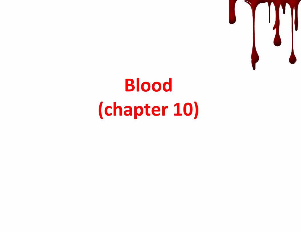
Blood(chapter 10)

1. Composition and function of blood
• Components of blood
• Physical characterist ics and volume
• Plasma
• Formed elements-‐(Erythrocytes*Leukocytes*Platelets)
• Hematopoiesis
• Blood groups and transfusion
2. Hemostasis

• It is a complex connective tissue in which blood cellsare suspended in plasma.
• It is the only fluid tissue.
• It has both solid and liquid compartments.
55% plasma
1% buffy coat
45% Erythrocytes
Blood Components:


Physical characteristics and volume:
• viscous, thick, opaque fluid.
• Slightly alkaline pH 7.35-‐7.45
• Its temp. is 38C
• Volume in Healthy males 5-‐6 liters.

Plasma:
• The fluid portion of blood.
• A solution (90% of it is water.) containing ions,
inorganic & organic molecules.
1)Transports substances around the body;
nutrients, salts, respiratory gases, hormones, Abs,
Plasma proteins, waste products of cell metabolism.
-‐Plasma proteins has a variety of functions.
e.g. Albumin maintain the osmotic pressure of plasma
Fibrinogen is essential for blood clotting.
Globulin participates in immune system.
-‐Most plasma proteins are made by the liver. Theyaren’t taken up by cells to be used as nutrients.
2) Plasma distributes body heat throughout the body.
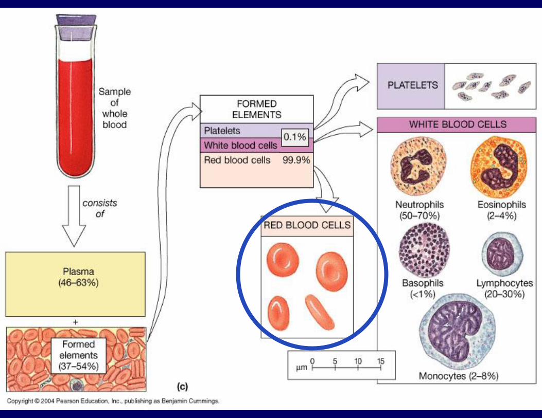

Erythrocytes (RBCs):• Structure-‐
o Anucleatedo mature RBCs circulating in the blood are filled with
hemoglobin.o Haemoglobin (Hb), an iron-bearing protein, that binds to
oxygen. Made up of four polypeptide chains.
o Erythrocytes are small, flexible cells shaped like biconcave discs—flattened discs with depressed centers on both sides .
o A single red blood cell contains about 250 million hemoglobin molecules, each capable of binding 4 molecules of oxygen, so each of these tiny cells can carry about 1 billion molecules of oxygen.
o Normal blood contains 12–18 grams (g) of hemoglobin per 100 milliliters (ml) of blood. The hemoglobin content is slightly higher in men.

Erythrocytes (RBCs):
Function-‐ ?
Perfect example on how structure fits function.
What determines how well the erythrocytes are performing their role of _5 million /mm3 (outnumber WBC 1000:1)

11

What is Haematocrit (Hct)?
Definition:◦ Is the proportion of blood volume that is
occupied by red blood cells.
OR◦ Percentage of red blood cells in whole blood.
Normal value:◦ Male = 40-52%.
◦ Female = 36-48%.
12

1
2
Take a blood sample
Centrifuge
3Read
13

Reading the result
14
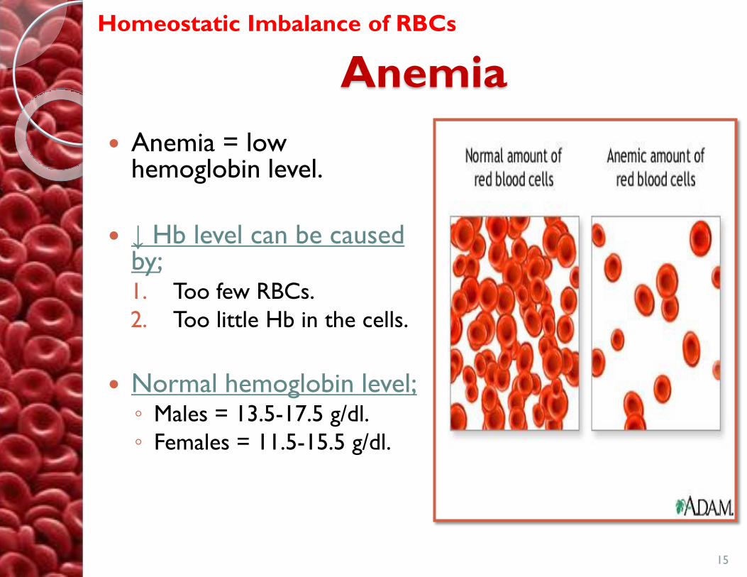
Anemia
Anemia = low hemoglobin level.
↓ Hb level can be caused by; 1. Too few RBCs.
2. Too little Hb in the cells.
Normal hemoglobin level;◦ Males = 13.5-17.5 g/dl.
◦ Females = 11.5-15.5 g/dl.
15
Homeostatic Imbalance of RBCs

16
Causes of Anemia
FactoryRaw material Circulation
Iron
Folic acid
& B12
Abnormal
Hb
Blood loss
Hemolysis
• Iron deficiency anemia.
• Folic acid deficiency.
•Vitamin B12 deficiency
)Pernicious anemia(.
• Aplastic anemia.
• Sickle cell anemia.
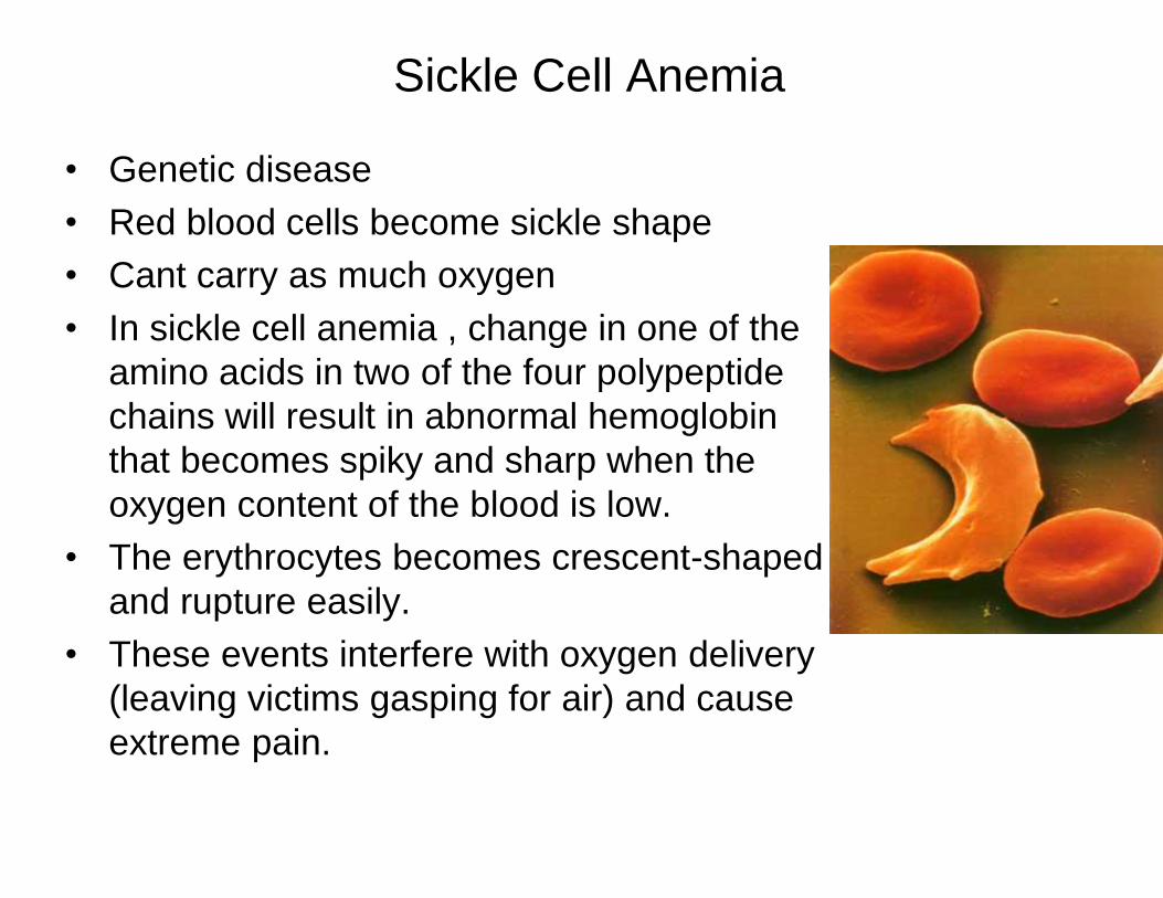
Sickle Cell Anemia
• Genetic disease
• Red blood cells become sickle shape
• Cant carry as much oxygen
• In sickle cell anemia , change in one of the
amino acids in two of the four polypeptide
chains will result in abnormal hemoglobin
that becomes spiky and sharp when the
oxygen content of the blood is low.
• The erythrocytes becomes crescent-shaped
and rupture easily.
• These events interfere with oxygen delivery
(leaving victims gasping for air) and cause
extreme pain.

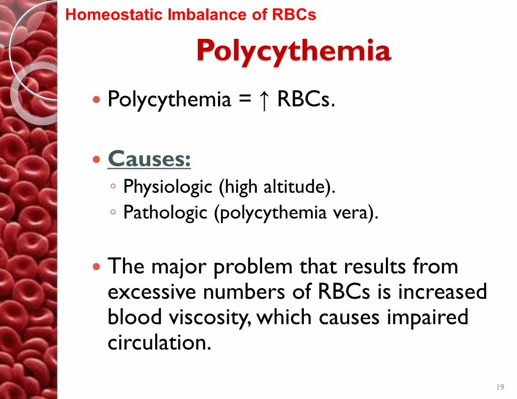
Polycythemia
Polycythemia = ↑ RBCs.
Causes:◦ Physiologic (high altitude).
◦ Pathologic (polycythemia vera).
The major problem that results from excessive numbers of RBCs is increased blood viscosity, which causes impaired circulation.
19

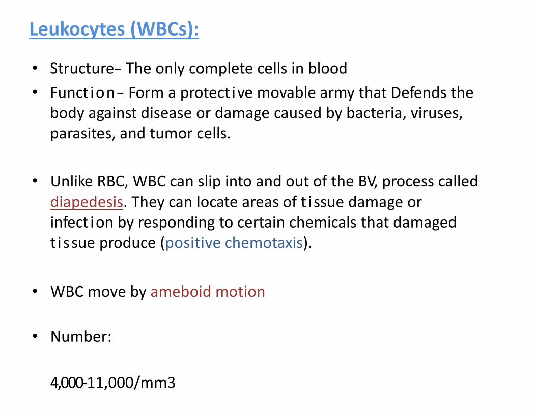
Leukocytes (WBCs):
• Structure-‐ The only complete cells in blood
• Funct ion-‐ Form a protect ive movable army that Defends thebody against disease or damage caused by bacteria, viruses,parasites, and tumor cells.
• Unlike RBC, WBC can slip into and out of the BV, process calleddiapedesis. They can locate areas of t issue damage or infect ion by responding to certain chemicals that damagedtissue produce (positive chemotaxis).
• WBC move by ameboid motion
• Number:
4,000‐11,000/mm3

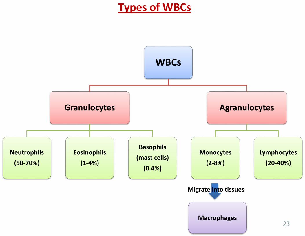
Types of WBCs
23
WBCs
Granulocytes
Neutrophils
(50-70%)
Eosinophils
(1-4%)
Basophils
(mast cells)
(0.4%)
Agranulocytes
Monocytes
(2-8%)
Lymphocytes
(20-40%)
Macrophages
Migrate into tissues
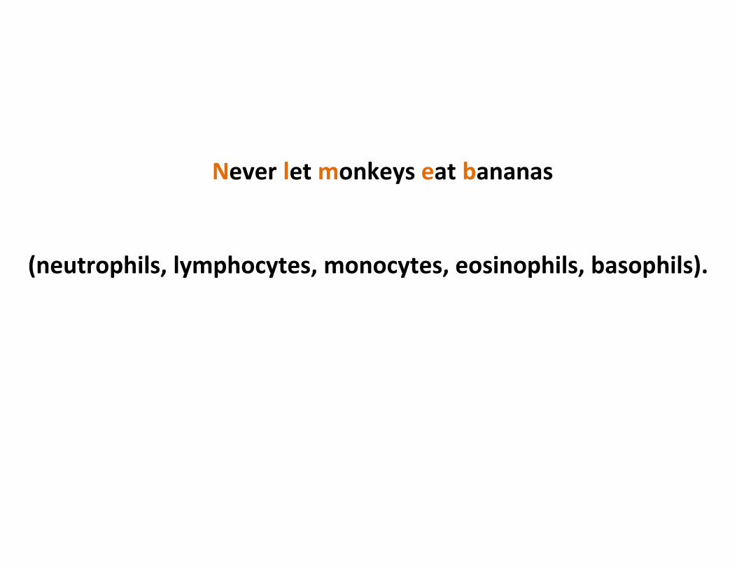
Never let monkeys eat bananas
(neutrophils, lymphocytes, monocytes, eosinophils, basophils).
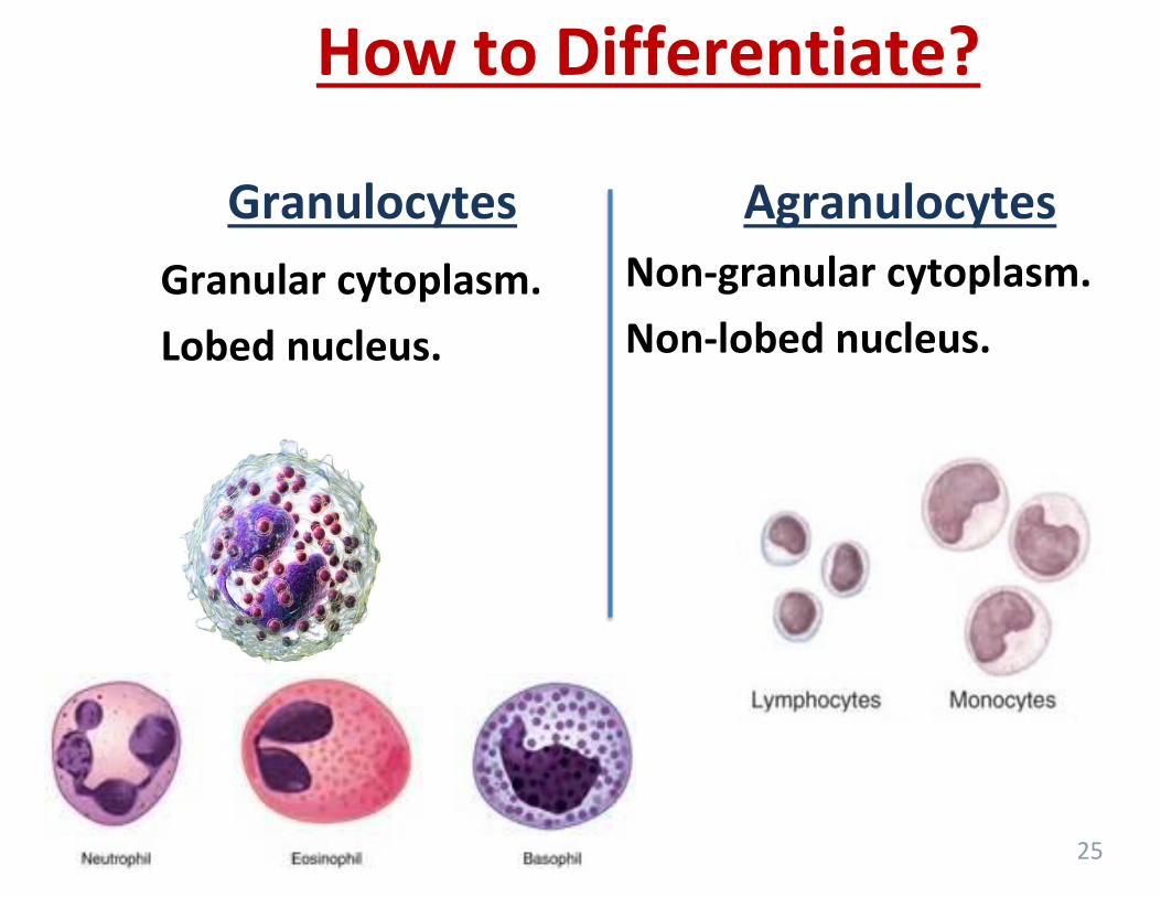
How to Differentiate?
Granulocytes Agranulocytes
Granular cytoplasm.
Lobed nucleus.
Non-granular cytoplasm.
Non-lobed nucleus.
25

How to Differentiate Between Granulocytes?
26
• Multilobed nucleus (2-7) lobes.
• Pale cytoplasm.
2-3 lobed nucleus.
Coarse granules which stain with acid dyes like eosin (red).
An S-shaped nucleus (2-3 lobes).
Coarse cytoplasmicgranules that stains with basic dyes (methyleneblue).

How to Differentiate Between Agranulocytes?
27
Large round nucleus. Scanty cytoplasm..
• Larger than other cells.• Oval, kidney, horse-shoe shaped nucleus.

28
A. NeutrophilB. EosinophilC. BasophilD. MonocyteE. Lymphocyte
On blood film

WBC Function
Neutrophils short-term or acute bacterial and fungal infections.
Eosinophils Parasitic infections and allergies .
Basophils inflammatory reactions, prevents blood from clotting and promotes blood flow to tissues..
Lymphocytes Part of immune system;(B lymphocytes)produces antibodies; (T lymphocytes) Involved in graft rejection, fighting tumors and viruses.
Monocytes (or macrophages in tissues)
chronic infections such as tuberculosis

Leukocytosis• an increase in the number of white cells in the blood,
especially during an infection• generally indicates that a bacterial or viral infection.
• Eosinophilia? Neutrophilia? Lymphocytosis?
Leukopenia• Is an abnormally low WBC count. • It is commonly caused by certain drugs, such as
corticosteroids and anticancer agents.
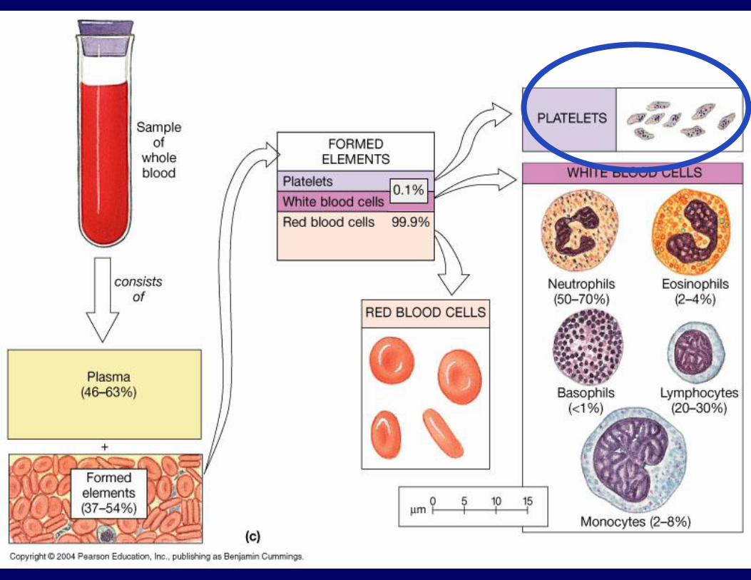

Platelets:
• Not a cell
• Fragments Derived from
ruptured multinucleated
megakaryocytes.
• function-‐Needed for theclotting process that occurs inplasma when BVs areruptured or broken.
• Live 2-4 days
• Number-‐300,000/mm3
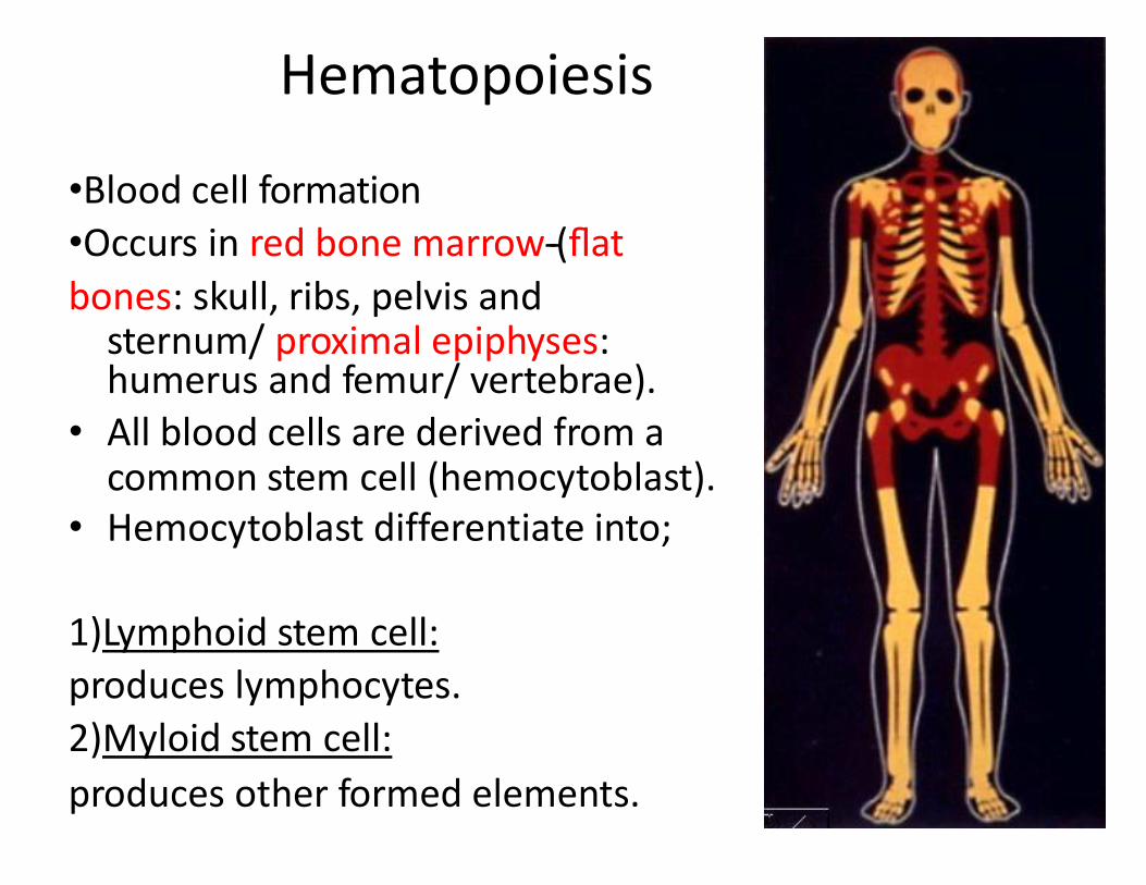
Hematopoiesis
•Blood cell formation
•Occurs in red bone marrow-‐(flat
bones: skull, ribs, pelvis andsternum/ proximal epiphyses: humerus and femur/ vertebrae).
• All blood cells are derived from acommon stem cell (hemocytoblast).
• Hemocytoblast differentiate into;
1)Lymphoid stem cell:
produces lymphocytes.
2)Myloid stem cell:
produces other formed elements.


Fate of Erythrocyte
• Unable to divide, grow, or synthesize proteins. (?)
• Wear out in 100 to 120 days.
• When worn out, they are eliminated by phagocytesin the spleen and liver.
• Lost cells are replaced by division of hemocytoblast.
Control of Erythrocyte production
• Rate of production is controlled by a hormonecalled (erythropoietin).
• Kidneys produce erythropoietin as a responseto reduced oxygen levels in blood for anyreason, that will then target the bone marrow .

Hemeostasis (balance)
Blood Groups and Transfusions

Objectives:
• Describe the blood clotting process.
2. Hemostasis

2. Hemostasis• Stoppage of blood flow when a blood vessel wall breaks.
• Fast and localized reaction.
Hemostasis involves three phases:
1) Vascular spasms
2) Platelet plug formation
3) coagulation (blood clot, fibrin clot)

1) Vascular Spasm
• direct injury to the smooth muscle cells, stimulation of local pain receptors, and release of serotonin by anchored platelets
• Spasms narrow the BV at that point decreasing bloodloss until clotting occur.

2) Platelet plug formation
• Collagen fibers are exposed by a break in a BV.
• Platelets become “st icky” and cling to fibers (damagedsite).
• Anchored platelets release chemicals to aqract more platelets tothe site.
•Platelets pile up to Form a platelet
plug.
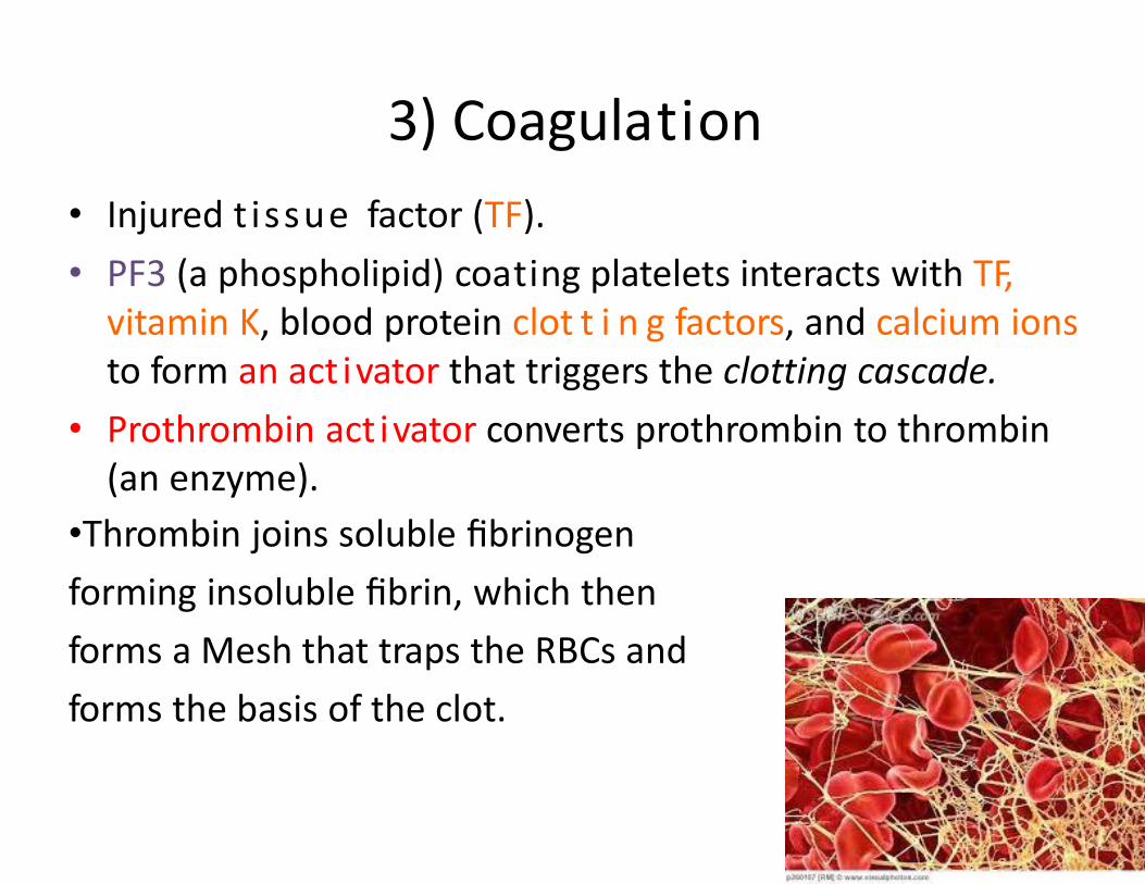
3) Coagulation
• Injured t issue factor (TF).
• PF3 (a phospholipid) coating platelets interacts with TF, vitamin K, blood protein clot t i n g factors, and calcium ionsto form an act ivator that triggers the clotting cascade.
• Prothrombin act ivator converts prothrombin to thrombin(an enzyme).
•Thrombin joins soluble fibrinogen
forming insoluble fibrin, which then
forms a Mesh that traps the RBCs and
forms the basis of the clot.

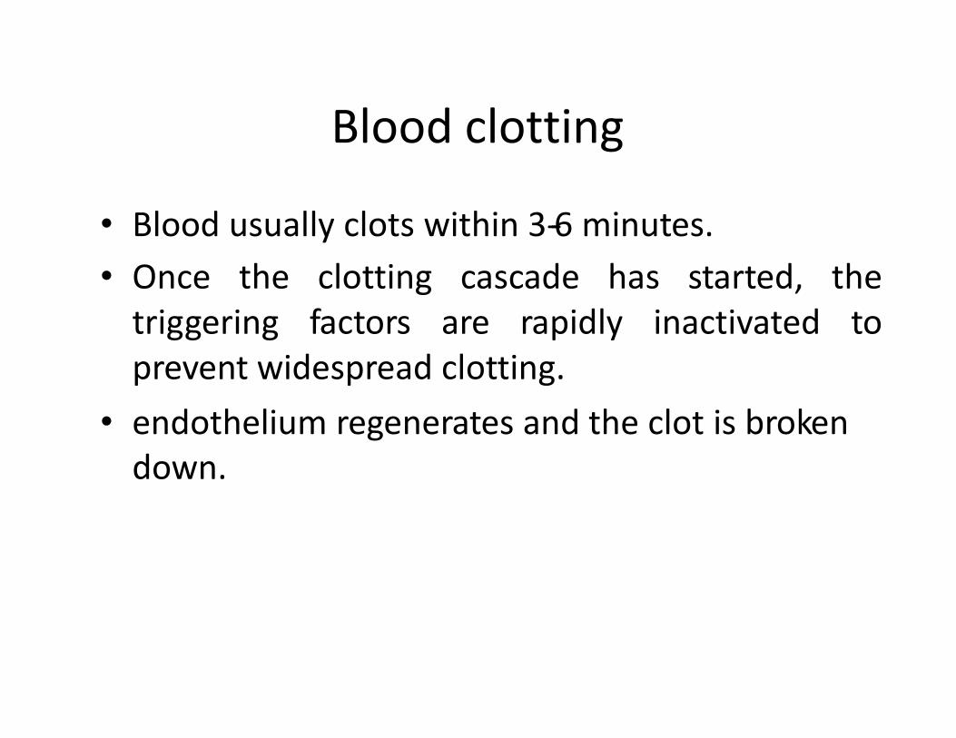
Blood clotting
• Blood usually clots within 3-‐6 minutes.
• Once the clotting cascade has started, thetriggering factors are rapidly inactivated toprevent widespread clotting.
• endothelium regenerates and the clot is brokendown.


Undesirable clotting
• Thrombus:
A clot in an unbroken BV. Can be deadly in areaslike the heart.
• Embolus:
A thrombus that breaks away and floats freely inthe bloodstream. Can later clog vessels incriFcal areas such as the brain

Name the parts of the blood?

Identify cells

Lymphatic System(chapter 12)

Introduction
– Components
• Lymph is the fluid.
• Network of lymphatic vessels (lymphatics).
• Lymphoid tissues and organs.
– Functions
• Return tissue fluid to the bloodstream.
• Transport fats from the digestive tract to the
bloodstream.
• Surveillance & defense (lymphoid tissues and
organs house phagocytic cells and lymphocytes).

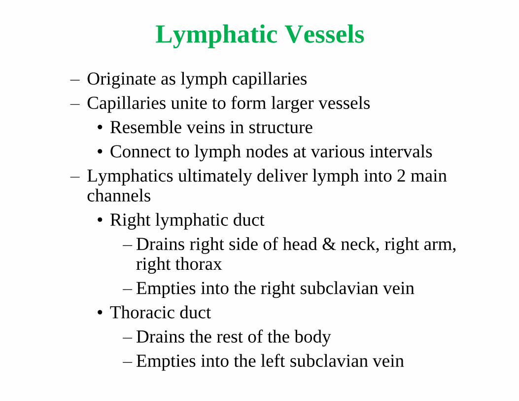
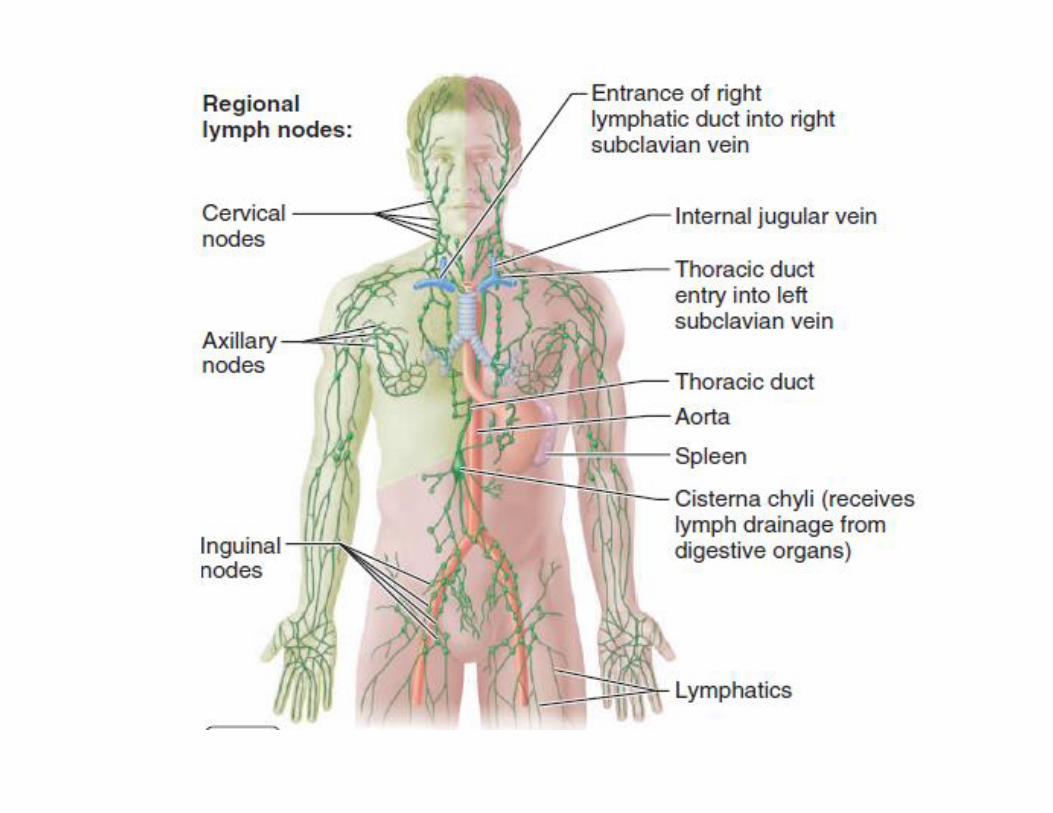
Lymphatic Vessels
– Originate as lymph capillaries
– Capillaries unite to form larger vessels
• Resemble veins in structure
• Connect to lymph nodes at various intervals
– Lymphatics ultimately deliver lymph into 2 main channels
• Right lymphatic duct
– Drains right side of head & neck, right arm, right thorax
– Empties into the right subclavian vein
• Thoracic duct
– Drains the rest of the body
– Empties into the left subclavian vein

Lymphatic Vessels
The function of the lymphatic vessels is to form a drainage system that picks up this excess tissue fluid.
This fluid is called lymph (clear water), and vessels returns it to the blood.
Lymphatic Vessels
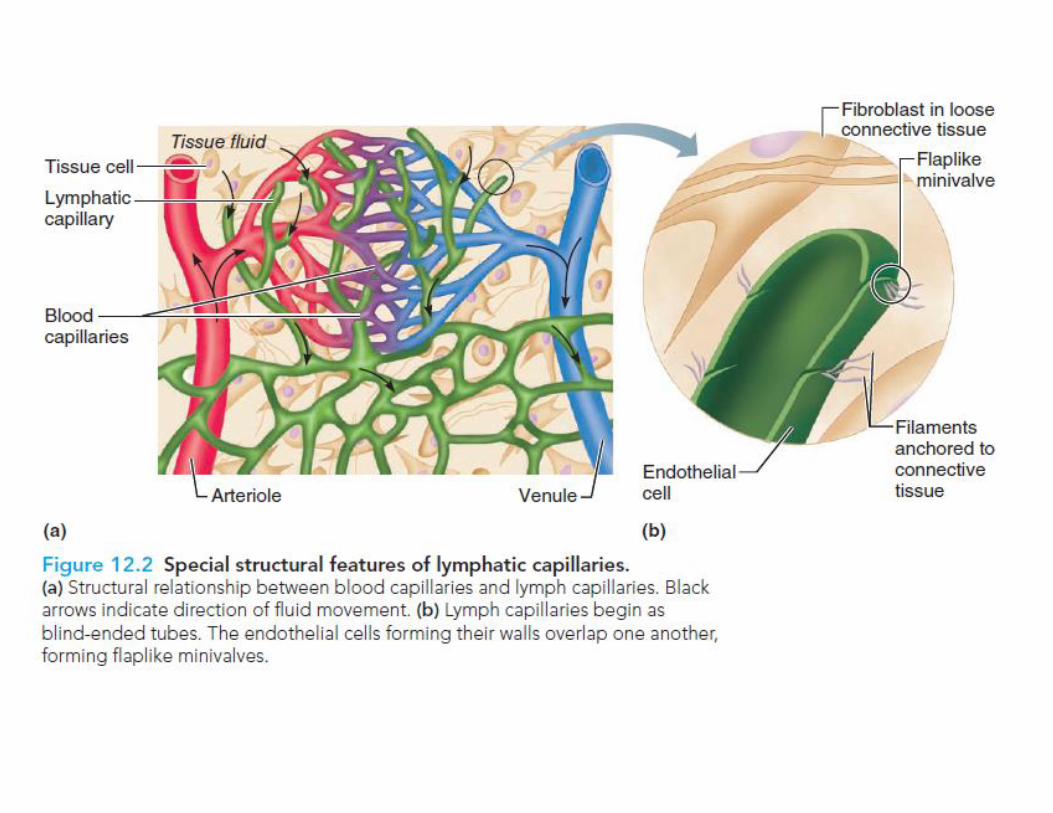
• lymph flows only toward the heart. • lymph capillaries sound between the tissue cells and blood
capillaries in the loose connective tissues of the body and absorb the leaked fluid.
• the edges of the endothelial cells forming their walls loosely overlap one another, forming flaplike minivalves that act as one-way door.
• gape open when the fluid pressure is higher in the interstitial space, allowing fluid to enter the lymphatic capillary.
• when the pressure is higher inside the lymphatic vessels, the endothelial cell flaps are forced together, preventing the lymph from leaking.
Lymph Nodes
– Oval structures located along lymphatics
– Enclosed by a fibrous capsule
– Cortex = outer portion
• Germinal centers produce lymphocytes
– Medulla = inner portion
• Medullary cords
– Lymph enters nodes through afferent lymphatics, flows through sinuses, exits through efferent lymhpatic

Lymph Nodes
– lymph nodes help protect the body by removing foreign material from the lymph (macrophages) and by producing lymphocytes that function in the immune response.

Other Lymphoid Organs
• spleen, thymus, tonsils, Peyer’s patches and appendix.
• The common feature of all these organs is a predominance of reticular connective tissue and lymphocytes.
• Although all lymphoid organs have roles in protecting the body, only the lymph nodes filter lymph.


Tonsils
– Multiple groups of large lymphatic nodules
– Location – mucous membrane of the oral and pharyngeal cavities

Spleen
– Largest lymphatic organ
– Located between the stomach & diaphragm
– Functions
• Filters blood
• Stores blood
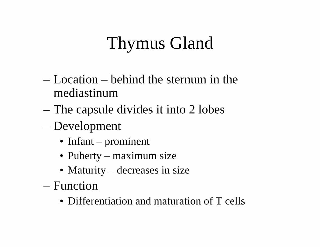
Thymus Gland
– Location – behind the sternum in the mediastinum
– The capsule divides it into 2 lobes
– Development
• Infant – prominent
• Puberty – maximum size
• Maturity – decreases in size
– Function
• Differentiation and maturation of T cells


Peyer’s patches & appendix
• found in the wall of the distal part of the small
intestine.
• The macrophages of Peyer’s patches and the appendix
are in an ideal position to capture and destroy bacteria
(always present in tremendous numbers in the
intestine), thereby preventing them from penetrating the
intestinal wall.
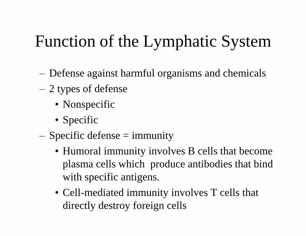
Function of the Lymphatic System
– Defense against harmful organisms and chemicals
– 2 types of defense
• Nonspecific
• Specific
– Specific defense = immunity
• Humoral immunity involves B cells that become
plasma cells which produce antibodies that bind
with specific antigens.
• Cell-mediated immunity involves T cells that
directly destroy foreign cells

BODY DEFENSES