Embed Size (px)
Citation preview

1
Hu Proteins Regulate Polyadenylation by Blocking Sites Containing
U-rich Sequences
Hui Zhu
1, Hua-Lin Zhou
1, Robert A. Hasman
1, and Hua Lou
1,2,3*
1Department of Genetics,
2Case Comprehensive Cancer Center,
3The Center for RNA Molecular
Biology, School of Medicine, Case Western Reserve University, 10900 Euclid Ave, Cleveland,
OH 44106
*Corresponding author: Department of Genetics, Case Western Reserve University, 10900
Euclid Ave., Cleveland, OH 44122, E-mail:[email protected], Phone: (216) 368-6419, Fax: (216)
368-0491
Running title: Hu proteins regulate polyadenylation
Summary
A recent genome-wide bioinformatic
analysis indicated that 54% of human genes
undergo alternative polyadenylation. Although it
is clear that differential selection of poly(A) sites
can alter gene expression, resulting in significant
biological consequences, the mechanisms that
regulate polyadenylation are poorly understood.
Here we report that the neuron-specific members
of a family of RNA-binding proteins, Hu proteins,
known to regulate mRNA stability and translation
in the cytoplasm, play an important role in
polyadenylation regulation. Hu proteins are
homologs of the Drosophila embryonic lethal
abnormal visual (ELAV) protein and contain three
RNA recognition motifs. Using an in vitro
polyadenylation assay with HeLa cell nuclear
extract and recombinant Hu proteins, we show that
Hu proteins selectively block both cleavage and
poly(A) addition at sites containing U-rich
sequences. Hu proteins have no effect on poly(A)
sites that do not contain U-rich sequences or sites
in which the U-rich sequences are mutated. All
three RRMs of Hu proteins are required for this
activity. Over-expression of HuR in HeLa cells
also block polyadenylation at a poly(A) signal that
contains U-rich sequences. Hu proteins block the
interaction between the polyadenylation factor
CstF64 and RNA, most likely through direct
interaction with poly(A) factors CstF64 and
CPSF160. These studies identify a novel group of
mammalian polyadenylation regulators.
Furthermore, they define a previously unknown
nuclear function of Hu proteins.
Introduction Polyadenylation, a process through which
a poly(A) tail of 150-250 adenosines is added to
the 3’-end of a newly synthesized mammalian pre-
mRNA, plays an essential regulatory role in
almost every aspect of gene expression including
transcription termination, splicing, mRNA
transport, mRNA stability and protein translation
(1). A recent genome-wide bioinformatic analysis
indicated that a significant number of human and
mouse genes undergo alternative polyadenylation
(54% and 32%, respectively) (2). Of the human
genes with multiple polyadenylation sites, 41%
have polyadenylation sites located on different
exons, indicating an important role for
polyadenylation in gene regulation. A well
characterized example of this type of regulation is
the human calcitonin/calcitonin gene-related
peptide (CGRP) gene, which has two alternative
3’-terminal exons that are selectively included in
two different tissues to produce two functionally
distinct polypeptides (3).
Polyadenylation of eukaryotic pre-mRNA
is a two-step reaction including cleavage and
addition of the poly(A) tail to the newly generated
3’-end (1,4). In mammals, two major cis-acting
elements define a poly(A) site: a highly conserved
AAUAAA hexanucleotide located 10-30
nucleotides upstream of the cleavage site, and a
G/U- or U-rich downstream element (DSE)
http://www.jbc.org/cgi/doi/10.1074/jbc.M609349200The latest version is at JBC Papers in Press. Published on November 26, 2006 as Manuscript M609349200
Copyright 2006 by The American Society for Biochemistry and Molecular Biology, Inc.
by guest on April 8, 2018
http://ww
w.jbc.org/
Dow
nloaded from

2
located 20-40 downstream of the cleavage site.
Some poly(A) sites also contain one or more U-
rich upstream sequence elements (USEs)
surrounding the AAUAAA. Trans-acting factors
are required to assemble on a poly(A) site to carry
out the polyadenylation reaction. The multi-
protein cleavage and polyadenylation specificity
factor (CPSF) complex binds at the AAUAAA
sequence through the CPSF160 subnuit. The
heterotrimeric complex cleavage stimulation factor
(CstF) binds at the DSE sequence through the
CstF64 subunit. In addition to these two
complexes, poly(A) polymerase (PAP), cleavage
factor I and II (CFI, CFII), the carboxy-terminal
domain of RNA polymerase II and nuclear
poly(A)-binding protein I are also involved in
polyadenylation (1,4).
Although nearly all of the components of
the basal polyadenylation machinery have been
identified and reconstituted in vitro, regulation of
polyadenylation is not well understood. The
factors that have been shown to regulate
polyadenylation include two basal polyadenylation
factors, CFIm68 and CstF64 (5-8), hnRNP
proteins (hnRNP H/H’, hnRNP F and PTB) and
the U1 snRNP associated proteins (8-12). Tissue-
specific regulation of polyadenylation is an under
studied area. While a testis-specific basal
polyadenylation factor, CstF64 protein, was
proposed to change the preference of the
machinery as a whole to favor poly(A) sites
containing non-canonical hexaucleotides (13,14),
factors that regulate transcript-specific events in a
tissue-specific manner have not been identified in
mammalian cells. Interestingly, this type of factor
has been demonstrated in flies. The embryonic
lethal abnormal visual (ELAV) protein was
recently shown to inhibit the non-neuronal 3’-end
processing, thereby promoting the neural splicing
of the erect wing (ewg) pre-mRNA (15,16).
Mammalian Hu proteins are a group of
RNA-binding proteins closely related to the
Drosophila ELAV protein. The three neuron-
specific members of the Hu family, HuB (HelN1
in human), HuC and HuD, were cloned as
autoimmune antigens of a paraneoplastic
neurodegenerative disorder (PND), called Hu
syndrome (17). All members of the Hu protein
family have been shown to interact with AU-rich
elements and regulate mRNA stability; the human
HuB protein HelN1 has also been shown to
modulate translation (18). Recently, we
demonstrated a nuclear function of Hu proteins as
regulators of alternative RNA processing (19),
although the underlying mechanism remains to be
determined. Given that ELAV and Hu proteins
belong to a well-conserved protein family, we
have investigated the possibility that Hu proteins
regulate polyadenylation in mammalian cells.
In this report, we provide evidence for a
novel function of the neuron-specific Hu proteins
as polyadenylation regulators. Using an in vitro
polyadenylation assay with HeLa cell nuclear
extract and recombinant Hu proteins, we show that
both cleavage and poly(A) addition at two of the
four tested poly(A) sites (SVL and the non-
neuronal alternative 3’-terminal exon 4 of the
calcitonin/CGRP pre-mRNA) are blocked by Hu
proteins. We demonstrate that the polyadenylation
inhibition is specific to poly(A) sites that contain
U-rich sequence near cleavage sites and depends
on binding of Hu proteins to the U-rich sequences.
Furthermore, we show that the third RRM on Hu
proteins, which is dispensable for RNA binding, is
required to block polyadenylation. We also
discover interactions between Hu proteins and two
poly(A) factors, CstF64 and CPSF160, and show
that Hu proteins block binding of CstF64 to the U-
rich sequence-containing poly(A) site. These
studies identify a novel nuclear function of Hu
proteins as polyadenylation regulators. They also
suggest a novel mechanism of regulated
alternative RNA processing by Hu proteins in
neurons.
Experimental Procedures
Plasmids. To generate cDNA sequences of the
mouse HuB and HuC, RT/PCR was carried out
using RNA isolated from the mouse F9 cells and
mHuB or mHuC-specific oligonucleotides. The
PCR products were digested with BamHI and
EcoRI and cloned into the BamHI and EcoRI sites
in pGEX-2TK vector (Amersham). The
HuA/HuR cDNA in pTet.myc.HuR (a gift from
Dr. Ann-Bin Shyu, Texas) was digested with
EcoRV and subcloned into pcDNA3.1HisB. hGH
plasmids were PCR cloned from HeLa cell
genomic DNA. The reporters used in cell
transfections contain the human metallothionein
2A exon 1 and 170 nucleotides of intron 1 fused
with 90 nucleotides of intron 2 and 120
nucleotides of exon 3 as well as either 200
by guest on April 8, 2018
http://ww
w.jbc.org/
Dow
nloaded from

3
nucleotides of the SVL poly(A) site or 265
nucleotides of the human growth hormone (hGH)
poly(A) site. The reporters were generated by
PCR-directed cloning.
Recombinant proteins. Glutathione S-transferase
(GST)-fusion proteins were prepared using B-PER
GST Spin Purification kit (Pierce).
Nuclear extract. Nuclear extracts were prepared
from HeLa cells and the mouse brain as previously
described (20,21).
In vitro assays. Polyadenylation cleavage,
poly(A) addition, UV cross-linking reactions and
gel mobility shift assay were carried out as
described previously (22-24). Cross-linked
polypeptides were immunoprecipitated using Hu
patient sera (a gift from Dr. Jerome Posner, New
York) or anti-CstF64 antibody 3A7.
In vitro translation, GST/His pull-down and co-
immunoprecipitation. In vitro translation was
performed with the TNT coupled transcription-
translation rabbit reticulacyte system (Promega).
GST/His pull-down and coimmunoprecipitation
experiments were carried out as described (23).
RNase A was included in the pull-down assays.
HeLa cell transfcetion. Transfection, RNA
isolation, RT/PCR analysis, protein isolation and
western blot analysis were carried out as
previously described (23). One micorgram of
reporter, 1 ug of LacZ plasmid, and 0.4 ug of HuR
or pcDNA3.1HisB plasmid were used in the
transfections. Radioactivity in RT/PCR products
was measured by PhosphorImager analysis.
RT/PCR products of the pre-mRNA and mRNA
were excised from a gel and sequenced to confirm
their identity.
Results
Hu proteins block polyadenylation at sites
containing U-rich sequences
A subset of mammalian cellular and viral
poly(A) sites contain U-rich USE sequences that
function to promote efficient polyadenylation at
these sites (23). The fact that Hu proteins
specifically bind to the AU-rich elements (AREs)
in the 3’-untranslated regions (3’-UTR’s) of a
number of mammalian mRNAs to regulate mRNA
stability prompted us to examine whether Hu
proteins might also play a role in regulating
polyadenylation.
To address this question, we carried out in
vitro polyadenylation assays using HeLa cell
nuclear extract and recombinant Hu proteins.
Both cleavage and poly(A) addition were assayed.
We chose poly(A) sites derived from four pre-
mRNAs based on U content in the vicinity of the
poly(A) sites. Of the four sites, SV40 late poly(A)
site (SVL) and the human calcitonin/CGRP exon 4
poly(A) site contain more than one U-run that
contains three or more U’s, while the hamster
adenine phosphoribosyl transferase (APRT) and
human growth hormone (GH) poly(A) sites do not
(Fig. 1A). The SVL poly(A) site has several U-
runs both upstream and downstream of the
cleavage site, while the calcitonin exon 4 poly(A)
site has one long U-run between the
hexanucleotide and cleavage site and another one
upstream of the hexanucleotide. The sizes of the
cleavage and pre-cleaved, poly(A) addition
substrates for all four poly(A) sites are shown in
Fig 1B. All but the GH poly(A) site were
previously documented in similar polyadenylation
cleavage assays (22). In establishing the poly(A)
addition assay, we initially confirmed that under
our assay conditions, poly(A) addition at the
calcitonin exon 4 poly(A) site is dependent on the
AAUAAA hexanucleotide (data not shown).
As shown in Fig. 2A and 2B, addition of
GST-mHuB blocks polyadenylation of SVL and
calcitonin exon 4 poly(A) sites at both the
cleavage and poly(A) addition steps but had no
effect on APRT and GH poly(A) sites. Addition
of the GST protein had no effect on any of the
poly(A) sites. Note that the low cleavage activity
of the calcitonin exon 4 poly(A) site made it
somewhat difficult to assess the effect of Hu
proteins on polyadenylation cleavage of this site.
However, the reproducible reduction of the
cleavage activity coupled with a significant
reduction of the poly(A) addition activity (Fig. 2B)
and the CstF64 binding activity (Fig. 5A) in the
presence of the Hu proteins clearly indicates that
Hu proteins affect polyadenylation at this site.
We tested all of the Hu proteins (HuR, mHuB,
mHuC, and hHuD) in polyadenylation cleavage
assays on the SVL poly(A) site and found no
difference in their ability to block polyadenylation
(Supplemental Fig. 1A). Results with only GST-
mHuB are shown in Figs. 2 and 3. The ability of
the Hu proteins to block polyadenylation does not
correlate with the strength of the poly(A) sites
since SVL and calcitonin exon 4 represent the two
extremes, the former being a very strong site and
by guest on April 8, 2018
http://ww
w.jbc.org/
Dow
nloaded from
4
the latter a weak site (Fig. 2A and 2B, compare the
product to precursor ratio in lane 1 of the SVL and
calcitonin exon 4 panels).
The polyadenylation blocking activity of Hu
proteins depends on their binding to U-rich
sequences surrounding the hexanucleotide.
Hu proteins have been shown to have
strong affinity for U-rich sequences (25-27). We
suspected that Hu proteins bind to the U-rich
sequences located close to the AAUAAA
hexanucleotide in SVL and the calcitonin exon
poly(A) site, which is the binding site of the 160
kD subunit of cleavage/polyadenylation specificity
factors (CPSF160) required for both steps of
polyadenylation. To determine if Hu proteins bind
to any of the four poly(A) sites, UV cross-
linking/immunoprecipitation (IP) assays were
carried out using HeLa cell nuclear extract
supplemented with the recombinant GST-mHuB
protein. Anti-Hu sera derived from patients who
suffer from the Hu syndrome that can detect all
three neuron-specific Hu proteins (HuB, HuC, and
HuD) but not the ubiquitously expressed HuA
(HuR), were used to immunoprecipitate the cross-
linked proteins. Pre-cleaved substrates that
contain only the sequences upstream of the
cleavage site were used in this assay.
Interestingly, strong binding of mHuB to both
SVL and calcitonin exon 4 poly(A) site, but not to
the APRT and GH sites, was detected (Fig. 2C).
A gel mobility-shift assay using the same RNA
substrate was also carried out, the result of which
is consistent with the cross-linking/IP assay
(Supplemental Fig. S2). These results imply that
Hu proteins preferentially block poly(A) sites
containing U-rich sequences. The stronger
binding of mHuB at SVL site than at the calcitonin
exon 4 site is most likely a reflection of the
existence of multiple U-runs at the SVL site (Fig.
1A). Consistent with cleavage assay, all of the Hu
protein members are capable of binding at the
SVL poly(A) site (Supplemental Fig. S1A and
S1B).
Because Hu proteins showed a robust
inhibition in the poly(A) addition assay and the
calcitonin exon 4 poly(A) site has only one
significant U-run between the hexanucleotide and
cleavage site, we next used this specific assay to
further test the specificity of the Hu effect in
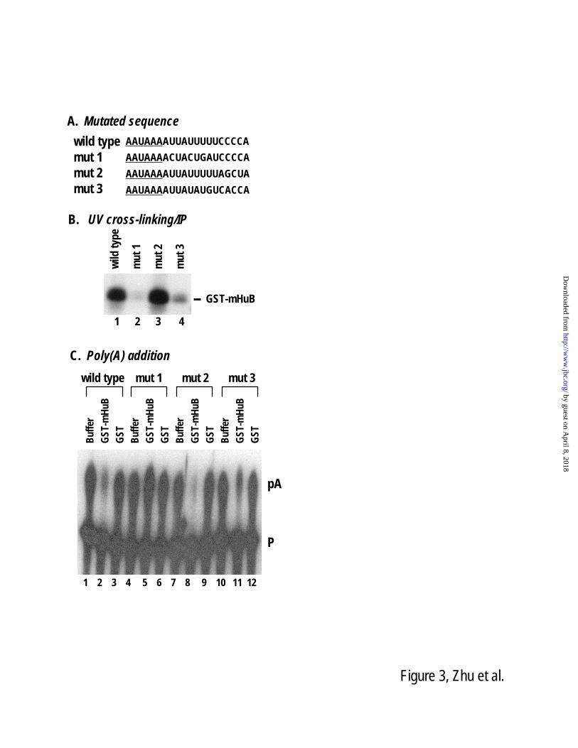
polyadenylation. We generated three mutant RNA
substrates that contain different point mutations in
the U-rich region (Fig. 3A). When we examined
these mutant substrates for binding to Hu proteins
and poly(A) addition activity, we observed a
strong inverse correlation between the two
activities. A U-rich sequence mutant (mutant 1 in
Fig. 3A) that completely abolished Hu protein
binding also lost its ability to be regulated by Hu
proteins in the poly(A) addition assay (Fig. 3B,
compare lane 2 to 1, and Fig. 3C, compare lane 5
to 2). A milder mutant (mutant 3 in Fig. 3A) that
shows reduced but still detectable Hu binding
activity was partially blocked by an Hu protein in
the poly(A) addition assay (Fig. 3B, compare lane
4 to 1, and Fig. 3C, compare lane 11 to 2).
Finally, in mutant 2, where the C-rich but not the
U-rich sequence was disrupted, Hu protein binding
was not affected, nor was poly(A) addition (Fig.
3B, compare lane 3 to 1, and Fig. 3C, compare
lane 8 to 2). These experiments establish a strong
correlation between binding of Hu proteins and
their ability to regulate polyadenylation.
All of the three RRMs on Hu proteins are
required to block polyadenylation
It is possible that Hu proteins block
polyadenylation by binding to the U-rich
sequences close to a poly(A) site and simply block
the access of poly(A) factors to the
polyadenylation signals. We carried out two sets
of experiments to address this issue. First, because
U-rich sequences can also be bound by other
proteins such as PTB and TIAR, we tested if these
proteins would block polyadenylation of the SVL
poly(A) site. Both proteins are similar to Hu
proteins in size. Addition of GST-TIAR did not
affect poly(A) cleavage at this site even though it
can bind to the SVL RNA with comparable
affinity as GST-mHuB (Fig. 4A and 4C). Addition
of GST-PTB did not affect polyadenylation at this
site either (data not shown). These results indicate
that the polyadenylation inhibitory effect is
specific to the Hu proteins. Second, specific RNA
binding was demonstrated previously for RRM1
and RRM2 domains of HuD and the RRM3
domain for HuB (Hel-N1/HelN2) (26-28). To test
which RRMs are required to block
polyadenylation, we generated two truncation
mutants of mHuB, one containing RRMs 1 and 2
and the other RRM3 (Fig. 4B). Neither of these
two mutants blocked SVL polyadenylation
by guest on April 8, 2018
http://ww
w.jbc.org/
Dow
nloaded from
5
cleavage (Fig. 4B). However, RRM12, but not the
RRM3 mutant, showed strong binding to RNA
(Fig. 4C). The exact same result was observed
with the hHuD full-length and RRM12 mutant
(data not shown). Taken together, these results
indicate that binding to RNA by itself is not
sufficient for Hu proteins to block
polyadenylation. It is likely that Hu proteins
interact with polyadenylation factors to block their
polyadenylation activity.
Hu proteins block binding of CstF64 to poly(A)
sites, and interact with two polyadenylation
factors
To probe the mechanism whereby Hu
proteins inhibit polyadenylation of SVL and
calcitonin exon 4, we first tested if addition of Hu
proteins to HeLa cell nuclear extract blocks
CstF64 binding to the GU/U-rich sequence
downstream of the cleavage sites. As shown in
Fig. 4A via UV cross-linking/IP analysis using the
cleavage substrate (Fig. 1B) and anti-CstF64
antibody, binding of CstF64 to the calcitonin exon
4 poly(A) sites is significantly reduced in the
presence of increasing amounts of GST-mHuB
protein, but not the control GST protein (Fig. 5A).
Surprisingly, a protein with a higher mobility was
also detected in this assay. A similar result was
observed with the SVL substrate (data not
shown)(27). We identified this protein as GST-
mHuB based on its size, intensity, and the fact that
when His-hHuD was used instead of GST-mHuB
in this assay, an extra band with the expected
molecular weight of the His-hHuD protein was
detected (data not shown). This result is
interesting in light of the observation that the
Drosophila ELAV protein is
coimmunoprecipitated with dCstF64 when added
to the non-neuronal nuclear extract to examine
binding of dCstF64 to the non-neuronal poly(A)
site of the ewg pre-mRNA. However, in that case,
binding of dCstF64 to the poly(A) site was not
reduced by ELAV protein (16).
The CstF64 binding result indicates that
Hu proteins block polyadenylation of SVL and the
calcitonin exon 4 by interfering with the
interaction of poly(A) factors to the poly(A) sites
and also suggests that CstF64 interacts with Hu
proteins. To test this hypothesis, we examined the
potential interaction between Hu proteins and
poly(A) factors by a GST/His pull-down assay
using recombinant Hu proteins (GST-mHuB,
GST-mHuC and His-hHuD) and 35
S-labeled
CstF64, CPSF160, or CFIm68. RNase was
included to ensure that any interaction detected is
not mediated by RNA. The result of this
experiment demonstrates that Hu proteins interact
with CstF64 and CSPF160, but not CFIm68 (Fig.
5B, and not shown). Thus, Hu proteins block
polyadenylation by interacting, mostly likely
directly, with CstF and CPSF, which are required
for both cleavage and poly(A) addition steps. To
test if the interaction between Hu proteins and
poly(A) factors occurs in vivo in cells where Hu
proteins are naturally expressed, we carried out a
co-immunoprecipitation assay using nuclear
extract isolated from the mouse brains. CstF64
was detected in the complex immunoprecipited
with the anti-Hu sera, while the control proteins
TIA-1/TIAR were not detected (Fig. 5C).
HuR blocks polyadenylation of SVL poly(A)
site in cultured cells
To demonstrate the biological relevance of
our findings in vitro, we tested whether the results
can be duplicated in a cell transfection experiment.
We made two reporter constructs that are identical
except for the poly(A) sites: one contains the SVL
poly(A) site and the other the hGH poly(A) site
(Fig. 6A). The two reporters were individually
transfected with either a vector control or the HuR
expression plasmid in HeLa cells. A third
plasmid, LacZ expression vector, was included as
an internal control. Semi-quantitative RT/PCR
was carried out using total RNA isolated from the
transfected cells and two sets of primers. One set
of primers is used to detect the precursor transcript
and the other set including an oligo-dT primer to
detect the polyadenylated mRNA. As shown in
Fig. 6B, the level of the SVL poly(A) site-
containing mRNA is decreased when HuR is over-
expressed. Importantly, this change in the mRNA
level is accompanied by an increase in the pre-
mRNA level, suggesting that the HuR effect is not
the result of a change in mRNA turnover. No
significant change of either the mRNA or the pre-
mRNA level was observed with the reporter
containing the hGH poly(A) site in the absence or
presence of over-expressed HuR.
Interestingly, we observed reduced
splicing when the transcript containing the SVL
poly(A) site was blocked at polyadenylation. This
by guest on April 8, 2018
http://ww
w.jbc.org/
Dow
nloaded from

6
result is consistent with the previously
demonstrated coupling of polyadenylation and
splicing during RNA processing {(29-31) and
reviewed in (32)}. It has been documented that
mutations of polyadenylation signal AAUAAA or
downstream sequences decreases not only
polyadenylation but also splicing efficiency.
Discussion
In this report, we demonstrate that the
mammalian Hu proteins regulate polyadenylation
by blocking poly(A) sites containing U-rich
sequences. Our studies expand the very short list
of polyadenylation regulators in mammals. Given
that more than 50% of human pre-mRNAs
undergo alternative polyadenylation and
alternative poly(A) sites are frequently associated
with AU-rich sequences within 100 nucleotides
from the cleavage site on both sides (2,33),
understanding how Hu proteins regulate
alternative polyadenylation is of great importance.
The polyadenylation regulating activity of
Hu proteins correlates with their binding to the U-
rich sequences upstream of the cleavage site.
These U-rich sequences are similar to the
previously characterized USEs present in some of
the cellular and viral poly(A) sites and necessary
for efficient polyadenylation at those sites
harboring them (23). In the case of the calcitonin
exon 4 poly(A) site, mutation of the U-rich
sequence did not reduce the polyadenylation
efficiency (Fig. 3C, compare lanes 1, 4, 7 and 10).
Thus, whether the U-rich sequence functions as a
classical USE in this poly(A) site remains to be
further determined. Recently, Kaufmann and
colleagues have demonstrated that a newly
discovered human CPSF subunit, Fip1, binds to
the L3 U-rich USEs surrounding AAUAAA and
stimulates poly(A) polymerase (23). Interestingly,
in this study, Fip1 was also found to bind to the
SVL poly(A) site, the same site that can be
blocked by Hu proteins (23). In a more recent
study by Zhao and colleagues, a 57-nucleotide
USE was identified in human papillomavirus 16
that interacts with several nuclear factors including
Fip1 (34). Taken together, these studies suggest
an intriguing potential mechanism in which Hu
proteins compete with and/or prevent Fip1 from
binding to the U-rich sequences. In light of
another recent discovery that CFIm68 binds to a
number of USE sequences containing UGUAN
repeats and promotes polyadenylation (7) and that
SVL poly(A) site contains such repeats, it remains
a formal possibility that Hu proteins and CFIm
may modulate polyadenylation activity of sites
that contain binding sites for both proteins through
their competing activities. Given that USEs have
been identified in an increasing number of cellular
poly(A) sites (23,35-40), it will be of particular
interests to further investigate the role of Hu
proteins in USE-mediated polyadenylation
regulation.
How do Hu proteins regulate
polyadenylation? We demonstrate that all three of
the RRMs of Hu proteins are required for the
polyadenylation blocking activity of these
proteins, suggesting that RNA-binding activity
alone is not sufficient. Presumably, RRM3 and
the hinge region are involved in interacting with
the poly(A) factors. We favor a model in which
Hu proteins bind to RNA and poly(A) factors
simultaneously. Such interactions may modulate
the structure of the poly(A) complex formed on
the U-rich sequence-containing sites in a way that
renders the complex non-functional. Presumably,
neither RNA binding nor interaction with poly(A)
factors alone can induce such a rearrangement.
Given that three of the Hu proteins are
neuron-specific and interact with CstF64 in brain
nuclear extract (Fig. 5C), our studies also suggest
an appealing mechanism for neuron-specific
alternative RNA processing regulation.
Calcitonin/CGRP pre-mRNA is differentially
processed in neurons, where the non-neuronal
calcitonin exon 4 is skipped and the exons 5 and 6
are included to produce CGRP (3). It was
previously demonstrated that inclusion of exon 4
is promoted by a number of factors bound at an
intronic element in non-neuronal cells
(20,24,41,42). Of these factors, U1 snRNP and
SRp20 were shown to promote polyadenylation of
this exon (20,22). Recently, we demonstrated that
Hu as well as Fox-1/Fox-2 proteins promote the
neuron-specific skipping of the calcitonin exon 4
(19,43). It is highly likely that in neurons, Hu
proteins block polyadenylation of exon 4, thereby
promoting the neuron-specific pathway. This
mechanism may function in conjunction with other
neuronal factors such as Fox-1/Fox-2 to regulate
the neuron-specific skipping of exon 4. A similar
example is the Drosophila ewg pre-mRNA.
ELAV protein, the Drosophila homolog of Hu
by guest on April 8, 2018
http://ww
w.jbc.org/
Dow
nloaded from

7
proteins, inhibits 3’-end processing within the
non-neuronal exon of the ewg pre-mRNA to
promote neural splicing (16).
Although both Hu proteins and ELAV
block polyadenylation, major differences exist
between the functions of these proteins. ELAV
binds to several runs of AU-rich sequence
downstream of the cleavage site of the non-
neuronal 3’-terminal exon, close to where CstF64
binds, whereas Hu proteins bind at sequences
adjacent to the AAUAAA hexanucleotide, the
CPSF160 binding site. However, it is possible that
Hu proteins function through binding at the
downstream U-rich sequences of those sites such
as SVL that contain such sequences (Fig. 1).
Furthermore, addition of ELAV to nuclear extract
isolated from non-neuronal cells did not block
interaction of dCstF64 and RNA, in contrast to
what we observed for the Hu proteins (Fig. 5)
(16). These results suggest potential distinct or
different variations of mechanisms for these two
putative orthologous proteins in regulating
polyadenylation.
Acknowledgements
This work was supported by an American
Heart Association grant (award number:
0365274B), an NSF grant (MCB-0237685) and an
NIH grant (NS-049103-01) to Hua Lou. Hui Zhu
is supported by a pre-doctoral fellowship from
American Heart Association.
We thank the following individuals for
providing antibody and plasmids: Jerome Posner
(Hu patient sera), Clinton MacDonald (anti-
CstF64 antibody), Henry Furneaux (GST-hHuD,
and GST-hHuD RRM12), Imed-Eddine Gallouzi
(GST-HuR plasmid), Walter Keller (CstF64,
CPSF160 and CFIm68 plasmids), Ite Laird-
Offringa (HuD plasmid), and Ann-bin Shyu (HuR
plasmid). We thank Helen Salz and Jo Ann Wise
for critical reading of the manuscript.
References
1. Zhao, J., Hyman, L., and Moore, C. (1999) Microbiol Mol Biol Rev 63(2), 405-445 2. Tian, B., Hu, J., Zhang, H., and Lutz, C. S. (2005) Nucleic Acids Res 33(1), 201-212 3. Amara, S. G., Jonas, V., Rosenfeld, M. G., Ong, E. S., and Evans, R. M. (1982) Nature
298(5871), 240-244 4. Edmonds, M. (2002) Prog Nucleic Acid Res Mol Biol 71, 285-389 5. Takagaki, Y., Seipelt, R. L., Peterson, M. L., and Manley, J. L. (1996) Cell 87(5), 941-
952 6. Brown, K. M., and Gilmartin, G. M. (2003) Mol Cell 12(6), 1467-1476 7. Venkataraman, K., Brown, K. M., and Gilmartin, G. M. (2005) Genes Dev 19(11), 1315-
1327 8. Shell, S. A., Hesse, C., Morris, S. M., Jr., and Milcarek, C. (2005) J Biol Chem 280(48),
39950-39961 9. Colgan, D. F., and Manley, J. L. (1997) Genes Dev 11(21), 2755-2766 10. Veraldi, K. L., Arhin, G. K., Martincic, K., Chung-Ganster, L. H., Wilusz, J., and
Milcarek, C. (2001) Mol Cell Biol 21(4), 1228-1238 11. Arhin, G. K., Boots, M., Bagga, P. S., Milcarek, C., and Wilusz, J. (2002) Nucleic Acids
Res 30(8), 1842-1850 12. Castelo-Branco, P., Furger, A., Wollerton, M., Smith, C., Moreira, A., and Proudfoot, N.
(2004) Mol Cell Biol 24(10), 4174-4183 13. Wallace, A. M., Dass, B., Ravnik, S. E., Tonk, V., Jenkins, N. A., Gilbert, D. J.,
Copeland, N. G., and MacDonald, C. C. (1999) Proc Natl Acad Sci U S A 96(12), 6763-6768
14. MacDonald, C. C., and Redondo, J. L. (2002) Mol Cell Endocrinol 190(1-2), 1-8 15. Lisbin, M. J., Qiu, J., and White, K. (2001) Genes Dev 15(19), 2546-2561
by guest on April 8, 2018
http://ww
w.jbc.org/
Dow
nloaded from

8
16. Soller, M., and White, K. (2003) Genes Dev 17(20), 2526-2538 17. Szabo, A., Dalmau, J., Manley, G., Rosenfeld, M., Wong, E., Henson, J., Posner, J. B.,
and Furneaux, H. M. (1991) Cell 67(2), 325-333 18. Okano, H. J., and Darnell, R. B. (1997) J Neurosci 17(9), 3024-3037 19. Zhu, H., Hasman, R. A., Barron, V. A., Luo, G., and Lou, H. (2006) Mol Biol Cell 20. Lou, H., Neugebauer, K. M., Gagel, R. F., and Berget, S. M. (1998) Mol Cell Biol 18(9),
4977-4985 21. Ashiya, M., and Grabowski, P. J. (1997) Rna 3(9), 996-1015 22. Lou, H., Gagel, R. F., and Berget, S. M. (1996) Genes Dev 10(2), 208-219 23. Kaufmann, I., Martin, G., Friedlein, A., Langen, H., and Keller, W. (2004) Embo J 23(3),
616-626 24. Zhu, H., Hasman, R. A., Young, K. M., Kedersha, N. L., and Lou, H. (2003) Mol Cell
Biol 23(17), 5959-5971 25. Chung, S., Eckrich, M., Perrone-Bizzozero, N., Kohn, D. T., and Furneaux, H. (1997) J
Biol Chem 272(10), 6593-6598 26. Levine, T. D., Gao, F., King, P. H., Andrews, L. G., and Keene, J. D. (1993) Mol Cell
Biol 13(6), 3494-3504 27. Gao, F. B., and Keene, J. D. (1996) J Cell Sci 109 ( Pt 3), 579-589 28. Chung, S., Jiang, L., Cheng, S., and Furneaux, H. (1996) J Biol Chem 271(19), 11518-
11524 29. Cooke, C., Hans, H., and Alwine, J. C. (1999) Mol Cell Biol 19(7), 4971-4979 30. Cooke, C., and Alwine, J. C. (2002) Mol Cell Biol 22(13), 4579-4586 31. Kyburz, A., Friedlein, A., Langen, H., and Keller, W. (2006) Mol Cell 23(2), 195-205 32. Proudfoot, N. J., Furger, A., and Dye, M. J. (2002) Cell 108(4), 501-512 33. Hu, J., Lutz, C. S., Wilusz, J., and Tian, B. (2005) Rna 11(10), 1485-1493 34. Zhao, X., Oberg, D., Rush, M., Fay, J., Lambkin, H., and Schwartz, S. (2005) J Virol
79(7), 4270-4288 35. Hall-Pogar, T., Zhang, H., Tian, B., and Lutz, C. S. (2005) Nucleic Acids Res 33(8),
2565-2579 36. Natalizio, B. J., Muniz, L. C., Arhin, G. K., Wilusz, J., and Lutz, C. S. (2002) J Biol
Chem 277(45), 42733-42740 37. Sachchithananthan, M., Stasinopoulos, S. J., Wilusz, J., and Medcalf, R. L. (2005)
Nucleic Acids Res 33(3), 1010-1020 38. Moreira, A., Wollerton, M., Monks, J., and Proudfoot, N. J. (1995) Embo J 14(15), 3809-
3819 39. Moreira, A., Takagaki, Y., Brackenridge, S., Wollerton, M., Manley, J. L., and
Proudfoot, N. J. (1998) Genes Dev 12(16), 2522-2534 40. Brackenridge, S., and Proudfoot, N. J. (2000) Mol Cell Biol 20(8), 2660-2669 41. Lou, H., and Gagel, R. F. (2001) Endocr Rev 22(2), 205-225 42. Lou, H., Helfman, D. M., Gagel, R. F., and Berget, S. M. (1999) Mol Cell Biol 19(1), 78-
85 43. Zhou, H. L., Baraniak, A. P., and Lou, H. (2006) Mol Cell Biol
by guest on April 8, 2018
http://ww
w.jbc.org/
Dow
nloaded from
9
Figure Legends
Figure 1. Sequence and structure of the polyadenylation substrates used in this study. (A)
Sequences upstream and downstream of the cleavage site of the four Poly(A) sites are shown. The
AAUAAA hexanucleotide and the CA dinucleotide preceding the cleavage sites are underlined. (B)
Diagram showing the cleavage and poly(A) addition (pre-cleaved) substrates. (C) Recombinant proteins
used in this study. 8 ug of each GST protein was run on a SDS-PAGE gel and stained with Gelcode Blue
Stain (Pierce).
Figure 2. Hu proteins block polyadenylation of and interact with two poly(A) sites. (A) Effect of
mHuB on in vitro polyadenylation cleavage. The 32
P-labeled in vitro transcribed RNA cleavage
substrates were assayed in HeLa cell nuclear extract under polyadenylation cleavage condition in the
presence of buffer alone (lane 1) or increasing amounts (0.3 and 1.2 μM) of GST (lanes 2 and 3) or GST-
mHuB (lanes 4 and 5) protein. P, precursor; C, cleavage product. (B) Effect of mHuB on in vitro
poly(A) addition. The 32
P-labeled in vitro transcribed pre-cleaved RNA substrates were assayed in HeLa
cell nuclear extract under poly(A) addition condition in the presence of buffer alone (lane 1), 1.2 μM of
GST (lanes 2) or GST-mHuB (lanes 3) protein. P, precursor; pA, poly(A) addition product. (C) Hu
proteins interact with poly(A) sites containing U-rich sequences. The 32
P-labeled in vitro transcribed pre-
cleaved RNA substrates were UV cross-linked in HeLa cell nuclear extract supplemented with GST-
mHuB protein (0.3 μM) and immunoprecipitated with antibodies specific to Hu proteins.
Figure 3. Binding of Hu protein is required for blocking poly(A) addition of the calcitonin exon 4
poly(A) site. (A) Sequences of the U-rich region of the wild type and mutant calcitonin exon 4 poly(A)
sites. (B) The 32
P-labeled in vitro transcribed cleavage precursor RNA substrates were UV cross-linked
in HeLa cell nuclear extract in the presence of 0.3 μM GST-mHuB and immunoprecipitated with
antibodies specific to Hu proteins. (C) Poly(A) addition analysis of wild type (lanes 1-3) or mutated
(lanes 4-12) exon 4 pre-cleaved precursor RNA substrates. The RNA substrates were assayed in HeLa
cell nuclear extract in the presence of buffer alone (lanes 1, 4, 7, and 10), 1.5 μM of GST-mHuB (lanes 2,
5, 8, and 11) or GST protein (lanes 3, 6, 9, and 12).
Figure 4. All of three RRMs of Hu proteins are required for blocking polyadenylation. (A)
Polyadenylation cleavage analysis of SVL poly(A) precursor. The RNA substrate was assayed in HeLa
cell nuclear extract in the presence of buffer alone (lane 1) or increasing amounts (0.3, 1.2 and 1.8 μM) of
GST (lanes 2-4) or GST-TIAR (lanes 5-7) protein. (B) Polyadenylation cleavage analysis of SVL
poly(A) precursor. The RNA substrate was assayed in HeLa cell nuclear extract in the presence of buffer
alone (lane 1) or increasing amounts (0.3, 0.6 and 1.2 μM) of GST-mHuB (lanes 2-4), GST-mHuB
RRM1,2 (lanes 5-7) or GST-mHuB RRM3 (lanes 8-10) protein. (C) Gel mobility-shift assay. The SVL
cleavage substrate RNA was incubated with no recombinant protein (lane 1) or increasing amounts (0.3,
1.2 or 4.8 nM) of GST-mHuB (lanes 2-4), GST-mHuB RRM1,2 (lanes 5-7), GST-mHuB RRM3 (lanes 8-
10), or GST-TIAR (lanes 11-13) protein.
Figure 5. Hu proteins interact with poly(A) factors. (A) Hu proteins block CstF64 binding to the exon
4 poly(A) site. The 32
P-labeled in vitro transcribed cleavage precursor RNA (Fig. 1B) was UV cross-
linked in HeLa cell nuclear extract in the presence of buffer alone (lane 1), increasing amounts (0.3 and
1.5 μM) of GST-mHuB (lanes 2 and 3) or GST (lanes 4 and 5) and immunoprecipitated with the antibody
specific to CstF64. (B) GST/His pull-down experiment with GST/His-Hu proteins and 35
S-labeled
CPSF160 (left panel) or CstF64 (right panel). 10% of input 35
S-labeled protein was included in lane 1 in
each panel. GST/His-tagged proteins (2 μg of each protein) used in this assay include: GST (lane 2),
GST-mHuB (lane 3 on left panel), GST-mHuC (lane 4 on left and lane 3 on right panel), and His-hHuD
by guest on April 8, 2018
http://ww
w.jbc.org/
Dow
nloaded from
10
(lane 5 on left and lane 4 on right panel). (C) Co-immunoprecipitation of CstF64 with Hu proteins. The
mouse brain nuclear extract was subjected to immunoprecipitation with the Hu patient sera. Proteins in
total nuclear extract (50% input) (lane 1), supernatant (5%) (lane 2) or pellet (lane 3) after
immunoprecipitation were separated on SDS-PAGE and probed with antibody specific to CstF64 (top
panel) or TIA-1/TIAR (bottom panel) in western blot analysis.
Figure 6. HuR blocks SVL polyadenylation in transfected cells. (A) Diagram showing the transfected
reporters. Sizes of exons and introns are indicated. Oligonucleotides used to analyze polyadenylation are
shown. (B) RT/PCR analysis of total RNA isolated from HeLa cells co-transfected with a reporter
containing the SVL or hGH poly(A) site together with a LacZ expression plasmid and a pcDNA 3.1HisB
vector or HuR expression plasmid. Fold change of the mRNA or pre-mRNA level upon increased level
of HuR was calculated as follows. The signals in the HuR and vector lanes were first normalized to the
LacZ level in the corresponding transfections. The normalized signal in the HuR lane was then divided
by the normalized signal in the vector lane. The numbers are an average of two transfections. (C)
Western blot analysis. Total protein lysate isolated from the transfected cells were probed with anti-
Xpress antibody (InVitrogen) and anti-U1 70K antibody.
by guest on April 8, 2018
http://ww
w.jbc.org/
Dow
nloaded from

A. Poly(A) site sequence:
SVL: GCUUUAUUUGUGAAAUUUGUGAUGCUAUUGCUUUAUUUGUAACCAUUAUAAGCUGC
AAUAAACAAGUUAACAACAACAAUUGCAUUCAUUUUAUGUUUCAGG
Calcitonin Ex4: AUCAUCAGAGCUCCUCUGUCCUGCUUCUGAAUGUGCUGAUUUGAGG
AAUAAAAUUAUUUUUCCCCAAAGAUCUGAGCUGUGGUGGUCAUUGCUCU
APRT: GAGUCCUGGAGCAGAGCAGAGCUACUGUGGGUUAUGACACAGCAGAUC
AAUAAAUAGUUUGGUACAUAUGGUGCUUCCUGUUGUCUUGUUGCAUGGAU
GH: AGUGCCUCUCCUGGCCCUGGAAGUUGCCACUCCAGUGCCCACCAGCCUUGUCCU
AAUAAAAUUAAGUUGCAUCAUUUUGUCUGACUAGGUGUCCUUCU
244 91
141 55
119 186
272 220
136
116
272
B. RNA substrates:
SVL
Calcitonin Ex4
APRT
GH
Cleavage substrate Pre-cleaved substrate
247
Figure 1, Zhu et al.
1 2 3 4 5
C. Recombinant proteins:
mar
ker
GS
T
Fu
ll le
ng
th
RR
m1,
2
RR
M3
GS
T-T
IAR
GST-mHuB
10481
48
36
27
by guest on April 8, 2018
http://ww
w.jbc.org/
Dow
nloaded from

P
C
Bu
ffer
A. Poly(A) cleavage assay
SVL Calcitonin Ex4
GST-
mHuBGST
Bu
ffer
P
C
APRT
P
C
P
C
bu
ffer
GS
T-m
Hu
B
GS
T
GH
B. Poly(A) addition assay
P
pA
P
pA
P
pA
bu
ffer
GS
T-m
Hu
B
GS
T bu
ffer
GS
T-m
Hu
B
GS
T
bu
ffer
GS
T-m
Hu
B
GS
T
APRTCalcitonin Ex4SVL
GST-
mHuBGST
Bu
ffer GST-
mHuBGST
Bu
ffer GST-
mHuBGST
GH
C. UV cross-linking/IP
Figure 2, Zhu et al.
P
pA
1 2 3 4 5
1 2 3 4 5
1 2 3 4 51 2 3 4 5
1 2 3 1 2 3
1 2 31 2 3
GST-
mHuB
SV
L
Ex4
AP
RT
GH
1 2 3 4
by guest on April 8, 2018
http://ww
w.jbc.org/
Dow
nloaded from
AAUAAAAUUAUUUUUCCCCA
AAUAAAACUACUGAUCCCCA
AAUAAAAUUAUUUUUAGCUA
AAUAAAAUUAUAUGUCACCA
wild
typ
e
mu
t 1
mu
t 2
mu
t 3
wild type
mut 1
mut 2
mut 3
GST-mHuB
Bu
ffer
GS
T-m
Hu
B
GS
T
Bu
ffer
GS
T-m
Hu
B
GS
T
Bu
ffer
GS
T-m
Hu
B
GS
T
Bu
ffer
GS
T-m
Hu
B
GS
T
wild type mut 1 mut 2 mut 3
1 2 3 4
1 2 3 4 5 6 7 8 9 10 11 12
A. Mutated sequence
C. Poly(A) addition
P
pA
B. UV cross-linking/IP
Figure 3, Zhu et al.
by guest on April 8, 2018
http://ww
w.jbc.org/
Dow
nloaded from

P
C
1 2 3 4 5 6 7 8 9 10
P
C
1 2 3 4 5 6 7
Bu
ffer GST-
mHuB
GST-
RRM1,2
Bu
ffer GST-
TIARGST
B.A.
RRM1 RRM2 RRM3
C.
hinge
RRM1 RRM2
RRM3
GST-
RRM3
1 2 3 4 5 6 7 8 9 10 11 12 13
No
pro
tein
GST-
TIARGST-
mHuBGST-
RRM1,2
GST-
RRM3
Free RNA
RNA-protein
complex
Figure 4, Zhu et al.
by guest on April 8, 2018
http://ww
w.jbc.org/
Dow
nloaded from

A. UV cross-linking/IP
C. Co-IP with brain nuclear extract
mHuB-GST GST-
CstF64mHuB-GST
NE
Su
p
Pel
let
CstF64
TIA-1/TIAR
1 2 3 4 5
1 2 3
10%
inp
ut
GS
T
GS
T-m
Hu
B
GS
T-m
Hu
C
His
-hH
uD
10%
inp
ut
GS
T
GS
T-m
Hu
C
His
-hH
uD
CstF64CPSF160
B. GST/His Pull-down
1 2 3 4 5 1 2 3 4
Figure 5, Zhu et al.
Calcitonin Ex4
by guest on April 8, 2018
http://ww
w.jbc.org/
Dow
nloaded from

260SVLRSV
A
120 142 58
Poly(A) cleavage site
260hGHRSV
120 171 84
Poly(A) cleavage site
An An
B
Pre-mRNA
mRNA
LacZ SVL hGH
mRNA Pre
vect
or
vect
or
vect
or
vect
or
Hu
R
Hu
R
Hu
R
Hu
RSVL hGH
vect
or
vect
or
Hu
R
Hu
R
precursor
polyadenylated
LacZ
81 81
C SVL hGH
vect
or
vect
or
Hu
R
Hu
R
Xpress-HuR
Figure 6, Zhu et al.
Fold change:
HuR/vector
0.3 1.4 1.2 1.0
mRNA Pre
U1 70K
by guest on April 8, 2018
http://ww
w.jbc.org/
Dow
nloaded from

Hui Zhu, Hua-Lin Zhou, Robert A. Hasman and Hua LouHu proteins regulate polyadenylation by blocking sites containing U-rich sequences
published online November 26, 2006J. Biol. Chem.
10.1074/jbc.M609349200Access the most updated version of this article at doi:
Alerts:
When a correction for this article is posted•
When this article is cited•
to choose from all of JBC's e-mail alertsClick here
Supplemental material:
http://www.jbc.org/content/suppl/2006/11/27/M609349200.DC1
by guest on April 8, 2018
http://ww
w.jbc.org/
Dow
nloaded from
![RNA structural dynamics regulate early embryogenesis ... · step of RNA processing, including splicing, polyadenylation, localization, translation, and degradation [21–29]. Hence,](https://img.dokumen.tips/doc/110x75/5fcbd55aebc53830683c0924/rna-structural-dynamics-regulate-early-embryogenesis-step-of-rna-processing.jpg)