Embed Size (px)
Citation preview

hSSB1 and hSSB2 Form Similar Multi-Protein Complexes that Participate in DNA Damage Response
Yongjiang Li1, Emma Bolderson2, Rakesh Kumar3, Parameswary A Muniandy4, Yutong Xue1, Derek Richard2, Michael Seidman4, Tej K. Pandita3, Kum Kum Khanna2, and Weidong Wang1* 1Laboratory of Genetics, 4Laboratory of Molecular Gerontology, National Institute on Aging, National Institutes of Health, NIH Biomedical Research Center, 251 Bayview Boulevard, Baltimore, Maryland
21224; 2Signal Transduction Laboratory, Queensland Institute of Medical Research, Brisbane, Queensland 4029,
Australia; 3Department of Radiation Oncology, Washington University School of Medicine, St Louis, Missouri
63108 Running head: hSSB1 and hSSB2 complexes in DNA damage response
* Address correspondence to: Weidong Wang, Ph.D., 251 Bayview Boulevard, RM 10B113, Baltimore, Maryland 21224. Fax: 410-558-8331; Email: [email protected]
hSSB1 (human single strand DNA binding
protein 1) has been shown to participate in homologous recombination (HR)-dependent repair of DNA double strand breaks (DSBs) and ATM-mediated checkpoint pathways. Here we present evidence that hSSB2, a homolog of hSSB1, plays a role similar to hSSB1 in DNA damage response pathways. This was evidenced by findings that hSSB2-depleted cells resemble hSSB1-depleted cells in hypersensitivity to DNA damaging reagents, reduced efficiency in HR-dependent repair of DSBs, and defective ATM-dependent phosphorylation. Notably, hSSB1 and hSSB2 form separate complexes with two identical proteins, INTS3 and hSSBIP1 (C9ORF80). Cells depleted of INTS3 and hSSBIP1 also exhibited hypersensitivity to DNA damage reagents, chromosomal instability, and reduced ATM-dependent phosphorylation. hSSBIP1 was rapidly recruited to laser-induced DSBs, a feature also similar to that reported for hSSB1. Depletion of INTS3 decreased the stability of hSSB1 and hSSBIP1, suggesting that INTS3 may provide a scaffold to allow proper assembly of the hSSB complexes. Thus, our data demonstrate that hSSB1 and hSSB2 form two separate complexes with similar structures, and both are required for efficient HR-dependent repair of DSBs and ATM-dependent signaling pathways.
OB-fold (oligonucleotide/oligosaccharide binding-fold) is a single-strand DNA or RNA binding domain that has been found in proteins from all species. Proteins containing this domain play essential roles in diverse cellular processes on
DNA, including replication, transcription, recombination, repair, telomere maintenance, and DNA damage-activated checkpoint pathways (1-6). OB-fold domains can also mediate protein-protein interactions, such that proteins containing these domains often associate with each other to form multi-OB-fold complexes. Examples of such complexes include RPA (replication protein A), TPP1-POT1, Cdc13-Stn1-Ten1, and RMI (RecQ-Mediated genome Instability) complex that consists of RMI1 and RMI2 (7-9). OB-fold containing proteins can also associate with other DNA processing enzymes to form complexes that coordinately remodel DNA structures generated during replication and/or repair. One example is the Bloom syndrome protein complex, which consists of BLM helicase, topoisomerase 3a, as well as two OB-fold complexes, RPA and RMI (7,10). RPA can stimulate the DNA unwinding activity of BLM (11), whereas RMI can promote double-Holliday dissolution, a reaction that requires coordinated action by both BLM and Topo 3a (7).
hSSB1 and hSSB2 are closely related human OB-fold proteins that are highly conserved during evolution. hSSB1 has been shown to be a ssDNA binding protein that plays essential roles in protecting genome stability (12). Cells depleted of hSSB1 display increased genomic instability, hypersensitivity to radiation, deficiency in activation of ATM-dependent checkpoint pathway following DNA damage, and reduced efficiency in homologous recombination (HR)-dependent repair of DNA double-strand breaks (DSBs). The exact mechanism of how hSSB1 protects genome stability remains unclear. The available evidence
1
http://www.jbc.org/cgi/doi/10.1074/jbc.C109.039586The latest version is at JBC Papers in Press. Published on July 14, 2009 as Manuscript C109.039586
Copyright 2009 by The American Society for Biochemistry and Molecular Biology, Inc.
by guest on July 12, 2018http://w
ww
.jbc.org/D
ownloaded from
suggests that it has at least two roles. It may directly participate in HR-dependent repair of DSBs by stimulating activity of RAD51 recombinase, and/or by recruiting RAD51 to the DNA damage sites (12-14). In addition, it may mediate the ATM-dependent signaling pathway, because its depletion results in defective phosphorylation of several ATM substrates (12).
Our group has previously identified RMI as an essential component of the BLM complex and shown that, like BLM, it plays a crucial role for BLM to maintain genome stability (7). During bioinformatic analyses of OB-fold domains of RMI, we noticed that they share a certain degree of similarity to the OB-fold domain of hSSB2. We therefore hypothesized that hSSB2, and perhaps hSSB1, may also be present in multiprotein complexes that participate in DNA damage response. Here we show that hSSB1 and hSSB2 are indeed present in two separate complexes with identical components, one of which is a novel protein, and both complexes participate in the DNA damage response.
Experimental Procedures
Cell cultures, antibodies, and siRNAs- HeLa, HEK293, and U2OS cells were cultured in Dulbecco’s modified Eagle’s medium (DMEM) supplemented with 10% fetal bovine serum (FBS). Neonatal foreskin fibroblast (NFF) cells were cultured in RPMI1640 medium supplemented with 10% FBS, 6 mM L-glutamine, 20 mM HEPES, and penicillin-streptomycin mixture. All cells were grown in a humidified 37 ºC incubator in an atmosphere containing 5% CO2.
INTS3 antibody was purchased from Bethyl (A300-427A-1). hSSB1, hSSB2, and hSSBIP1 polyclonal antibodies were raised in rabbits against fusion proteins containing maltose binding protein (MBP) and human full length hSSB1, hSSB2, and hSSBIP1 proteins, respectively. Antibodies were affinity-purified using their respective MBP-fusion proteins as affinity-matrix.
SMARTpool siRNA oligos for INTS3, hSSBIP1, hSSB2 were purchased from Dharmacon Inc. The siRNA for hSSB1 (12), BRCA2 (Dharmacon CHEYV-000001), and non-targeting control (Dharmacon D-001810-01) siRNAs were synthesized by Dharmacon Inc.
Nuclear extract fractionation- Preparation of cell nuclear extracts followed the procedure described before (15), excepted for that no dialysis against buffer D was performed.
HeLa nuclear extract was applied to a Superose 6 column (HR 16/50, Amersham) pre-equilibrated with column buffer (20 mM HEPES (pH7.9), 200 mM NaCl, 1 mM DTT, 0.1 mM PMSF, and 5% glycerol). Fractions were collected at 1.5 ml per fraction.
Immunoprecipitation (IP) and mass spectrometric analysis- hSSB1 and hSSB2 complexes were isolated from HeLa nuclear extract by a protocol described before (16). The eluates were subjected to Tris-Glycine polyacrylamide gel for separation, and bands were visualized by either silver-staining or Coomassie-Blue staining, excised and analyzed by mass spectrometry.
siRNA knockdown- Cells were transfected with siRNA oligos and control oligo using Oligofectamine (Invitrogen). Briefly, cells were grown until 30% confluence, and then were transfected twice with siRNA oligos at 24 hour intervals. Another 24 or 48 hours later, cells were collected and were subjected for subsequent assays.
Cell survival assay- HeLa cells were transfected twice with siRNA 24 hours apart. 48 hours after the second transfection, 200 cells were seeded into 6-well plates in duplicates. Cells were treated with ionizing radiation or camptothecin at indicated doses or concentrations. The cells were incubated in camptothecin for 1 hour. Cells were then grown for 8-10 days before staining with methylene blue.
Assay for metaphase chromosome aberration analysis- Exponentially growing knockdown cells and AT cells (GM5823) were monitored for the chromosome abnormalities (breaks, gaps, exchanges) after irradiation. Metaphase chromosome spreads were prepared by procedures described (17). Assay for Homologous recombination (HR)-dependent repair of DSB- DR-U2OS, a U2OS cell line stably transfected with DR-GFP reporter gene, was used in HR-dependent DSB repair assay. The cell line and assay have been previously described and used for the hSSB1 study (12,18,19). In this
2
by guest on July 12, 2018http://w
ww
.jbc.org/D
ownloaded from

assay, a DSB was induced by I-SceI restriction enzyme. GFP can be expressed only when HR occurs and uses truncated GFP gene as a template to repair the DSB. Therefore, GFP expression level can be used as an indicator for HR efficiency. We followed a published protocol for this assay (19). Briefly, 48 hours after siRNA knock down, cells were transfected with pCBASce, a I-SceI expression plasmid, using lipofectamine (Invitrogen). Another 72 hours later, cells were trypsinized and resuspended in PBS. Single cells were then subjected for GFP expression analysis by flow cytometry in a Guava EasyCyte mini system (Guava technologies). BRCA2 siRNA was used as positive control.
RESULTS
hSSB2 forms a complex with INTS3 and
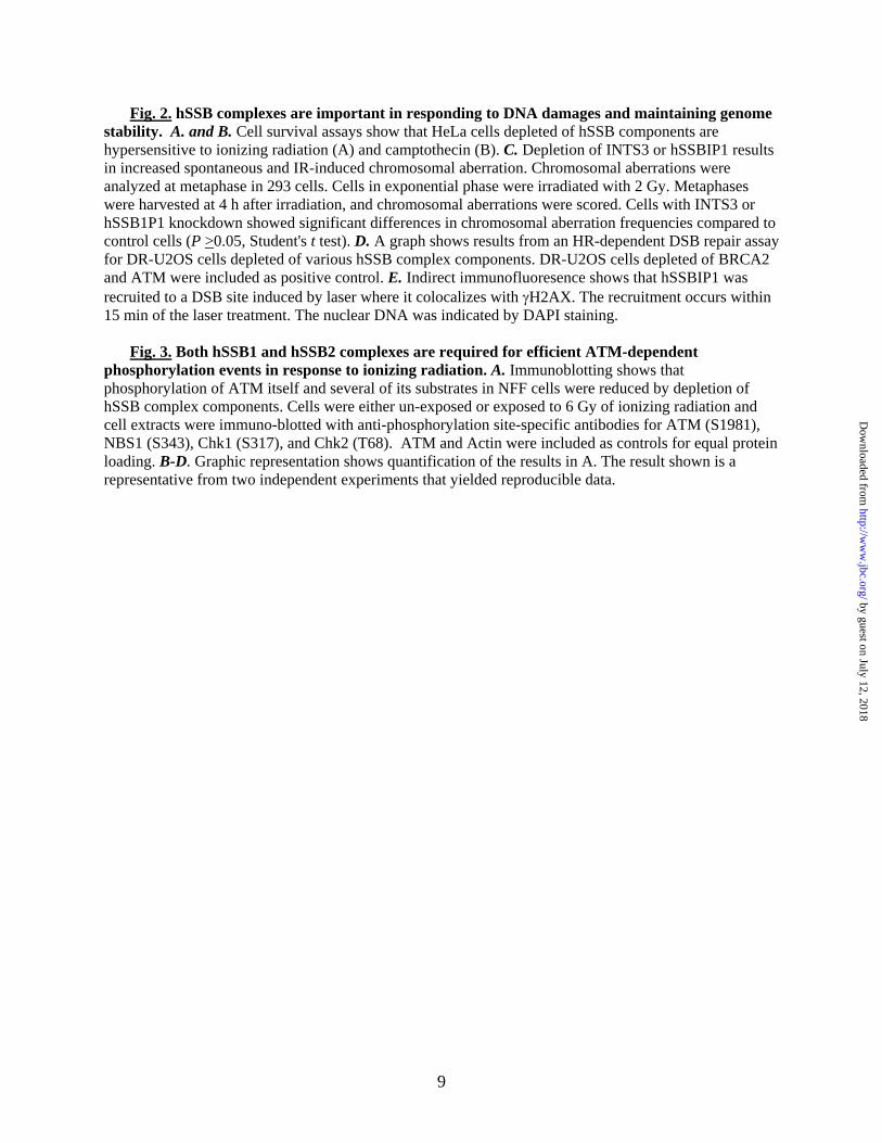
hSSBIP1 (C9ORF80). Based on sequence similarity between the OB-fold domains of hSSB2 and RMI2, we predicted that hSSB2 may resemble RMI2 as part of a multiprotein complex that participates in DNA damage responses. To investigate this hypothesis, we constructed a HEK293 cell line stably expressing FLAG-tagged hSSB2 and immunoprecipitated hSSB2-associated complex with a Flag antibody. To distinguish hSSB2 complex components from contaminating polypeptides, we also performed a mock immunoprecipitation (IP) from regular HEK293 cells that do not express tagged hSSB2. SDS-PAGE and silver-staining analyses revealed the presence of three specific major polypeptides in the Flag-hSSB2 IP, with apparent molecular weights of about 115, 25, and 10 KDa (Fig. 1A, compare lanes 3 with 2). By mass spectrometry the 25 KDa polypeptide was identified as hSSB2. The 115 KDa protein was identified as INTS3, a subunit of the integrator complex that interacts with C-terminal repeats of RNA polymerase II and mediates RNA processing (20-22). The 10 KDa protein was identified as a protein with unknown function, C9ORF80 (Genbank accession number: CAH71936). We renamed this protein hSSBIP1 (hSSB-Interacting Protein 1) as a part of the hSSB complex.
To demonstrate that hSSB2, INTS3 and hSSBIP1 are components of an endogenous complex rather than artifacts due to over-
expression of Flag-hSSB2, we used an hSSB2 antibody to immunoprecipitate the endogenous form of the protein and its associated polypeptides. Silver staining revealed the presence of about 10 major polypeptides, three of which displayed molecular weights similar to components immunoprecipitated by the Flag antibody (Fig. 1B). These three polypeptides were found to be hSSB2, INTS3, and hSSBIP1 by both mass spectrometry (data not shown) and immunoblotting (Fig. 1C, lanes 13-15). The other polypeptides were likely contaminants due to antibody crossreactivity (data not shown). The fact that INTS3 and hSSBIP1 can be co-immunopurified by two independent hSSB2 antibodies strongly suggests that they are components of an hSSB2 complex.
Reciprocal IP with INTS3 and hSSBIP1 antibodies yielded not only these two proteins, but also hSSB2, as revealed by immunoblotting (Fig. 1C, Lanes 4-6, and 7-9). The findings that hSSB2, INTS3 and hSSBIP1 can be co-immunoprecipitated by multiple antibodies provide further evidence that they constitute one complex.
hSSB1 forms a separate complex with INTS3 and hSSBIP1. IP-coupled immunoblotting analysis is a biased approach that may not reveal major components of a complex. To avoid the bias, we used silver staining and mass spectrometry to analyze polypeptides immunoprecipitated by the INTS3 antibody. Three major polypeptides of about 115, 33, and 10 KDa were obtained (Fig. 1D). Mass spectrometry identified the 115 and 10 KDa polypeptides as INTS3 and hSSBIP1, consistent with their association in an hSSB2 complex (Fig. 1C, lanes 4-9). Interestingly, the 33 KDa polypeptide was identified as hSSB1, indicating that INTS3 and hSSBIP1 not only form a complex with hSSB2, but also with hSSB1. In accord with this notion, immunoblotting showed that polypeptides immunoprecipitated by INTS3 or hSSBIP1 antibody contained not only hSSB2, but also hSSB1 (Fig. 1C, lane 6, 9). Moreover, reciprocal IP by hSSB1 antibody yielded both INTS3 and hSSBIP1 (Fig. 1C, lanes 10-12). Together, the data indicate that INTS3 and hSSBIP1 associate with both hSSB1 and hSSB2.
To distinguish whether hSSB1 and hSSB2 are present in a single or two separate complexes with
3
by guest on July 12, 2018http://w
ww
.jbc.org/D
ownloaded from
INTS3 and hSSBIP1, we performed IP-Western analyses and found that hSSB1 and hSSB2 are mutually excluded from one another’s immunoprecipitate (Fig. 1C, lane 12, 15). The data indicate that hSSB1 and hSSB2 form two separate complexes, each of which contains INTS3 and hSSBIP1. Notably, although both hSSB1 and hSSB2 were detected in the INTS3 immunoprecipitate by immunoblotting (Fig. 1C, lane 6), only hSSB1 (about 35 KDa), but not hSSB2 (about 25 KDa), was observed as an abundant polypeptide by the silver-staining (Fig. 1D). The data suggest that hSSB1 is more abundant than hSSB2, so that majority of INTS3 in the extract is present in the hSSB1 complex. This interpretation is consistent with the data that the stability of INTS3 is strongly dependent on the presence of hSSB1, but not hSSB2 (see below).
We fractionated the HeLa nuclear extracts by Superose 6 gel-filtration chromatography, and found that both hSSB1 and hSSB2 co-fractionated with INTS3 and hSSBIP1, with the peak of each protein near fraction 36 (Fig. 1E). The data are consistent with the notion that the two hSSB proteins form two separate complexes of the same size with INTS3 and hSSBIP1. Fraction 36 corresponds to a globular complex of about 500 KDa, which is much larger than the sum of the calculated molecular weight of hSSB, INTS3 and hSSBIP1 (about 150 KDa). The results imply that the hSSB complex may contain more than one copy of each subunit, or may not be globular. Notably, both hSSB1 and hSSB2 fractionated in an additional peak corresponding to complexes of about 150 and 100 kDa, respectively. These molecular weights are about 4-times of the calculated mass of each protein, hinting that hSSB may form homotetramers. This notion is supported by previous findings that bacterial SSB exists as homotetramers. A portion of INTS3 (about 30% of less) also fractionated in an additional peak (faction 26) corresponding to a complex of about 1 MDa, which could be derived from the integrator complex reported previously (20).
INTS3 and hSSB1 are essential for stability of the hSSB1 complex. The stability of a component of multiprotein complexes often depends on the presence of the other components, so that when one protein is depleted, the others become unstable and show reduced cellular levels (7,23,24).
Usually, components with direct interactions show the strongest interdependence for their stability, perhaps because their proper folding requires interacting partners. We depleted various components of hSSB1 and hSSB2 complexes and examined their interdependence for protein stability. When INTS3 was depleted in neonatal foreskin fibroblast (NFF) cells, the levels of both hSSB1 and hSSBIP1 were reduced about 60% (Fig. 1F, lane 2, and Fig. 1G). Similarly, when hSSB1 was depleted, the levels of INTS3 and hSSBIP1 were also reduced about 50% and 30%, respectively (Fig. 1F, lane 4, and Fig. 1G). These data are consistent with the notion that these three proteins are components of one complex, and that stability of this complex strongly depends on INTS3 and hSSB1. The depletion of hSSBIP1 reduced the level of INTS3 by about 30%, but did not significantly affect the level of hSSB1. It was noted that hSSBIP1 siRNA had poor depletion efficiency compared INTS3 siRNA, as they depleted their targets by 70% and 90%, respectively. It is possible that the different effects of INTS3 and hSSBIP1 siRNAs may be partially attributed to their differential depletion efficiency. While INTS3 depletion strongly reduced the level of hSSB1, it did not significantly affect the level of hSSB2 (Fig. 1F, lane 2, and Fig. 1G). Likewise, deletion of hSSB2 had no significant effect on the levels of INTS3 and hSSBIP1 (Fig. 1F, lane 3, and Fig. 1G). The data are consistent with the INTS3 immunoprecipitation data (Fig. 1D), and we infer that the majority of INTS3 and hSSBIP1 that forms hSSB complexes are in the complex with hSSB1 but not hSSB2. This may help to explain the observation that the hSSB2 level was only marginally affected by INST3 knockdown because such knockdown will generate misfolded hSSB1 in larger amounts than hSSB2, so that the former will be more easily recognized and degraded than the latter when they compete for the limited capacity of cellular protein degradation machinery. We noticed that INTS3 knockdown reduced levels of both hSSB1 and hSSB2 in two cancer cell lines, U2OS and HeLa (Supplementary Figure 1). Perhaps, these cancer cell lines have more active protein degradation machinery so that can efficiently remove both misfolded hSSB proteins.
4
by guest on July 12, 2018http://w
ww
.jbc.org/D
ownloaded from

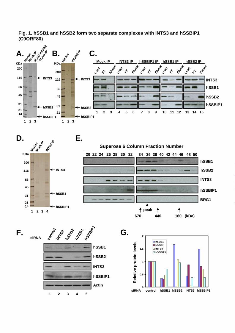
hSSB complexes are required for cellular resistance to ionizing radiation (IR) and camptothecin. The fact that hSSB1 and hSSB2 complexes share two identical components implies that they may have similar functions. We examined whether cells depleted of these two proteins have common phenotypes. hSSB1-depleted cells have previously been shown to be hypersensitive to IR, which can generate DSBs (12). We found that cells depleted of hSSB2 also were hypersensitive to IR (Fig. 2A). In addition, cells depleted of hSSB complex components, INTS3 and hSSBIP1, displayed similar level of IR sensitivity as judged by clonogenic survival (Fig. 2A).
We found that cells depleted of various SSB complex components also displayed sensitivity to camptothecin (Fig. 2B), a topoisomerase I (Top1) inhibitor that can block transcription and replication, and also induce DNA single or double strand breaks (25-27). The data suggest that hSSB complexes may participate in repair of several types of DNA damages.
hSSB complexes are required for maintenance of genome stability
Cells depleted hSSB1 and hSSB2 have been shown to exhibit elevated levels of both spontaneous and IR-induced chromosomal aberrations (12)(K. K. Khanna, manuscript submitted). We found that cells depleted of INTS3 and hSSBIP1 also showed elevated levels of chromosomal aberrations (chromosome and chromatid breaks with fragments) in the absence or presence of IR (Fig. 2C). These data are consistent with the notion that both hSSB complexes are required for genome stabilization and cellular resistance to IR.
hSSB1 and hSSB2 depleted cells have reduced efficiency of HR-dependent DNA repair. hSSB1-depleted cells have been shown to display decreased efficiency in HR-dependent repair of a DSB induced by the I-SceI restriction enzyme (12). Using the same assay, we found that hSSB2-depleted cells exhibited a 60% reduction in HR-dependent repair of DSB (Fig. 2D). This reduction is comparable to that of cells depleted of hSSB1, suggesting that both hSSB proteins participate in HR-dependent DSB repair.
We found that depletion of either INTS3 or hSSBIP1 slightly reduced the HR-dependent repair of DSB (about 10-20%) (Fig. 2D), implying that these components may be largely dispensable for HR-dependent repair. Their effects on HR might be indirect, due to destabilization of hSSB proteins.
hSSBIP1 colocalizes with H2AX at laser-induced DSBs. It has been reported that hSSB1 can be recruited to DNA damage foci, where it colocalizes with γH2AX, a phosphorylated histone variant that specifically marks the region of DSBs (12). This recruitment occurs rapidly within 30 min of IR. We found that one component of hSSB complexes, hSSBIP1, was similarly recruited to a DNA damage region generated by a narrow laser beam within nucleus (Fig. 2E). This type of damage should generate DSBs, as H2AX was recruited to the site with 5 min of laser treatment (data not shown). The recruitment of hSSBIP1 was also rapid, within 15 min of laser treatment. Together with the published data on hSSB1 (12), these findings suggest that whole hSSB complexes may rapidly relocate to DSBs, where they participated in repair and signaling reactions.
We were unable to perform similar analyses for other components of hSSB complexes, due to high non-specific backgrounds of corresponding antibodies (data not shown).
Both hSSB1 and hSSB2 function in ATM-dependent signaling pathway. ATM-dependent phosphorylation of many checkpoint proteins is essential for IR-induced checkpoint activation (28). hSSB1 depleted cells have been shown to have impaired ATM-dependent phosphorylation of its substrates and itself (12) following IR. We found that hSSB2 depleted cells also had reduced ATM autophosphorylation (S1981), as well as basal and IR-induced phosphorylation of substrates NBS1 (S343) and Chk1 (S317) (Fig. 3A, compare lanes 5-6 with lanes 1-2). The reduced phosphorylation in hSSB2-depleted cells was not as drastic as that in hSSB1-depleted cells in response to IR, with the former cells always displaying a higher level of the phosphorylated protein than the latter: ATM (45% vs. 35%) (Fig. 3B), NBS (40% vs. 26%) (Fig. 3C), Chk1 (66% vs. 8%) (Fig. 3D), and Chk2 (109% vs. 30%) (Fig. 3E). The results suggest that both hSSB1 and hSSB2 participate in ATM-
5
by guest on July 12, 2018http://w
ww
.jbc.org/D
ownloaded from

dependent signaling pathways, but hSSB1 may play a relatively more important role.
We found that cells-depleted of INTS3 and hSSBIP1 also had reduced levels of ATM autophosphorylation and some of its substrates in the absence or presence of IR (Fig. 3A, compare lanes 7-10 to 1-2; Fig. 3B-E). Thus, the entire hSSB complexes may participate in ATM-dependent signaling. The reduction in phosphorylation when INTS3 or hSSBIP1 is depleted is weaker than that when hSSB1 or hSSB2 is depleted (Fig. 3B-E), hinting that the former two proteins may play a lesser role than the latter two during this process. Because depletion of INTS3 decreases stability of hSSB1 and hSSB2 (Fig. 1F), some effect of INTS3 depletion could be indirect.
DISCUSSION
OB-fold containing proteins often assemble
into multiprotein complexes and function critically in DNA damage response pathways. A previous study has shown that one highly conserved OB-fold protein, hSSB1, participates in HR-dependent repair of DSBs and ATM-mediated signaling pathway (12). Here we show that hSSB1 and its homolog, hSSB2, form separate complexes with two identical proteins, INTS3 and hSSBIP1 (C9ORF80). The fact that hSSB1 and hSSB2 are homologous and associate with identical partners implies that their respective complexes likely have similar structures and functions. Indeed, we showed that hSSB2-depleted cells resemble hSSB1-depleted cells in several phenotypes, including hypersensitivity to DNA damaging reagents, reduced efficiency in HR-dependent repair of DSBs, and defective ATM-dependent phosphorylation pathway in response to IR, suggesting that the two SSB proteins may have non-overlapping functions in DNA damage response. Moreover, hSSBIP1 was rapidly recruited to DSBs (Fig. 2E), similar to previous findings for hSSB1 (12). Furthermore, cells depleted of INTS3 or hSSBIP1 showed increased chromosomal aberrations in the absence or presence of IR, similar to those depleted of hSSB1 and hSSB2 (12)(K.K. Khanna, manuscript submitted). Our data suggest that the two hSSB complexes may employ similar mechanisms in
mediating repair of DSBs and ATM-dependent damage response pathways.
Although hSSB1 and hSSB2 are homologous and form complexes with the same partners, the two proteins appear to have some interesting differences. First, hSSB1 complex may be more abundant than the hSSB2 complex, based on the silver-staining data that hSSB1, but not hSSB2, is one of the major polypeptides co-immunoprecipitating with INTS3 (Fig. 1D), and that the stability of INTS3 also strongly depends on hSSB1, but not hSSB2. Second, the hSSB1 complex may be more important than the hSSB2 complex in mediating ATM-dependent signaling events, as cells depleted of hSSB1 have a stronger reduction in ATM-dependent phosphorylation for several substrates than do those depleted of hSSB2 (Fig. 3D, 3E). The data suggest that the two hSSB complexes also have overlapping functions in DNA damage response.
How may INTS3 and hSSBIP1 facilitate the functions of hSSB proteins? Bioinformatic analyses failed to reveal domains of known function within these proteins. INTS3 has previously been identified as a component of an integrator complex that interacts with the CTD domain of RNA polymerase II, but its exact role in the integrator complex remains unclear (20). Cells depleted of INTS3 or hSSBIP1 display hypersensitivity to DNA damage reagents, increased genomic instability, and a modest reduction in ATM-mediated phosphorylation, suggesting that both proteins may play a role in DNA repair and signaling within the hSSB complexes. One possible role for INTS3 may be to serve as a scaffold protein to allow assembly of hSSB complexes. INTS3 (about 115 KDa) is considerably larger than hSSB (about 30 KDa) and hSSBIP1 (about 10 KDa), consistent with such a role. Also, INTS3 strongly decreased the stability of the other two partners, whereas depletion of either hSSB1 or hSSBIP1 only led to a modest effect on the stability of each other. Thus, INTS3 may interact with both hSSB1 and hSSBIP1 to assist their proper folding and assembly into the complex. The effect of INTS3 depletion on DNA repair and ATM signaling could then be indirect---through destabilization of hSSB1 and hSSB2. Alternatively, INTS3 may possess an as yet unidentified activity in binding DNA or other proteins.
6
by guest on July 12, 2018http://w
ww
.jbc.org/D
ownloaded from
7
Unlike INTS3, whose depletion strongly reduced the levels of hSSB1 and hSSBIP1 (about 60% each), depletion of hSSBIP1 did not significantly affect the level of hSSB1 and only modestly reduced levels of INTS3 (30%). Nevertheless, cells depleted of hSSBIP1 show sensitivity to DNA damaging agents indistinguishable from that of cells depleted of INTS3. The role of hSSBIP1 may thus be not simply through stabilization of other components
of the complex. It remains to be determined if INTS3 and hSSBIP1 interact with DNA and other proteins.
The intrinsic potential of INTS3 interactions is emphasized by findings that it is one of the genes frequently amplified in hepatocellular carcinoma (29). Over-expression of INTS3 might alter the normal DNA damage response pathway, contributing to cancer development.
REFERENCES
1. Arcus, V. (2002) Curr Opin Struct Biol 12, 794-801 2. Bochkarev, A., Bochkareva, E., Frappier, L., and Edwards, A. M. (1999) EMBO J 18,
4498-4504 3. Iftode, C., Daniely, Y., and Borowiec, J. A. (1999) Crit Rev Biochem Mol Biol 34, 141-
180 4. Theobald, D. L., Mitton-Fry, R. M., and Wuttke, D. S. (2003) Annu Rev Biophys Biomol
Struct 32, 115-133 5. Wold, M. S. (1997) Annu Rev Biochem 66, 61-92 6. Yang, H., Jeffrey, P. D., Miller, J., Kinnucan, E., Sun, Y., Thoma, N. H., Zheng, N.,
Chen, P. L., Lee, W. H., and Pavletich, N. P. (2002) Science 297, 1837-1848 7. Xu, D., Guo, R., Sobeck, A., Bachrati, C. Z., Yang, J., Enomoto, T., Brown, G. W.,
Hoatlin, M. E., Hickson, I. D., and Wang, W. (2008) Genes Dev 22, 2843-2855 8. Lei, M., Podell, E. R., Baumann, P., and Cech, T. R. (2003) Nature 426, 198-203 9. Xin, H., Liu, D., Wan, M., Safari, A., Kim, H., Sun, W., O'Connor, M. S., and Songyang,
Z. (2007) Nature 445, 559-562 10. Liu, Y., and West, S. C. (2008) Genes Dev 22, 2737-2742 11. Brosh, R. M., Jr., Li, J. L., Kenny, M. K., Karow, J. K., Cooper, M. P., Kureekattil, R. P.,
Hickson, I. D., and Bohr, V. A. (2000) J Biol Chem 275, 23500-23508. 12. Richard, D. J., Bolderson, E., Cubeddu, L., Wadsworth, R. I., Savage, K., Sharma, G. G.,
Nicolette, M. L., Tsvetanov, S., McIlwraith, M. J., Pandita, R. K., Takeda, S., Hay, R. T., Gautier, J., West, S. C., Paull, T. T., Pandita, T. K., White, M. F., and Khanna, K. K. (2008) Nature 453, 677-681
13. New, J. H., and Kowalczykowski, S. C. (2002) J Biol Chem 277, 26171-26176 14. Sugiyama, T., and Kowalczykowski, S. C. (2002) J Biol Chem 277, 31663-31672 15. Dignam, J. D., Martin, P. L., Shastry, B. S., and Roeder, R. G. (1983) Methods Enzymol
101, 582-598 16. Meetei, A. R., Sechi, S., Wallisch, M., Yang, D., Young, M. K., Joenje, H., Hoatlin, M.
E., and Wang, W. (2003) Mol Cell Biol 23, 3417-3426 17. Pandita, R. K., Sharma, G. G., Laszlo, A., Hopkins, K. M., Davey, S., Chakhparonian, M.,
Gupta, A., Wellinger, R. J., Zhang, J., Powell, S. N., Roti Roti, J. L., Lieberman, H. B., and Pandita, T. K. (2006) Mol Cell Biol 26, 1850-1864
18. Pierce, A. J., Johnson, R. D., Thompson, L. H., and Jasin, M. (1999) Genes Dev 13, 2633-2638
19. Xia, B., Sheng, Q., Nakanishi, K., Ohashi, A., Wu, J., Christ, N., Liu, X., Jasin, M., Couch, F. J., and Livingston, D. M. (2006) Mol Cell 22, 719-729
by guest on July 12, 2018http://w
ww
.jbc.org/D
ownloaded from

20. Baillat, D., Hakimi, M. A., Naar, A. M., Shilatifard, A., Cooch, N., and Shiekhattar, R. (2005) Cell 123, 265-276
21. Jacobs, E. Y., Ogiwara, I., and Weiner, A. M. (2004) Mol Cell Biol 24, 846-855 22. Uguen, P., and Murphy, S. (2003) EMBO J 22, 4544-4554 23. Meetei, A. R., Levitus, M., Xue, Y., Medhurst, A. L., Zwaan, M., Ling, C., Rooimans, M.
A., Bier, P., Hoatlin, M., Pals, G., de Winter, J. P., Wang, W., and Joenje, H. (2004) Nat Genet 36, 1219-1224
24. Yin, J., Sobeck, A., Xu, C., Meetei, A. R., Hoatlin, M., Li, L., and Wang, W. (2005) EMBO J 24, 1465-1476
25. Avemann, K., Knippers, R., Koller, T., and Sogo, J. M. (1988) Mol Cell Biol 8, 3026-3034
26. Pommier, Y., Redon, C., Rao, V. A., Seiler, J. A., Sordet, O., Takemura, H., Antony, S., Meng, L., Liao, Z., Kohlhagen, G., Zhang, H., and Kohn, K. W. (2003) Mutat Res 532, 173-203
27. Pommier, Y., Pourquier, P., Fan, Y., and Strumberg, D. (1998) Biochim Biophys Acta 1400, 83-105
28. Khanna, K. K., and Jackson, S. P. (2001) Nat Genet 27, 247-254 29. Inagaki, Y., Yasui, K., Endo, M., Nakajima, T., Zen, K., Tsuji, K., Minami, M., Tanaka,
S., Taniwaki, M., Itoh, Y., Arii, S., and Okanoue, T. (2008) Cancer Genet Cytogenet 180, 30-36
FOOTNOTES
We thank Dr. David Schlessinger for critical reading of the manuscript. This work was supported in part by the Intramural Research Program of the National Institute on Aging (Z01: AG000657-09), National Institute of Health, by NIH:CA123232 and CA129537 (to T.K.P.), and National Health and Medical Research Council of Australia (to K.K.K.).
FIGURE LEGENDS
Fig. 1. hSSB1 and hSSB2 form two separate complexes with INTS3 and hSSBIP1 (C9ORF80). A. A silver-stained SDS-gel showing major polypeptides immunoprecipitated by Flag antibody from HEK-293 cells stably expressing Flag-hSSB2. A Mock IP was done using cells that do not express FLAG-hSSB2 (Mock). B. A silver-stained gel showing major polypeptides immunoprecipitated by a polyclonal antibody against hSSB2 from HeLa nuclear extract. Arrows indicate the three components of the hSSB2 complex that have been identified by mass spectrometry. C. Immunoblotting to show co-IP of hSSB complex components from HeLa nuclear extract. A control IP using rabbit preimmune serum was included (Mock). The nuclear extract (Load), flowthrough (FT), and eluted fractions are indicated at the top. D. A silver-stained SDS-gel showing major polypeptides immunoprecipitated by an INTS3 antibody from Superose 6 fractions of HeLa extract that are enriched of hSSB complexes (fractions 34-38, see Fig. 1E). E. Immunoblotting shows that components of hSSB complexes co-fractionate near fraction 36 on a Superose 6 gel-filtration column. BRG1, a component of 1MDa SWI/SNF complex, was included as an internal control. The fractions corresponding to calibration proteins with known molecular weight are indicated at the bottom. F. Immunoblotting to show that the levels of various hSSB complex components in NFF cells were reduced by siRNA depletion of its partners, indicating that hSSB complex components are inter-dependent for their stability. Immunoblotting of actin was used as loading control. G. Graphic presentation showing quantification of the immunoblotting data in F.
8
by guest on July 12, 2018http://w
ww
.jbc.org/D
ownloaded from
9
Fig. 2. hSSB complexes are important in responding to DNA damages and maintaining genome stability. A. and B. Cell survival assays show that HeLa cells depleted of hSSB components are hypersensitive to ionizing radiation (A) and camptothecin (B). C. Depletion of INTS3 or hSSBIP1 results in increased spontaneous and IR-induced chromosomal aberration. Chromosomal aberrations were analyzed at metaphase in 293 cells. Cells in exponential phase were irradiated with 2 Gy. Metaphases were harvested at 4 h after irradiation, and chromosomal aberrations were scored. Cells with INTS3 or hSSB1P1 knockdown showed significant differences in chromosomal aberration frequencies compared to control cells (P >0.05, Student's t test). D. A graph shows results from an HR-dependent DSB repair assay for DR-U2OS cells depleted of various hSSB complex components. DR-U2OS cells depleted of BRCA2 and ATM were included as positive control. E. Indirect immunofluoresence shows that hSSBIP1 was recruited to a DSB site induced by laser where it colocalizes with γH2AX. The recruitment occurs within 15 min of the laser treatment. The nuclear DNA was indicated by DAPI staining.
Fig. 3. Both hSSB1 and hSSB2 complexes are required for efficient ATM-dependent
phosphorylation events in response to ionizing radiation. A. Immunoblotting shows that phosphorylation of ATM itself and several of its substrates in NFF cells were reduced by depletion of hSSB complex components. Cells were either un-exposed or exposed to 6 Gy of ionizing radiation and cell extracts were immuno-blotted with anti-phosphorylation site-specific antibodies for ATM (S1981), NBS1 (S343), Chk1 (S317), and Chk2 (T68). ATM and Actin were included as controls for equal protein loading. B-D. Graphic representation shows quantification of the results in A. The result shown is a representative from two independent experiments that yielded reproducible data.
by guest on July 12, 2018http://w
ww
.jbc.org/D
ownloaded from
E.
hSSB2
hSSB1
Moc
k IP
INTS
3 IP
hSSBIP1
20 22 24 26 28 30 32 34 36 38 40 42 44 46 48 50
Superose 6 Column Fraction Number
670 440 160 (kDa)
peak
INTS3
Mar
ker
45
31
21
200
116
66
KDa
14
BRG1
INTS3
0
0.5
1
1.5
2
hSSB1
hSSB2
INTS3
hSSBIP1
1 2 3 4 5
1 2 3 4
Fig. 1. hSSB1 and hSSB2 form two separate complexes with INTS3 and hSSBIP1 (C9ORF80)
siRNA cont
rol
INTS
3hS
SB2
hSSB
1hS
SBIP
1
hSSB2
Actin
hSSB1
hSSBIP1
INTS3
Rel
etiv
ep
rote
in le
vels
siRNA hSSB1 hSSB2 INTS3 hSSBIP1control
D.
F. G.
hSSB1
hSSBIP1
hSSB
2 IP
Mar
ker
1 2 3
B.
INTS3
hSSB2
hSSBIP1
45
31
21
200
116
66
KDa
45
31
200
116
66
KDa
14
1 2 3
INTS3
hSSB2
hSSBIP1
Moc
k IP
FLAG
-hSS
B2
FLAG
-IP
Mar
ker
Load
FT Elua
te
Load
FT Elua
te
Load
FT Elua
te
Load
FT Elua
te
Load
FT Elua
te
Mock IP INTS3 IP hSSBIP1 IP hSSB1 IP hSSB2 IP
C.
1 2 3 4 5 6 7 8 9 10 11 12 13 14 15
A.
hSSB2
hSSB1
hSSBIP1
INTS3
21
by guest on July 12, 2018http://w
ww
.jbc.org/D
ownloaded from

DAPI H2AX hSSBIP1 Merged
A.
E.
B.
Fig. 2. Cells depleted of hSSB complexes have DNA damage sensitivity, reduced HR-dependent DNA repair efficiency, increased chromosomal instability; hSSBIP1 rapidly localizes at DSBs
0
1
2
3
4
5
Control
Control siRNA
INTS3 siRNA
hSSBIP1 siRNA
A-T
C. D.
IR (Gy)
0 2
Ab
erra
tio
ns
per
met
aph
ase
0.00
0.20
0.40
0.60
0.80
1.00
1.20
HR
rep
air
effi
cien
cy
cont
rol
BRCA2
ATMhS
SB1
hSSB
2IN
TS3
hSSB
IP1
siRNA
0.1
1.0
0 100 200
Control siRNA
hSSB2 siRNA
INTS3 siRNA
hSSBIP1 siRNA0.1
1.0
0 2 4 6 8
Control siRNA
hSSB2 siRNA
INTS3 siRNA
hSSBIP1 siRNA
IR (Gy) Camptothecin (nM)
Su
rviv
ing
fra
ctio
n
Su
rviv
ing
fra
ctio
n
by guest on July 12, 2018http://w
ww
.jbc.org/D
ownloaded from

E.D.
B. C.
NBS1-S343-
ATM
Chk2-T68-
ATM-S1981-
Chk1-S317-
IR (6 Gy) - + - + - + - + - +control hSSB1 hSSB2 INTS3 hSSBIP1siRNA
1 2 3 4 5 6 7 8 9 10
A.
0
0.5
1
1.5
Actin
Chk2-T68- P
0
0.5
1
1.5Chk1-S317- P
0
0.5
1
1.5
0
0.5
1
1.5
no IR IR (6Gy)
sico
ntr
ol
sih
SS
B1
sih
SS
B2
siIN
TS
3si
hS
SB
IP1
Ph
op
ho
ryla
ted
pro
tein
leve
l
NBS-S343- PATM-S1981- P
Fig. 3. Both hSSB1 and hSSB2 are required for normal phosphorylation events in response to DNA damage
sico
ntr
ol
sih
SS
B1
sih
SS
B2
siIN
TS
3si
hS
SB
IP1
no IR IR (6Gy)
sico
ntr
ol
sih
SS
B1
sih
SS
B2
siIN
TS
3si
hS
SB
IP1
Ph
op
ho
ryla
ted
pro
tein
leve
l
sico
ntr
ol
sih
SS
B1
sih
SS
B2
siIN
TS
3si
hS
SB
IP1
no IR IR (6Gy)
sico
ntr
ol
sih
SS
B1
sih
SS
B2
siIN
TS
3si
hS
SB
IP1
Ph
op
ho
ryla
ted
pro
tein
leve
l
sico
ntr
ol
sih
SS
B1
sih
SS
B2
siIN
TS
3si
hS
SB
IP1
no IR IR (6Gy)
sico
ntr
ol
sih
SS
B1
sih
SS
B2
siIN
TS
3si
hS
SB
IP1
Ph
op
ho
ryla
ted
pro
tein
leve
l
sico
ntr
ol
sih
SS
B1
sih
SS
B2
siIN
TS
3si
hS
SB
IP1
P
P
P
P
by guest on July 12, 2018http://w
ww
.jbc.org/D
ownloaded from

Derek Richard, Michael Seidman, Tej K. Pandita, Kum Kum Khanna and Weidong WangYongjiang Li, Emma Bolderson, Rakesh Kumar, Parameswary A. Muniandy, Yutong Xue,
damage responsehSSB1 and hSSB2 form similar multi-protein complexes that participate in DNA
published online July 14, 2009J. Biol. Chem.
10.1074/jbc.C109.039586Access the most updated version of this article at doi:
Alerts:
When a correction for this article is posted•
When this article is cited•
to choose from all of JBC's e-mail alertsClick here
Supplemental material:
http://www.jbc.org/content/suppl/2009/07/14/C109.039586.DC1
by guest on July 12, 2018http://w
ww
.jbc.org/D
ownloaded from