Embed Size (px)
Citation preview

p
Perspectives on Brain DevelopmentMr. Higgins Builds a HouseLinking Brain and Behavioral Development
The Development of the Child’s BrainThe Gross Development of the Human
Nervous SystemThe Origins of Neurons and GliaThe Growth and Development of NeuronsFocus on Disorders: Cerebral PalsyGlial Development
Correlating Behavior with Brain DevelopmentMotor BehaviorsLanguage DevelopmentThe Development of Problem-Solving Ability
Brain Development and the EnvironmentExperience and Cortical OrganizationExperience and Neural ConnectivityCritical Periods for Experience and Brain
DevelopmentAbnormal Experience and Brain DevelopmentFocus on Disorders: Romanian OrphansHormones and Brain DevelopmentInjury and Brain DevelopmentOther Kinds of Abnormal Brain DevelopmentFocus on Disorders: SchizophreniaMental Retardation
How Do Any of Us Develop a Normal Brain?
234 ■
How Does the BrainDevelop?
C H A P T E R
7
Myrleen F. Cate/Photo Network/Picture Quest
Oliver Meckes/Ottawa/Photo Researchers

p
Amonarch butterfly begins life as a fertilized egg
and develops into a caterpillar. After a time, the
caterpillar spins a cocoon, inside of which it lives
as it undergoes a process that transforms it into a butterfly.
The stages of development in the life cycle of a monarch
butterfly are collectively called metamorphosis and are
shown in Figure 7-1. Consider how formidable this insect’s
development is. First, the egg must develop a body,
including a nervous system. This nervous system has to
produce caterpillar-like movements
and control the animal’s feeding
apparatus, which is designed for
munching leaves. Then, during meta-
morphosis, the original nervous sys-
tem has to be reconstructed to control
the flight, feeding, and reproductive
behaviors of a butterfly. The addition
of flying behavior is remarkable
because it requires the use of entirely
different muscles from those used in
crawling. Furthermore, adult monarch
butterflies fly very long distances in
their annual migration and must navi-
gate to the correct geographical loca-
tion. In contrast, the caterpillar’s
main challenge is to find an appropri-
ate food source as it crawls slowly
around in a limited area. It seems that
a caterpillar would need a major
overhaul of its brain to control the
completely reconfigured body and
brand-new behaviors that go with
being a butterfly.
We humans do not metamor-
phose into a different life form in the
course of our development, but the
developmental problems that we face are similar to those
of the monarch. We also begin life as a fertilized egg that
develops a body and a nervous system. When we are
born, however, we are not able to fend for ourselves. Hu-
man offspring are virtually helpless for an extended period
of time. The behavioral demands on the brain of a new-
born include relatively simple actions such as searching
for and recognizing a nipple with which to feed and sig-
naling hunger or discomfort to caregivers. But soon a hu-
man infant undergoes an enormous
transformation. The child’s brain be-
comes able to control a variety of new
behaviors such as crawling and, later,
walking, eating solid foods, using
tools, and learning a language. At
school age, the child’s brain becomes
able to formulate complex ideas,
solve challenging problems, and re-
member large quantities of informa-
tion. And changes in a person’s ner-
vous system do not end with
graduation from college. As the adult
brain begins to age in the third
decade of life, it starts to lose cells
and grows few new ones. The loss
forces the middle-aged brain to recon-
struct some of its parts to forestall the
effects of aging. Brain development,
then, is a continuous process that is
central to our functioning. Changes in
the brain allow us to adapt to the en-
vironment throughout our life cycle.
This chapter answers many ques-
tions about the development of the
human brain. How did your brain
manage to develop from a single em-
bryonic cell into an organ made up of
billions of cells? This question paral-
lels one asked in Chapter 1—namely,
how did the brain evolve from a small
and simple organ into a large and
Figure 7-1 In metamorphosis, the nervous system ofthe monarch butterfly must undergosignificant changes as the insect developsfrom a larva into a caterpillar into abutterfly.
■ 235
Larva
Caterpillar
Cocoon
Adultbutterfly
PERSPECTIVES ON BRAIN DEVELOPMENTTo begin to understand how the brain is constructed, we start with an analogy ofbuilding a house. Do not take this analogy too literally. It is used here simply as a wayof introducing the topic of brain development and some important principles relatedto it.
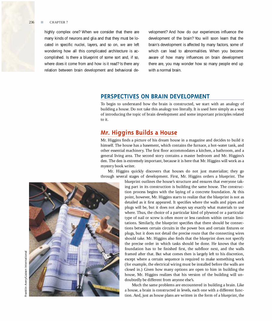
Mr. Higgins Builds a HouseMr. Higgins finds a picture of his dream house in a magazine and decides to build ithimself. The house has a basement, which contains the furnace, a hot-water tank, andother essential machinery. The first floor accommodates a kitchen, a bathroom, and ageneral living area. The second story contains a master bedroom and Mr. Higgins’sden. The den is extremely important, because it is here that Mr. Higgins will work as amystery book writer.
Mr. Higgins quickly discovers that houses do not just materialize; they gothrough several stages of development. First, Mr. Higgins orders a blueprint. The
blueprint outlines the house’s structure and ensures that everyone tak-ing part in its construction is building the same house. The construc-tion process begins with the laying of a concrete foundation. At thispoint, however, Mr. Higgins starts to realize that the blueprint is not asdetailed as it first appeared. It specifies where the walls and pipes andplugs will be, but it does not always say exactly what materials to usewhere. Thus, the choice of a particular kind of plywood or a particulartype of nail or screw is often more or less random within certain limi-tations. Similarly, the blueprint specifies that there should be connec-tions between certain circuits in the power box and certain fixtures orplugs, but it does not detail the precise route that the connecting wiresshould take. Mr. Higgins also finds that the blueprint does not specifythe precise order in which tasks should be done. He knows that thefoundation has to be finished first, the subfloor next, and the wallsframed after that. But what comes then is largely left to his discretion,except where a certain sequence is required to make something work(for example, the electrical wiring must be installed before the walls areclosed in.) Given how many options are open to him in building thehouse, Mr. Higgins realizes that his version of the building will un-doubtedly be different from anyone else’s.
Much the same problems are encountered in building a brain. Likea house, a brain is constructed in levels, each one with a different func-tion. And, just as house plans are written in the form of a blueprint, the
p
highly complex one? When we consider that there are
many kinds of neurons and glia and that they must be lo-
cated in specific nuclei, layers, and so on, we are left
wondering how all this complicated architecture is ac-
complished. Is there a blueprint of some sort and, if so,
where does it come from and how is it read? Is there any
relation between brain development and behavioral de-
velopment? And how do our experiences influence the
development of the brain? You will soon learn that the
brain’s development is affected by many factors, some of
which can lead to abnormalities. When you become
aware of how many influences on brain development
there are, you may wonder how so many people end up
with a normal brain.
Fran
klin
Ave
ry/L
iais
on
Inte
rnat
ion
al236 ■ CHAPTER 7
plans for a brain are encoded in genes. As Mr. Higgins learned, architects do notspecify every detail in a blueprint; nor do genes include every instruction for how abrain is assembled and wired. The process of building a brain is just too complex tobe encoded entirely and precisely in genes. For this reason, the fate of billions of braincells is left partly open, especially when it comes to the massive undertaking of form-ing appropriate connections between cells.
If the structure and fate of each brain cell are not specified in advance, what fac-tors do control brain development? Many factors are at work, including special mole-cules, such as hormones. Brain development is also influenced by the experiences thatpeople have both in the womb and after they are born. We return to these influenceslater in this chapter, after examining the major stages in brain development. But firstwe explore how scientists go about studying the interconnected processes of brainand behavioral development.
Linking Brain and Behavioral DevelopmentIn the course of development, changes take place both in the brain and in behavior.Scientists assume that these two lines of development are closely linked. As the braindevelops, neurons become more and more intricately connected, and these increas-ingly complex interconnections underlie increased behavioral complexity.
We can study the relation between brain and behavioral development in three ba-sic ways. First, we can look at the structural development of the nervous system andcorrelate it with the emergence of specific behaviors. For example, we can link the de-velopment of certain brain structures to the development of, say, grasping or crawlingin infants. As the brain structures develop, their functions emerge; these functions aremanifested in behaviors that we can observe.
Structures that develop quickly exhibit their functions sooner than structuresthat develop more slowly. Because the human brain continues to develop well intoadolescence, you should not be surprised that some behavioral abil-ities emerge rather late in development. For example, the frontallobes continue to develop well into adolescence, reaching maturityat about 16 years of age. It follows that certain behaviors controlledby the frontal lobes also are slow to develop.
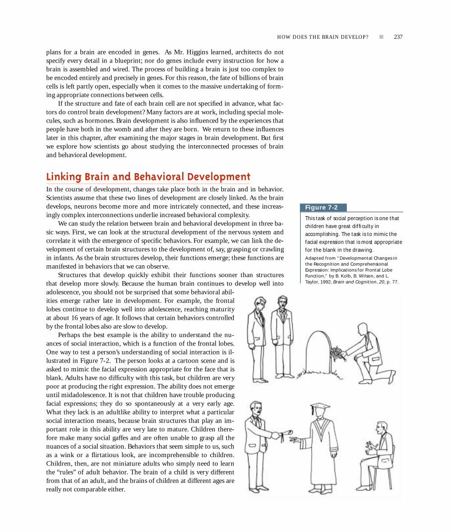
Perhaps the best example is the ability to understand the nu-ances of social interaction, which is a function of the frontal lobes.One way to test a person’s understanding of social interaction is il-lustrated in Figure 7-2. The person looks at a cartoon scene and isasked to mimic the facial expression appropriate for the face that isblank. Adults have no difficulty with this task, but children are verypoor at producing the right expression. The ability does not emergeuntil midadolescence. It is not that children have trouble producingfacial expressions; they do so spontaneously at a very early age.What they lack is an adultlike ability to interpret what a particularsocial interaction means, because brain structures that play an im-portant role in this ability are very late to mature. Children there-fore make many social gaffes and are often unable to grasp all thenuances of a social situation. Behaviors that seem simple to us, suchas a wink or a flirtatious look, are incomprehensible to children.Children, then, are not miniature adults who simply need to learnthe “rules” of adult behavior. The brain of a child is very differentfrom that of an adult, and the brains of children at different ages arereally not comparable either.
HOW DOES THE BRAIN DEVELOP? ■ 237
p
Figure 7-2
This task of social perception is one thatchildren have great difficulty inaccomplishing. The task is to mimic thefacial expression that is most appropriatefor the blank in the drawing. Adapted from “Developmental Changes inthe Recognition and ComprehensionalExpression: Implications for Frontal LobeFunction,” by B. Kolb, B. Wilson, and L.Taylor, 1992, Brain and Cognition, 20, p. 77.

The second way to examine the relation between brain and behavioral develop-ment is to turn our sequence of observations around. First we scrutinize behavior forthe emergence of new abilities, and then we make inferences about underlying neuralmaturation. For example, as language emerges in the young child, we expect to findcorresponding changes in neural structures that control language. In fact, this is whatwe do find. At birth, children do not speak, and even extensive speech training wouldnot enable them to do so. The neural structures that control speech are not yet ma-ture enough. As language emerges, we can conclude that the speech-related structuresin the brain are undergoing the necessary maturation. The same reasoning can be ap-plied to frontal-lobe development. As frontal-lobe structures mature in adolescence,we look for related changes in behavior, but we can also do the reverse: because weobserve new abilities emerging in the teenage years, we infer that they must be con-trolled by late-maturing neural structures.
The third way to study the relation between brain and behavioral development isto identify and study factors that influence both. From this perspective, the mereemergence of a certain fully developed brain structure is not enough; we must alsoknow the events that shape how that structure functions and produce certain kinds ofbehaviors. Some of the events that influence brain function are sensory experience,injuries, and the actions of hormones and abnormal genes. Logically, if behavior is in-fluenced by one of these experiences, then structures in the brain that are changed bythat experience are responsible for the behavioral outcomes. For example, we mightstudy how the abnormal secretion of a hormone affects both a certain brain structureand a certain behavior. We can then infer that, because the observed behavioral ab-normality results from the abnormal functioning of the brain structure, that structuremust normally play some role in controlling the behavior.
By applying each of these three approaches to the study of brain and behavioraldevelopment, we can shed much light on the nature of brain organization and func-tion. We begin by considering the anatomical development of the child’s brain. Wethen explore the behavioral correlates of brain development. Finally, we explore somefactors that influence the development of both the brain and behavior.
In ReviewBrain development can be approached from three different perspectives. First, the struc-tural development can be studied and correlated with the emergence of behavior. Sec-ond, behavioral development can be analyzed and predictions can be made about whatunderlying circuitry must be emerging. Finally, those factors that influence brain andbehavioral development, such as an injury to the brain, can be studied. In this lastapproach, the idea is that events that alter behavioral development should similarly alterstructural development.
THE DEVELOPMENT OF THE CHILD’S BRAINSome 2000 years ago the Roman philosopher Seneca proposed that a human embryowas a miniature person. According to him, the task of development was simply togrow bigger. This idea, known as preformation, was so appealing that, until fairly re-cently, it was widely believed to be true. In fact, even with the development of the mi-croscope, the appeal of preformation was so strong that biologists claimed to be ableto see microscopic horses in horse semen.
238 ■ CHAPTER 7
p

By the middle of the nineteenth century, the idea of preformation began to waneas people realized that embryos looked nothing like the adults that they become. Infact, it was obvious that embryos of different species more closely resembled one an-other than their respective parents. Figure 7-3 shows the strikingsimilarity in the early embryos of species as diverse as salamanders,chickens, and humans. Early in development, all species have a similar-looking primitive head, which is a region with bumps or folds,and all possess a tail. It is only as the embryo develops that it ac-quires the distinctive characteristics of its species. The similarity ofyoung embryos is so great that many nineteenth-century biologistssaw it as evidence for Darwin’s view that vertebrates arose from acommon ancestor millions of years ago.
Although not shown in Figure 7-3, embryos are structurallysimilar in their nervous systems as well as in their bodies. Figure 7-4reveals that the nervous system of a young vertebrate embryo al-ways has three regions: the forebrain, the brainstem (with the mid-brain and hindbrain clearly visible), and the remaining neural tube,which forms the spinal cord. Where do these three regions comefrom? We can answer this question by tracing events as the embryomatures.
HOW DOES THE BRAIN DEVELOP? ■ 239
p
Salamander Chick Human Figure 7-3
The similarity of embryos of differentspecies is striking in the earliest stages ofdevelopment, as these salamander, chick,and human embryos show. This similarityled to the conclusion that embryos arenot miniature versions of adults.
Brainstem
Head
Body
Forebrain
Midbrain
Hindbrain
Neural tube(forms spinalcord)
Figure 7-4
The basic brain regions of the forebrain,the midbrain, and the hindbrain arevisible at about 28 days, as is theremaining neural tube, which will formthe spinal cord.

The Gross Development of the Human Nervous SystemAt the time an egg is fertilized by a sperm, a human zygote consists of just a singlecell. But this cell soon begins to divide; by the 15th day, the embryo resembles a friedegg. It is made of several sheets of cells with a raised area in the middle, as shown inFigure 7-5. The raised area is called the primitive body. By 3 weeks after conception,there is primitive neural tissue, known as the neural plate, which is part of the outer-most layer of embryonic cells. The neural plate first folds to form a groove, called theneural groove, as illustrated in Figure 7-6. The neural groove then curls to form theneural tube, much as a flat sheet of paper can be curled to make a cylinder. Micro-graphs of the neural tube closing in a mouse embryo can be seen in Figure 7-7. Thecells that form the neural tube can be thought of as the “nursery” for the rest of thenervous system. The open region in the center of the tube remains open and becomesthe brain’s ventricles and the spinal canal.
240 ■ CHAPTER 7
p
Neural plate. The thickened region ofthe ectodermal layer that gives rise to theneural tube.
Neural tube. A structure in the earlystage of brain development from whichthe brain and spinal cord develop.
Visit the Web site at www.worthpublishers.com/kolb/chapter7 to link to visual tours of humanfetal development.
Figure 7-5
Development begins at fertilization (day 1), with theformation of the zygote. On day 2, the zygote beginsto divide. On day 15, the embryo begins to form parts.On day 21, the primitive brain and neural groove arevisible. On day 23, the neural tube is forming. On day51, the embryo has a distinctly human form. Adapted from The Developing Human: Clinically OrientedEmbryology (4th ed., p. 61), by K. L. Moore, 1988,Philadelphia: Saunders.
Day 1: Fertilization Day 2: Division Day 15
Day 51
Day 23
Day 21
Embryonicdisc
Precursor ofeye and earpresent
Neuraltube
Developingbrain
Neuralgroove

HOW DOES THE BRAIN DEVELOP? ■ 241
p
Neural plate
Neural groove
21 daysNeural plate(primitive neural tissue)
18 days
Neural groove (closing to form neural tube)
Neural tube Ventricle
22 days
Developingforebrain
Developingheart
Neural tube
24 days
Anterior neural folds (close to form brain)
Neural tube
23 days
Figure 7-6
In the formation of the neural tube, the precursor of thenervous system, a long depression (the neural groove) is firstformed in the neural plate. The neural plate collapsesinward, forming a tube along the length of the dorsalsurface of the embryo. The embryo is shown in aphotograph at 24 days.
Figure 7-7
Scanning electronmicrographs show the closingof the neural tube in a mouseembryo. Reproduced with the permissionof Dr. R. E. Peolman, Laboratoryof Anatomy, University ofLeyden.
(A) Day 9 (B) Day 10 (C) Day 11
SPL/
CM
SP
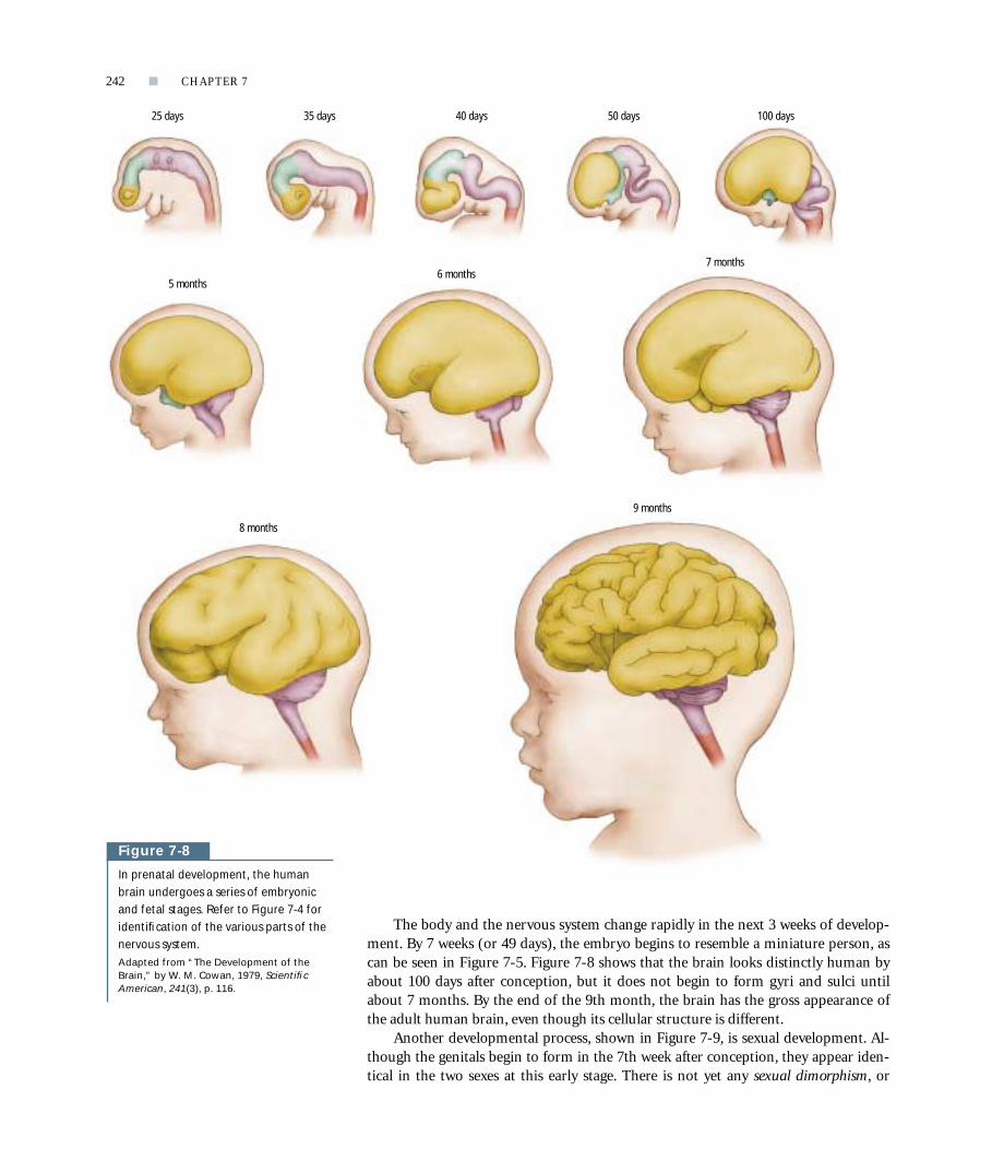
The body and the nervous system change rapidly in the next 3 weeks of develop-ment. By 7 weeks (or 49 days), the embryo begins to resemble a miniature person, ascan be seen in Figure 7-5. Figure 7-8 shows that the brain looks distinctly human byabout 100 days after conception, but it does not begin to form gyri and sulci untilabout 7 months. By the end of the 9th month, the brain has the gross appearance ofthe adult human brain, even though its cellular structure is different.
Another developmental process, shown in Figure 7-9, is sexual development. Al-though the genitals begin to form in the 7th week after conception, they appear iden-tical in the two sexes at this early stage. There is not yet any sexual dimorphism, or
242 ■ CHAPTER 7
p
100 days25 days 35 days 40 days 50 days
8 months
9 months
5 months6 months
7 months
Figure 7-8
In prenatal development, the humanbrain undergoes a series of embryonicand fetal stages. Refer to Figure 7-4 foridentification of the various parts of thenervous system. Adapted from “The Development of theBrain,” by W. M. Cowan, 1979, ScientificAmerican, 241(3), p. 116.

structural difference between the two sexes. Then, about 60 days after conception,male and female genitals start to become distinguishable. But what does this sexualdifferentiation have to do with brain development? The answer is that sexual differen-tiation is stimulated by the presence of the hormone testosterone in male embryos.Testosterone changes the genetic activity of certain cells, most obviously those thatform the genitals. However, genital cells are not the only cells influenced by testos-terone. The brain also has cells that respond to this hormone, so certain regions of theembryonic brain also may begin to show sexual dimorphism, beginning about 60days after conception.
The Origins of Neurons and GliaThe cells lining the neural tube, the nursery for the brain, are known as neural stemcells. A stem cell is a cell with an extensive capacity for self-renewal. It divides andproduces two stem cells, which both can divide again. In adulthood, one stem cell diesafter division, leaving a constant number of dividing stem cells. In an adult, the neuralstem cells line the ventricles and thus form what is called the ventricular zone.
If lining the ventricles were all that stem cells did throughout the decades of a hu-man life, they would seem like odd kinds of cells to possess. But stem cells also have
HOW DOES THE BRAIN DEVELOP? ■ 243
p
Neural stem cells. Cells that give rise toall neurons in the nervous system.
Ventricular zone. The zone surroundingthe ventricles in which stem cells reside.
Figure 7-9
Sexual differentiation in the humaninfant. Early in development (indifferentstage), the human male and femaleembryos are identical. In response totestosterone in male embryos, thegenitalia begin to develop into the malestructure at about 60 days. In theabsence of testosterone, the femalestructure emerges. Parallel changes takeplace in the brain in response to theabsence or presence testosterone.
Developingpenis
Urogenitalmembrane
Urethralfolds
Penis
Scrotum
Clitoris
Urethralfolds
Scrotalfolds
Labialfolds
Anus
Analmembrane
Indifferent stage
Developing male genitalia Developing female genitalia
DevelopingclitorisDevelopingclitoris
Penis
Scrotum
Anus
Anus
Anus
Clitoris
LabialfoldsLabialfolds

another function: they give rise to so-called progenitor (precursor) cells. These pro-genitor cells also can divide and, as shown in Figure 7-10, they eventually producenondividing cells known as neuroblasts and glioblasts. In turn, neuroblasts andglioblasts become neurons and glia when they mature. Neural stem cells, then, are thecells that give rise to all the many specialized cells of the brain and spinal cord.
Sam Weiss and his colleagues (1996) discovered that stem cells remain capable ofproducing neurons and glia not just into early adulthood, but even in an aging brain.This discovery is important because it implies that neurons that die in an adult brainshould be replaceable. We do not yet know how to instruct stem cells to carry out thisreplacement process, however. Consequently, injury to central nervous system tissueusually remains permanent.
An important question in the study of brain development is how cells are gener-ated to form stem cells, progenitor cells, neuro- and glioblasts, and finally neurons andglia. In other words, how does a cell “know” to become a neuron rather than a skincell? Recall that each human cell has 23 chromosome pairs containing the approxi-mately 100,000 genes of the human genome. In each cell, certain genes are “turned on”by a signal, and those genes then produce a particular cell type. “Turned on” meansthat a formerly dormant gene becomes activated, which results in the cell making aspecific kind of protein. You can easily imagine that certain types of proteins areneeded to produce skin cells, whereas other types of proteins are needed for neurons.The specific signals for turning on genes are largely unknown, but these signals areprobably chemical. Thus, the chemical environment of a cell in the brain is differentfrom that of a cell forming skin, and so different genes in these cells are activated, pro-ducing different proteins and different cell types. The different chemical environmentsneeded to trigger this cellular differentiation could be caused by the activity of other
244 ■ CHAPTER 7
p
Progenitor cell. A cell that is derivedfrom a stem cell and acts as a precursorcell that migrates and produces a neuronor a glial cell.
Neuroblast. A progenitor cell that givesrise to all the different types of neurons.
Glioblast. A progenitor cell that givesrise to different types of glial cells.
Figure 7-10
Cells in the brain begin as multipotentialstem cells, which become precursor cells,which become blasts, which finallydevelop into specialized neurons or glia.
Self-renewal
Precursorproduced
Neuroblastsand glioblasts
produced
Neuronsand glia
differentiate
Neural Glial
Stem
Precursor
Blast
Specialized
Projectingneuron
Interneuron Oligodendroglia Astrocyte
Cell type Process

neighboring cells or by chemicals, such as hormones, that are transported in thebloodstream.
You can see that the differentiation of stem cells into neurons must require a se-ries of signals and the resulting activation of genes. A chemical signal must induce thestem cells to produce progenitor cells, and then another chemical signal must inducethe progenitor cells to produce either neuroblasts or glioblasts. Finally, a chemical sig-nal, or perhaps even a set of signals, must induce the genes to make a neuron or a par-ticular type of neuron.
One class of compounds that signal cells to develop in particular ways comprisesso-called neurotrophic factors. By removing stem cells from the brain of an animaland placing those cells in solutions that keep them alive, researchers can study howneurotrophic factors function. When one compound, known as epidermal growth fac-tor (EGF), is added to the cell culture, it stimulates stem cells to produce progenitorcells. Another compound, basic fibroblast growth factor (bFGF), stimulates progenitorcells to produce neuroblasts. At this point, the destiny of a given neuroblast is not pre-determined. A neuroblast can become any type of neuron if it receives the rightchemical signal. The body relies on a “general-purpose neuron” that, when exposed tocertain neurotrophic factors, matures into the specific type of cell that the nervoussystem requires in a particular location. This process makes brain development sim-pler than it would be if each different kind of cell, and the number of cells of eachtype, had to be precisely specified in an organism’s genes. In the same way, building ahouse from “all-purpose” two-by-fours that can be cut to any length as needed is eas-ier than specifying in a blueprint a precise number of precut pieces of lumber that canbe used only in a certain location.
The Growth and Development of NeuronsIn humans, approximately 109 cells are needed to form just the cortex of a singlehemisphere. To produce such a large number of cells, about 250,000 neurons must beborn per minute at the peak of brain development. But, as Table 7-1 shows, this rapidformation of neurons and glia is just the first step in the growth of a brain. These cellsmust travel to their correct locations (a process called migration), they must differen-tiate into the right type of neuron or glial cell, and the neurons must grow dendritesand axons and subsequently form synapses. It may surprise you to learn that the brainmust also prune back unnecessary cells and connections, sculpting itself according tothe experiences and needs of the particular person. In the following subsections, wewill consider each of these stages in brain development. We will focus our attentionon the development of the cerebral cortex because more is known about cortical de-velopment than about the development of any other area of the human brain. How-ever, the developmental principles derived from our examination of the cortex applyto other brain regions as well.
HOW DOES THE BRAIN DEVELOP? ■ 245
p
Neurotrophic factors. A class of com-pounds that act to support growth and dif-ferentiation in developing neurons andmay act to keep certain neurons alive inadulthood.
Table 7-1 The Stages of Brain Development
1. Cell birth (neurogenesis; gliogenesis)
2. Cell migration
3. Cell differentiation
4. Cell maturation (dendrite and axon growth)
5. Synaptogenesis (formation of synapses)
6. Cell death and synaptic pruning
7. Myelogenesis (formation of myelin)

NEURAL GENERATION, MIGRATION, AND DIFFERENTIATIONIn humans, as in other vertebrates, the brain begins as part of the neural tube, thepart that contains the cells from which the brain will form. Figure 7-11 shows thatthe generation of the cells that will eventually form the cortex begins about 7 weeksafter conception and is largely complete by 20 weeks. In other words, the process offorming neurons (called neurogenesis) is largely finished by about 5 months of gesta-tion, approximately the time at which prematurely born infants have some chance ofsurviving.
During the next 4 months, until full-term birth, the brain is especially delicateand is extremely vulnerable to injury or trauma, including asphyxia, as explained in“Cerebral Palsy” on page 248. Apparently, the brain can more easily cope with injuryduring the time of neuron generation than it can during the time of cell migration ordifferentiation. One reason may be that, when neurogenesis has stopped, it is veryhard to start it again. If neurogenesis is still progressing, it may be possible to makemore neurons to replace injured ones or perhaps existing neurons can be allocateddifferently. The same is true in supplying the lumber for a house. If some of the lum-ber is damaged during milling, it is possible to make more to replace the damagedpieces. But if the lumber is damaged in transit or on site, it is not so easy to replace,especially if the mill is closed. Replacement is even more difficult if the lumber has al-ready has been cut to size for a specific use.
Cell migration begins shortly after the first neurons are generated, but it contin-ues for about 6 weeks after neurogenesis is complete. At this point, the process of celldifferentiation, in which neuroblasts become specific types of neurons, begins. Celldifferentiation is essentially complete at birth, although neuron maturation, which in-cludes the growth of dendrites, axons, and synapses, goes on for years and, in someparts of the brain, may continue into adulthood.
As you learned in Chapter 2, the cortex is organized into various areas that aredistinctly different from each other in their cellular makeup. How is this arrangementof differentiated areas created during development? Pasko Rakic and his colleagueshave been finding answers to this question for the past 30 years. Apparently, the ven-tricular zone contains a primitive map of the cortex that predisposes cells formed in acertain ventricular region to migrate to a certain cortical location. For example, oneregion of the ventricular zone may produce cells destined to migrate to the visual cor-tex, whereas another region produces cells destined to migrate to the frontal lobes.
246 ■ CHAPTER 7
p
Figure 7-11
The major developmental events in theontogenesis of the human cerebralcortex. The cortex begins to form atabout 6 weeks, with neurogenesis largelycomplete by 20 weeks. Neural migrationbegins at about 8 weeks and is largelycomplete by about 29 weeks. Neuronmaturation, including axon and dendritegrowth, begins at about 20 weeks andcontinues until well after birth. Bothbrain and body weight grow rapidly, andin parallel, during the prenatal period. Adapted from “Pathogenesis of Late-Acquired Leptomeningeal Heterotopias andSecondary Cortical Alterations: A GolgiStudy,” by M. Marin-Padilla, in Dyslexia andDevelopment: Neurobiological Aspects ofExtraordinary Brains (p. 66), edited by A. M.Galaburda, 1993, Cambridge, MA: HarvardUniversity Press.
200
2000
5001000
40003000
400300
20
50100
4030
10
ConceptionAge (weeks)
Brain weight
Body weight
18 279
Rela
tive
size
(gra
ms)
36Birth
45
Neuronal migration Neuronal maturation
Neurogenesis

But how do the cells know where these different parts of the cortex are located?This problem is solved by having a road of sorts for the cells to follow. The road ismade up of cells known as radial glial cells; a radial glial cell has a fiber that extendsfrom the ventricular zone to the surface of the cortex, as illustrated in Figure 7-12.The cells from a given region of the ventricular zone need only follow the glial roadand they will end up in the right location. The advantage of this system is that, as thebrain grows, the glial fibers stretch but they still go to the same place. Figure 7-12 alsoshows a cell that is migrating perpendicularly to the radial glial fibers. Although mostcortical neurons follow the radial glial fibers, a small number of neurons appear tomigrate by seeking some type of chemical signal. We do not yet know why these cellsfunction in this different way.
Perhaps the most obvious characteristic of the cortex is its layered appearance,also discussed in Chapter 2. The layers develop from the inside out, much like addinglayers to a ball. The neurons of layer VI, which is the innermost layer, migrate to theirlocations first, followed by those destined for layer V, and so on. In this way, successivewaves of neurons pass earlier-arriving neurons to assume progressively more exteriorpositions in the cortex. The formation of the cortex is a bit like building the groundfloor of a house first, then the second floor, and so on, until you reach the roof. Thematerials needed to build higher floors must pass through lower floors to get to theirdestinations.
HOW DOES THE BRAIN DEVELOP? ■ 247
p
Radial glial cells. Cells that form minia-ture “highways” that provide pathways formigrating neurons to follow to their ap-propriate destinations.
Primitivecortex
Ventricularzone
(A) (B) (C)
Radialglia
Brain surfaceBrain surface
Direction ofmovement
Radial glialprocess
Migratingneuron
Radial glialcell body
Migrating neuron
Radial glialprocesses
Non-radiallymigratingneuron
Ventricle
Ventricularzone
Figure 7-12
(A) The map for the cortex ishypothesized to be represented in theventricular zone. (B) Radial glial fibersextend from the ventricular zone to thecortical surface. (C) Neurons migratealong the radial glial fibers, which takethem from the protomap in theventricular zone to the respective regionin the cortex. Adapted from “Neurons in Rhesus MonkeyCerebral Cortex: Systematic RelationBetween Time of Origin and EventualDisposition,” by P. Rakic, 1974, Science, 183,425.

One thing that facilitates the building of a house is that eachnew story has a blueprint-specified dimension, such as 8 feethigh. But how do neurons determine how thick a cortical layershould be? This is a tough question, especially when you con-sider that the layers of the cortex are not all the same thickness.Probably the answer is partly related to timing. Cells that aredestined to be located in a certain layer are generated at a certaintime in the ventricular zone, and so they migrate together in thatparticular time frame. The mechanisms that govern this timingare not yet understood, however. In addition, there are likelysome local environmental signals—chemicals produced by othercells—that also influence the way in which cells form layers inthe cortex. These signals progressively restrict the choice of traits
that a cell can express, as illustrated in Figure 7-13. Thus, the emergence of distincttypes of cells in the brain does not result from the unfolding of a specific genetic pro-gram. Instead, it is due to the interaction of genetic instructions, timing, and local sig-nals from other cells.
248 ■ CHAPTER 7
p
Cerebral Palsy
Focus on Disorders
We first encountered Patsy when she took our introductory
course on brain and behavior. She walked with a peculiar
shuffle; her handwriting was almost illegible; and her speech
was at times almost unintelligible. She got an A in the
course. Patsy had cerebral palsy.
It was William Little, an English physician, who first no-
ticed in 1853 that difficult or abnormal births could lead to
later motor difficulties in children. The disorder that Little de-
scribed was cerebral palsy, although it has also been called Lit-
tle’s disease. Cerebral palsy is relatively common worldwide,
with an incidence estimated to be 1.5 in every 1000 births.
Among surviving babies who weigh less than 2.5 kilograms at
birth, the incidence is much higher—about 10 in every 1000.
The most common cause of cerebral palsy is birth in-
jury, especially due to anoxia, a lack of oxygen. Anoxia may
result from a defect in the placenta, the organ that allows
oxygen and nutrients to pass from mother to child, or it may
be caused by an entanglement of the umbilical cord during
birth, which may reduce the oxygen supply to the infant.
Other causes include infections, hydrocephalus, seizures,
and prematurity. All produce a defect in the immature brain
either before, during, or just after birth.
Most children with cerebral palsy appear normal in the
first few months of life but, as the nervous system develops,
the motor disturbances become progressively more notice-
able. The most common symptom, which afflicts about half
of those affected, is spasticity, or exaggerated contraction of
muscles when they are stretched. Not surprisingly, spasticity
often interferes with other motor functions. For example,
people with cerebral palsy may have an odd gait, sometimes
dragging one foot. A second common symptom is dyskine-
sia, or involuntary extraneous movements. Examples are
tremors and uncontrollable jerky twists, called athetoid
movements, which often occur during activities such as
walking. A third common symptom is rigidity, or resistance
to passive movement. For example, the patient’s fingers may
resist being moved passively by an examiner, even though
the person is able to move the fingers voluntarily. In addition
to these motor symptoms, people with cerebral palsy are at
risk for retardation, although many of them, Patsy included,
function at a high intellectual level and earn college and
postgraduate degrees.
Uncommittedprecursor
Cells withsome segregationof determinants
Furthersegregation
of determinants
Intercellularsignals
Diversecells
Figure 7-13
Precursor cells have an unlimited cell-fatepotential but, as they develop, theybecome increasingly committed to aparticular cell type.

NEURAL MATURATIONAfter neurons have migrated to their final destinations and differentiated into specificneuron types, they must begin the process of growing dendrites to provide the surfacearea for synapses with other cells. They must also extend their axons to appropriatetargets to initiate the formation of other synapses. These processes are part of neuralmaturation.
Two events take place in the development of a dendrite: dendritic arborization(branching) and the growth of dendritic spines. As illustrated in Figure 7-14, den-drites begin as individual processes protruding from the cell body. Later, they developincreasingly complex extensions that look much like the branches of trees visible inwinter; that is, they undergo arborization. The dendritic branches then begin to formspines, which are the location of most synapses on the dendrites.
Although dendritic development begins prenatally in humans, it continues for along time after birth, as Figure 7-14 shows. Dendritic growth proceeds at a relativelyslow rate, on the order of micrometers per day. This rate contrasts with that for thedevelopment of axons, which grow on the order of a millimeter per day. The disparatedevelopmental rates of axons and dendrites are important because the faster-growingaxon can contact its target cell before the dendrites of that cell are completely formed.In this way, the axon may play a role in dendritic differentiation.
The development of an axon presents a significant “engineering” problem be-cause the axon must find its way through a complex cellular terrain to make appro-priate connections that may be millimeters or even centimeters away. Such a taskcould not possibly be specified in a rigid genetic program. Rather, the formation ofaxonic connections is guided by various molecules that attract or repel the developingaxon.
Santiago Ramón y Cajal in the early twentieth century was the first to describethis developmental process. He called the growing tips of axons growth cones. Figure7-15 shows that, as these growth cones extend, they send out shoots that are similarto fingers reaching out to find a pen on a cluttered desk. When one shoot, known asa filopod (plural, filopodia), reaches an appropriate target, the others follow. Thegrowth cones are responsive to two types of cues. One cue consists of a variety ofcell-manufactured molecules that either lie on the cell surface or are secreted intothe space between cells. Some of these molecules provide a surface to which the
HOW DOES THE BRAIN DEVELOP? ■ 249
p
Growth cone. The growing tip of anaxon.
Filopod. A process at the end of a devel-oping axon that reaches out to search fora potential target.
Figure 7-14
In postnatal differentiation of thehuman cerebral cortex around Broca’sarea, the neurons begin with simpledendritic fields, which becomeprogressively more complex until a childreaches about 2 years of age. Adapted from Biological Foundations ofLanguage (pp. 160–161), by E. Lenneberg,1967, New York: Wiley.
Newborn 1 3 6Age (months)
15 24
Click on your CD to review the struc-ture of dendrites. Find the area on thestructure of a neuron in the module onNeural Communication.

growth cones can adhere and are thus called cell-adhesion molecules (CAMs),whereas others serve to attract or repel the growth cones. The second cue to whichgrowth cones respond is chemicals, known as tropic molecules, that are produced bythe targets being sought by the axons. (Tropic molecules, which guide axons, shouldnot be confused with the trophic molecules that support the growth of neurons andtheir processes.) These tropic molecules essentially tell growth cones to “come overhere.” They likely also tell other growth cones seeking different targets to “keepaway.” Although Ramón y Cajal predicted the presence of tropic molecules morethan 100 years ago, they have proved difficult to find. Only one group of tropic mol-ecules, known as netrins (from Sanskrit meaning “to guide”), has so far been identi-fied. Given the enormous number of connections in the brain and the great com-plexity in wiring them, it seems likely that many other types of tropic molecules arestill to be found.
SYNAPTIC DEVELOPMENTThe number of synapses in the human cerebral cortex is staggering, on the order of1014. This huge number could not possibly be determined by a genetic program thatassigns each synapse a specific location. Instead, it is more likely that only the generaloutlines of neural connections in the brain are predetermined. The vast array of spe-cific synaptic contacts is then guided into place by a variety of cues and signals.
In humans, simple synaptic contacts exist in the fifth gestational month. By theseventh gestational month, synaptic development on the deepest cortical neurons isextensive. After birth, the number of synapses increases rapidly. In the visual cortex,
250 ■ CHAPTER 7
p
Cell-adhesion molecule (CAM). Achemical to which specific cells can ad-here, thus aiding in migration.
Tropic molecule. A signaling moleculethat attracts or repels growth cones.
Netrins. A class of tropic molecules.
Figure 7-15
At the tip of this axon growing in cultureis a growth cone that sends out filopodiaseeking specific molecules that will guideaxon direction. At top are drawingsshowing the growth in the axon tip overtime. The growth cone is at the end ofthe axon.
(B)
0.1
0
Time (minutes)
Size
(mm
)
3831231490
Growth cone
Filopodia
Co
urt
esy
Den
nis
Bra
y
(A)
(B)

synaptic density almost doubles between age 2 months and age 4 months and thencontinues to increase until age 1 year.
CELL DEATH AND SYNAPTIC PRUNINGPerhaps the most surprising events in vertebrate brain development are cell death andsynaptic pruning. These terms mean that there is first an overproduction of neuronsand synapses and then a subsequent loss of them. For example, as already stated, thenumber of synapses in the visual cortex increases rapidly after birth, reaches a peak atabout 1 year, and begins to decline as the brain apparently prunes out unnecessary orincorrect synapses. The graph in Figure 7-16 plots this rise and fall in synaptic den-sity. Pasko Rakic estimated that, at the peak of synapse loss in humans, as many as100,000 synapses may be lost per second. We can only wonder what the behavioralconsequence of this rapid synaptic loss might be. It is probably no coincidence thatchildren seem to change moods and behaviors quickly.
How does the brain accomplish this elimination of neurons? The simplest expla-nation is competition, sometimes referred to as neural Darwinism. Charles Darwinbelieved that the key to evolution was the production of variation in the traits that aspecies possesses. Certain traits can then be selected by the environment for their fa-vorableness in aiding survival. According to a Darwinian perspective, then, more ani-mals are born than can survive to adulthood, and environmental pressures “weedout” the less fit ones. Similar pressures cause neural Darwinism.
But what exactly is causing this weeding out of cells in the brain? It turns outthat, when neurons form synapses, they become somewhat dependent on their targetsfor survival. In fact, if deprived of synaptic targets, they eventually die. This neurondeath occurs because target cells produce signaling molecules—the neurotrophic fac-tors that we encountered earlier—that are absorbed by the axon terminals and func-tion to regulate neuronal survival. If many neurons are competing for a limitedamount of a neurotrophic factor, only some of those neurons can survive. The deathof neurons deprived of a neurotrophic factor is different from the cell death caused byinjury or disease. It seems that, when neurons are deprived of a neurotrophic factor,
HOW DOES THE BRAIN DEVELOP? ■ 251
p
Neural Darwinism. The idea that theprocess of cell death and synaptic pruningis not random but is the outcome of com-petition between neurons for connectionsand metabolic resources.
Figure 7-16
This estimate of the total number ofsynapses in the human visual cortex as afunction of age shows that the synapsenumber rises rapidly, peaking at about 1year. Then the number declines untilabout 10 years of age, at which pointsynapse number levels off until earlyadulthood, when it begins to drop again. Adapted from “Synaptogenesis in HumanCerebral Cortex,” by P. R. Huttenlocher, inHuman Behavior and the Developing Brain(p. 142), edited by G. Dawson and K. W.Fischer, 1994, New York: Guilford Press.
Num
ber o
fsy
naps
es (�
1011
)
7months
gestation
Birth
2 4 6 8 10 12 2 5 10 20 30 50 70
8
4
12
16
20
24
0
Months
Year 1 Subsequent yearsPrenatal
Years
certain genes are “turned on” that result in a message for the cell to die. This process iscalled apoptosis.
Apoptosis accounts for the death of overabundant neurons, but it does not ac-count for the pruning of synapses from cells that survive. In 1976, the French neuro-biologist Jean-Pierre. Changeux proposed a theory for synapse loss that also is basedon competition. According to Changeux, synapses persist into adulthood only if theyhave become members of functional neural networks. If they have not, they areeventually eliminated from the brain.
An example will help explain this mechanism of synaptic pruning. Considerneural input to the midbrain from the eyes and ears. The visual input goes to the su-perior colliculus, and the auditory input goes to the inferior colliculus. Some errantaxons from the auditory system will likely end up in the visual midbrain and formsynapses with the same cells as those connected to axons coming from the visualpathway. However, the auditory axons are not part of functional networks in this lo-cation. Whereas inputs from an eye are apt to be active at the same time as one an-other, inputs from an ear are unlikely to be active along with the visual ones. Thepresence of simultaneous electrical activity in a set of visually related synapses leadsto the formation of a neural circuit comprising those synapses. In contrast, the errantauditory inputs, because they are not active at the same time as the visual inputs, be-come unstable and are eventually eliminated. We can speculate that factors such ashormones, drugs, and experience would influence the formation of active neural cir-cuits and thus influence the processes of synapse stabilization and pruning. In fact, asyou will see shortly, experience can have truly massive effects on the organization ofthe nervous system.
In addition to outright errors in synapse formation that give rise to synapticpruning, more subtle changes in neural circuits may trigger the same process. An in-stance of this accounts for the findings of Janet Werker and Richard Tees (1992), whostudied the ability of infants to discriminate speech sounds taken from widely dis-parate languages, such as English, Hindi (from India), and Salish (a Native Americanlanguage). Their results showed that young infants can discriminate speech sounds ofdifferent languages without previous experience, but their ability to do so declinesover the first year of life. One explanation of this declining ability is that synapses en-coding speech sounds not normally encountered in the infant’s daily environment arenot active simultaneously with other speech-related synapses. As a result, they becomeunstable and are eliminated.
Synapse elimination is quite extensive. Peter Huttenlocher (1994) estimated it tobe on the order of 42 percent of all synapses in the human cortex. Synapse elimina-tion is much less extensive in smaller-brained animals, however. In the rat cortex, itis about 10 percent, and, in the cat cortex, about 30 percent. The reason for these dif-ferences may be that, the larger the brain, the more difficult it is to make precise con-nections and so the greater the need for synaptic pruning. Synaptic pruning may alsoallow the brain to adapt more flexibly to environmental demands. Human culturesare probably the most diverse and complex environments with which any animalmust cope. Perhaps the flexibility in cortical organization that is achieved by themechanism of selective synaptic pruning is a necessary precondition for developingthis kind of environment. It may also be a precursor to disputes related to differentperceptions of the world.
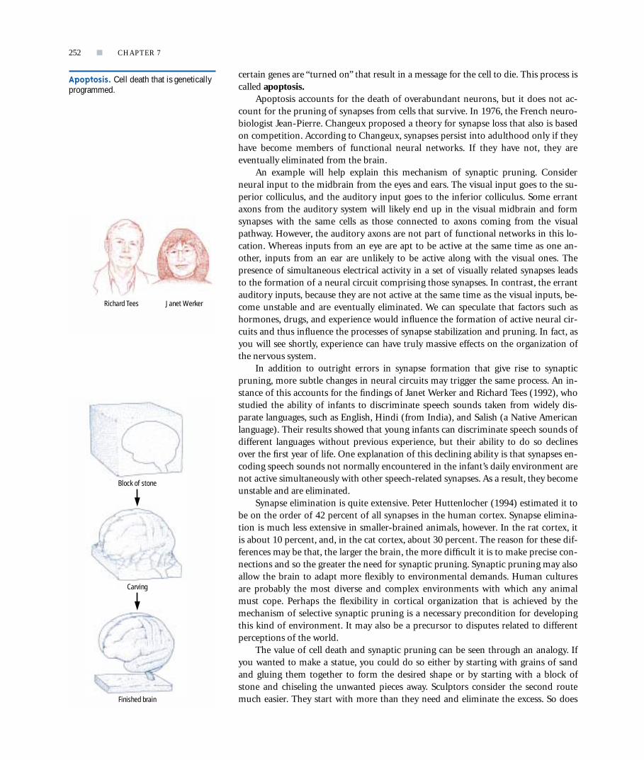
The value of cell death and synaptic pruning can be seen through an analogy. Ifyou wanted to make a statue, you could do so either by starting with grains of sandand gluing them together to form the desired shape or by starting with a block ofstone and chiseling the unwanted pieces away. Sculptors consider the second routemuch easier. They start with more than they need and eliminate the excess. So does
252 ■ CHAPTER 7
p
Apoptosis. Cell death that is geneticallyprogrammed.
Block of stone
Carving
Finished brain
Richard Tees Janet Werker

the brain. It makes too many neurons and too many connections and then gets rid ofthe unessential ones. The “chisel” in the brain could be of several forms, including agenetic signal, experience, reproductive hormones, and stress.
Glial DevelopmentThe birth of astrocytes and oligodendrocytes begins after most neurons areborn and continues throughout life. As you know from Chapter 3, oligo-dendroglia form the myelin that surrounds axons in the spinal cord andbrain. Although axons can function before they are encased by myelin, nor-mal adult function is attained only after myelination is complete. Conse-quently, myelination is useful as a rough index of cerebral maturation.
In the early 1920s, Paul Flechsig noticed that myelination of the cortex begins justafter birth and continues until nearly 18 years of age. He also noticed that some corti-cal regions were myelinated by age 3 to 4 years, whereas others showed virtually nomyelination at that time. Figure 7-17 shows one of Flechsig’s maps of the brain, withareas shaded according to the age at which myelination takes place. Flechsig hypothe-sized that the earliest-maturing areas control relatively simple movements or sensoryanalyses, whereas the late-myelinating areas control the highest mental functions.
In ReviewBrain development begins with the growth of the first neural stem cell in the third weekof embryonic development. The nervous system begins as a sheet of cells that folds tobecome a tube, known as the neural tube. Brain formation then proceeds rapidly; byabout 100 days after conception, the brain begins to look human in form. The neuronsand glia of the brain develop through a series of seven stages: birth, migration, differenti-ation, maturation, synaptic formation, death, and myelination. Neurons begin to processinformation before they are completely mature, but their activity is much simpler than itwill be with full maturation. Behavioral development is therefore constrained by the mat-uration of brain cells. For example, although infants and children are capable of complexmovements, it is not until the completion of myelin formation in adolescence that adultlevels of coordination and fine motor control are reached. By studying how the nervoussystem develops and matures, we are able to make predictions about when behaviorswill emerge. Conversely, by studying the stages of behavioral development, we can makepredictions about developments taking place in the brain.
CORRELATING BEHAVIOR WITH BRAIN DEVELOPMENTIt is reasonable to assume that, as a particular brain area matures, a person exhibitsbehaviors corresponding to that particular mature brain structure. The strongest ad-vocate of this view has been Eric Lenneberg, who, in 1967, published a seminal booktitled Biological Foundations of Language. A principal theme of this book is that chil-dren’s acquisition of language is tied to the development of the critical language areasin the cerebral cortex. This idea immediately stimulated debate over the merits of cor-relating brain and behavioral development. Now, 30-some years later, the relation be-tween brain development and behavior is widely accepted, although the influence ofexperience and learning on behavior is still considered critical. Psychologists believe
HOW DOES THE BRAIN DEVELOP? ■ 253
p
Figure 7-17
A map of how myelination progresses inthe human cortex, based on Flechsig’sresearch. The light-colored zones arevery late to myelinate, which led Flechsigto propose that they are qualitativelydifferent in function from those thatmature earlier.

that behaviors cannot emerge until the neural machinery for them has developed,but, when that machinery is in place, related behaviors develop quickly and areshaped significantly by experience. The new behaviors then alter brain structure bythe processes of neural Darwinism presented earlier. Researchers have studied theseinteracting changes in the brain and behavior, especially in regard to the emergence ofmotor skills, language, and problem solving in children. We will explore each of thesetopics separately.
Motor BehaviorsThe development of locomotion in human infants is easy to observe. At first, babiesare unable to move about independently but, eventually, they learn to crawl and thento walk. Other motor skills develop is less obvious but no less systematic ways. Forexample, Tom Twitchell studied and described the development of the ability toreach for and grasp objects. This development progresses in a series of stages, illus-trated in Figure 7-18. Shortly after birth, an infant is capable of flexing the joints ofan arm in such a way that he or she could scoop something toward the body, but, atthis age, infants do not seem to direct their arm movements toward any specificthing. Then, between 1 and 3 months of age, a baby begins to orient a hand towardan object that the hand has touched and gropes to hold that object. For example, ifthe baby’s hand touches a stick, the fingers will flex to grasp it. At this stage, however,all the fingers flex together. Between 8 and 11 months, infants’ grasping becomesmore sophisticated as the “pincer grasp,” which uses the index finger and the thumb,
develops. The pincer grasp is a significant develop-ment because it allows babies to make the very pre-cise finger movements needed to manipulate smallobjects. What we see, then, is a sequence in the de-velopment of grasping: first scooping, then grasp-ing with all of the fingers, and then grasping by us-ing independent finger movements.
If the development of increasingly well-coordi-nated grasping depends on the emergence of certainneural machinery, anatomical changes in the brainshould accompany the emergence of these behaviors.Probably many such changes take place, especially in
the development of dendritic arborizations. However, a correlation between myelinformation and the ability to grasp has been found. In particular, a group of axons from motor-cortex neurons becomes myelinated at about the same time that reachingand grasping with the whole hand develop. Similarly, another group of motor-cortexneurons, which are known to control finger movements, becomes myelinated at aboutthe time that the pincer grasp develops.
We can now make a simple prediction. If specific motor-cortex neurons are es-sential for adultlike grasping movements to emerge, removal of those neurons shouldmake an adult’s grasping ability similar to that of a young infant, which is in fact whathappens. One of the classic symptoms of damage to the motor cortex is the perma-nent loss of the pincer grasp.
Language DevelopmentThe acquisition of speech follows a gradual series of developments that has usuallyprogressed quite far by the age of 3 or 4. According to Lenneberg, children reach cer-tain important speech milestones in a fixed sequence and at relatively constantchronological ages. These milestones are summarized in Table 7-2.
254 ■ CHAPTER 7
p
2 months 10 months4 months
Orients hand towardan object and gropesto hold it.
Grasps appropriately shaped object with entire hand.
Uses pincer grasp with thumb and indexfinger opposed.
Figure 7-18
Development of the grasping responseof infants. Adapted from “The Automatic GraspingResponse of Infants,” by T. E. Twitchell, 1965,Neuropsychologia, 3, p. 251.
Link to the Web site at www.worthpublishers.com/kolb/chapter7 to see some more examples ofmotor development during childhood.
Visit the CD to review myelination ofaxons and how this process affects neuraltransmission. Find the area on the con-duction of the action potential in themodule on Neural Communication.
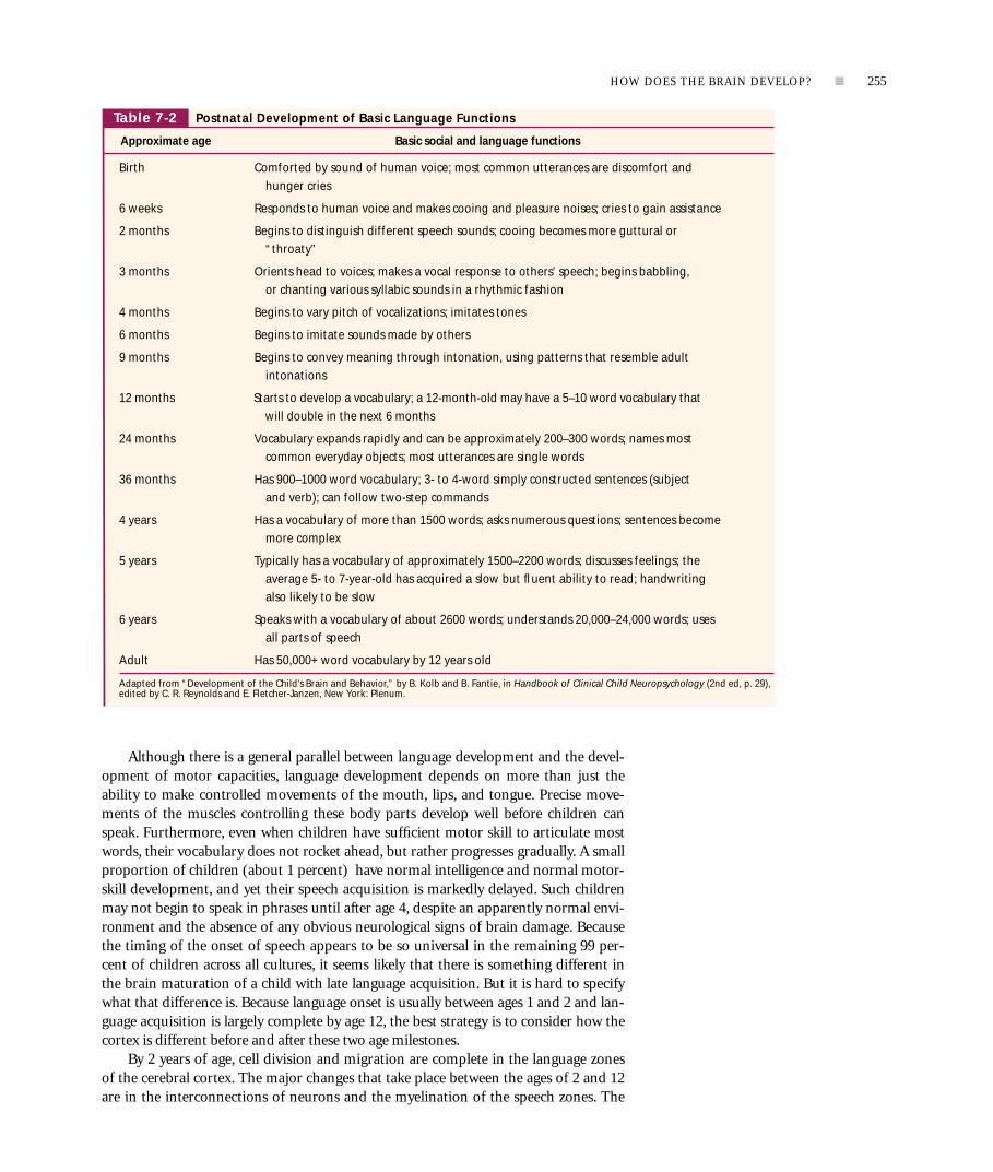
Although there is a general parallel between language development and the devel-opment of motor capacities, language development depends on more than just theability to make controlled movements of the mouth, lips, and tongue. Precise move-ments of the muscles controlling these body parts develop well before children canspeak. Furthermore, even when children have sufficient motor skill to articulate mostwords, their vocabulary does not rocket ahead, but rather progresses gradually. A smallproportion of children (about 1 percent) have normal intelligence and normal motor-skill development, and yet their speech acquisition is markedly delayed. Such childrenmay not begin to speak in phrases until after age 4, despite an apparently normal envi-ronment and the absence of any obvious neurological signs of brain damage. Becausethe timing of the onset of speech appears to be so universal in the remaining 99 per-cent of children across all cultures, it seems likely that there is something different inthe brain maturation of a child with late language acquisition. But it is hard to specifywhat that difference is. Because language onset is usually between ages 1 and 2 and lan-guage acquisition is largely complete by age 12, the best strategy is to consider how thecortex is different before and after these two age milestones.
By 2 years of age, cell division and migration are complete in the language zonesof the cerebral cortex. The major changes that take place between the ages of 2 and 12are in the interconnections of neurons and the myelination of the speech zones. The
HOW DOES THE BRAIN DEVELOP? ■ 255
p
Table 7-2 Postnatal Development of Basic Language Functions
Approximate age Basic social and language functions
Birth Comforted by sound of human voice; most common utterances are discomfort and hunger cries
6 weeks Responds to human voice and makes cooing and pleasure noises; cries to gain assistance
2 months Begins to distinguish different speech sounds; cooing becomes more guttural or “throaty”
3 months Orients head to voices; makes a vocal response to others’ speech; begins babbling, or chanting various syllabic sounds in a rhythmic fashion
4 months Begins to vary pitch of vocalizations; imitates tones
6 months Begins to imitate sounds made by others
9 months Begins to convey meaning through intonation, using patterns that resemble adult intonations
12 months Starts to develop a vocabulary; a 12-month-old may have a 5–10 word vocabulary that will double in the next 6 months
24 months Vocabulary expands rapidly and can be approximately 200–300 words; names most common everyday objects; most utterances are single words
36 months Has 900–1000 word vocabulary; 3- to 4-word simply constructed sentences (subject and verb); can follow two-step commands
4 years Has a vocabulary of more than 1500 words; asks numerous questions; sentences become more complex
5 years Typically has a vocabulary of approximately 1500–2200 words; discusses feelings; the average 5- to 7-year-old has acquired a slow but fluent ability to read; handwriting also likely to be slow
6 years Speaks with a vocabulary of about 2600 words; understands 20,000–24,000 words; uses all parts of speech
Adult Has 50,000+ word vocabulary by 12 years old
Adapted from “Development of the Child’s Brain and Behavior,” by B. Kolb and B. Fantie, in Handbook of Clinical Child Neuropsychology (2nd ed, p. 29),edited by C. R. Reynolds and E. Fletcher-Janzen, New York: Plenum.

changes in dendritic complexity in these areas are among the most impressive in thebrain. As illustrated in Figure 7-13, the axons and dendrites of the speech zone calledBroca’s area are simple at birth but become dramatically more dense between 15 and24 months of age. This development correlates with an equally dramatic change inlanguage ability, given that this age is when a baby’s vocabulary starts to expandrapidly. We can therefore infer that language development may be constrained, at leastin part, by the maturation of language areas in the cortex. Individual differences inthe speed of language acquisition may be accounted for by differences in this neuraldevelopment. Children with early language abilities may have early maturation of thespeech zones, whereas children with delayed language onset may have later speech-zone maturation.
The Development of Problem-Solving AbilityThe first person to try to identify stages of cognitive development was the Swiss psy-chologist Jean Piaget. He realized that the behavior of children could be used to makeinferences about their understanding of the world. For example, a baby who lifts acloth to retrieve a hidden toy is showing an understanding that objects continue to ex-ist even when out of sight. This understanding, called the concept of object permanence,is revealed by the behavior of the infant in the upper photographs of Figure 7-19. Anabsence of understanding also can be seen in children’s behavior, as shown by the ac-tions of the 5-year-old girl in the lower photographs of Figure 7-19. She was showntwo beakers with identical volumes of liquid in each, and then watched as one beaker’sliquid was poured into a skinnier beaker. When asked which beaker contained more
p
Figure 7-19
Stages of cognitivedevelopment. (Top) Theinfant illustrates that sheunderstands that thingscontinue to exist when they are out of sight.(Bottom) This girl does notyet understand theprinciple of conservation ofvolume. Beakers withidentical volumes seem tohold different amounts.
Dou
g G
oodm
an/M
onkm
eyer
Cour
tesy
Don
and
San
dy H
ocke
nbur
y
256 ■ CHAPTER 7

liquid, she pointed to the taller, skinnier beaker, not understanding that the amount ofliquid remains constant despite the difference in appearance. An understanding of thisprinciple, called conservation of liquid volume, is not displayed until about age 7.
By studying children engaged in such tasks, Piaget concluded that cognitive de-velopment is a continuous process. Children’s strategies for exploring the world, andtheir understanding of it, are constantly changing. These changes are not simply theresult of acquiring specific pieces of new knowledge. Rather, at certain points in de-velopment, fundamental changes take place in the organization of a child’s strategiesfor learning about the world, and with these changes come new understandings.
Piaget identified four major stages of cognitive development, which are summarizedin Table 7-3. Stage I is the sensorimotor period, from birth to about 18 to 24 months ofage. During this time, babies learn to differentiate themselves from the external world,they come to realize that objects exist even when out of sight, and they gain some under-standing of cause-and-effect relations. Next is stage II, the preoperational period, fromage 2 to 6 years. This stage is when children become able to form mental representationsof things in their world and to represent those things in words and drawings. Stage III isthe period of concrete operations, from age 7 to 11 years. At this stage, children are ableto mentally manipulate concrete ideas such as volumes of liquid and dimensions of ob-jects. Finally, stage IV is the period of formal operations, which is reached after age 11.The child is now able to reason in the abstract, not just in concrete terms.
If we take Piaget’s stages as rough approximations of qualitative changes that takeplace in children’s thinking as they grow older, we can ask what changes in the brainmight underlie them. One place to look for brain changes is in the relative rate ofbrain growth. After birth, the brain does not grow uniformly; rather, it tends to in-crease its mass during irregularly occurring periods commonly called growth spurts.In his analysis of brain-to-body-weight ratios, Herman Epstein found consistentspurts in brain growth between 3 and 10 months (accounting for an increase of 30percent in brain weight by the age of 11⁄2 years) as well as from the ages of 2 to 4, 6 to8, 10 to 12, and 14 to 16+ years. The increments in brain weight were from about 5 to10 percent in each of these 2-year periods. The brain growth takes place without aconcurrent increase in the number of neurons, so it is most likely due to the growthof glial cells and synapses. Although synapses themselves would be unlikely to addmuch weight to the brain, the growth of synapses is accompanied by increased meta-bolic demands, which cause neurons to become larger, new blood vessels to form, andnew astrocytes to be produced.
HOW DOES THE BRAIN DEVELOP? ■ 257
p
Table 7-3 Piaget’s Stages of Cognitive Development
Typical age range Description of the stage Developmental phenomena
Birth to Stage I: Sensorimotor Object permanence18–24 months Experiences the world through senses and actions (looking, Stranger anxiety
touching, mouthing)
About Stage II: Preoperational Pretend play2–6 years Represents things with words and images but lacks logical Egocentrism
reasoning Language development
About Stage III: Concrete operational Conservation7–11 years Thinks logically about concrete events; grasps concrete analogies Mathematical transformations
and performs arithmetical operations
About Stage IV: Formal operational Abstract logic12+ years Reasons abstractly Potential for mature moral reasoning
Growth spurt. A sudden growth in de-velopment that lasts for a finite time.

We would expect such an increase in the complexity of the cortex to generatemore complex behaviors, so we might predict that there would be significant, per-haps qualitative, changes in cognitive function during each of the growth spurts.The first four brain-growth spurts coincide nicely with the four main stages of cog-nitive development described by Piaget. This correspondence suggests that there may be significant alterations in neural functioning with the onset of each of Pi-
aget’s stages. At the same time, differences in the rate of brain development or perhaps in the rate atwhich specific groups of neurons mature may ac-count for individual differences in the age at whichthe various cognitive advances that Piaget identifiedemerge. Although Piaget did not identify a fifthstage of cognitive development in later adolescence,the presence of a growth spurt then implies thatthere may, in fact, be one.
One difficulty in linking brain-growth spurts tocognitive development is that growth spurts are su-perficial measures of changes taking place in thebrain. We need to know what neural events are con-tributing to brain growth and just where they aretaking place. A way to find this out is to observe chil-dren’s attempts to solve specific problems that are di-agnostic of damage to discrete brain regions inadults. If children perform a particular task poorly,then whatever brain region regulates the perfor-mance of that task in adults must not yet be maturein children. Similarly, if children can perform onetask but not another, the tasks apparently require dif-ferent brain structures and these structures mature atdifferent rates.
258 ■ CHAPTER 7
p
Question: In what sequence do the forebrain structures required for learning and memory mature?
Both human and monkey infants learn the concurrent-discrimination task at a younger age than the nonmatching-to-sample task, implying that the neural structures underlying the former task mature sooner than those underlying the latter.
Conclusion
EXPERIMENT
Concurrent-discrimination learning task
For a period of days, the subject must learn and remember which object in each pair must be displaced to receive a food reward.
� �
��
� �
� �
�� � �
� �
�
� �
� �
�
Pair 1
Pair 2
Pair 3
Pair 4
Pair 20
Day 1 Day 2
24-h
our d
elay
24-h
our d
elay
Procedurerepeated
Nonmatching-to-sample learning task
Subject is shown object that can be displaced for a food reward.
Preceding object and new object are presented.
Displacement of new object is rewarded with food.
�
� �
15 seconds
Procedures
Figure 7-20
An experiment designed to show the order in whichforebrain structures involved in learning and memorymature. In these versions of the Wisconsin General TestApparatus, the subject’s task is to displace an object toreveal a food reward. The non matching-to-sample taskrequires maturation of the temporal lobes, while theconcurrent-discrimination task requires maturation of thebasal ganglia. Both human and monkey infants learn theconcurrent task at a younger age than the matching task,implying that the neural structures underlying the formertype of learning mature sooner than those underlying thelatter. Adapted from “Object Recognition Versus Object Discrimination:Comparison Between Human Infants and Infant Monkeys,” by W. H. Overman, J. Bachevalier, M. Turner, and A. Peuster, 1992,Behavioral Neuroscience, 106, p. 18.

Bill Overman and Jocelyn Bachevalier used this logic to study the development offorebrain structures required for learning and memory in young children and mon-keys. Figure 7-20 shows the tests that they presented to their subjects. The first taskwas simply to learn to displace an object to obtain a food reward. When the subjectshad learned this task, they were trained in two more tasks that are believed to measurethe functioning of the temporal lobes and the basal ganglia, respectively. In the first ofthese two additional tasks, the subjects were shown an object, which they could dis-place to receive a food reward. After a brief (15-second) delay, two objects were pre-sented: the first object and a novel object. The subjects then had to displace the novelobject to obtain the food reward. This task, called nonmatching to sample, is thoughtto measure object recognition, which is a function of the temporal lobes. The subjectcan find the food only by recognizing the original object and not choosing it. In thesecond of the two additional tasks, the subjects were presented with a pair of objectsand had to learn that one object in that pair was always associated with a food reward,whereas the other object was never rewarded. The task was made more difficult by se-quentially giving the subjects 20 different object pairs. Each day, they were presentedwith one trial per pair. This task, called concurrent discrimination, is thought to mea-sure trial-and-error learning of specific object information, which is a function of thebasal ganglia.
Adults easily solve both tasks, but they say that the concurrent task is more dif-ficult because it requires remembering far more information than the nonmatch-ing-to-sample task. The key question developmentally is whether there is a differ-ence in the age at which children (or monkeys) can solve these two tasks. It turnsout that children can solve the concurrent task by about 12 months of age, but notuntil about 18 months of age can they solve what most adults believe to be the eas-ier task. These results imply that the basal ganglia, which is the critical site for theconcurrent-discrimination task, mature more quickly than the temporal lobe,which is the critical region for the nonmatching-to-sample task.
In ReviewAs children develop, increasingly mature behaviors emerge in a predictable sequence.This behavioral development is probably related to neural changes in the brain. Forexample, as the cortex and basal ganglia develop, different motor abilities and cognitivecapacities emerge. As you will see in the next section, these developing behaviors areshaped not only by the emergence of brain structures but also by the experiences thateach person has.
BRAIN DEVELOPMENT AND THE ENVIRONMENTBrain plasticity refers to the lifelong changes in the structure of the brain that ac-company experience. This term suggests that the brain is pliable, like plastic, and canbe molded into different forms, at least at the microscopic level. Brains exposed todifferent environmental experiences are molded in different ways. Culture is part ofthe human environment, so culture helps to mold the human brain. We would there-fore expect people in different cultures to acquire differences in brain structure thatwould have a lifelong effect on their behavior.
The brain is plastic not only in response to external events but also in response toevents within a person’s body, including the effects of hormones, injury, and abnormal
HOW DOES THE BRAIN DEVELOP? ■ 259
p
Brain plasticity. The capacity of thebrain to change in response to chemicals,activity, or experience.
Bill Overman

genes. The developing brain early in life is especially responsive to these internal fac-tors, which in turn alter the way that the brain reacts to external experiences. In thissection, we explore a whole range of environmental influences on brain development,including both external and internal ones. We start with the question of exactly howexperience manages to alter brain structure.
Experience and Cortical OrganizationResearchers can study the effects of experience on the brain and behavior by placinglaboratory animals in different environments and observing the results. In one of theearliest such studies, Donald Hebb took one group of young laboratory rats homeand let them grow up in his kitchen. A control group grew up in standard laboratorycages at McGill University. The “home rats” had many experiences that the caged ratsdid not, including being chased with a broom by Hebb’s less-than-enthusiastic wife.Subsequently, Hebb gave all the rats a rat-specific “intelligence test” that consisted oflearning to solve a series of mazes, collectively known as Hebb-Williams mazes. An ex-ample of a Hebb-Williams maze is shown in Figure 7-21. The home rats performedfar better on these tasks than the caged rats did. Hebb therefore concluded that intelli-gence must be influenced by experience.
On the basis of his research, Hebb reasoned that people reared in stimulating en-vironments would maximize their intellectual development, whereas people raised inimpoverished environments would not reach their intellectual potential. Althoughthis seems to be a logical conclusion, there is a problem in defining what stimulatingand impoverished environments are. People living in slums with little education arenot in what we would normally call an enriched setting, but that does not necessarilymean that the environment offers no cognitive stimulation or challenge. Certainly,people raised in this setting would be better adapted for survival in a slum than peo-ple raised in upper-class homes. Does this make them more intelligent in a certainway? Perhaps. In contrast, slum dwellers are not likely to be well adapted for collegelife, which was probably closer to what Hebb had in mind when he referred to such anenvironment as limiting intellectual potential. Indeed, it was Hebb’s logic that led tothe development of preschool television programs such as Sesame Street, which triedto provide a form of enrichment for children who would otherwise have little
preschool exposure to reading.The idea that early experience can change later behavior seems
sensible enough, but we are left with the question of why experi-ence should make such a difference. One reason is that experiencechanges the structure of neurons in the brain, especially in the cor-tex. Neurons in the brains of animals raised in complex environ-ments, such as the environment shown in Figure 7-22, are largerand have more synapses than do those of animals reared in barrencages. Presumably, the increased number of synapses results fromincreased sensory processing in a complex and stimulating envi-ronment. There are also more (and larger) astrocytes in the brainsof animals raised in complex settings.
Although complex-rearing studies do not address the effectsof human culture directly, it is easy to make predictions about hu-man development on the basis of their findings. We know that ex-perience can modify the brain, so we can predict that different ex-periences might modify the brain differently, which seems to bethe case in language development. Recall that exposure to differ-ent languages in infancy alters a child’s subsequent ability to dis-
260 ■ CHAPTER 7
p
Figure 7-21
In this version of the Hebb-Williamsmaze, a rat is placed in the start box (S)and must learn to find the food in thegoal box (G). The walls of the maze canbe moved to create new problems. Ratsraised in complex environments solvesuch mazes much more quickly than dorats raised in standard laboratory cages.
S
G

criminate language-specific speech sounds. A similar process is likely to occur formusic. People exposed to Western music since childhood usually find Eastern musicpeculiar, even nonmusical, on first encountering it when they are adults. Presumably,cells in the language- and music-analysis systems of the auditory cortex are alteredby early experience and lose much of their plasticity in adulthood.
This loss of plasticity does not mean that the human brain becomes fixed and un-changeable in adulthood, however. There is little doubt that the brains of adults are in-fluenced by exposure to new environments and experiences, although probably moreslowly and less extensively than the brains of children are. Animal studies have shownplasticity in the adult brain. In fact, there is evidence that the brain is affected by expe-rience well into old age, which is good news for those of us who are no longer children.
Experience and Neural ConnectivityIf experience can influence the structure of the cerebral cortex after a person is born,can it also do so prenatally? It can. This prenatal influence of experience is very clearlyillustrated in studies of the developing visual system.
Consider the problem of connecting the eyes to the rest of the visual system. Theproblem can be understood with a simple analogy. Imagine that students in a largelecture hall are each viewing the front of the room (the visual field) through a small
HOW DOES THE BRAIN DEVELOP? ■ 261
p
Figure 7-22
(A) In a complex housing environment for a group of about sixrats. The animals have an opportunity to move about and tointeract with toys that are changed weekly. (B) Representativeneurons from the parietal cortex of a laboratory-housed ratand of a complex-environment-housed rat. The neuron fromthe rat raised in the enriched environment is more complex andhas about 25 percent more dendritic space for synapses.
Laboratory housed Complex environment housed
(A) (B)

cardboard tube, such as that for a paper-towel roll. If each student looks directlyahead, he or she will each see only a small bit of the visual field. This is essentially howthe photoreceptors in the eyes act. Each of these cells sees only a small bit of the visualfield. The problem is to put all the bits together to form a complete picture. To do so,receptors that see adjacent views (analogous to students sitting side by side) mustsend their information to adjacent regions in the various parts of the brain’s visualsystem, such as the midbrain. How do they accomplish this feat?
Roger Sperry suggested that specific molecules exist in different cells in the vari-ous regions of the midbrain, giving each cell a distinctive chemical identity. Each cell,in other words, has an identifiable biochemical label. This idea is called thechemoaffinity hypothesis. Presumably, incoming axons seek out a specific chemical,such as the tropic factors discussed earlier, and consequently land in the correct gen-eral region of the midbrain. Many experiments have shown this process to take place.But the problem is that chemical affinity “directs” incoming axons to only the generallocation in which they need to be. To return to our two adjacent retinal cells, how dothey now place themselves in the precisely correct position?
This fine-tuning of placement is believed to be activity dependent. Because adja-cent receptors tend to be activated at the same time, they tend to form synapses onthe same neurons in the midbrain, after chemoaffinity has drawn them to a generalmidbrain region. This process is shown in Figure 7-23. Neurons A and G are unlikelyto be activated by the same stimulus and so they seldom fire synchronously. NeuronsA and B, in contrast, are apt to be activated by the same stimuli, as are B and C.Through this simultaneous activity, cells eventually line up correctly in the connec-tions that they form.
Now consider what happens to axons coming from differenteyes. Although the inputs from the two eyes may be active simulta-neously, the activity of cells in the same eye are more likely to be ac-tive together than are cells in different eyes. The net effect is that in-puts from the two eyes tend to organize themselves into bands, orcolumns, that represent the same region of space in each of the eyes,as shown in Figure 7-24. The formation of these segregated bandstherefore depends on the patterns of coinciding electrical activity onthe incoming axons.
The importance of coinciding electrical activity and the forma-tion of neural columns in the brain are demonstrated beautifully in aclever experiment by Martha Constantine-Paton. She knew that, be-cause the optic nerves of frogs are completely crossed, the optic tec-tum on each side has input from only one eye. She wondered whatwould happen if a third eye were transplanted in the embryonic froghead. Probably this eye would send connections to one of the tecta,which would now have to accommodate to the new input. This ac-commodation is exactly what happened, as shown in Figure 7-25.
262 ■ CHAPTER 7
p
Chemoaffinity hypothesis. The ideathat cells or their axons and dendrites aredrawn toward a signaling chemical thatindicates the correct direction in which togo.
Amblyopia. A condition in which visionin one eye is reduced as a result of disuse;usually caused by a failure of the two eyesto point in the same direction.
Time
AB
FG
E
CD
AB
FG
E
CD
Eye
Retina
Optic tectumin midbrain
Optic tectum
Figure 7-23
Experience has a role in organizing connections in the brain. Various neurons(labeled A–G) project from the retina to the tectum. The activities of adjacentneurons (for example, C and D) are more likely to coincide than the activitiesof neurons that are far apart (for example, A and G). As a result, the adjacentneurons are more likely to establish permanent synapses on the same tectalneurons. Axons grow to the approximate location in the tectum by usingchemical signals (top), but there is a lack of precision. The connections aremade more precise by the correlated activity.

The new eye sent connections to one of the tecta, which produced competition withone of the ungrafted eyes sending axons there. This competition resulted in the forma-tion of one neural column for each eye. We can only imagine what this frog made ofthe world with its three eyes.
To summarize, the details of neural connections are modified by experience. Anorganism’s genetic blueprint is vague regarding exactly which connections in thebrain go to exactly which neurons. It is experience that fine-tunes neural connectivity.If experience is abnormal, such as would happen if one eye were covered during de-velopment, then the connections will not be guided appropriately by experience. Infact, this is exactly what happens to children who have a “lazy eye.” The visual inputfrom the lazy eye does not contribute to the fine-tuning of connections as it should,so the details of those connections do not develop normally. The result is a loss ofsharpness in vision known as amblyopia.
HOW DOES THE BRAIN DEVELOP? ■ 263
p
Figure 7-24
In the postnatal development of oculardominance columns in the cat, axonsenter the cortex where they grow largeterminal arborizations. In infancy, theprojections of both eyes overlap (L, left eye; R, right eye). In adulthood, a nonoverlapping pattern of terminalarborizations from each of the eyes isnormal. If one eyelid of a kitten is sewnshut during a critical week ofdevelopment, the terminations from thateye retract and those from the open eyeexpand.
Figure 7-25
(A) The third eye of this three-eyed frogwas grafted into the frog embryo. (B) The third eye forms connections withone optic tectum, in this case the right.Because the connections of the third eyeare shared with another eye, these twoeyes compete for synaptic space. Thiscompetition leads to the formation ofalternating bands of connections.
Normal
Infant
Adolescent
Adult
Abnormal
L R L R L L R L R L
Rightoptic tectum
Leftoptic tectum
(B)
Retina
Grafted third eye
From Martha Constantine-Paton and Margaret I. Law, “Eye Specific
Termination Bands in Tecta of Three-Eyed Frogs,” Science, November 10, 1978,
vol. 202, pp. 639–641. ©1978 by the American Association for the
Advancement of Science.
(B)(A)

Critical Periods for Experience and Brain DevelopmentThere seem to be particular times in the course of brain development when specificexperiences are especially important for normal development. In kittens, for example,the effect of suturing one eye closed has the most disruptive effect on cortical organi-zation between 30 and 60 days after birth. A period during which brain developmentis most sensitive to a specific experience is often called a critical period. The absenceof the appropriate sensory experience during a critical period may result in abnormalbrain development, leading to abnormal behavior that endures even into adulthood.
Richard Tees offered an analogy to help explain the concept of critical periods. Hepictured the developing animal as a little train traveling past an environmental setting,perhaps the Rocky Mountains. All the windows are closed at the beginning of the jour-ney (prenatal development), but, at particular stages of the trip, the windows in certaincars open, exposing the occupants (different parts of the brain) to the outside world.Some windows open to expose the brain to specific sounds, others to certain smells,others to particular sights, and so on. This exposure affects the brain’s developmentand, in the absence of any exposure through an open window, that development is se-verely disturbed. As the journey continues, the windows become harder to open until,finally, they are permanently closed. This closure does not mean that the brain can nolonger change, but changes become much harder to induce. Now, imagine two differ-ent trains, one headed through the Rocky Mountains and another, the Orient Express,traveling across eastern Europe. The “views” from the windows are very different, andthe effects on the brain are correspondingly different. In other words, not only is the
brain altered by the experiences that it has during a critical period,but the particular kinds of experiences encountered matter, too.
An extensively studied behavior that relates to the concept ofcritical periods is imprinting. In imprinting, an animal learns, dur-ing a critical period, to restrict its social preferences to a specific classof objects, usually the members of its own species. In birds, such aschickens or waterfowl, the critical period for imprinting is oftenshortly after hatching. Normally, the first moving object that a younghatchling sees is one of its parents or a sibling, so the hatchling’sbrain appropriately imprints to its own species. This appropriate im-printing is not inevitable, however. Konrad Lorenz demonstratedthat, if the first animal or object that baby goslings encounter is ahuman, the goslings imprint to the human as though it were theirmother. Figure 7-26 shows a flock of goslings that imprinted toLorenz and followed him about wherever he went. This incorrectimprinting has long-term consequences for the hatchlings, whichwill often direct their subsequent sexual behavior inappropriately to-ward humans. For instance, a Barbary dove that had become im-printed to Lorenz directed its courtship toward his hand and eventried to copulate with the hand if it were held in a certain orienta-tion. Interestingly, birds inappropriately imprint not just to humans,but to inanimate objects, too, especially if they are moving. Chickenshave been induced to imprint to a milk bottle sitting on the back of atoy train that was moving around a track. But the brain is not en-tirely “clueless” when it comes to selecting a target to which to im-print. Given a choice, young chicks choose a real chicken to which toimprint over any other stimulus.
264 ■ CHAPTER 7
p
Critical period. A period in develop-ment during which some event has along-lasting influence on the brain; oftenreferred to as a sensitive period.
Imprinting. The process in which an an-imal is predisposed to learning an attach-ment to objects or animals at a critical pe-riod in development.
Figure 7-26
The ethologist Konrad Lorenz is beingfollowed by goslings that imprinted onhim. Because he was the first “object”that the geese experienced afterhatching, he became their “mother.”
Tho
mas
D. M
cAvo
y/Ti
me
Mag
azin
e

The fact that imprinting is rapid and has permanent behavioral consequencessuggests that, during imprinting, the brain makes a rapid change of some kind, prob-ably a structural change, given the permanence of the new behavior. Gabriel Hornand his colleagues at Cambridge University tried to identify this change in the brainsof chicks during imprinting. Apparently, the change takes place in a specific region ofthe forebrain, known as the IMHV. The results of electron microscopic studies showthat the synapses in this region enlarge with imprinting. Imprinting, then, seems to bea good model for studying brain plasticity during development, in part because thechanges are rapid, are related to specific experience, and are localized.
Abnormal Experience and Brain DevelopmentIf complex experiences can stimulate brain growth and influence later behavior, itseems likely that severely restricted experiences might retard both brain growth andbehavior. To study the effects of such restrictions, Donald Hebb and his colleaguesplaced Scottish terriers in a dark environment with as little stimulation as possibleand compared their behavior with that of dogs raised in a normal environment.When the dogs raised in the impoverished environment were later removed from thatenvironment, their actions were very unusual. They showed virtually no reaction topeople or other dogs, and they appeared to have lost their sense of pain. Even stickingpins in them produced no response. When given a dog version of the Hebb-Williamsintelligence test for rats, these dogs performed very poorly and were unable to learnsome tasks that dogs raised in more stimulating settings could learn easily.
The results of subsequent studies have shown that depriving young animalsspecifically of visual input or even of maternal contact has devastating conse-quences for their behavioral development and, presumably, for the development ofthe brain. For instance, Austin Riesen and his colleagues extensively studied animalsraised in the dark and found that, even though the animals’ eyes still work, theymay be functionally blind after early visual deprivation. The absence of visual stim-ulation results in an atrophy of dendrites on cortical neurons, which is essentiallythe opposite of the results observed in the brains of animals raised in complex andstimulating environments.
Not only does the absence of specific sensory inputs adversely affect brain devel-opment, so do more complex kinds of abnormal experiences. This can be seen in theretarded intellectual development of children raised in dreadful circumstances in Ro-manian orphanages, as described in “Romanian Orphans” on page 266. In the 1950s,Harry Harlow began the first systematic laboratory studies of analogous deprivationin laboratory animals. Harlow showed that infant monkeys raised without maternal(or paternal) contact have grossly abnormal intellectual and social behaviors in adult-hood. Harlow separated baby monkeys from their mothers shortly after birth andraised them in individual cages. Perhaps the most stunning effect was that, in adult-hood, these animals were totally unable to establish normal relations with other ani-mals. Unfortunately, Harlow did not analyze the brains of the deprived monkeys. Wewould predict atrophy of cortical neurons, especially in the frontal-lobe regionsknown to be related to normal social behavior.
The importance of the environment in brain development cannot be overempha-sized. Children exposed to impoverished environments or to abuse or neglect can beexpected to be at a serious disadvantage later in life. Although it is often thought thatchildren can succeed in school and in life if they really want to, it is clear that abnor-mal developmental experiences can alter the brain irrevocably. As a society, we cannotbe complacent about the environments to which our children are exposed.
HOW DOES THE BRAIN DEVELOP? ■ 265
p

Hormones and Brain DevelopmentThe determination of sex is largely genetic. In mammals, the Y chromosome presentin males controls the process by which an undifferentiated primitive gonad developsinto testes, as illustrated in Figure 7-9. The testes subsequently secrete testosterone,which stimulates the development of male reproductive organs and, during puberty,the growth of male secondary sexual characteristics.
Gonadal hormones also influence the development of neurons. Testosterone is re-leased in males during a brief period in the course of brain development, and it subse-quently acts to alter the brain, much as it alters the sex organs. This process is calledmasculinization. Just as testosterone does not affect all body organs, it does not affectall regions of the brain. It does, however, affect many brain regions and in many differ-ent ways. For instance, it affects the number of neurons formed in certain brain areas,reduces the number of neurons that die, increases cell growth, increases or reducesdendritic branching, increases or reduces synaptic growth, and regulates the activity ofsynapses. As a result of these effects due to exposure to testosterone, a male brain and afemale brain are not the same.
266 ■ CHAPTER 7
p
Romanian Orphans
Focus on Disorders
In the 1970s, the Communist regime then governing Roma-
nia outlawed all forms of birth control and abortion. The
natural result was thousands of unwanted pregnancies.
More than 100,000 unwanted children were placed in or-
phanages where the conditions were appalling. Children
had virtually no environmental stimulation. In most in-
stances, they were confined to cots. There were few, if any,
playthings and virtually no personal interaction with care-
givers. Bathing often consisted of being hosed down with
cold water. After the Communist government fell and the
outside world was able to intervene, hundreds of these chil-
dren were rescued and placed in adoptive homes through-
out the world, especially in the United States, Canada, and
the United Kingdom.
There have now been several studies of the fate of these
severely deprived children (see Ames, 1997; Rutter et al.,
1998). When the children arrived in their new homes, they
were in a poor physical state. They were malnourished; they
had chronic respiratory and intestinal infections; and they
were severely developmentally impaired. A British study by
Michael Rutter and his colleagues found them to be two
standard deviations below age-matched children for weight,
height, and head circumference. Assessments with the use of
scales of motor and cognitive development showed most of
the children to be in the retarded range.
The improvement in these children in the first 2 years
after placement in their adoptive homes was nothing short of
spectacular. Average height and weight became nearly nor-
mal, although head circumference remained below normal.
(Head circumference can be taken as a very rough measure
of brain size.) Many of the children were now in the nor-
mal range of motor and cognitive development. A signifi-
cant number, however, were still considered retarded. Why
were there individual differences in recovery from the past
deprivation?
The key factor in predicting recovery was age at adop-
tion. Those children adopted before 6 months of age did sig-
nificantly better than those adopted later. In a Canadian
study by Elenor Ames, Romanian orphans who were
adopted before 4 months of age had an average Stanford-
Binet IQ of 98 when tested at 41⁄2 years of age. In compari-
son, age-matched Canadian controls had an average IQ of
109, whereas Romanian children adopted at a median age
of 19 months had an average IQ of only 90. Brain-imaging
studies showed the children adopted at an older age to have
smaller-than-normal brains. Although there are no formal
Masculinization. A process by whichexposure to androgens alters the brain,rendering it “malelike.”

It was once believed that testosterone’s effects on brain development were not allthat important, because this hormone was thought to primarily influence regions ofthe brain regarding sexual behavior, not regions of “higher” functions. We now knowthat this belief is false. Testosterone changes the structure of cells in many regions ofthe cortex, with diverse behavioral consequences that include influences on cognitiveprocesses.
Consider one example. Jocelyn Bachevalier trained infant male and female mon-keys in the concurrent-discrimination task described earlier, in which the subject hasto learn which of two objects in a series of object pairs conceals a food reward. In ad-dition, Bachevalier trained the animals in another task, known as object-reversal learn-ing. The task here is to learn that one particular object always conceals a food reward,whereas another object never does. After this pattern has been learned, the rewardcontingencies are reversed so that the particular object that has always been rewardedis now never rewarded, whereas the formerly unrewarded object now conceals the re-ward. When this new pattern has been learned, the contingencies are reversed again,and so on, for five reversals. Bachevalier found that 21⁄2-month-old male monkeys
HOW DOES THE BRAIN DEVELOP? ■ 267
p
studies of large groups of these children as they approach
adolescence, anecdotal reports of individual children who
were adopted at an older age and are now adolescents indi-
cate continuing problems. Some of these youngsters have
significant learning disabilities in school, suffer from hyper-
activity, and have not developed normal patterns of social
interaction.
The inescapable conclusion emerging from the Roman-
ian orphanage experience is that the brain may be able to re-
cover from a brief period of extreme deprivation in early in-
fancy, but periods longer than 6 months produce significant
abnormalities in brain development that cannot be com-
pletely repaired. This conclusion is supported by the case
study of an American girl named Genie, who experienced se-
vere social and experiential deprivation as well as chronic
malnutrition at the hands of her psychotic father (see Curtis,
1978). She was discovered at the age of 13, after having spent
much of her life in a closed room, during which time she was
punished for making any noise. After her rescue, she, too,
showed rapid growth and cognitive development, although
her language development remained severely retarded.
To summarize, studies of the Romanian orphans, of or-
phans from other highly impoverished settings, and of cases
such as that of Genie make it clear that the developing brain
requires stimulation for normal development. Although the
brain may be able to catch up after a short period of depri-
vation, more than a few months of severe deprivation results
in a smaller-than-normal brain and associated behavioral
abnormalities, especially in cognitive and social skills.
The situation depicted in this photo was not unusual forRomanian orphans in the 1970s and 1980s: children werehoused and clothed, but had no other forms of stimulation,either from caregivers or an enriched environment. Studieson this population have shown that the lack of stimulationhas hampered normal brain development.
Joh
nso
n/G
amm
a-Li
aiso
n

were superior to female monkeys on the object-reversal task, but females did betteron the concurrent task. Apparently, the different brain areas required for these twotasks matured at different rates in the male and female monkeys. Bachevalier latertested additional male monkeys whose testes had been removed at birth and so wereno longer exposed to testosterone. These animals performed like females on the tasks,implying that testosterone was influencing the rate of brain development in areas re-lated to certain cognitive behaviors.
Bachevalier and her colleague Bill Overman then repeated the experiment, thistime using as their subjects children from 15 to 30 months old. The results were thesame: boys were superior at the object-reversal task and girls were superior at the con-current task. There were no such male–female differences in performance amongolder children (32–55 months of age). Presumably, by this older age, the brain regionsrequired for each task had matured in both boys and girls. At the earlier age, however,gonadal hormones seemed to be influencing the rate of maturation in certain regionsof the brain, just as they had in the baby monkeys.
Although the biggest effects of gonadal hormones may be during early develop-ment, their role is by no means finished at the end of childhood. Gonadal hormones(including both testosterone and estrogen, the latter of which is produced in largequantities by the ovaries in females) continue to influence the structure of the brainthroughout an animal’s life. In fact, removal of the ovaries in middle-aged laboratoryrats leads to marked growth of dendrites and the production of more glial cells in thecortex. This finding of widespread neural change in the cortex associated with loss ofestrogen has implications for the treatment of postmenopausal women.
Gonadal hormones also affect how the brain responds to events in the environ-ment. For instance, among rats housed in complex environments, males show moredendritic growth in neurons of the visual cortex than do females (see Juraska, 1990).In contrast, females housed in this setting show more dendritic growth in the frontalcortex than do males. Apparently, the same experience can affect the male and femalebrain differently owing to the mediating influence of gonadal hormones. This findingmeans that, as females and males develop, their brains continue to become more andmore different from each other. It is much like coming to a fork in a road. Once hav-ing chosen to go down one path, your direction of travel is forever changed as theroads diverge and become increasingly farther apart.
To summarize, gonadal hormones alter the basic development of neurons, shapethe nature of experience-dependent changes in the brain, and influence the structureof neurons throughout our lifetime. These effects of sex hormones need to be consid-ered by those who believe that behavioral differences between males and females aresolely the result of environmental experiences. In part, it is true that environmentalfactors exert a major influence. But one reason that they do may be that male and fe-male brains are different to start with, and even the same events, when experienced bystructurally different brains, may lead to different effects on the brain. In our view, theimportant task is not to deny the presence of sex differences in brain organization andfunction, but rather to understand the degree to which those neurological differencescontribute to observed differences in behavior.
Injury and Brain DevelopmentIf the brain is damaged in the course of development, is it irrevocably altered? Inthe 1930s, Donald Hebb studied children with major birth-related injuries to thefrontal lobes and found that such children had severe and permanent behavioralabnormalities in adulthood. He concluded that severe brain damage early in life can
268 ■ CHAPTER 7
p

alter the subsequent development of the rest of the brain, leading to chronic behav-ioral disorders.
To what extent have other studies confirmed Hebb’s conclusion? There are fewanatomical studies of humans with early brain injuries, but we can make some gen-eral predictions from the study of laboratory animals. In general, early brain injuriesdo produce abnormal brains, especially at certain critical periods of development. Forhumans, the worst time appears to be during the last half of the intrauterine periodand the first couple of months after birth. Rats that suffer injuries at a comparativetime have significantly smaller brains than normal, and their cortical neurons show ageneralized atrophy relative to normal brains, as illustrated in Figure 7-27. Behav-iorally, these animals appear cognitively retarded, deficient in a wide range of skills.
The effect of injury to the developing brain is not always devastating, however.For example, we have known for more than 100 years that children with brain injuriesin the first couple of years after birth almost never have the severe language distur-bances common to adults with equivalent injuries. Animal studies help explain why.Whereas damage to the brain in the period comparable to the last few months of ges-tation in humans produces widespread cortical atrophy, damage at a time roughlycomparable to age 6 months to 2 years in humans actually produces more dendriticdevelopment, as also seen in Figure 7-27. Furthermore, these animals show dramaticrecovery of functions, implying that the brain has a capacity during development tocompensate for injury.
HOW DOES THE BRAIN DEVELOP? ■ 269
p
Cortical neuronin adult
Frontal-cortexinjury
Damage onday 1
Damage onday 10
Other Kinds of Abnormal Brain DevelopmentThe nervous system need not be damaged by external forces for it to develop abnor-mally. For instance, many genetic abnormalities are believed to result in abnormalitiesin the development and, ultimately, the structure of the brain. You may have heard ofspina bifida, a condition in which the genetic blueprint goes awry and the neural tubedoes not close completely, leading to an incompletely formed spinal cord. After birth,
Figure 7-27
Cortical injury at different times in thecourse of development has differentanatomical and behavioralconsequences. In the rat, damage to thefrontal cortex on the day of birth leadsto the development of cortical neuronswith simple dendritic fields and a sparcegrowth of spines. In contrast, damage tothe frontal cortex at 10 days of age leadsto the development of cortical neuronswith expanded dendritic fields anddenser spines than normal. Adapted from “Possible Anatomical Basis ofRecovery of Function After Neonatal FrontalLesions in Rats,” by B. Kolb and R. Gibb,1993, Behavioral Neuroscience, 107, p. 808.

children with spina bifida usually have serious motor problems because of this spinal-cord abnormality. But imagine what would happen if some genetic abnormalitycaused the front end of the neural tube not to close properly. Because the front end ofthe neural tube forms the brain, this failure would result in gross abnormalities inbrain development. Such a condition exists and is known as anencephaly. Infants af-fected by this condition die soon after birth.
Abnormal brain development can be much subtler than anencephaly. For exam-ple, if cells do not migrate to their correct locations and these mispositioned cells donot subsequently die, they can disrupt brain function and may lead to disorders rang-ing from seizures to schizophrenia (see “Schizophrenia” below). There are also a vari-ety of conditions in which neurons fail to differentiate normally. In certain cases, the
270 ■ CHAPTER 7
p
Anencephaly. Failure of the forebrain todevelop.
Schizophrenia
Focus on Disorders
When Mrs. T. was 16 years old, she began to
experience her first symptom of schizophre-
nia: a profound feeling that people were star-
ing at her. These bouts of self-consciousness
soon forced her to end her public piano per-
formances. Her self-consciousness led to
withdrawal, then to fearful delusions that oth-
ers were speaking about her behind her back,
and finally to suspicions that they were plot-
ting to harm her. At first Mrs. T.’s illness was
intermittent, and the return of her intelligence,
warmth, and ambition between episodes
allowed her to complete several years of col-
lege, to marry, and to rear three children. She
had to enter a hospital for the first time at age
28, after the birth of her third child, when she
began to hallucinate.
Now, at 45, Mrs. T. is never entirely well.
She has seen dinosaurs on the street and live
animals in her refrigerator. While hallucinat-
ing, she speaks and writes in an incoherent,
but almost poetic way. At other times, she is
more lucid, but even then the voices she
hears sometimes lead her to do dangerous
things, such as driving very fast down the
highway in the middle of the night, dressed
only in a nightgown. . . . At other times and
without any apparent stimulus, Mrs. T. has
bizarre visual hallucinations. For example,
she saw cherubs in the grocery store. These
experiences leave her preoccupied, confused,
and frightened, unable to perform such every-
day tasks as cooking or playing the piano.
(Gershon & Rieder, 1992, p. 127)
Schizophrenia is obviously an extraordinary disorder,
with symptoms that are hard to generalize. It has always
been easier to identify schizophrenic behavior than to define
what schizophrenia is. Perhaps the one universally accepted
criterion for diagnosing schizophrenia is the absence of
other neurological disturbances or affective disorders that
could cause a person to lose touch with reality. This is a def-
inition by default. Other authors have emphasized the pres-
ence of bizarre hallucinations and disturbances of thought,
much like those displayed by Mrs. T. However, the symp-
toms of schizophrenia are heterogeneous, suggesting that
the biological abnormalities vary from person to person.
In 1913, Emil Kraepelin first proposed that schizophre-
nia follows a progressively deteriorating course with a dis-
mal final outcome. This opinion about the disorder was
dominant through most of the twentieth century. Today,
however, a consensus is emerging that this view is probably
incorrect. Most patients appear to stay at a fairly stable level
after the first few years of displaying schizophrenic symp-
toms, with little evidence of a decline in neuropsychological
functioning. The symptoms come and go, much as for Mrs.
T., but the severity is relatively constant after the first few
episodes.

neurons fail to produce long dendrites or spines. As a result, connectivity in the brainis abnormal, leading to retardation. The opposite condition also is possible: neuronscontinue to make dendrites and form connections with other cells to the point atwhich these neurons become extraordinarily large. The functional consequences of allthe newly formed connections can be devastating. Excitatory synapses in the wronglocation effectively short-circuit a neuron’s function.
One curious consequence of abnormal brain development is that the behavioraleffects may emerge only as the brain matures and the maturing regions begin to playa greater role in behavior. This consequence is especially true of frontal-lobe injuries.The frontal lobes continue to develop into adolescence, and often not until adoles-cence do the effects of frontal-lobe abnormalities begin to be noticed. Schizophrenia
HOW DOES THE BRAIN DEVELOP? ■ 271
p
Numerous studies have investigated the brains of schiz-
ophrenics, both in autopsies and in MRI and CT scans. Al-
though the results vary, most agree that schizophrenics have
brains that are lighter than normal and that have enlarged
ventricles. There are also suggestions that schizophrenics
have both smaller frontal lobes (or at least a reduction in the
number of neurons in the prefrontal cortex) and thinner
parahippocampal gyri. One of the most interesting discover-
ies is that of Joyce Kovelman and Arnold Scheibel (1984),
who found pronounced abnormalities in the orientation of
neurons in the hippocampi of schizophrenics. Rather than
the consistently parallel orientation of neurons in this region
characteristic of normal brains, the schizophrenics had a
more haphazard organization, as shown in the accompany-
ing drawing.
There is increasing evidence that the abnormalities ob-
served in schizophrenic brains are associated with distur-
bances of brain development. William Bunney and his col-
leagues (1997) suggest that at least a subgroup of
schizophrenics experience either environmental insults or
some type of abnormal gene activity in the fourth to sixth
month of fetal development. These events are thought to re-
sult in abnormal cortical development, particularly in the
frontal lobes. Later in adolescence, as the frontal lobes com-
plete development, the person begins to experience symp-
toms of this abnormal prior development.
Examples of pyramidal cell orientation from thehippocampus of (A) a normal brain and (B) a schizophrenicbrain. In the schizophrenic brain these pyramidal neuronsare much more disorganized.
Adapted from “A Neurohistologic Correlate of Schizophrenia,” by J. A. Kovelman and A. B. Scheibel, 1984, Biological Psychiatry, 19, p. 1613.
Hippocampus
(A) Organized (normal)pyramidal neurons
(B) Disorganized (schizophrenic)pyramidal neurons
To learn more about abnormal braindevelopment, visit the Web site atwww.worthpublishers.com/kolb/chapter7.

is a disease that is characterized by slow development, usually not becoming obviousuntil late adolescence. The schizophrenic brain has many abnormalities, some ofwhich are in the frontal lobes.
Mental RetardationMental retardation refers to an impairment in cognitive functioning that accompaniesabnormal brain development. Mental retardation may range in severity from mild, al-lowing an almost normal life style, to severe, requiring constant care. As summarizedin Table 7-4, mental retardation can result from chronic malnutrition, genetic abnor-malities such as Down’s syndrome, hormonal abnormalities, brain injury, or neurolog-ical disease. Different causes produce different abnormalities in brain organization, butthe critical similarity across all types of retardation is that the brain is not normal.
272 ■ CHAPTER 7
p
Table 7-4 Causes of Mental Retardation
Cause Example mechanism Example condition
Genetic abnormality Error of metabolism Phenylketonuria (PKU)Chromosomal abnormality Down’s syndrome
Abnormal embryonic Exposure to a toxin Fetal alcohol syndromedevelopment
Prenatal disease Infection Rubella (also called German measles)
Birth trauma Anoxia (oxygen deprivation) Cerebral palsy
Malnutrition Abnormal brain development Kwashiorkor
Environmental abnormality Sensory deprivation Children in orphanages
A study by Dominique Purpura provides an example of one of the few systematicinvestigations of the brains of retarded children. Purpura used Golgi stain to examineneurons of children who had died from accident or disease unrelated to the nervoussystem. When he examined the brains of children with various forms of retardation,he found that the dendrites were stunted in growth and the spines were very sparse, asillustrated in Figure 7-28. The simple structure of these neurons was probably indica-tive of a marked reduction in the number of connections in the brain, which presum-ably caused the retardation. Variation in both the nature and the extent of neuronalabnormality in different children would lead to different behavioral syndromes.
In ReviewThe brain is plastic during its development and can therefore be molded by experienceinto different forms, at least at the microscopic level. The sensitivity of the brain to expe-rience varies with time, however. There are critical periods in the course of developmentwhen different parts of the brain are particularly sensitive to different experiences. Notonly is the brain plastic in response to external events, but it is changed by internalevents as well, including the effects of hormones, injury, and abnormal genes. If experi-ences are abnormal, then the brain’s development is abnormal, possibly leading to disor-ders such as mental retardation or schizophrenia.
Retardedchild
Normalchild
Figure 7-28
Representative dendritic branches fromcortical neurons in a normal child (left)and a retarded child (right). The dendritebranch from the retarded child has fewer spines. Adapted from “Dendritic Spine ‘Dysgenesis’and Mental Retardation,” by D. P. Purpura,1974, Science, 186, p. 1127.

HOW DO ANY OF US DEVELOP A NORMAL BRAIN?When we look at the complexity of the brain, the less-than-precise process of braindevelopment, and the large number of factors that can influence it, we are left mar-veling at how so many of us end up with brains that pass for “normal.” After all, wemust all have had neurons that migrated to wrong locations, made incorrect connec-tions, and were exposed to viruses or other harmful substances. If the brain were asfragile as it might seem, it would be almost impossible to end up with a normal brain.
Apparently, animals have evolved a substantial capacity to repair minor abnor-malities in brain development. Most people have developed in the range that we call“normal” because the human brain’s plasticity and regenerative powers are successfulin overcoming minor developmental deviations. Recall that one stage in brain devel-opment consists of cell death and synaptic pruning. By initially overproducing neu-rons and synapses, the brain has the capacity to correct any errors that might havearisen accidentally.
These same plastic properties of the brain later allow us to cope with the ravages ofaging. Neurons are dying throughout our lifetimes and, by age 50, we ought to be ableto see significant effects of all of this cell loss, especially considering the cumulative re-sults of exposure to environmental toxins, drugs, closed head injuries, and so on. Butthis is not what happens. Although teenagers may not believe it, very few 50-year-oldsare demented. By most criteria, the 50-year-old who has been intellectually activethroughout adulthood is likely to be much wiser than the 18-year-old whose brain haslost relatively few neurons. Clearly, we must have some mechanism to compensate forloss and minor injury to our brain cells. This capacity for plasticity and change is oneof the most important characteristics of the human brain, not only during develop-ment but through the rest of life as well. We return to this idea in Chapter 13.
SUMMARY1. What are the stages of neural development? The process of brain maturation is
long, lasting until 16 or 18 years of age. Neurons, the elementary components ofthe brain, are born, develop a neuronal phenotype, migrate, and, as theirprocesses elaborate, establish connections with other neurons. Because the braincontains such a large number of cells, and an even larger number of connections,the brain produces more neurons and connections than it needs and then prunesback to a stable adult level.
2. How does behavior develop? The infant and child go through stages of behavioraldevelopment that are similar in children across all cultural spectrums. For exam-ple, as infants develop, motor behaviors emerge in a predictable sequence. Infantsfirst make clumsy movements towards objects but they are poorly directed. Byabout 4 months, the motor system has matured sufficiently so that the infant cangrasp objects with the whole hand, and by around 11 months children are able tomake pincer grasps to pick up objects like pencils. Other motor behaviors emergeover the ensuing months and years, such as walking, throwing, catching, and soon. Similarly, cognitive behaviors emerge through a series of stages in which chil-dren acquire principles that allow them to solve problems. Researchers such asJean Piaget have identified and characterized four or more distinct stages of cog-nitive development, each of which can be identified by special behavioral tests.
3. How do behavioral and neural maturation relate to one another? The emergence ofbehaviors is correlated with the development of the neural systems that producethe behaviors. Behavioral and cognitive capacities follow a similar sequence ofdevelopment from the rudimentary to the complex. The relationship between
HOW DOES THE BRAIN DEVELOP? ■ 273
p

274 ■ CHAPTER 7
p
KEY TERMS
amblyopia, p. 263anencephaly, p. 270apoptosis, p. 252brain plasticity, p. 259cell-adhesion molecule
(CAM), p. 250 chemoaffinity hypothesis,
p. 262 critical period, p. 264
filopod, p. 249glioblast, p. 244growth cone, p. 249growth spurt, p. 257imprinting, p. 264masculinization, p. 266netrins, p. 250neural Darwinism, p. 251neural plate, p. 240
neural stem cells, p. 243neural tube, p. 240neuroblast, p. 244neurotrophic factors, p. 245progenitor cells, p. 244radial glial cells, p. 247tropic molecules, p. 250ventricular zone, p. 243
REVIEW QUESTIONS1. Describe the gross development of the nervous system. Summarize and explain
the steps in brain development.
2. What roles do different factors such as molecules, genetics, and experience play indevelopment?
3. How does behavioral development relate to neural development?
4. How does experience affect brain development?
FOR FURTHER THOUGHT1. Experience plays an important role in brain development. How might an interac-
tion between sex and environment account for behavioral differences in adult-hood?
neuroscience interact ive
There are many resources available for
expanding your learning on line:
■ www.worthpublishers.com/kolb/
chapter7.
Try some self-tests to reinforce your
mastery of the material. Look at some
of the news updates reflecting current
research on the brain. You’ll also be
able to link to other sites which will re-
inforce what you’ve learned.
■ www.embryo.mc.duke.edu
Link here to see a remarkable collection
of human embryos through magnetic
resonance microscopy.
■ www.nads.org.
Investigate current research about
Down syndrome at the National Asso-
ciation for Down Syndrome.
■ www.ucpa.org.
Learn more about cerebral palsy at the
home page of United Cerebral Palsy.
On your CD-ROM you’ll be able to
quiz yourself on your comprehension
of the chapter. The module on Neural
Communication also provides impor-
tant review on the neural structure.
brain structure and function can be inferred by matching the developmentaltimetables of brain anatomy and physiology with that of behavior. For example,motor behaviors emerge in synchrony with the maturation of motor circuits inthe cerebral cortex, basal ganglia, and cerebellum, as well as in the connectionsfrom these areas to the spinal cord. Similar correlations between behavioral emer-gence and neuronal development can be seen in the development of other behav-iors, including cognitive behaviors. For example, different types of memory abili-ties emerge as circuits in the frontal and temporal lobes mature.
4. What factors influence neural maturation? The brain is modifiable during its de-velopment and the structure of neurons and their connections can be molded byvarious factors throughout the period of development. These factors include ex-ternal events, gonadal hormones, and injury. The sensitivity of the brain to thesefactors varies with time as there are periods during the course of developmentwhen different brain regions are particularly sensitive to different events. If expe-riences are abnormal, then the brain’s development is abnormal, as well, and canlead to disorders such as retardation.
5. How sensitive is the developing brain to injury? Perturbations of the brain duringdevelopment, such as from anoxia, trauma, or toxins can significantly alter braindevelopment and result in severe behavioral abnormalities including retardationand cerebral palsy. The brain does have a substantial capacity to repair or correctminor abnormalities, however, allowing most people to develop a normal behav-ioral repertoire.

2. How can the principles of behavioral development help to explain why eachbrain is unique?
RECOMMENDED READINGEdelman, G. M. (1987). Neural darwinism: The theory of neuronal group selection. New York:
Basic Books. You have heard of Darwinism and the idea of survival of the fittest. Edel-man applies this Darwinian concept to the nervous system’s shedding of neurons in thecourse of development and throughout a person’s lifetime. Although not universally ac-cepted, the ideas in the book are amusing to read.
Greenough, W. T., & Chang, F. F. (1988). Plasticity of synapse structure and pattern in thecerebral cortex. In A. Peters and E. G. Jones (Eds.), Cerebral cortex: Vol. 7. Developmentand maturation of the cerebral cortex (pp. 391–440). New York: Plenum. Greenough isone of the world leaders in the study of experience-dependent change in the nervoussystem. This chapter not only provides a nice historical review, but also lays out seminalideas on the developmental plasticity of the nervous system.
Hebb, D. O. (1949). The organization of behavior. New York: Wiley. Although 1949 may seemlike a long time ago for a book to be still relevant today, Hebb’s book may be the mostimportant single volume on brain and behavior. It was the first serious attempt to out-line a neuropsychological theory of how the brain could produce behavior and, espe-cially, thought. Development is an important theme in the book because Hebb believedthat experience plays an essential role in developing the cognitive and neural structuresnecessary for adulthood. This book is mandatory reading for any student going on tograduate school in behavioral neuroscience.
Michel, G. F., & Moore, C. L. (1995). Developmental psychobiology. Cambridge, MA: MITPress. Most neural development books are thin on behavioral development, but thisbook strikes a nice balance in its analysis of both brain and behavioral development.
Purves, D., & Lichtman, J. W. (1985). Principles of neural development. Sunderland, MA: Sin-auer. Although not primarily about the development of the cortex, the book providessufficient background to enable a thorough understanding of the principles that guidenervous system development.
HOW DOES THE BRAIN DEVELOP? ■ 275
p